mass spectroscopy of natural products xxii—the negative ion mass spectra of brassinosteroids
TRANSCRIPT

BIOMEDICAL AND ENVIRONMENTAL MASS SPECTROMETRY, VOL. 13, 663-666 (1986)
Mass Spectroscopy of Natural Products XXII-The Negative Ion Mass Spectra of Brassinosteroids?
J. Schmidt, H.-M. Vorbrodt and G. Adam Institut fur Biochemie der Pflanzen der Akademie der Wissenschaften der DDR, DDR-4050 Halle/Saale, Weinberg 3, GDR
The negative ion mass spectra of some brawinosteroids are discussed in comparison with their positive ones. In contrast to the EI-mass spectra the negative ion mass spectra show strong peaks in the molecular ion region. The nature of an intense [M - 41-' ion was investigated on the base of model compounds and deuterium labeled derivatives.
INTRODUCTION
Recently, a new plant growth-promoting steroid, brassinolide (l), has been isolated from rape pollen (Brussicu nupus L.). The structure was elucidated as (22R,23R,24S)-2a,3a,22,23-tetra-hydroxy-24-methyl-B- homo-7-oxa-5a-cholestan-6-one' representing the first member of a new naturally occurring group of steroidal plant growth regulators, the brassino~teroids.~
Mass spectrometry is a very useful tool both for struc- ture elucidation and identification of brassinosteroids: Grove et ul. used high-resolution FD-MS to detehine the molecular formula of natural brassinolide.' The FD- mass spectrum showed an [M+ 13' ion at m/z 481 and fragment ions resulting from side chain cleavages (m/z 101, f (looo/,), m / z 131, e, see also Scheme 1). In the emitter CI-MS of 1 the ion at m/z481 represents the base peak and predominant fragments are [ M + 1 - H,O]+ and [M + 1 - 2H20]+4. Furthermore, the EI- and CI-mass spectra of methanboronates of brassinosteroids are well The methanboronates seem to be the best derivatives for the GC-analysis.'
This communication presents results obtained by negative ion mass spectroscopy of brassinolide (l), 24- epi-brassinolide (2) and 22S,23S-homobrassinolide (3).
OH R
1: R.24 S-Me; 22R,23R 2: R.24 R-Me; 22R,23R 3: R.2LS-Et; 22S,23S
~~
EXPERIMENTAL
The positive (10-16 eV) and negative (2-4 eV) ion mass spectra were recorded on an electron attachment mass
t For part XXI see: M. H. A. Elgarnal, D. Voigt and G. Adam, J. prakt. Chem., in press.
0887-6134/86/ 120663-04 $03.00 @ 1986 by John Wiley & Sons Ltd
spectrograph of the Research Institute 'Manfred von Ardenne', Dresden, GDR (unheated ion source, direct inlet system, inlet temperature 130-150 "C, electron cur- rent 10 mA, accelerating voltage 40 kV). The low energy electrons were produced in a low pressure plasma (dis- charge gas: k, source pressure: 1.33 Pa) of a duoplas- matron ion s o ~ r c e . ~
The syntheses of the brassinosteroids 2 and 3 are described elsewhere: 26, 3'. The model compounds 4 and 5 are prepared as follows,
4 ((24R) -2a,3a- Dihydroxy-24-ethyl-5a-cholestan-6- one): (24S)-2a,3a-dihydroxy-24-ethyl-5a-cholest-22E- en-6-one' (44 mg, 0.1 mmol) was hydrogenated in MeOH (10 ml) in the presence of 10% Pd/CaCO, (5 mg) by shaking for 12 h at room temperature. Filtration and crystallization yielded 44mg 4 of mp 236" (MeOH);
[+I294 0, [$1310-2089, a=-54; UVA,,, nm (log&): 290 (1.48); IR v:':' cm-': 3450 (br), 1705 (s); 'H-NMR (200 MHz, CDC1,-TMS): 6 0.59,0.85 (2s, H3-18, H3-19), 0.74 (d, H2, J = 7 Hz) 0.77 (d, H2, J = 7 Hz) 0.8 (d, H2,
and 4 Hz), 2.6 (br. d. H2-7, J = 13 Hz), 3.68 (H1-2, br. d, J = 13 Hz), 3.97 (br. s, H1-3); MS m/z (YO): 446 (M", loo), 431 (42), 428 (70), 413 (17), 410 (25), 401 (lo), 349 (14), 331 ( l l ) , 305 (50), 287 (29), 278 (27), 263 (66), 246 (56), 245 (60), 228 (37), 188 (30), 175 (37), 155 (68), 137 (43), 122 (36), 108 (51), 98 (79).
5 ((22S,23S,24S)-3a,5-Cyclo-22,23-dihydroxy-6$- methoxy-24-ethyl-5a-cholestane): (24S)-3a,5-cyclo-6P- methoxy-24-ethyl-5a-cholest-22E-ene' (4.3 g, 10 mmol) in THF (80 ml) was hydroxylated by adding of a solution of OsO, (300mg) in t-BuOH (12ml), N-methyl- morpholin N-oxide (NMO) (4 g) and H,O (1 5 ml)? The mixture was stirred for 4 days at room temperature under k. Additional amounts of NMO (2 x 1 g) were added during this time. Then excess OsO, was reduced with Na,S,04 solution. The mixture was filtered through Celite. Evaporation and separation on SiO, (Merck) by flash-chromatography (petrol: ethyl acetate/80 : 20) yiel- ded 1.9 (41%) 5 as gum: [a]g+32.5, (c=O.2, CHC13); IR v2;cm-l : 3440 (br), 1095 (s); 'H-NMR (200 MHz, CDC13-TMS): 6 0.76 (s, H3-18), 0.88 (d, H3, J = 7 Hz), 0.94 (d, H3, J = 7 Hz), 0.99 (d, H3, J = 7 Hz), 1.02 (s, H3-19), 1.03 (d, H3, J = 7 Hz), 2.78 (m, H,-6), 3.33 (s, H3-OCH3), 3.66 (m, H2-22 and -23); MS m/z (YO): 460
[CY]'03+ 10.2" (CDC13: C=O.45); ORD: [+]266+3313, MeOH
J=7Hz), 0.85 (d, H2, J=6Hz), 2.2 (dd, HI-5, J=13
Received 12 March 1986 Accepted 22 May 1986
664 J. SCHMIDT, H.-M. VORBRODT AND G. ADAM
(M+'; 15), 445 ( l l ) , 428 ( l l ) , 405 (21), 375 (lo), 346 (54), 331 (24), 314 (83), 313 (84), 295 ( 5 8 ) , 291 (29), 288 (31), 284 (47), 255 (59,227 (42), 213 (62), 199 (30), 187 (30), 173 (31), 159 (59,145 (75), 133 (59,127 ( 5 5 ) , 115 (41), 109 (80), 95 (80), 85 (100).
The deuteriated derivatives 2a and 5a ('H/'H- exchange of the hydroxyl hydrogens) were obtained by repeated (twice) recrystallization of 2 and 5, respectively, from CH302H/2H20. 5a: 7% 'Ha, l8%, *HI, 75% 2H2.
The deuterium content of 5a was calculated on the M+' peak by a procedure described elsewhere." A quan- titative calculation of the deuterium content of 2a was not possible.
RESULTS AND DISCUSSION
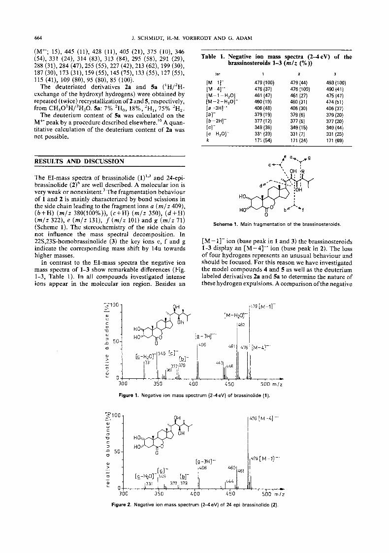
The EI-mass spectra of brassinolide (l)Is3 and 24-epi- brassinolide (2)6 are well described. A molecular ion is very weak or nonexi~tent.~ The fragmentation behaviour of 1 and 2 is mainly characterized by bond scissions in the side chain leading to the fragment ions a ( m / z 409), (b+H) ( m / z 380(100%)), ( c f H ) ( m / z 350), ( d + H ) ( m / z 322), e ( m / z 131), f ( m / z 101) and g ( m / z 71) (Scheme 1). The stereochemistry of the side chain do not influence the mass spectral decomposition. In 22S,23S-homobrassinolide (3) the key ions e, f and g indicate the corresponding mass shift by 14u towards higher masses.
In contrast to the EI-mass spectra the negative ion mass spectra of 1-3 show remarkable differences (Fig. 1-3, Table 1). In all compounds investigated intense ions appear in the molecular ion region. Besides an
-100 s & U C
Table 1. Negative ion mass spectra ( W e V ) of the brassinosteroids 1-3 ( m / z ("%))
Ion 1 2 3
[M-11- [M-4]-' [M-l-H,O]- [M - 2 - H,O]- [ a -3H]-' [bl-
ICI- [b-ZH]-
[ c - H,O]- k
479 (1 00) 476 (37) 461 (47) 460 (1 9) 406 (48)
317 ( 1 2) 349 (36) 331 (23) 171 (54)
379 (1 9)
479 (44) 476 (1 00) 461 (27) 460 (31) 406 (30) 379 (6)
349 (1 5) 331 (7) 171 (24)
377 (5)
493 (1 00)
475 (47)
379 (20) 377 (20) 349 (44)
490 (41)
474 (51) 406 (37)
331 (25) 171 (69)
Scheme 1. Main fragmentation of the brassinosteroids.
[M - 11- ion (base peak in 1 3 ) and the brassinosteroids 1-3 display an [ M - 41-' ion (base peak in 2). The loss of four hydrogens represents an unusual behaviour and should be focused. For this reason we have investigated the model compounds 4 and 5 as well as the deuterium labeled derivatives 2a and 5a to determine the nature of these hydrogen expulsions. A comparison of the negative
r9 ['-'l- [M- H20]-'
iiFiZ I
300 350 400 5 0 0 m l z
Figure 1. Negative ion mass spectrum (2-4eV) of brassinolide (1)
Y
C
C
a, u 0
0 479 [ M -11 -. [g-3H]-'
> 460 406 461
50
W
[GI- ._ 0
L4L - 331
300 350 4 0 0 450 500 m l z
Figure 2. Negative ion mass spectrum (2-4 eV) of 24-epi-brassinolide (2).
" 479 [ M -11 -.
W [g-3H]-' > 460 406 461 [GI- ._ 0
L4L - 331
300 350 4 0 0 450 500 m l z
Figure 2. Negative ion mass spectrum (2-4 eV) of 24-epi-brassinolide (2)

MASS SPECTROSCOPY OF NATURAL PRODUCTS. XXII
Y
W 0
73 C
n 50 0 [M-l-H20]' 0 ill L 2 7 > .-
0 [M-1-2 +O]- - 3 4 5 373
665
4 4 5 LM-11-
OH R l-'
0 M -. [ M - 4 ] - .
Scheme 2. Formation of t h e [M-4]-' ion in 1 3 .
& 4 4Q
._
OMe
ion mass spectra of 4 and 5 clearly show that only the side chain hydroxylated compound 5 forms an [ M - 41-' ion (Fig. 4 and 5 ) . Therefore, the four hydrogens elimi- nated during this process should stem from the side chain hydroxyls and the carbon atoms 22 and 23. This
is supported by the negative ion mass spectra of 2a and 5a. While in 5a the [M - 41-' ion is not shifted, this ion in 2a shows a mass shift by 2u towards higher masses. From these data one can conclude, that the [M-4]-' ion should have the structure figured in Scheme 2. The
4 4 2 [M-HzOI-'
456[M-L]-'
f ' T
3 0 0 3 5 0 4 00
493[M-l]-
450 500 m l z
Figure 3. Negative ion m a s s spectrum ( 2 4 eV) of 22S,23S-homobrassinolide (3).
Figure 4. Negative ion m a s s spectrum ( 2 4 eV) of compound 4.
0
L O 3 00
[M-h
414 I / , I , l!lll /Ill! , , , ,
I ' w 3 5 0 4 0 0 4 5 0 5 0 0 m l z
Figure 5. Negative ion mass spectrum ( 2 4 eV) of compound 5.

666 J. SCHMIDT, H.-M. VORBRODT A N D G. ADAM
formation of the [ M - 41-' ion is favoured by resonance stabilization of the negative charge." A similar reaction of the ring A hydroxy groups is probably predicted by the rigid ring system.
Therefore, the appearance of a [M-4]-' ion seems to be an evidence for the presence of two vicinal hydroxy groups in the side chain.
A further resonance-stabilized negative ion appears at m / z 171, which is formed by scissions through ring B (Table 1):
k , m / z 171
Acknowledgements
The authors are indebted to Prof. N. Ikekawa, Department o f Chemistry, Tokyo Institute o f Technology, for kindly providing the samples 1 and 2 as well as to Prof. K. Mori, Department of Agricultural Chemistry o f the University of Tokyo, for a gift o f compound 3.
REFERENCES
1. M. D. Grove, G. F. Spencer, W. K. Rohwedder. N. Mandava, J. F. Worley, J. D. Warthen, jr., G. L. Steffens, J. L. Flippen- Anderson and J. Cook, jr., Nature (London) 281, 216 (1979).
2. For a review see: G. Adam and V. Marquardt, fhytochemistry W, 1787 (1 896).
3. N. lkekawa and S. Takatsuto, Mass Spectrosc. 32, 55 (1984). 4. S. Takatsuto, B. Ying, M. Morisaki and N. Ikekawa,
J. Chromatogr. 239, 233 (1982). 5. M. v. Ardenne, K. Steinfelder and R. Tummler, Elektronenan-
lagerungsmassenspektrographie organischer Substanzen, Springer-Verlag, Berlin (1971).
6. S. Takatsuto and N. Ikekawa, Chem. fharm. Bull. (Tokyo), 32, 2001 (1984).
7. K. Mori, M. Sakakibara, Y. Ichikawa, H. Ueda, K. Okada, T. Umemura, G. Yabuta, S. Kuwahara, M. Kondo, M. Minobe and A. Sogabe, Tetiahedron 38, 2099 (1982).
8. J. J. Partridge, S. Faber and M. R. Uskokovic, Helv. Chim. Acta 57, 764 (1974).
9. V. Van Rheenen, R. C. Kelly and D. Y. Cha, Tetrahedron Lett. 23, 1973 (1976).
10. K. Biemann, Mass Spectrometry Organic Chemical Application, Chapter 5, McGraw-Hill, New York (1962).
11. H. Budzikiewicz, Angew. Chem. 93, 635 (1981).