mass spectrometry technologies in lipid...
TRANSCRIPT

Mass spectrometryTechnologies in Lipid chemistry
Rabah Soliymani
University Of HelsinkiProtein Chemistry UnitBiomedicum – Helsinki

Complex_&_dynamic_mixtures(few copies to >30%, modifications)
Extraction in organic solvents
Fractionation
MassSpectrometry
Local databaseSearch
Separation(LC-MS)
shotgun lipidomics
Separation(TLC, gas chroma-
tography, etc…)
LipidIdentifications

Lipids
• All Lipids are hydrophobic at one end and hydrophilic at the other end.
• That’s the one property they have in common
• Lipids includes fats and oils, waxes, phospholipids, steroids (like cholesterol), and some other related compounds

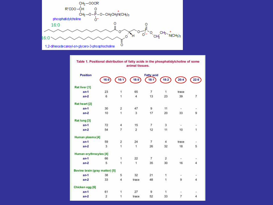
Phospholipids
phospho-, glycero-, or sphingolipidsmajor component of all cell membranes asthey can form lipid bilayerscontain a diglyceride, a phosphate group, and an organic molecule such as cholineexcept sphingomyelin (sphingosineinstead of glycerol)

Phospholipids

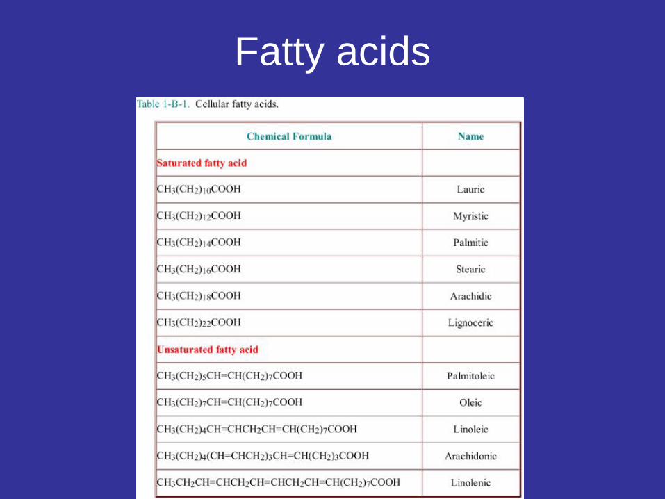
Fatty acids
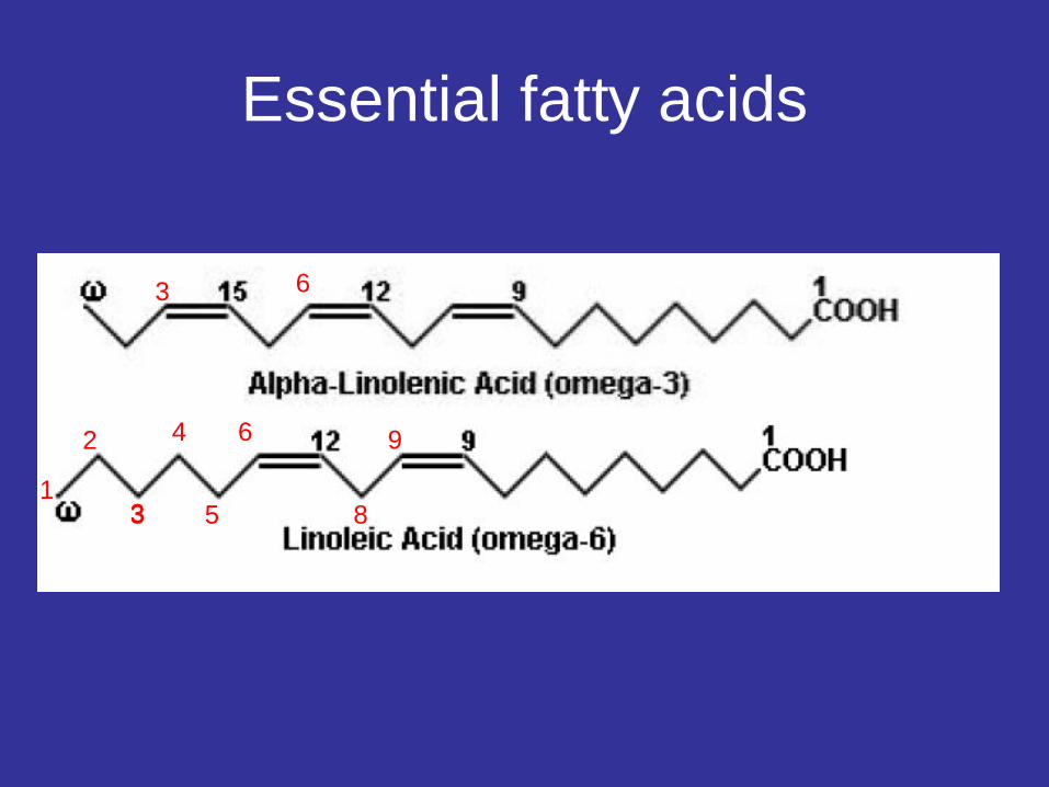
Essential fatty acids
1
2 4
3 5
6
83
3 6
9
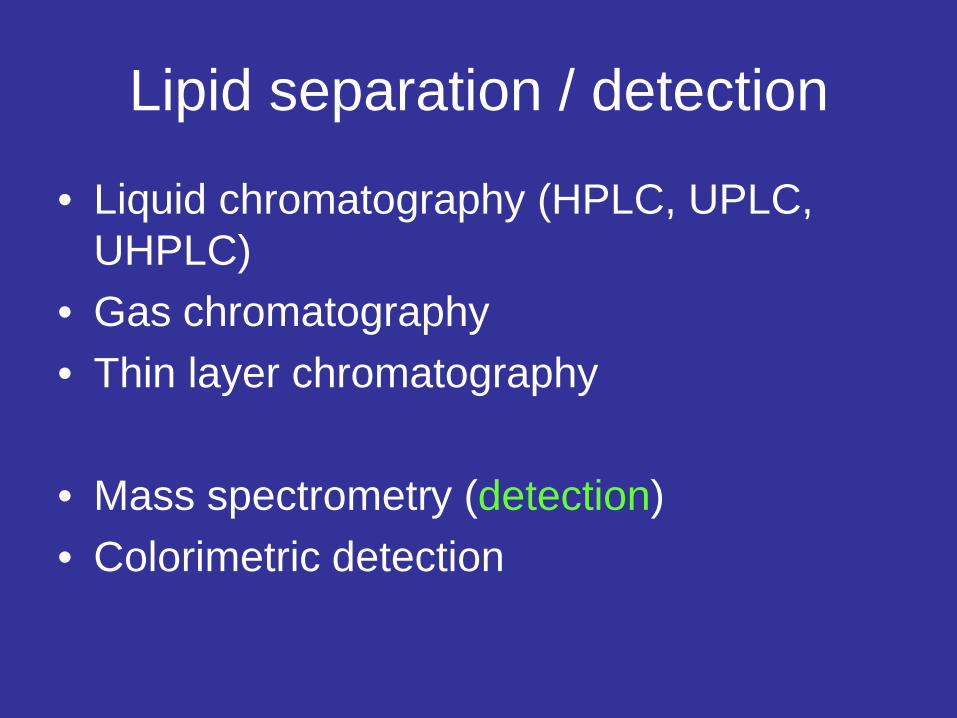
Lipid separation / detection
• Liquid chromatography (HPLC, UPLC, UHPLC)
• Gas chromatography• Thin layer chromatography
• Mass spectrometry (detection)• Colorimetric detection

TLC, a separation technique on the basis of components differences in polarity
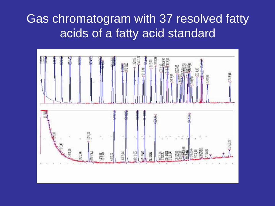
Gas chromatogram with 37 resolved fatty acids of a fatty acid standard

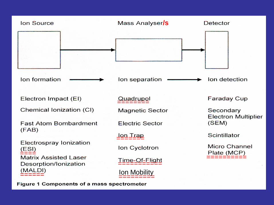
Mass spectrometry as a tool to analyze lipids
======
====
========
=======
=====================
Ion Mobility=========
/s

•Matrix-Assisted Laser Desorption Ionization (MALDI):
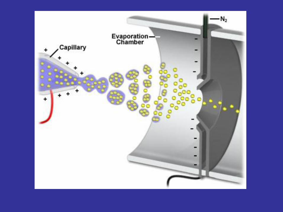
• ElectroSpray Ionization (ESI):
• both ESI and MALDI belongs to the "soft" ionization techniques.
MALDI vs ESI
Biomolecule sample requirements for MS
For better ionization, sample should preferably be in a volatile solvent
- e.g. ACN (acetonitrile e.g. 50%), methanol, propanol, etc…
- Samples from liquid chromatograph

Electrospary Ionization
High vacuum


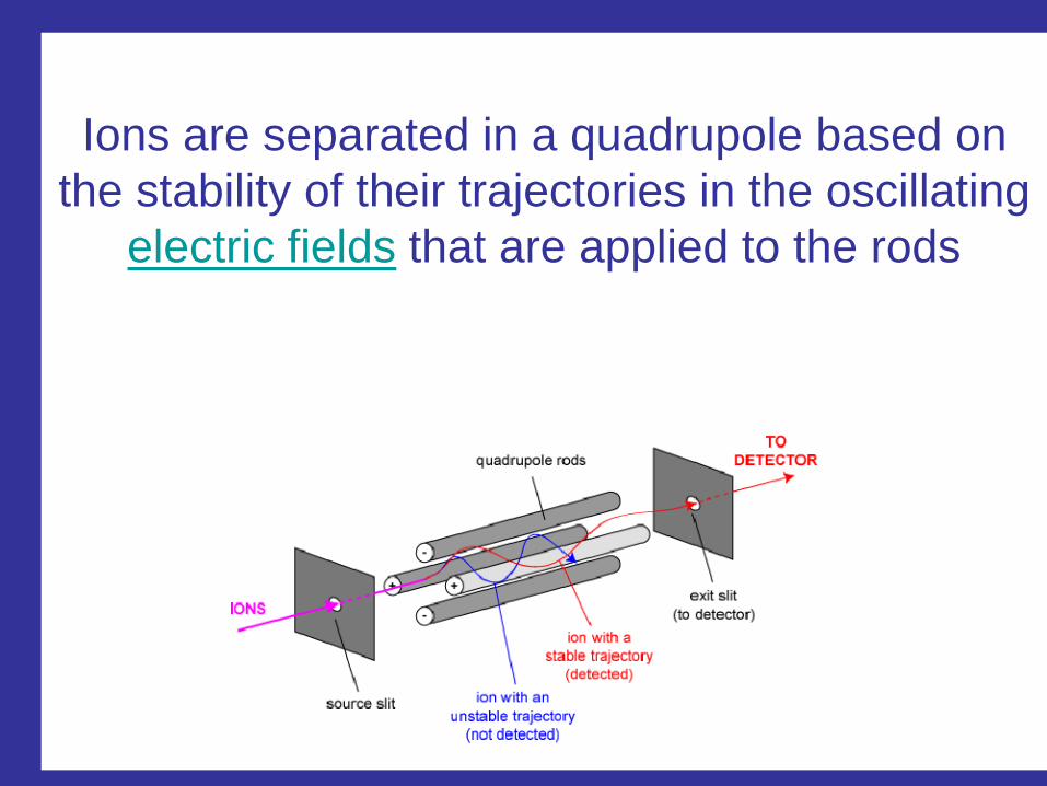
Ions are separated in a quadrupole based on the stability of their trajectories in the oscillating
electric fields that are applied to the rods

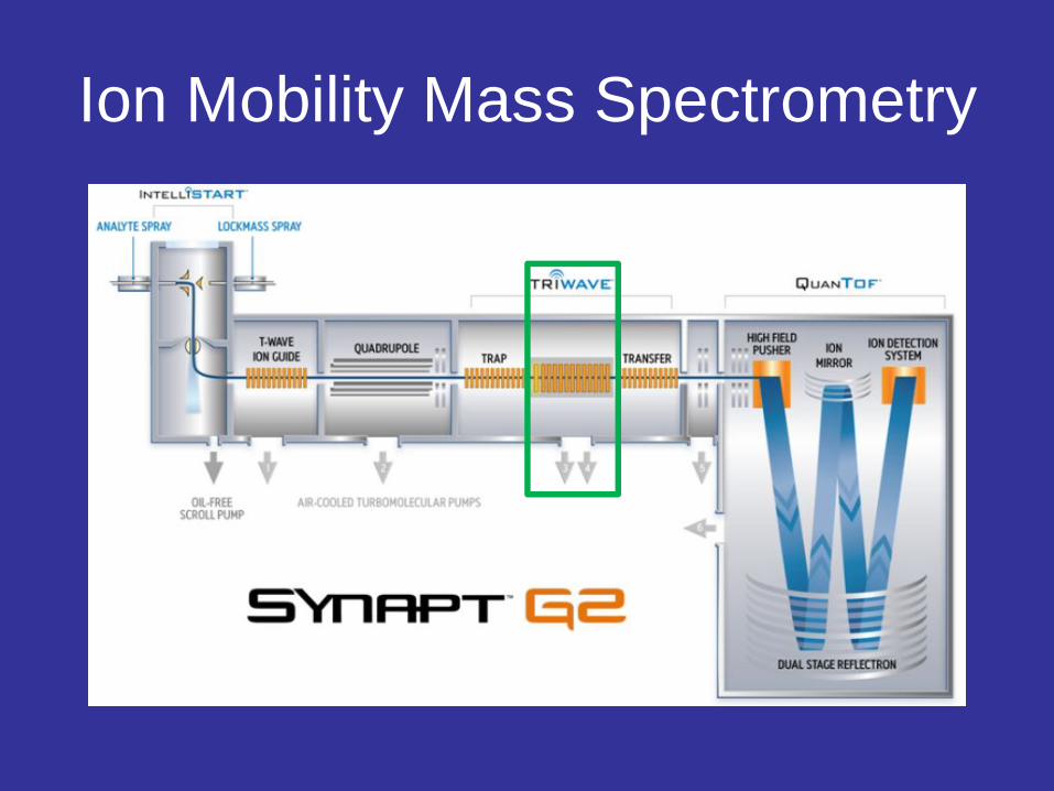
Ion Mobility Mass Spectrometry
Ion Mobility Mass Spectrometry
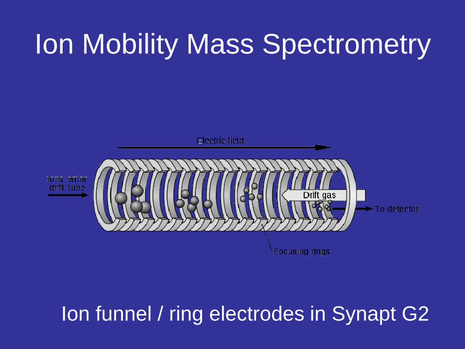
Ion funnel / ring electrodes in Synapt G2

Cross-sectional view of the ion mobility spectrometer sample inlet, ionization chamber, drift tube, and detector.
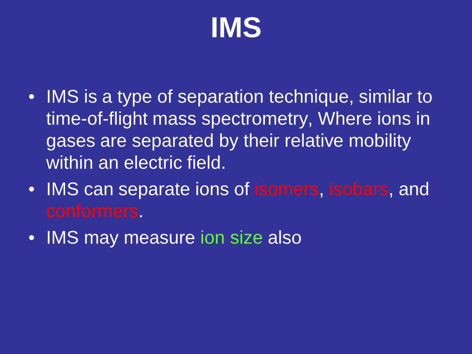
IMS
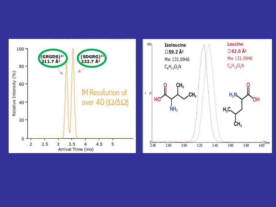
• IMS is a type of separation technique, similar to time-of-flight mass spectrometry, Where ions in gases are separated by their relative mobility within an electric field.
• IMS can separate ions of isomers, isobars, and conformers.
• IMS may measure ion size also
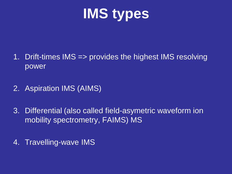
1. Drift-times IMS => provides the highest IMS resolving power
2. Aspiration IMS (AIMS)
3. Differential (also called field-asymetric waveform ion mobility spectrometry, FAIMS) MS
4. Travelling-wave IMS
IMS types


• Eg. PeptideKLFTSY MH+ 757.42 Da and
• Bradykinin peptideRPPGFSP MH+ 757.40 Da
• These 2 peptides are of the same size, but the structures are different.
• ION MOBILITY can separate them from each other
16:0
16:0
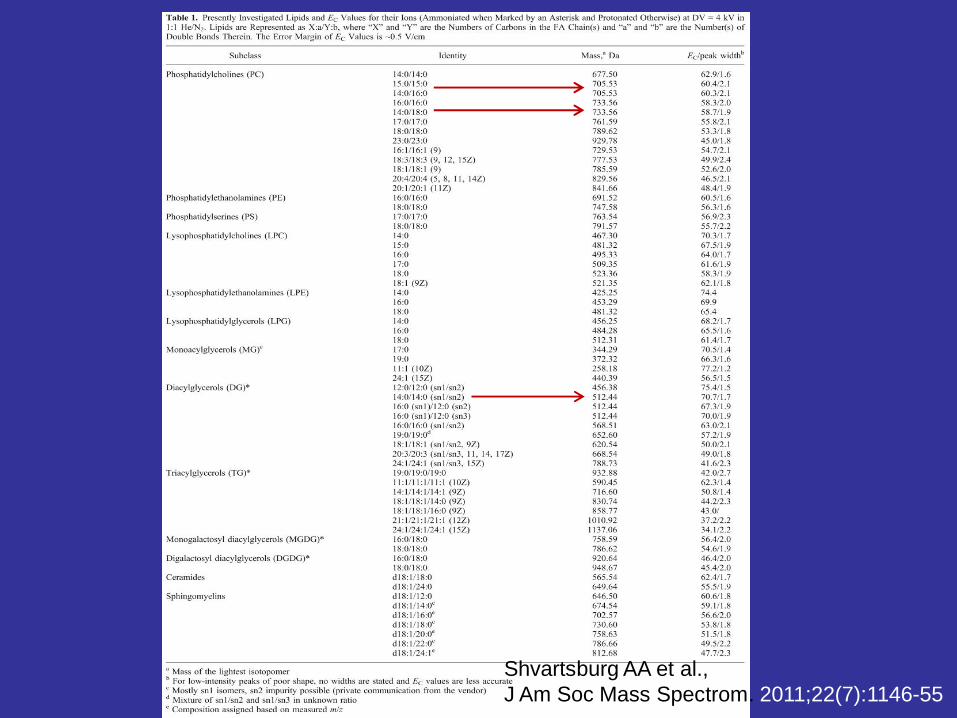
Shvartsburg AA et al., J Am Soc Mass Spectrom. 2011;22(7):1146-55

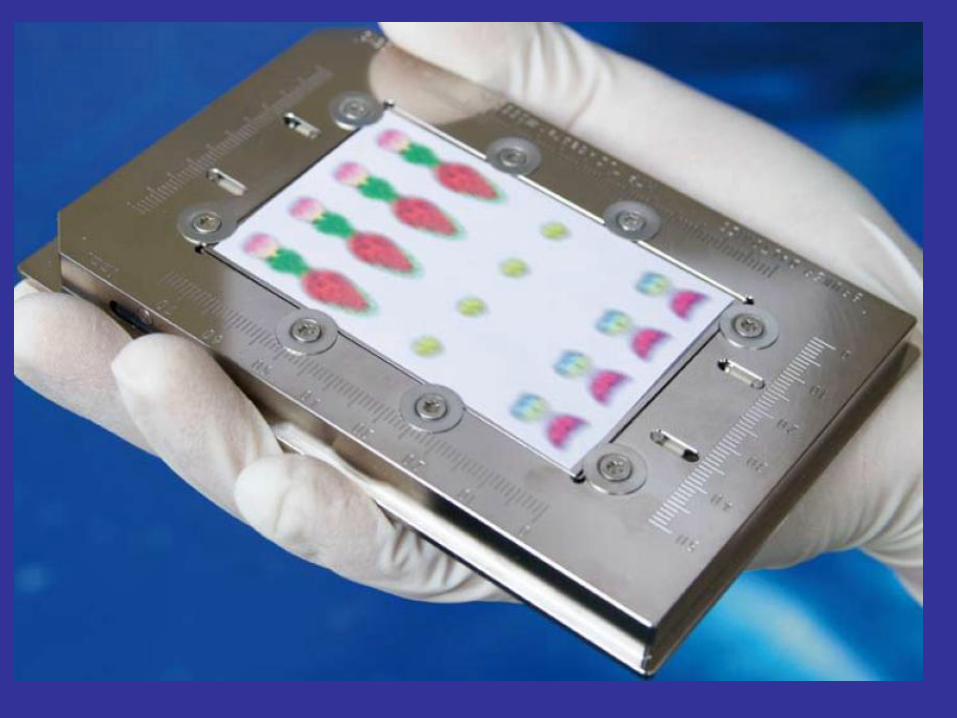
MALDI Imaging (MALDI-MSI)
• TLC MALDI
• MALDI Imaging (MALDI-MSI)– Application: Tissue imaging
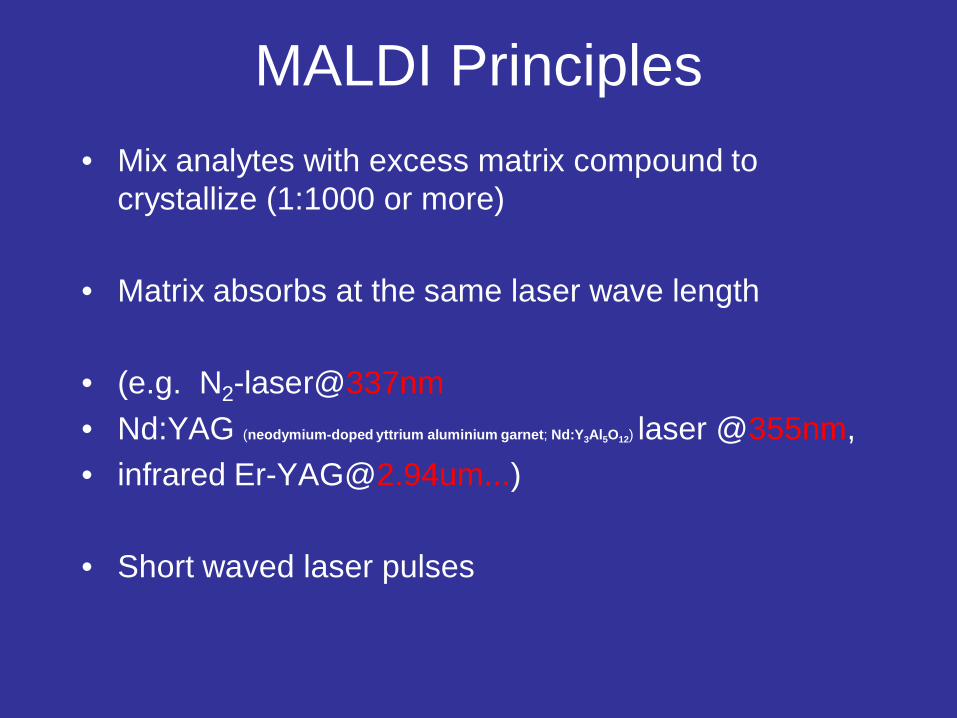
MALDI Principles• Mix analytes with excess matrix compound to
crystallize (1:1000 or more)
• Matrix absorbs at the same laser wave length
• (e.g. N2-laser@337nm• Nd:YAG (neodymium-doped yttrium aluminium garnet; Nd:Y3Al5O12) laser @355nm,• infrared [email protected]...)
• Short waved laser pulses
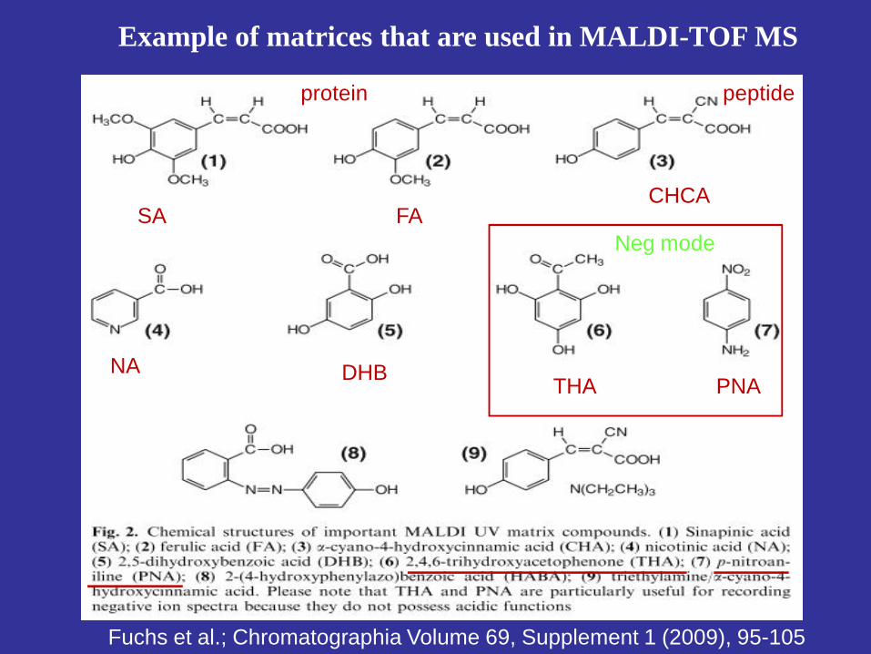
SA FACHCA
NA DHB THA PNA
protein peptide
Neg mode
Example of matrices that are used in MALDI-TOF MS
Fuchs et al.; Chromatographia Volume 69, Supplement 1 (2009), 95-105


Gas phase
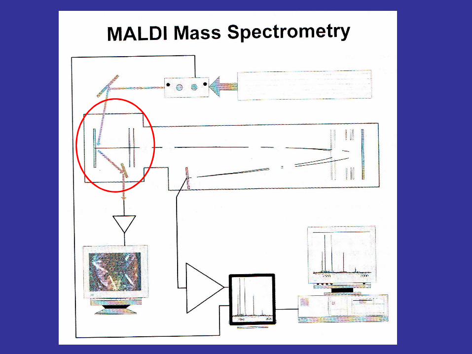
Principles of MALDI process
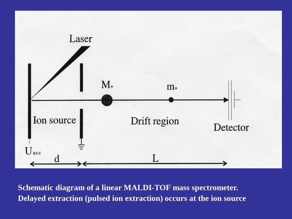
Schematic diagram of a linear MALDI-TOF mass spectrometer.Delayed extraction (pulsed ion extraction) occurs at the ion source

Schematic diagram of a Reflector MALDI-TOF mass spectrometer.MALDI-TOF = Matrix Assisted Laser Desorption/Ionization - Time Of Flight
N2 , 337nm
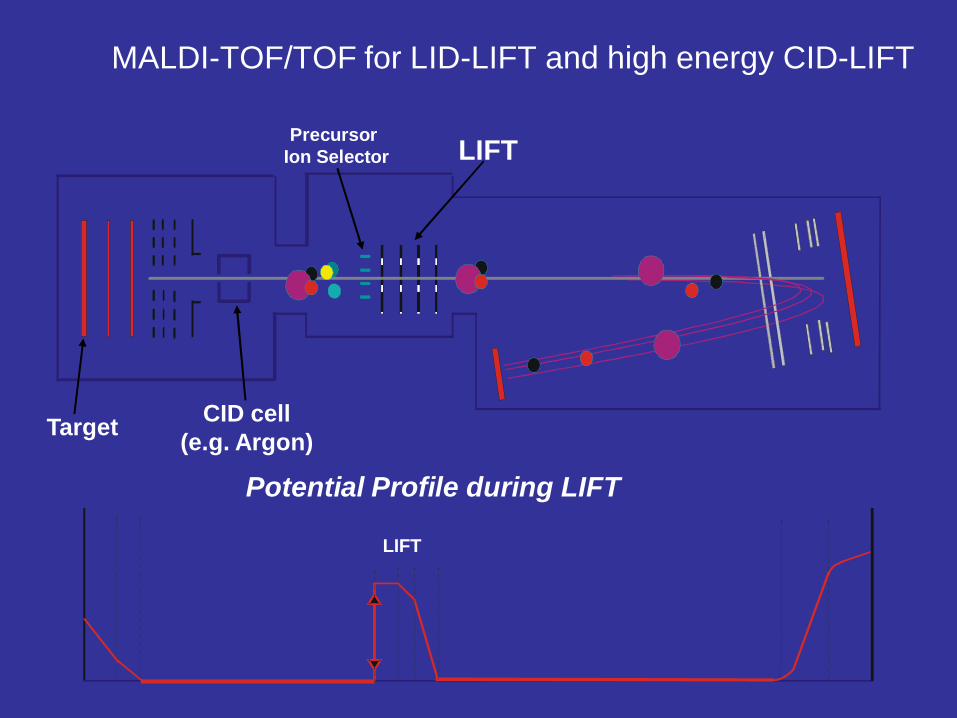
MALDI-TOF/TOF for LID-LIFT and high energy CID-LIFT
Target
LIFTPrecursor Ion Selector
CID cell(e.g. Argon)
Potential Profile during LIFT
LIFT

1. TLC separation (nmol level)2. Staining with primuline3. Densitometry4. Spot Marking5. Matrix solution (eg. DHB)6. Mount and acquire spectra
primuline

Fuchs et al.; Chromatographia Volume 69, Supplement 1 (2009), 95-105
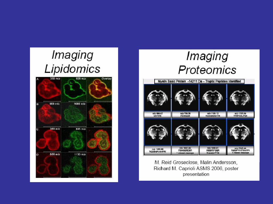
Definitions:MALDI Imaging (MALDI-IMS)
• allows spatial localization of multiple different compounds that are recorded in parallel without the need of a label
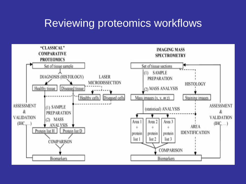
Reviewing proteomics workflows
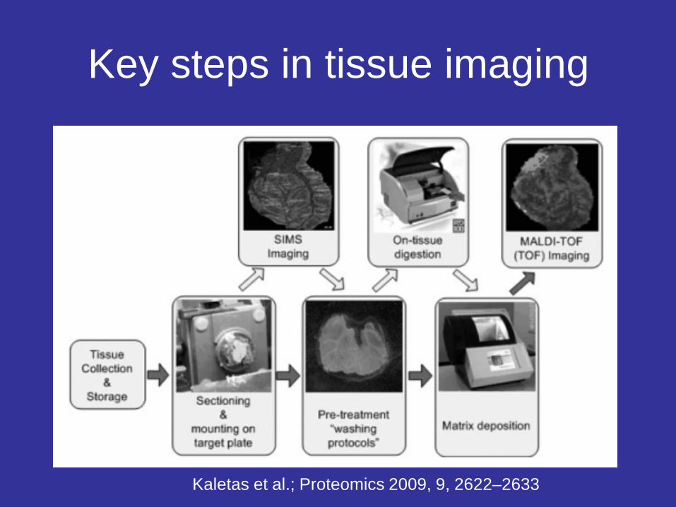
Key steps in tissue imaging
Kaletas et al.; Proteomics 2009, 9, 2622–2633

Tissue section (mouse brain)
2,000 15,000 30,000
Ion
inte
nsity
Acquisition x
Acq
uisi
tion
y
m/z
12mm
Principles
• A laser is rastered over a defined area while acquiring a complete mass spectrum from each position, resulting in molecular images for multiple analytes Cornett, et al., Nature Methods 2007

Matrix Application
• Matrix application is vital for quality image resolution
• Must contact sample as fine, liquid mist
• Current procedure involves manual application with airbrush 100µm raster step
Barrett-Wilt, G., USA
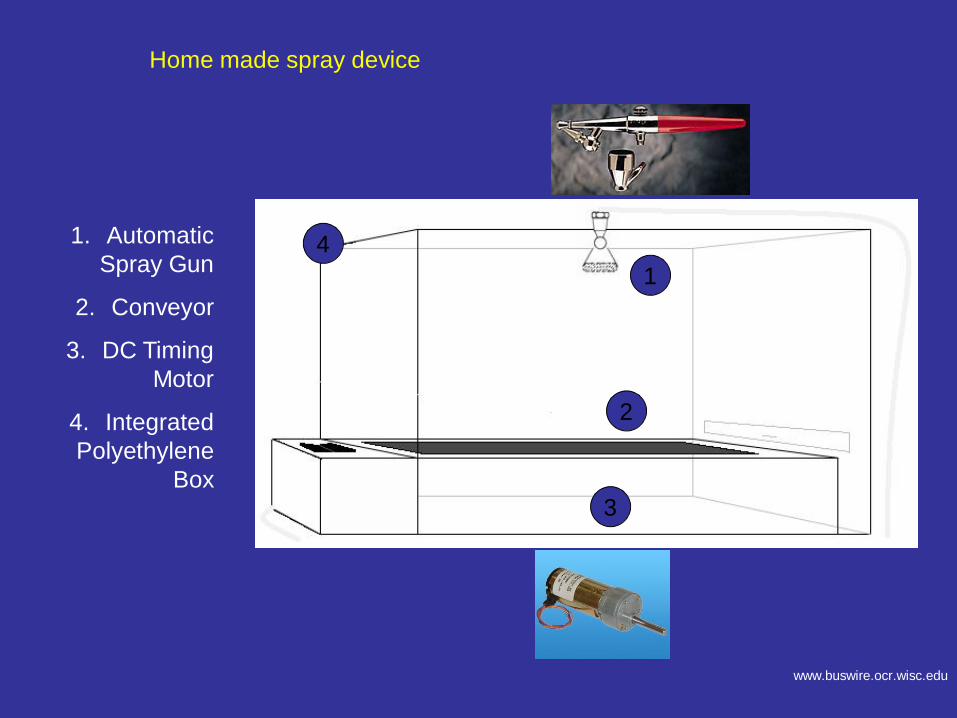
1. Automatic Spray Gun
2. Conveyor
3. DC Timing Motor
4. Integrated Polyethylene
Box
14
3
2
Home made spray device
www.buswire.ocr.wisc.edu
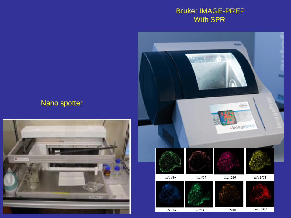
Bruker IMAGE-PREPWith SPR
Nano spotter

Sublimation vs dry coating

MSI workflow
Stoeckli, M, et al., Nature Medicine 2001; vol 7, 4, p493-496

Consecutive tissue sections
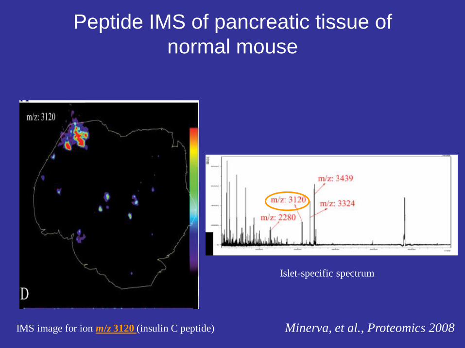
Peptide IMS of pancreatic tissue of normal mouse
IMS image for ion m/z 3120 (insulin C peptide)
Islet-specific spectrum
Minerva, et al., Proteomics 2008

IMS vs. histochemical stain
IMS image for ion m/z 3120 (insulin C peptide) Overlay image of ion m/z 3120 and Methylene blue stained of the pancreas
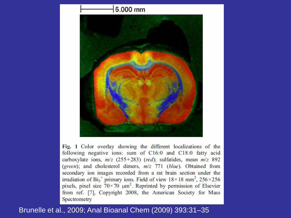
Brunelle et al., 2009; Anal Bioanal Chem (2009) 393:31–35

Chaurand et al., 2011; Molecular & Cellular Proteomics 10.2: p 1-11

Comparing Regions of interest
Control vs. patient

2
Application
.
Ifa et al.; Science. 321(5890):805,
Fig. 1 . (A) DESI image of distribution of cocaine on a LFP blotted on glass. (B) Computer-generated fingerprint from DESI image. (C) Ink fingerprint blotted on paper and optically scanned. (D) Computer-generated fingerprint from optical image. Some of the automatically detected points of interest (minutiae) are represented by dots in (B) and (D).

Special applications
• Immuno-MS-IMS
• Microchips on IMS

Thiery, G., et al., Proteomics 2008
Imaging of regions immunoreactive with anti-synaptophysin Ab in healthy human pancreas.
(A) Localization of synaptophysin positive cells by TAMSIM. The monoclonal rabbit anti-synaptophysin is conjugated with the tag El 307 (498 m/z). The false color green points in the section show the presence of the tag El 307 and thus synaptophysin positive cells.
(B) Classical IHC image with the anti-insulin Ab. The dark pink spots correspond to Langerhans islets and so the synaptophysin-positive cells. The distribution of synaptophysin positive cells in (A) is very similar to that in (B).
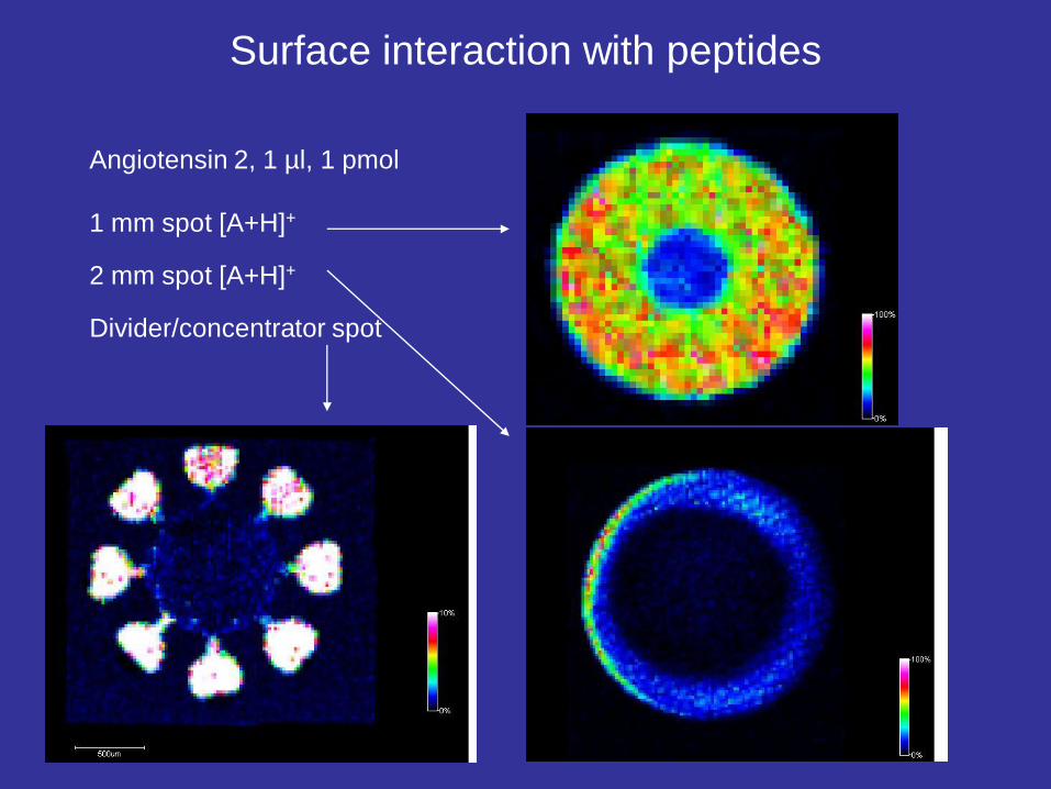
Surface interaction with peptides
Angiotensin 2, 1 µl, 1 pmol
1 mm spot [A+H]+
2 mm spot [A+H]+
Divider/concentrator spot
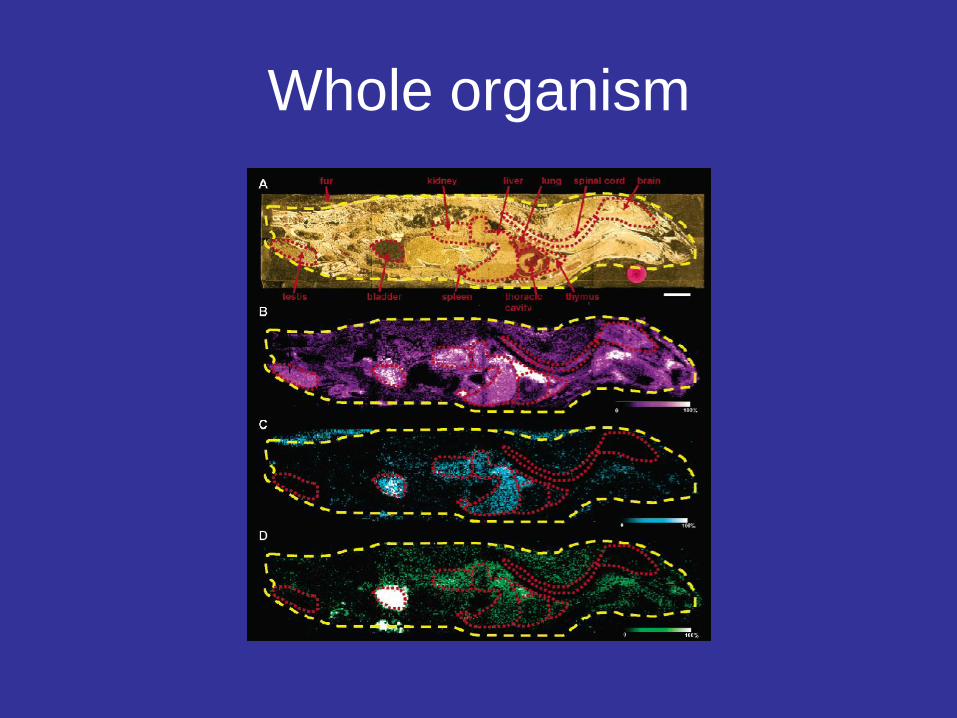
Whole organism

Advantages of MALDI-IMS• Analysis of entire tissue/organ/whole organism section at once
• Prior detailed knowledge of tissue molecular composition is not necessary
• Comparison of disease versus healthy may be possible
• Biomolecules are in native state (no modification / no label applied)
• Imaging of biomolecular modifications PTM’s, Metabolites is possible
• Detailed information on molecular identity
• Large scope of different elements and molecules
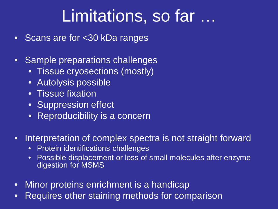
Limitations, so far …• Scans are for <30 kDa ranges
• Sample preparations challenges• Tissue cryosections (mostly)• Autolysis possible• Tissue fixation• Suppression effect• Reproducibility is a concern
• Interpretation of complex spectra is not straight forward• Protein identifications challenges• Possible displacement or loss of small molecules after enzyme
digestion for MSMS
• Minor proteins enrichment is a handicap• Requires other staining methods for comparison

“It is always wise to look ahead, but difficult to look further than you can see.” (Winston Churchill)

• Thank you for your attention
• Questions / comments