biological mass spectrometry in protein...
TRANSCRIPT

1
Tuula NymanInstitute of [email protected]
Biological Mass spectrometryin Protein Chemistry
ION SOURCE: molecules of interest are ionized
MASS ANALYZER:ions are separated according to their m/z-ratios
DETECTOR: separated ions are detected
MASS SPECTROMETRYis an analytical technique that identifies thechemical composition of a compound or samplebased on the mass-to-charge ratio of chargedparticles

2
MS of small molecules
-In 1918, Arthur Jeffrey Dempster developed the first modernmass spectrometer, and established the basic theory anddesign of mass spectrometers that is still used to this day
-1919 Francis Aston constructs the first velocity focusing massspectrograph with mass resolving power of 130 (1922 NobelPrize in chemistry)
-The use of a mass spectrometer as the detector in gaschromatography was developed during the 1950s by RolandGohlke and Fred McLafferty
-Ionization modes: chemical ionization (CI) and electronionization (EI), not suitable for labile biomolecules
Biological mass spectrometry
-two modes of ionization:MALDI (matrix assisted laser desorption ionization)and ESI (electrospray ionization)
-developed in 1980’s by Michael Karas & FranzHillenkamp, Koichi Tanaka, John Fenn & MatthiasMann, Peter Roespstorff…
Nobel-price in Chemistry 2002 to Tanaka and Fenn’For their development of soft desorption ionisationmethods for mass spectrometric analyses ofbiological macromolecules’

3
TAR
GE
TPULSEDLASER
+
++
+ +
++
+20 kV
MALDI= matrix assisted laser desorption ionization
The analyte substance is embedded in acrystallized matrix, which is irradiated by alaser. The power of the laser beam is usuallyadjusted in a way that it has enough energy toionize the biomolecules and matrix moleculesbut does not split the large analyte molecule.
A 384 position MALDI-TOFsample target plate.
Commonly used matrixes for MALDI-TOF mass spectrometry
================peptides
===================proteins

4
Electrospray ionization (ESI):Creation of ions by spraying a solution into an electrical field.This process, which belongs to the "soft" ionizationtechniques, enables the analysis of intact biomolecules, suchas e.g. proteins and peptides by mass spectrometry.
Electrospray of peptides in 0.1% FA/ACN from a 15 um I.D. fused silicaglass needle. The liquid flow is 200nl/min and the needle has a potentialof 2000V as compared to the coneinlet of the mass spectrometer.
Mass analyser types
+orbitrap
All mass analyzers work in high vacuum

5
Common MS-instrument types:-MALDI-TOF and MALDI-TOF/TOF-ESI-triple quadrupole-ESI-ion trap-ESI-hybrid quadrupole TOF-ESI-ion trap-ESI-orbitrap
A tandem mass spectrometer is a mass spectrometerthat has more than one analyser, in practice usually two.
Tandem mass spectrometry (MS/MS) is used toproduce structural information about a compound byfragmenting specific sample ions inside the massspectrometer and identifying the resulting fragment ions
MALDI TOF spectrum of a peptide mixture
1834.022
1215.597
1969.958
1461.748 2398.323
2335.083
1110.626
952.4722203.004
1269.549
1367.765 2145.0282026.977842.548 1891.0411574.915
0.0
0.5
1.0
1.5
4x10
Inte
ns.[
a.u.
]
800 1000 1200 1400 1600 1800 2000 2200 2400m/z
1 215.597
1216.5 92
1 217.600
0.0
0.2
0.4
0.6
0.8
1.0
4x 10
Inte
ns.[
a.u.
]
1214 1215 1216 1217 1218 1219m /z
2 399.312
2 398.323
2400.33 2
0
1000
2000
3000
4000
Inte
ns.[
a.u.
]
2398 2399 2400 2401 2402m /z
1215.597
1216.592
1217.600
2398.323
2399.312
2400.332

6
Monoisotopic mass is calculated using themass of the most abundant naturalisotope of each constituent element
1215.597
1216.592
1217.600
0.0
0.2
0.4
0.6
0.8
1.0
4x10
Inte
ns.
[a.u
.]
121 4 1 215 1216 12 17 12 18 1219m /z
1215.597
1216.592
1217.600
12C and 14N
the mass difference between the isotopic peaks is1 amu (1 Da) as isotopes differ in mass by theaddition of 1 neutron which weighs 1 amu
Singly charged ion
Doubly charged ion
MALDI-TOF spectrum of a protein

7
739.844
740.347
m/z difference 0.5 Da= doubly charged ion
ESI MS spectrum of a peptide mixture
+20+19
+18
+17
+21+22
+16
+15
+14
+23
+24
+25
p1
p2
-Protein is observed as aseries of different charges-Protein MW can be calculatedbased on adjacent m/z-values
ESI MS spectrum of a protein
8
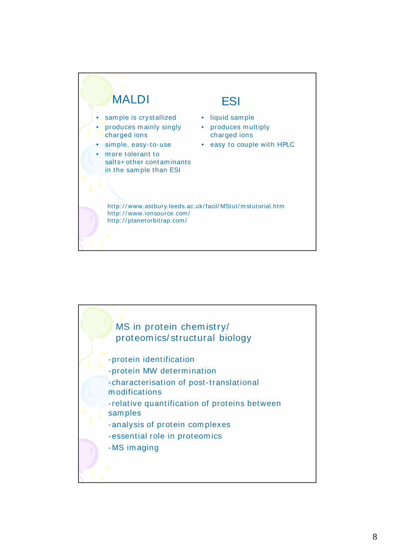
• sample is crystallized• produces mainly singly
charged ions• simple, easy-to-use• more tolerant to
salts+other contaminantsin the sample than ESI
• liquid sample• produces multiply
charged ions• easy to couple with HPLC
MALDI ESI
http://www.astbury.leeds.ac.uk/facil/MStut/mstutorial.htmhttp://www.ionsource.com/http://planetorbitrap.com/
MS in protein chemistry/proteomics/structural biology
-protein identification-protein MW determination-characterisation of post-translationalmodifications-relative quantification of proteins betweensamples-analysis of protein complexes-essential role in proteomics-MS imaging

9
Protein MW determination by MS(NOT= identification)
-for MW determination the protein needs to bein solution without salts and detergents
-usually proteins are first purified withRP chromatography before MW measurement
-MALDI TOF MS, linear mode-accuracy is not as good with ESI MS
Singly charged ion
Doubly charged ion

10
+20+19
+18
+17
+21+22
+16
+15
+14
+23
+24
+25
Protein MW determination, ESI MS
p1
p2
-Protein is observed as aseries of different charges-Protein MW can be calculatedbased on adjacent m/z-values
p=m/zp1=(Mr+z1)/z1p2=[Mr+(z1-1)]/(z1-1)
p= a peak in the mass spectrumm= total mass of an ionz= total chargeMr= average mass of the protein
Protein MW calculation from ESI spectra

11
If p1=942.753 and p2=998.067
p=m/zp1=(Mr+z1)/z1p2=[Mr+(z1-1)]/(z1-1)
942.753 =(Mr+z1)/z1942.753z1 = Mr+z1941.753z1 = Mr
998.067 = (941.753z1+z1-1)/z1-1998.067z1-997.067 = 942.753z1-1(998.067-942.753)z1 = 996.06755.314z1= 996.067 => z1=18.0075
Mr = 941.753z1 = 16 951.6 Da
Deconvoluted electrospray mass spectrum of myoglobin

12
Protein identification methods:•Peptide mass fingerprinting (PMF)•Identification based on MS/MS data from oneor more peptides
Protein identification bymass spectrometry
•protein of interest is cleaved into peptideswith a specific enzyme•peptides are analyzed by MS (and MS/MS)followed by a database search with theacquired MS (and MS/MS) data
A mass spectrum of the peptide mixture resultingfrom the digestion of a protein by an enzyme,usually measured by MALDI-TOF
Identification based on peptide MW information only
Database search engines create theoretical PMFs forall the proteins in the database and compare these tothe measured PMF from protein X
Peptide mass fingerprint (PMF)

13
Protein identification based on MS/MS datafrom one or more peptides
Database search engines create theoreticalpeptide fragmentation patterns for all theproteins in the database and compare theseto the measured MS/MS data
SS
N
C
N
CAlkylation
K
R
KK
K
R
KRTrypsin
intact protein
Reduction
alkylated protein tryptic fragments from the protein
Protein identification by MS
•Protein has to be digested into peptides•Disulphide bridges need to be reduced and alkylatedbefore digestion

14
Protein digestion into peptidesbefore MS analysis
Digestion can be done in-solution or in-gelThe enzyme has to be as specific as possibleTrypsin is most commonly used enzyme:
-very specific, quite cheap-cleaves peptide bond after lysines and arginines-tryptic peptides are ’good’ for MS analysisbecause they end up with basic amino acid-works for both in-solution and in-gel digestion
Other enzymes:-LysC, cleaves after lysines-LysN, cleaves before lysines-AspN, cleaves before aspartic acid residues-V8 protease, cleaves peptide bondsexclusively on the carbonyl side of aspartateand glutamate residues-possible to do double-digestions
Chemical cleavage:Cyanogen bromide, cuts after methionines

15
In-gel digestion
-works with low-femtomolar amounts of proteins
BUT:-easy to contaminate samples with keratin-the gel staining protocol needs to becompatible with MS-silver-staining: fixing with glutaraldehy cross-linksprotein into gel matrix after which identificationis not possible
Peptide mass fingerprinting
-usually peptides need to be desalted andconcentrated before MALDI analysis-> ZipTips, peptide elution directly onto MALDItarget plate-MALDI matrixincluded in theelution solution

16
Peptide mass fingerprintingMALDI, Positive ion reflector mode
-silver-stained spots from 2-DE gels, in-gel digestion+ZipTipping
1590.760
1574.775
1434.642
1606.761
1475.7281450.637
1657.772
0
1
2
3
4
5
4x10
Inte
ns.[
a.u.
]
1425 1450 1475 1500 1525 1550 1575 1600 1625 1650m /z
1574.775
0
1
2
3
4
5
4x10
Inte
ns.[
a.u.
]
1568 1570 1572 1574 1576 1578 1580 1582m /z
Peak picking parameters important in PMF
Label monoisotopic massesCheck for correct isotope!

17
Databases
Publicly available search engines for PMF
•Mascot•ProteinProspector/ MS-Fit•PROWL/ ProFound•Aldente
PMF/ database search
www.matrixscience.com

18

19
-check for known contaminants(these should be removed before search)-possibility to do 2nd pass search
Mass accuracy critical
Usually not manymissed cleavages

20
’Normal’ search,max 1missed cleavageallowed
Max 4 missed cleavages allowed
PEPTIDE MASS FINGERPRINTING
• ’quick and easy’• requires
– a very spesific enzyme– optimized digestion+ desalting protocols– internal/close external calibration of MALDI
spectra• works only for proteins which are already
in the databases as protein sequences• not suitable for complex protein mixtures

21
Protein identification based on MS/MS datafrom one or more peptides
-MALDI-TOF/TOF or nanoLC-ESI-MS/MS analysis-suitable for (complex) protein mixtures,especially when combined with LC separationof peptides before MS/MS
Tandem mass spectrometry scan types

22
Product ion scanning
C H I L PLH
IIL
Inte
nsity
,cou
nts
m/z, amu
TOF product 745,4
MS scan:
MS/MS scan:Peptides with certain m/z-ratio areselected and fragmented inside the
mass analyzer, and the m/z-ratios of thefragment ions are measured
The ions are first separatedaccording to their m/z ratios
Peptide Fragmentation Nomenclature
Peptides do not fragment sequentially, the fragmentationevents are somewhat random.
The most common peptide fragments observed in lowenergy collisions are a, b and y ions. The b ions appear toextend from the amino terminus (N-terminus), and y ionsappear to extend from the carboxyl terminus (C-terminus).

23
Protein identification with MALDI-TOF/TOF
-first, PMF with MALDI-TOF-next, selected precursor ions can be fragmented(TOF/TOF analysis)-MALDI produces singly charged parent ions
product ion spectra not as easy to interpretas in ESI-MS/MS-database search with both PMF and MS/MSinformation
874.168
1060.282340.943
1953.011243.966
479.932111.963
664.960
1825.969
972.188746.0871613.623
1158.227 1448.409
194.917
0.00
0.25
0.50
0.75
1.00
1.25
5x10
Inte
ns.[
a.u.
]
250 500 750 1000 1250 1500 1750 2000 2250m /z
1060.282
0.0
0.2
0.4
0.6
0.8
1.0
1.2
5x10
Inte
ns.[
a.u.
]
1040 1050 1060 1070 1080 1090m /z
MALDI-TOF/TOF fragment ion spectrafrom parent ion m/z 1952

24
Sequence Query: One or more peptide mass values associatedwith information such as partial or ambiguous sequence strings,amino acid composition information, MS/MS fragment ion masses, etc.

25
Protein identification using nanoLC-MS/MS
-50-75 um i.d. RP-columns, 200 nl/minno need to split the effluent before MS
-DDA= data dependent analysis, can be fullyautomated-suitable for complex protein mixtures,possibility to identify hundreds of proteins inone run
DDA =Data Dependent Acquisition(IDA =Information Dependent Acquisition)
-fully automated experiment, first MS scanfollowed by two or more product ion scans-the acquisition software is set to choosecertain types of ions for fragmentation andto use ’suitable’ collision energy for this ion-in ESI tryptic peptides have usually 2-4 charges

26
DDA experiment
Exp 1 = TOF MSExp 2 = Product ion scan from parent IExp 3 = Product ion scan from parent II
TIC = total ion current

27
Exp 1 = TOF MS
Exp 2 = Product ion scan I
Exp 3 = Product ion scan II
Search engines for (LC-)MS/MS data
-Mascot, Sequest, OMSSA etc-the programs take the fragment ion spectrum ofa peptide as input and score it against theoreticalfragmentation patterns constructed for peptidesfrom the searched database.
-in practise the user is often limited to usethose search engines which accept thedata format from the mass spec used
-mzXML is a open data format for storage andexchange of mass spec data-raw, proprietary file formats from most vendorscan be converted to the open mzXML format

28
Ion scores from individualMS/MS spectra

29
-produces huge amounts of raw data-requires efficient data processing tools-different database search programs can producedifferent results from the same raw data-false discovery rate estimation
-peptide level-protein level
Protein identification from complex mixturesusing nanoLC-MS/MS
Protein fragmentation-protein separation by SDS-PAGE/2-DEfollowed by in-gel digestion and MS analysis
Now you’re protein is identifiedWhat else can we find in the data?

30
Cytokeratin-18 is cleaved during viral infection,and fragments localize onto mitochondria
Mitochondrial protemes
Based on 2-DE and MS data:N-terminal fragment of theidentified protein

31
TAP-tag purification of protein complexes,on-beads digestion with double enzyme,first LysC 2h, then trypsin o/n
Post-translational modifications

32
KIN2 phosphopeptide

33
-if enough material is available classical Edmandegradation is still a vey good method-partial peptide sequencing is possible basedon MS/MS data
De novo =peptide sequencing performed withoutprior knowledge of the amino acid sequence
De novo sequencing

34
Publishing protein ID data
-most biological journals do not have anyguidelines yet
Mat+Met:-MS instrument, identification type-data acquisition+processing software-database search engine
Results: Table from ID results
Mol Cell Proteomicswww.mcponline.org

35
In the experimental section:

36

37

38

39
http://www.ebi.ac.uk/pride/archive/