manual pmal protein fusion and purification system … or manuals... · instruction manual pmal ™...
TRANSCRIPT
Instruction Manual
pMAL™ Protein Fusion & Purification System
PROTEIN EXPRESSION & ANALYSIS
NEB #E8200S1 set Version 5.0 5/18
be INSPIRED drive DISCOVERY stay GENUINE

This product is intended for research purposes only. This product is not intended to be used for therapeutic or diagnostic purposes in humans or animals.
ISO 9001Registered
QualityManagement
ISO 14001Registered
EnvironmentalManagement
ISO 13485Registered
Medical Devices
This product is covered by one or more patents, trademarks and/or copyrights owned or controlled by New England Biolabs, Inc. For more information about commercial rights, please email us at [email protected]. While NEB develops and validates its products for various applications, the use of this product may require the buyer to obtain additional third party intellectual property rights for certain applications.
© Copyright 2018, New England Biolabs, Inc.; all rights reserved.
1
Table of Contents:Kit Components . . . . . . . . . . . . . . . . . . . . . . . . . . . . . . . . . . . . . . . . . . . . . . . . . . . . . . . . . . . . . . . . . . . . . . . 2
Quick Start Guide . . . . . . . . . . . . . . . . . . . . . . . . . . . . . . . . . . . . . . . . . . . . . . . . . . . . . . . . . . . . . . . . . . . . . 3
Introduction. . . . . . . . . . . . . . . . . . . . . . . . . . . . . . . . . . . . . . . . . . . . . . . . . . . . . . . . . . . . . . . . . . . . . . . . . .4-5
Construction of the Fusion Plasmid. . . . . . . . . . . . . . . . . . . . . . . . . . . . . . . . . . . . . . . . . . . . . . .5-7 Choice of Vector . . . . . . . . . . . . . . . . . . . . . . . . . . . . . . . . . . . . . . . . . . . . . . . . . . . . . . . . . . . . . . . . . . 6
Subcloning Strategy. . . . . . . . . . . . . . . . . . . . . . . . . . . . . . . . . . . . . . . . . . . . . . . . . . . . . . . . . . . . . . 6
Creating a Blunt-ended PCR fragment . . . . . . . . . . . . . . . . . . . . . . . . . . . . . . . . . . . . . . . . . 6
Cloning a PCR Fragment . . . . . . . . . . . . . . . . . . . . . . . . . . . . . . . . . . . . . . . . . . . . . . . . . . . . . . . . . 7
Affinity Chromatography . . . . . . . . . . . . . . . . . . . . . . . . . . . . . . . . . . . . . . . . . . . . . . . . . . . . . . . . . 9-11 Method I: Fusion Protein from Total Cell Extract . . . . . . . . . . . . . . . . . . . . . . . . . . 9-10
Method II: Exported Fusion Protein . . . . . . . . . . . . . . . . . . . . . . . . . . . . . . . . . . . . . . . . . . .11
Regenerating the Amylose Resin Column . . . . . . . . . . . . . . . . . . . . . . . . . . . . . . . . . . . .11
Cleavage of the Fusion Protein . . . . . . . . . . . . . . . . . . . . . . . . . . . . . . . . . . . . . . . . . . . . . . . . . . . . .12 Denaturing the Fusion Protein . . . . . . . . . . . . . . . . . . . . . . . . . . . . . . . . . . . . . . . . . . . . . . . . .12
Separating the Protein of Interest from MBP after Protease Cleavage. . . . .13-14 Method I: Anion exchange chromatography. . . . . . . . . . . . . . . . . . . . . . . . . . . . . . . . . .13
Method II: Anion exchange chromatography variation . . . . . . . . . . . . . . . . . .13-14
Pilot Experiment. . . . . . . . . . . . . . . . . . . . . . . . . . . . . . . . . . . . . . . . . . . . . . . . . . . . . . . . . . . . . . . . . .14-16
Appendices A. Media and Solutions . . . . . . . . . . . . . . . . . . . . . . . . . . . . . . . . . . . . . . . . . . . . . . . . . . . . .16-17
B. Details of the pMAL vectors . . . . . . . . . . . . . . . . . . . . . . . . . . . . . . . . . . . . . . . . . . . . .17-18
C. Western Protocol . . . . . . . . . . . . . . . . . . . . . . . . . . . . . . . . . . . . . . . . . . . . . . . . . . . . . . . . . . . . .18
D. Troubleshooting and Tips . . . . . . . . . . . . . . . . . . . . . . . . . . . . . . . . . . . . . . . . . . . . . . .19-28
E. Protein Fusion and Purification Strain List . . . . . . . . . . . . . . . . . . . . . . . . . . . .29-30
Molecular Weights of pMAL Proteins. . . . . . . . . . . . . . . . . . . . . . . . . . . . . . . . . . . . . . . . . . . . . .31
References . . . . . . . . . . . . . . . . . . . . . . . . . . . . . . . . . . . . . . . . . . . . . . . . . . . . . . . . . . . . . . . . . . . . . . . .31-32
Ordering Information . . . . . . . . . . . . . . . . . . . . . . . . . . . . . . . . . . . . . . . . . . . . . . . . . . . . . . . . . . . . . . . .33
pMAL Protein Fusion & Purification System
2
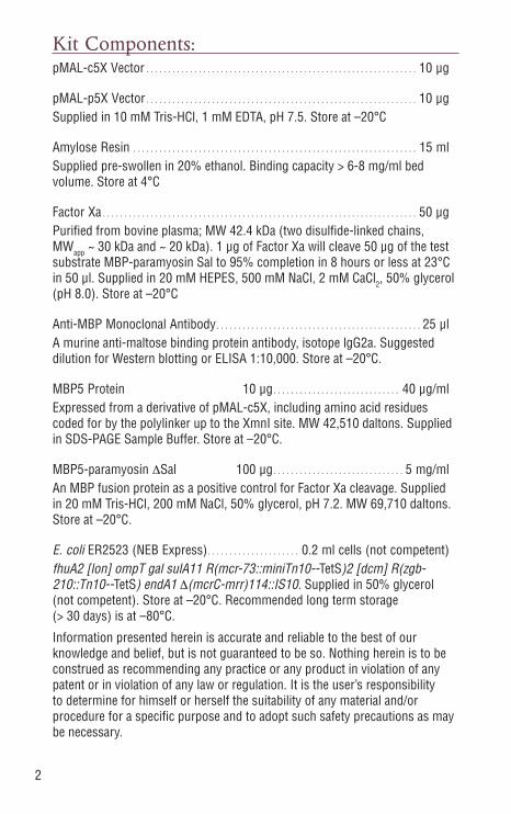
Kit Components:pMAL-c5X Vector . . . . . . . . . . . . . . . . . . . . . . . . . . . . . . . . . . . . . . . . . . . . . . . . . . . . . . . . . . . . . . 10 µg
pMAL-p5X Vector . . . . . . . . . . . . . . . . . . . . . . . . . . . . . . . . . . . . . . . . . . . . . . . . . . . . . . . . . . . . . . 10 µgSupplied in 10 mM Tris-HCl, 1 mM EDTA, pH 7.5. Store at –20°C
Amylose Resin . . . . . . . . . . . . . . . . . . . . . . . . . . . . . . . . . . . . . . . . . . . . . . . . . . . . . . . . . . . . . . . . . 15 mlSupplied pre-swollen in 20% ethanol. Binding capacity > 6-8 mg/ml bed volume. Store at 4°C
Factor Xa . . . . . . . . . . . . . . . . . . . . . . . . . . . . . . . . . . . . . . . . . . . . . . . . . . . . . . . . . . . . . . . . . . . . . . . . 50 µgPurified from bovine plasma; MW 42.4 kDa (two disulfide-linked chains, MWapp ~ 30 kDa and ~ 20 kDa). 1 µg of Factor Xa will cleave 50 µg of the test substrate MBP-paramyosin Sal to 95% completion in 8 hours or less at 23°C in 50 µl. Supplied in 20 mM HEPES, 500 mM NaCl, 2 mM CaCl2, 50% glycerol (pH 8.0). Store at –20°C
Anti-MBP Monoclonal Antibody. . . . . . . . . . . . . . . . . . . . . . . . . . . . . . . . . . . . . . . . . . . . . . . 25 µlA murine anti-maltose binding protein antibody, isotope lgG2a. Suggested dilution for Western blotting or ELISA 1:10,000. Store at –20°C.
MBP5 Protein 10 µg. . . . . . . . . . . . . . . . . . . . . . . . . . . . . 40 µg/mlExpressed from a derivative of pMAL-c5X, including amino acid residues coded for by the polylinker up to the XmnI site. MW 42,510 daltons. Supplied in SDS-PAGE Sample Buffer. Store at –20°C.
MBP5-paramyosin ΔSal 100 µg. . . . . . . . . . . . . . . . . . . . . . . . . . . . . . 5 mg/mlAn MBP fusion protein as a positive control for Factor Xa cleavage. Supplied in 20 mM Tris-HCl, 200 mM NaCl, 50% glycerol, pH 7.2. MW 69,710 daltons. Store at –20°C.
E. coli ER2523 (NEB Express). . . . . . . . . . . . . . . . . . . . . 0.2 ml cells (not competent)fhuA2 [lon] ompT gal sulA11 R(mcr-73::miniTn10--TetS)2 [dcm] R(zgb-210::Tn10--TetS) endA1 Δ(mcrC-mrr)114::IS10. Supplied in 50% glycerol (not competent). Store at –20°C. Recommended long term storage (> 30 days) is at –80°C.
Information presented herein is accurate and reliable to the best of our knowledge and belief, but is not guaranteed to be so. Nothing herein is to be construed as recommending any practice or any product in violation of any patent or in violation of any law or regulation. It is the user’s responsibility to determine for himself or herself the suitability of any material and/or procedure for a specific purpose and to adopt such safety precautions as may be necessary.
3
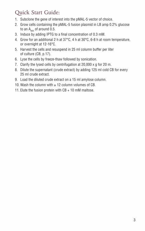
Quick Start Guide:1. Subclone the gene of interest into the pMAL-5 vector of choice.2. Grow cells containing the pMAL-5 fusion plasmid in LB amp 0.2% glucose
to an A600 of around 0.5.3. Induce by adding IPTG to a final concentration of 0.3 mM.4. Grow for an additional 2 h at 37°C, 4 h at 30°C, 6-8 h at room temperature,
or overnight at 12-16°C.5. Harvest the cells and resuspend in 25 ml column buffer per liter
of culture (CB, p.17).6. Lyse the cells by freeze-thaw followed by sonication. 7. Clarify the lysed cells by centrifugation at 20,000 x g for 20 m.8. Dilute the supernatant (crude extract) by adding 125 ml cold CB for every
25 ml crude extract.9. Load the diluted crude extract on a 15 ml amylose column.10. Wash the column with ≥ 12 column volumes of CB.11. Elute the fusion protein with CB + 10 mM maltose.
4
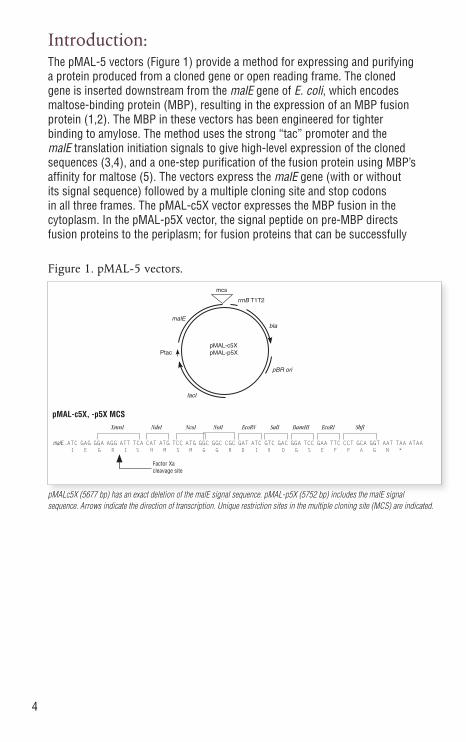
Introduction:The pMAL-5 vectors (Figure 1) provide a method for expressing and purifying a protein produced from a cloned gene or open reading frame. The cloned gene is inserted downstream from the malE gene of E. coli, which encodes maltose-binding protein (MBP), resulting in the expression of an MBP fusion protein (1,2). The MBP in these vectors has been engineered for tighter binding to amylose. The method uses the strong “tac” promoter and the malE translation initiation signals to give high-level expression of the cloned sequences (3,4), and a one-step purification of the fusion protein using MBP’s affinity for maltose (5). The vectors express the malE gene (with or without its signal sequence) followed by a multiple cloning site and stop codons in all three frames. The pMAL-c5X vector expresses the MBP fusion in the cytoplasm. In the pMAL-p5X vector, the signal peptide on pre-MBP directs fusion proteins to the periplasm; for fusion proteins that can be successfully
pMALc5X (5677 bp) has an exact deletion of the malE signal sequence. pMAL-p5X (5752 bp) includes the malE signal sequence. Arrows indicate the direction of transcription. Unique restriction sites in the multiple cloning site (MCS) are indicated.
Figure 1. pMAL-5 vectors.
malEbla
pBR ori
lacI
Ptac
rrnB T1T2
pMAL-c5X
mcs
pMAL-p5X
malE...ATC GAG GGA AGG ATT TCA CAT ATG TCC ATG GGC GGC CGC GAT ATC GTC GAC GGA TCC GAA TTC CCT GCA GGT AAT TAA ATAA I E G R I S H M S M G G R D I V D G S E F P A G N *
pMAL-c5X, -p5X MCS
Factor Xa cleavage site
XmnI NdeI NcoI NotI EcoRV SalI BamHI EcoRI SbfI
5
exported, this allows folding and disulfide bond formation to take place in the periplasm of E. coli, as well as allowing purification of the protein from the periplasm (6). The vectors carry the lacIq gene, which codes for the Lac repressor. This keeps expression from Ptac low in the absence of IPTG induction. The pMAL-5 vectors also include a sequence coding for the Factor Xa recognition site, located just 5´ to the polylinker insertion sites. This allows MBP to be cleaved from the protein of interest after purification (7,8). Factor Xa cleaves after its four amino acid recognition sequence, so that few or no vector-derived residues are attached to the protein of interest, depending on the site used for cloning.
In the large majority of cases, fusion protein expressed from pMAL-c5X constitutes 20–40% of the total cellular protein, while fusion protein expressed from pMAL-p5X constitutes 1-20% of the total cellular protein. For pMAL-c5X, a band corresponding to the fusion protein can usually be seen by running a small sample of induced cells on an SDS-PAGE gel. The yield of fusion protein from the affinity purification ranges up to 200 mg/liter culture, with typical yields in the range of 10–40 mg/liter. The yield varies greatly depending upon the sequences fused to malE. In E. coli K12 strains such as TBI, the pMAL-c5X (no signal sequence) gives approximately 10-fold more protein in the affinity purification than pMAL-p5X; in E. coli B strains such as NEB Express, the difference can be much less. The pMAL-p5X vector is useful for cases where export to the periplasm is desirable, e.g. for purification or disulfide bond formation. Greater than 75% of the fusions made so far have worked in the affinity purification. In the cases that have not worked, the fusion binds to the column poorly or not at all, is degraded by E. coli proteases, or is insoluble.
Construction of the Fusion Plasmid:To produce a fusion protein in the pMAL-5 vectors, the gene or open reading frame of interest must be inserted into the pMAL-5 vectors so that it is in the same translational reading frame as the vector’s malE gene. The vectors have a multiple cloning site (MCS) containing a restriction site for cloning fragments directly downstream of the sequence encoding the site recognized by Factor Xa. A number of other restriction sites are also available for cloning fragments downstream of the primary site, or for directional cloning of a blunt/sticky-ended fragment. A subset of these sites are shared with pTYB21 and pKLAC2. If an insert is subcloned using these sites, it can be subcloned into any of the vectors either in parallel or as a subsequent experiment. This simplifies an examination of expression in the IMPACT system, and in Kluveromyces lactis, respectively. Inserts cloned into the primary site produce a protein of interest that, after cleavage, contains no vector-derived amino acids (7,8). Factor Xa discriminates against fusion proteins that have a proline or a glutamate immediately following the arginine of its recognition site, so, when cloning into the primary site, the first three bases of the insert should not encode Pro or Glu.
6
Choice of Vector
The pMAL vectors come in two versions, for expressing fusion proteins in either the cytoplasm or the periplasm. Choice between the cytoplasmic and periplasmic versions can be guided by the descriptions below.
pMAL-c5X: The malE gene on these vectors has a deletion of the signal sequence, leading to cytoplasmic expression of the fusion protein. These vectors generally produce more fusion protein than pMAL-p5X. Fusion proteins that cannot be exported are more stable in pMAL-c5X.
pMAL-p5X: The signal sequence of the malE gene on this vector is intact, potentially allowing fusion proteins to be exported to the periplasm. The pMAL-p5X vector is often the best choice if it is known that the protein of interest does not fold properly in the E. coli cytoplasm, or that it requires disulfide bonding to fold correctly (6,9). The pMAL-p5X vector may also be the best choice if it is known that the protein of interest is a secreted protein, or is the extracellular domain of a transmembrane protein.
Subcloning Strategy
The MCS of the pMAL-5 vectors is designed to simplify three strategies for subcloning the gene of interest. The first is to insert a fragment with a blunt end at the beginning of the gene of interest; the 3´ end of the gene can be engineered to contain a restriction site that is shared with the MCS, to facilitate ligation. This creates a fragment that can be inserted into a linearized vector which has a blunt primary site (e.g. XmnI for pMAL-c5X) and a matching 5´ overhang (e.g. SbfI). The fusion protein produced by such a construct, when cleaved by the specific protease, yields a protein of interest with no extra residues at its N-terminus.The second strategy is to use the NdeI or NcoI site, along with a downstream site in the MCS, to transfer a gene from another expression vector. The third strategy is to use a pair of sites chosen from the core set of the MCS, which are present on the pTYB21 and pKLAC2 from NEB. This allows the same insert to be subcloned into vectors for expression in E. coli and K. lactis. All three of these strategies can be used in conjunction with the NEBuilder® HiFi DNA Assembly Kit, by designing PCR primers for the gene of interest that add sequences that overlap with the pMAL vector. The NEBuilder web app can simplify design of the necessary primers (NEBuilder.neb.com).
Creating a Blunt-ended Fragment
An example is presented for using PCR to create a fragment that has a blunt end at the 5´ end starting with the first codon of the gene, and a site at the 3´ end that can be cleaved with a restriction endonuclease to create a 5´ overhang. This creates a fragment that can be inserted into a linearized vector which has been cut with XmnI and a matching 5´ overhang at the end of the gene (e.g. SbfI). The fusion protein produced by such a construct, when
7
cleaved by Factor Xa, yields a protein of interest with no extra residues at its N-terminus. Factor Xa discriminates against fusion proteins that have a proline or a glutamate immediately following the arginine of its recognition site, so, when cloning into the primary site, the first three bases of the insert should not encode Pro or Glu.
Example: Create a blunt end using a 5´ primer that starts with the first codon of the gene of interest (underlined).
primer 1 5´ CTA ACC ATC TTC AGG AGG CGC GCA 3´5´...GACC CTA ACC ATC TTC AGG AGG CGC GCA....3´...CTGG GAT TGG TAG AAG TCC TCC GCG CGT.... gene of interest ...CCT CCT ATC TCG CCC GAT GTA TGA...3´ ...GGA GGA TAG AGC GGG CTA CAT ACT...5´ 3´ GGA GGA TAG AGC GGG CTA CAT ACT GGACGTCCNNNN 5´
primer 2 SbfI
Perform PCR reaction, preferably using a proofreading polymerase such as Q5® High-Fidelity DNA Polymerase (NEB #M0491). If using Taq DNA Polymerase (NEB #M0273), remove any 3´ extensions using the NEB Quick Blunting Kit (NEB #E1201). Clean up the PCR fragment with a commercially available kit or by phenol extraction and ethanol precipitation, then cut the PCR fragment with SbfI. Purify the cut PCR fragment by gel or with a kit that will remove the cut off end. Digest the pMAL-5X vector with XmnI and SbfI, mix with the prepared PCR fragment, and ligate (detailed ligation protocol in Cloning a PCR fragment, page 8).
Cloning a PCR FragmentThe procedure below is for cloning a fragment produced by PCR into a pMAL-5 vector. It is assumed that the PCR fragment is approximately 1 kb, begins with a blunt end, and has a SbfI overhang at the 3´ end (see Creating a blunt-ended fragment, page 7).1. Digest 0.5 µg of the pMAL vector DNA in 20 µl of 1X CutSmart® Buffer (supplied
as a 10X stock) with 10 units of XmnI and 10 units of SbfI at 37°C for 1 hour. Heat inactivate the enzymes by incubating at 65°C for 10 minutes.
2. Check for complete digestion of the pMAL digest by running 4 µl on an agarose gel. At the same time, run a sample of the PCR fragment to estimate its concentration.
3. Digest 0.5 µg of the PCR fragment in 20 µl of 1X CutSmart Buffer with 10 units of SbfI.
4. Purify the pMAL vector backbone and the cut PCR fragment by gel, or with a kit that will remove the MCS fragment of the vector and the cut off end of the PCR fragment.
5. Run a sample of the PCR insert and the vector backbone on a gel to check the concentrations.

8
6. Mix: 20 – 40 ng vector backbone 20 ng insert Add H2O to 10 µl Add 10 µl 2X Quick Ligation Reaction Buffer Add 1 µl Quick T4 DNA Ligase (NEB #M2200) Incubate for 10 minutes at room temperature7. Mix 2 µl of the ligation reaction with 50 µl competent NEB Express and incubate
on ice for 5 minutes. Heat to 42°C for 30 seconds.8. Add 0.2 ml SOC (or LB) and incubate at 37°C for 20 minutes. Spread on an LB
plate containing 100 µg/ml ampicillin (do not plate on IPTG). Incubate overnight at 37°C.
9. Screen for the presence of inserts in one or more of the following ways: A. Perform colony PCR on a number of the transformants using appropriate
primers (see Appendix D item 1.2, page 21). B. Prepare miniprep DNA (10). Digest with an appropriate restriction
endonuclease to determine the presence and orientation of the insert (11). C. i) Innoculate a number of transformants into 5 ml of LB amp broth and
grow to 2 x 10⁸ cells/ml (A600 of ~0.5). ii) Split each sample into two 2.5 ml cultures. iii) Add IPTG one of the cultures to a final concentration of 0.3 mM, for
example 7.5 µl of a 0.1 M stock solution. Incubate at 37°C with good aeration for 2 hours.
iv) Withdraw a 0.5 ml sample from each cluture. Microcentrifuge for 1 minute, discard the supernatant and resuspend the cells in 100 µl SDS-PAGE sample buffer.
v) Place samples in a boiling water bath for 5 minutes. Electrophorese 10 µl of each sample on a 10% SDS-PAGE gel along with a set of protein MW standards and 15 µl of the supplied MBP5 in SDS-PAGE Sample Buffer. Stain the gel with Coomassie brilliant blue (12).
For pMAL-c5X constructs, an induced band should be visible at a position corresponding to the molecular weight of the fusion protein. For pMAL-p5X constructs, the induced band may or may not be visible; sometimes a Western developed with anti-MBP antibody is necessary to visualize the fusion protein. A band at or around the position of MBP5 (MW 42.5 kDa) indicates either an out of frame fusion or a severe protein degradation problem. These can usually be distinguished by performing a Western blot using the Anti-MBP Monoclonal Antibody (Appendix C); even with severe protein degradation, a full length fusion protein can usually be detected on the Western.
9
Affinity Chromatography:Two protocols for purification of a fusion protein from a 1 liter culture are detailed below. Method I is designed for purification of a soluble fusion protein expressed in the cytoplasm from pMAL-c5X. However, the crude extract prepared by this method also contains periplasmic proteins, so it can be used to prepare secreted fusion proteins expressed from the pMAL-p5X vector as well. Method II is designed for purification of a secreted fusion protein expressed from the pMAL-p5X vector (13). It results in a periplasmic fraction that contains many fewer E. coli proteins than the total cell crude extract prepared by method I, but the procedure is more difficult to scale up.
Method I – Total cell extract1. Inoculate 1 liter rich broth + glucose & ampicillin (see Media and Solutions, page
17) with 10 ml of an overnight culture of cells containing the fusion plasmid. Glucose is necessary in the growth medium to repress the maltose genes on the
chromosome of the E. coli host, one of which is an amylase which can degrade the amylose on the affinity resin.
2. Grow to 2 x 10⁸ cells/ml (A600 ~0.5). Add IPTG to a final concentration of 0.3 mM, e.g. 72 mg or 3 ml of a 0.1 M stock in H2O (see Media and Solutions). Incubate the cells at 37°C for 2 hours.
The period of time and the temperature to use during expression depends on several factors (stability of the protein, host strain, etc.), and variations can be tried to find optimum conditions.
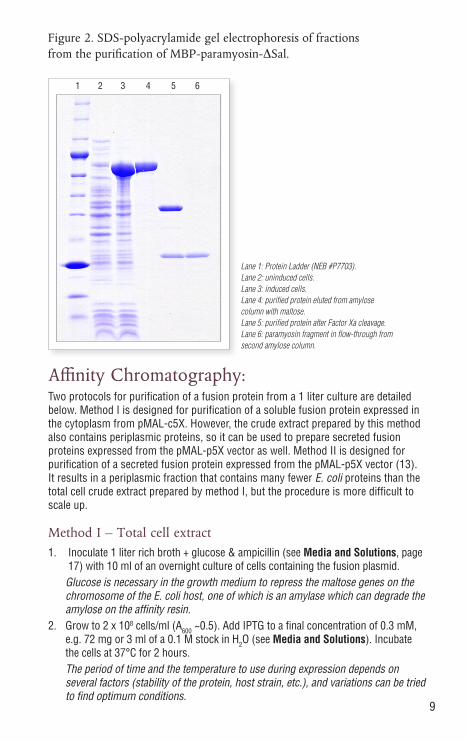
Lane 1: Protein Ladder (NEB #P7703). Lane 2: uninduced cells. Lane 3: induced cells. Lane 4: purified protein eluted from amylose column with maltose. Lane 5: purified protein after Factor Xa cleavage. Lane 6: paramyosin fragment in flow-through from second amylose column.
Figure 2. SDS-polyacrylamide gel electrophoresis of fractions from the purification of MBP-paramyosin-ΔSal.
1 2 3 4 5 6

10
3. Harvest the cells by centrifugation at 4000 x g for 20 minutes and discard the supernatant. Resuspend the cells in 25 ml Column Buffer (see Media and Solutions).
For many unstable proteins, most of the degradation happens during harvest and cell breakage. Therefore, it is best to harvest the cells quickly and keep them chilled. 25 ml of Column Buffer is based on the expectation of about 4-5 grams cells/liter, i.e. 5 ml for every gram of cells (wet weight).
The EDTA in the lysis buffer is to help inhibit proteases that have a Ca++ cofactor. Addition of PMSF (phenyl methylsulfonyl fluoride) and/or other protease inhibitors may help in some cases.
DTT or β–mercaptoethanol can be included to prevent oxidative damage to the fusion protein and interchain disulfide bond formation upon lysis (disulfide bonds usually do not form intracellularly in E. coli). For more about variations and additions to the Column Buffer, see Media & Solutions, page 17.
4. Freeze sample in a dry ice-ethanol bath or several hours to overnight at –20°C; freezing at –20°C is more effective than –70°C, but takes longer. Thaw in cold water.
This is a good place to stop the protocol overnight – the frozen cells can be stored for a week or more at –20°C, depending on the particular fusion.
5. Place sample in an ice-water bath and sonicate in short pulses of 15 seconds or less. Monitor the release of protein using the Bradford assay (14), adding 5 µl of the sonicate to 1 ml Bradford reagent and mixing. Continue sonication until the released protein reaches a maximum (usually about 2 minutes sonication time).
6. Centrifuge at 20,000 x g for 20 minutes. Save the supernatant (crude extract). Dilute the crude extract 1:6 with Column Buffer.
This is another good place to stop the protocol – the crude extract can be stored for a week or more at –20°C, depending on the particular fusion. It is usually more convenient to store the crude extract before diluting with column buffer, then to dilute it after removal from storage and thawing.
7. Pour the amylose resin in a 2.5 x 10 cm column. Wash the column with 5 column volumes of Column Buffer.
The amount of resin depends on the amount of fusion protein produced. The resin binds about 6-8 mg/ml bed volume, so a column of about 15 ml should be sufficient for a yield of up to 100 mg fusion protein/liter culture.
8. Load the diluted crude extract at a flow rate of no more than [50 x (diameter of column in cm)2] ml/hour. This is a maximum of 5 ml/minute for a 2.5 cm column.
9. Wash with 12 column volumes of Column Buffer at a rate of no more than [100 x (diameter of column in cm)2] ml/hour. This is a maximum of 10 ml/minute for a 2.5 cm column.
The column can be washed overnight, if it has a safety loop to prevent it from running dry (see Figure 3, page 14). In this case, it is better to restart the column with elution buffer (step 10), rather than continuing the wash. Avoid loading the column overnight.
11
10. Elute the fusion protein with Column Buffer + 10 mM maltose. Collect 10 to 20 fractions of 3 ml each (fraction size = 1/5th column volume). The fusion protein usually starts to elute within the first 5 fractions, and should be easily detected by UV absorbance at 280 nm or the Bradford protein assay (14).
11. Pool the protein-containing fractions. If necessary, concentrate to about 1 mg/ml in a Vivaspin Centrifugal Concentrator (Sartorius), or the equivalent.
Method II – Periplasmic extract1. Inoculate 1 liter rich broth + glucose & ampicillin with 10 ml of an overnight
culture of cells containing the fusion plasmid.2. Grow to 2 to 4 x 10⁸ cells/ml (A600 ~0.5). Add IPTG to a final concentration of
0.3 mM, e.g. 72 mg/l or 3 ml of a 0.1 M stock in H2O. Incubate the cells at 37°C for 2 hours.
The period of time and the temperature to use during expression depends on several factors (stability of the protein, host strain, etc.), and variations can be tried to find optimum conditions (see Pilot Experiment). In addition, partial induction of exported proteins may lead to higher yields, since protein export in E. coli may not be able to keep up with full level Ptac expression.
3. Harvest the cells by centrifugation at 4000 x g for 20 minutes and discard the supernatant. Resuspend the cells in 400 ml 30 mM Tris-HCl, 20% sucrose, pH 8.0 (80 ml for each gram of cells wet weight). Add EDTA to 1 mM and incubate for 5–10 minutes at room temperature with shaking or stirring.
4. Centrifuge at 8000 x g for 20 minutes at 4°C, remove all the supernatant, and resuspend the pellet in 400 ml of ice-cold 5 mM MgSO4. Shake or stir for 10 minutes in an ice bath.
5. Centrifuge at 8000 x g for 20 minutes at 4°C. The supernatant is the cold osmotic shock fluid.
6. Add 8 ml of 1 M Tris-HCl, pH 7.4 to the osmotic shock fluid. 7. Continue from Method I, step 7 (page 11).
Regenerating the Amylose Resin ColumnThe resin may be reused three to five times when regenerated with the following sequence of washes: Water: 3 column volumes 0.1% SDS: 3 column volumes Water: 1 column volumes Column Buffer: 3 column volumesPlease note that, although the column can be washed at 4°C, 0.1% SDS will eventually precipitate at that temperature. It is therefore recommended that the SDS solution be stored at room temperature until needed, and rinsed out of the column promptly.
Upon repeated use, trace amounts of amylase in the E. coli extract decrease the binding capacity of the column. It is recommended that the column be washed promptly after each use.
12
Cleavage of the Fusion Protein:For Factor Xa, fusion protein cleavage is carried out at a w/w ratio of 1% the amount of fusion protein (e.g., 1 mg Factor Xa for a reaction containing 100 mg fusion protein). The reaction mixture can be incubated for 3 hours to several days, at room temperature or 4°C. Depending on the particular fusion protein, the amount of protease can be adjusted within the range of 0.1–5.0%, to get an acceptable rate of cleavage. Factor Xa will cleave at non-canonical sites in some proteins; for some fusions, there is a correlation between instability of the protein of interest in and cleavage at additional sites (unpublished observations). Presumably this cleavage activity depends on the three dimensional conformation of the fusion protein. For fusions that are resistant to cleavage, two strategies can sometimes help. Inclusion of small amounts of SDS (0.005–0.05%) in the reaction appears to relax the fusion enough to allow for cleavage in some cases (15). The window of SDS concentrations that work can be small, so a pilot titration with different SDS concentrations is necessary. Another strategy that sometimes helps is to denature the fusion to render the protease site accessible to cleavage (see page 13 and reference 8). 1. If necessary, concentrate the fusion protein to at least 1 mg/ml.2. Do a pilot experiment with a small portion of your protein. Example: Mix 20 µl fusion protein at 1 mg/ml with 1 µl Factor Xa diluted to
200 µg/ml, or 0.2 ng Enterokinase In a separate tube, place 5 µl fusion protein with no protease (mock digestion).
Incubate the tubes at room temperature. At 2, 4, 8, and 24 hours, take 5 µl of the reaction, add 5 µl 2x SDS-PAGE Sample Buffer, and save at 4°C. Prepare a sample of 5 µl fusion protein + 5 µl 2X sample buffer (uncut fusion).
3. Boil the 6 samples for 5 minutes and run on an SDS-PAGE gel (12).4. Scale the pilot experiment up for the portion of the fusion protein to be cleaved.
Save at least a small sample of the uncut fusion as a reference.5. Check for complete cleavage by SDS-PAGE.
Denaturing the Fusion Protein1. Either dialyze the fusion against at least 10 volumes 20 mM Tris-HCl, 6 M
guanidine hydrochloride, pH 7.4 for 4 hours, or add guanidine hydrochloride directly to the sample to give a final concentration of 6 M.
2. Dialyze against 100 volumes Column Buffer, 2 times at 4 hours each. During refolding, one has to balance between two objectives. For the protease
to cleave it must be present before the protein has completely refolded, so removing the denaturant quickly is desirable. However, when the denaturant is removed quickly some proteins will fail to refold properly and precipitate. Stepwise dialysis against buffer containing decreasing amounts of guanidine hydrochloride can prevent precipitation of the fusion protein; halving the guanidine concentration at each step is convenient, but cases where 0.1 M steps are necessary have been reported. However, if the fusion protein is able to refold into a protease-resistant conformation, it may be better to dialyze away the denaturant in one step and take the loss from precipitation in order to maximize the amount of cleavable fusion protein recovered.
Go to step 2 or 4 above, as appropriate.

13
Separating the Protein of Interest from MBP after Protease Cleavage:Method I: Anion exchange chromatographyThis method potentially purifies the target protein away from MBP and the protease, but also provides an additional purification step for removing trace contaminants. A disadvantage is that occasionally the peak containing the protein of interest overlaps with MBP or the protease, resulting in poor separation. The procedure is written for quantities < 25 mg, and can be scaled up for larger amounts. The procedure calls for an anion exchange column such as the HiTrap Q FF (GE Life Sciences #17-5156-01).1. Dialyze the fusion protein cleavage mixture vs. 20 mM Tris-HCl, 25 mM NaCl,
pH 8.0 (2 or 3 changes of 100 volumes, at least 2 hours each).2. Wash the column with 15 ml of the same buffer.3. Load the fusion protein cleavage mixture onto the column. Collect 2.5 ml
fractions of the column flow-through.4. Wash the column with 3–5 column volumes of the same buffer. Continue
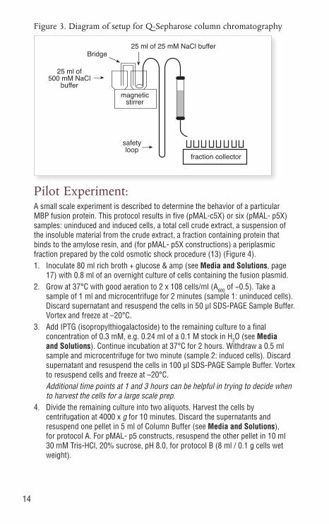
collecting 2.5 ml fractions.5. Start a gradient of 25 mM NaCl to 500 mM NaCl (25 ml each) in 20 mM Tris-
HCl, pH 8.0 (Figure 3). Collect 1 ml fractions.6. Determine which fractions contain protein by measuring A280, or by the
Bradford method. The MBP elutes as a sharp peak at 100–150 mM NaCl. Factor Xa elutes at about 400 mM NaCl. The target protein may flow through the column, or it may elute during the gradient. Electrophorese the relevant fractions on an SDS-PAGE gel (12). Pool the fractions containing the target protein free of MBP and concentrate as desired.
If your protein is not separated from MBP using anion exchange chromatography, other chromatography resins can be tried. HiTrap SP (phosphate buffer at pH 7.0; MBP flows through) and gel filtration are good alternatives.
Method II: Anion exchange chromatography variationThis method takes advantage of the fact that MBP and Factor Xa will bind to a positively-charged resin at pH 5.5, where most other proteins do not bind. The procedure calls for an anion exchange column such as the HiTrap Q FF (GE Life Sciences #17-5156-01).1. Dialyze the cleavage mixture vs. 20 mM sodium phosphate buffer, 25 mM NaCl,
pH 5.5 (2 or 3 changes of 100 volumes, at least 2 hours each)2. Wash the column with 15 ml of the same buffer.3. Load the fusion protein cleavage mixture onto the column. Collect 2.5 ml
fractions of the column flow-through.4. Wash the column with 3–5 column volumes of the same buffer. Continue
collecting 2.5 ml fractions.5. Check the flow-through fractions for protein by A280 or Bradford. In most cases,
the protein of interest will flow through the column.
14
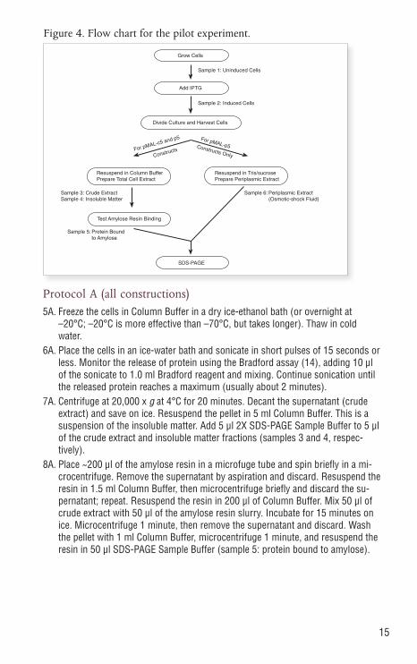
Pilot Experiment:A small scale experiment is described to determine the behavior of a particular MBP fusion protein. This protocol results in five (pMAL-c5X) or six (pMAL- p5X) samples: uninduced and induced cells, a total cell crude extract, a suspension of the insoluble material from the crude extract, a fraction containing protein that binds to the amylose resin, and (for pMAL- p5X constructions) a periplasmic fraction prepared by the cold osmotic shock procedure (13) (Figure 4).1. Inoculate 80 ml rich broth + glucose & amp (see Media and Solutions, page
17) with 0.8 ml of an overnight culture of cells containing the fusion plasmid.2. Grow at 37°C with good aeration to 2 x 10⁸ cells/ml (A600 of ~0.5). Take a
sample of 1 ml and microcentrifuge for 2 minutes (sample 1: uninduced cells). Discard supernatant and resuspend the cells in 50 µl SDS-PAGE Sample Buffer. Vortex and freeze at –20°C.
3. Add IPTG (isopropylthiogalactoside) to the remaining culture to a final concentration of 0.3 mM, e.g. 0.24 ml of a 0.1 M stock in H2O (see Media and Solutions). Continue incubation at 37°C for 2 hours. Withdraw a 0.5 ml sample and microcentrifuge for two minute (sample 2: induced cells). Discard supernatant and resuspend the cells in 100 µl SDS-PAGE Sample Buffer. Vortex to resuspend cells and freeze at –20°C.
Additional time points at 1 and 3 hours can be helpful in trying to decide when to harvest the cells for a large scale prep.
4. Divide the remaining culture into two aliquots. Harvest the cells by centrifugation at 4000 x g for 10 minutes. Discard the supernatants and resuspend one pellet in 5 ml of Column Buffer (see Media and Solutions), for protocol A. For pMAL- p5 constructs, resuspend the other pellet in 10 ml 30 mM Tris-HCl, 20% sucrose, pH 8.0, for protocol B (8 ml / 0.1 g cells wet weight).
Figure 3. Diagram of setup for Q-Sepharose column chromatography
magneticstirrer
25 ml of 25 mM NaCl buffer
fraction collector
safetyloop
Bridge
25 ml of500 mM NaCl
buffer
15
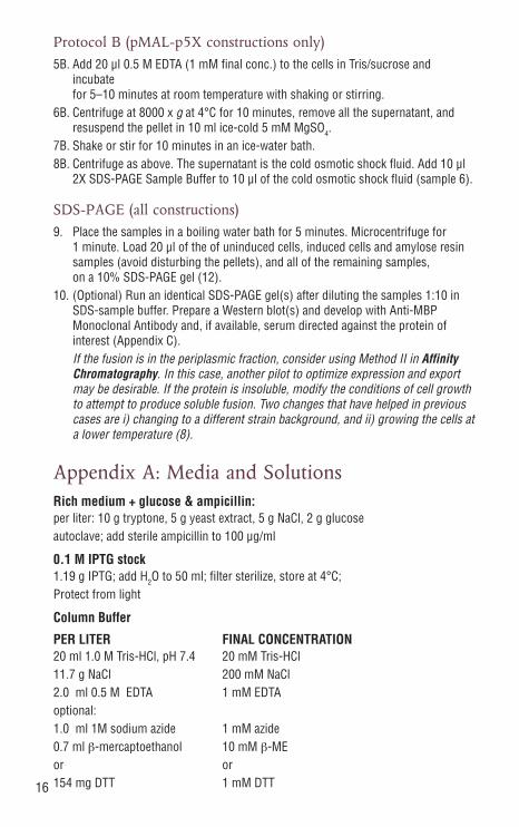
Protocol A (all constructions)5A. Freeze the cells in Column Buffer in a dry ice-ethanol bath (or overnight at
–20°C; –20°C is more effective than –70°C, but takes longer). Thaw in cold water.
6A. Place the cells in an ice-water bath and sonicate in short pulses of 15 seconds or less. Monitor the release of protein using the Bradford assay (14), adding 10 µl of the sonicate to 1.0 ml Bradford reagent and mixing. Continue sonication until the released protein reaches a maximum (usually about 2 minutes).
7A. Centrifuge at 20,000 x g at 4°C for 20 minutes. Decant the supernatant (crude extract) and save on ice. Resuspend the pellet in 5 ml Column Buffer. This is a suspension of the insoluble matter. Add 5 µl 2X SDS-PAGE Sample Buffer to 5 µl of the crude extract and insoluble matter fractions (samples 3 and 4, respec-tively).
8A. Place ~200 µl of the amylose resin in a microfuge tube and spin briefly in a mi-crocentrifuge. Remove the supernatant by aspiration and discard. Resuspend the resin in 1.5 ml Column Buffer, then microcentrifuge briefly and discard the su-pernatant; repeat. Resuspend the resin in 200 µl of Column Buffer. Mix 50 µl of crude extract with 50 µl of the amylose resin slurry. Incubate for 15 minutes on ice. Microcentrifuge 1 minute, then remove the supernatant and discard. Wash the pellet with 1 ml Column Buffer, microcentrifuge 1 minute, and resuspend the resin in 50 µl SDS-PAGE Sample Buffer (sample 5: protein bound to amylose).
Figure 4. Flow chart for the pilot experiment.
Grow Cells
Add IPTG
Divide Culture and Harvest Cells
Sample 1: Uninduced Cells
Sample 2: Induced Cells
For pMAL-c5 and p5
Constructs
For pMAL-p5Constructs Only
Resuspend in Column BufferPrepare Total Cell Extract
Resuspend in Tris/sucrosePrepare Periplasmic Extract
Sample 3: Crude ExtractSample 4: Insoluble Matter
Sample 6: Periplasmic Extract(Osmotic-shock Fluid)
Test Amylose Resin Binding
SDS-PAGE
Sample 5: Protein Boundto Amylose
16
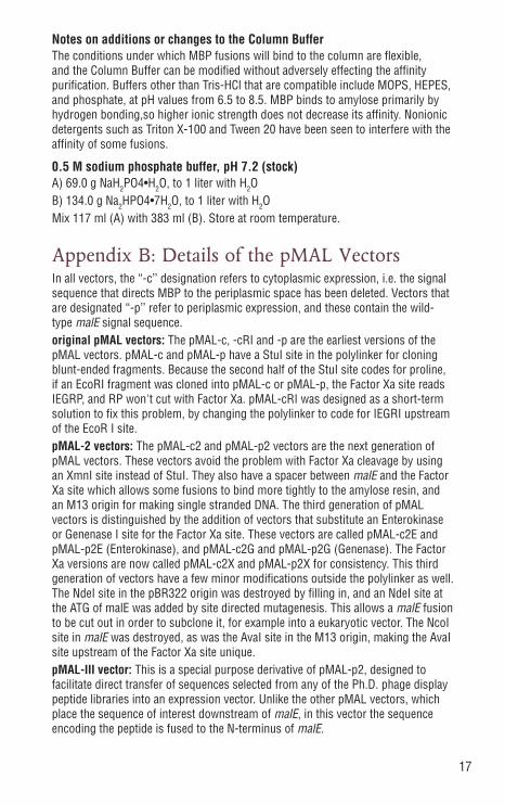
Protocol B (pMAL-p5X constructions only)5B. Add 20 µl 0.5 M EDTA (1 mM final conc.) to the cells in Tris/sucrose and
incubate for 5–10 minutes at room temperature with shaking or stirring.
6B. Centrifuge at 8000 x g at 4°C for 10 minutes, remove all the supernatant, and resuspend the pellet in 10 ml ice-cold 5 mM MgSO4.
7B. Shake or stir for 10 minutes in an ice-water bath.8B. Centrifuge as above. The supernatant is the cold osmotic shock fluid. Add 10 µl
2X SDS-PAGE Sample Buffer to 10 µl of the cold osmotic shock fluid (sample 6).
SDS-PAGE (all constructions)9. Place the samples in a boiling water bath for 5 minutes. Microcentrifuge for
1 minute. Load 20 µl of the of uninduced cells, induced cells and amylose resin samples (avoid disturbing the pellets), and all of the remaining samples, on a 10% SDS-PAGE gel (12).
10. (Optional) Run an identical SDS-PAGE gel(s) after diluting the samples 1:10 in SDS-sample buffer. Prepare a Western blot(s) and develop with Anti-MBP Monoclonal Antibody and, if available, serum directed against the protein of interest (Appendix C).
If the fusion is in the periplasmic fraction, consider using Method II in Affinity Chromatography. In this case, another pilot to optimize expression and export may be desirable. If the protein is insoluble, modify the conditions of cell growth to attempt to produce soluble fusion. Two changes that have helped in previous cases are i) changing to a different strain background, and ii) growing the cells at a lower temperature (8).
Appendix A: Media and SolutionsRich medium + glucose & ampicillin: per liter: 10 g tryptone, 5 g yeast extract, 5 g NaCl, 2 g glucose autoclave; add sterile ampicillin to 100 μg/ml
0.1 M IPTG stock 1.19 g IPTG; add H2O to 50 ml; filter sterilize, store at 4°C; Protect from light
Column Buffer
PER LITER FINAL CONCENTRATION 20 ml 1.0 M Tris-HCl, pH 7.4 20 mM Tris-HCl 11.7 g NaCl 200 mM NaCl 2.0 ml 0.5 M EDTA 1 mM EDTA optional: 1.0 ml 1M sodium azide 1 mM azide 0.7 ml β-mercaptoethanol 10 mM β-ME or or 154 mg DTT 1 mM DTT
17
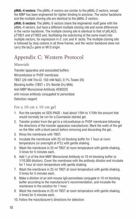
Notes on additions or changes to the Column Buffer The conditions under which MBP fusions will bind to the column are flexible, and the Column Buffer can be modified without adversely effecting the affinity purification. Buffers other than Tris-HCl that are compatible include MOPS, HEPES, and phosphate, at pH values from 6.5 to 8.5. MBP binds to amylose primarily by hydrogen bonding,so higher ionic strength does not decrease its affinity. Nonionic detergents such as Triton X-100 and Tween 20 have been seen to interfere with the affinity of some fusions.
0.5 M sodium phosphate buffer, pH 7.2 (stock) A) 69.0 g NaH2PO4•H2O, to 1 liter with H2O B) 134.0 g Na2HPO4•7H2O, to 1 liter with H2O Mix 117 ml (A) with 383 ml (B). Store at room temperature.
Appendix B: Details of the pMAL VectorsIn all vectors, the “-c’’ designation refers to cytoplasmic expression, i.e. the signal sequence that directs MBP to the periplasmic space has been deleted. Vectors that are designated “-p’’ refer to periplasmic expression, and these contain the wild-type malE signal sequence.original pMAL vectors: The pMAL-c, -cRI and -p are the earliest versions of the pMAL vectors. pMAL-c and pMAL-p have a StuI site in the polylinker for cloning blunt-ended fragments. Because the second half of the StuI site codes for proline, if an EcoRI fragment was cloned into pMAL-c or pMAL-p, the Factor Xa site reads IEGRP, and RP won't cut with Factor Xa. pMAL-cRI was designed as a short-term solution to fix this problem, by changing the polylinker to code for IEGRI upstream of the EcoR I site.pMAL-2 vectors: The pMAL-c2 and pMAL-p2 vectors are the next generation of pMAL vectors. These vectors avoid the problem with Factor Xa cleavage by using an XmnI site instead of StuI. They also have a spacer between malE and the Factor Xa site which allows some fusions to bind more tightly to the amylose resin, and an M13 origin for making single stranded DNA. The third generation of pMAL vectors is distinguished by the addition of vectors that substitute an Enterokinase or Genenase I site for the Factor Xa site. These vectors are called pMAL-c2E and pMAL-p2E (Enterokinase), and pMAL-c2G and pMAL-p2G (Genenase). The Factor Xa versions are now called pMAL-c2X and pMAL-p2X for consistency. This third generation of vectors have a few minor modifications outside the polylinker as well. The NdeI site in the pBR322 origin was destroyed by filling in, and an NdeI site at the ATG of malE was added by site directed mutagenesis. This allows a malE fusion to be cut out in order to subclone it, for example into a eukaryotic vector. The NcoI site in malE was destroyed, as was the AvaI site in the M13 origin, making the AvaI site upstream of the Factor Xa site unique.pMAL-III vector: This is a special purpose derivative of pMAL-p2, designed to facilitate direct transfer of sequences selected from any of the Ph.D. phage display peptide libraries into an expression vector. Unlike the other pMAL vectors, which place the sequence of interest downstream of malE, in this vector the sequence encoding the peptide is fused to the N-terminus of malE.
18
pMAL-4 vectors: The pMAL-4 vectors are similar to the pMAL-2 vectors, except the MBP has been engineered for tighter binding to amylose. The vector backbone and the multiple cloning site are identical to the pMAL-2 vectors.pMAL-5 vectors: The pMAL-5 vectors share the engineered malE gene with the pMAL-4 vectors, but have a different multiple cloning site and some differences in the vector backbone. The multiple cloning site is identical to that of pKLAC2, pTYB21 and pTYB22 and, facilitating the subcloning of the same insert into multiple vectors, for expression in E. coli, and K. lactis. The multiple cloning site is followed by stop codons in all three frames, and the vector backbone does not carry the lacZα gene or M13 origin.
Appendix C: Western ProtocolMaterialsTransfer apparatus and associated buffersNitrocellulose or PVDF membraneTBST (20 mM Tris-Cl, 150 mM NaCl, 0.1% Tween 20)Blocking buffer (TBST + 5% Nonfat Dry Milk)Anti-MBP Monoclonal Antibody #E8032Santi-mouse antibody conjugated to peroxidaseDetection reagent
For a 10 cm x 10 cm gel1. Run the samples on SDS-PAGE - load about 1/5th to 1/10th the amount that
would normally be run for a Coomassie stained gel.2. Transfer protein from the gel to a nitrocellulose or PVDF membrane following
the directions of the transfer apparatus manufacturer. Mark the wells of the gel on the filter with a blunt pencil before removing and discarding the gel.
3. Rinse the membrane with TBST. 4. Incubate the membrane with 25 ml blocking buffer for 1 hour at room
temperature (or overnight at 4°C) with gentle shaking.5. Wash the membrane in 25 ml TBST at room temperature with gentle shaking,
3 times for 5 minutes each.6. Add 1 µl of the Anti-MBP Monoclonal Antibody to 10 ml blocking buffer (a
1/10,000 dilution). Cover the membrane with the antibody dilution and incubate for 1 hour at room temperature with gentle shaking.
7. Wash the membrane in 25 ml TBST at room temperature with gentle shaking, 3 times for 5 minutes each.
8. Make a dilution of an anti-mouse IgG-peroxidase conjugate in 10 ml blocking buffer according to the manufacturer’s recommendation, and incubate the membrane in the solution for 1 hour.
9. Wash the membrane in 25 ml TBST at room temperature with gentle shaking, 3 times for 5 minutes each.
10. Follow the manufacturer’s directions for detection.
19
Appendix D: Troubleshooting and Tips1. Cloning and Transformation1.1 Strain recommendations1.2 Sequencing primers1.3 Inability to clone1.4 Low yield of pMAL in plasmid preps1.5 Obtaining pMAL sequences
2. Expression2.1 Fusion protein is degraded2.2 Fusion protein is insoluble2.3 Fusion protein not expressed2.4 Only MBP-sized protein expressed2.5 Effect of export (secretion, using pMAL-p vectors) on solubility and stability of the fusion2.6 Expressing small peptides
3. Affinity Purification3.1 Fusion protein does not bind to amylose column3.2 Amylose resin regeneration3.3 Nonionic detergents and binding3.4 Different buffers and salt concentrations
3. Affinity Purification (cont.)3.5 Degradation of the fusion upon cell lysis3.6 Extra (unexpected) bands upon SDS-PAGE3.7 Batch purification3.8 Purification in the presence of denaturants3.9 Storage of resin at –20°C
4. Factor Xa Cleavage4.1 Fusion protein is cleaved internally4.2 Control substrates4.3 Inactivation of Factor Xa4.4 Removal of Factor Xa4.5 Fusion will not cleave4.6 Molecular weight and calculated pI4.7 Cleavage in glycerol4.8 Cleavage in urea, guanidine hydrochloride and SDS4.9 Cleavage with fusion bound to the column
20
5. Separation of Fusion Protein Domains and Storage5.1 Difficulties with removal of maltose by dialysis5.2 Alternate separation protocols5.3 How to store proteins
6. MBP56.1 What is MBP5?6.2 Crystal structure of MBP6.3 How much of MBP is dispensable for binding?6.4 Kd, pI and extinction coefficient for MBP6.5 Origin of the MBP region of the pMAL vectors6.6 Is MBP a monomer?
1.Cloning and Transformation1.1 What strain(s) do you recommend as hosts for the pMAL vectors?
The strain we recommend is NEB Express (NEB #C2523). This is an E. coli B strain similar to T7 Express and BL21 (DE3), except that it lacks the T7 RNA Polymerase (the pMAL vectors use the E. coli RNA Polymerase). NEB Express, T7 Express (NEB #C2566), BL21 (NEB #C2530) and BL21(DE3) (NEB #C2527) all give similar results - the presence of the T7 RNA Polymerase doesn't seem to have any effect. Another strain that has been used extensively is TB1 (NEB #E4122), which is JM83 hsdR. There is nothing special about it with respect to the pMAL system, but it has given good results when considering plasmid stability, expression and purification. TB1 is a good choice for proteins that express and/or fold better in E. coli K12 strains. Other successful strains include NEB 10-beta (NEB #C3019), NEB 5-alpha (NEB #C2991) and NEB Turbo (NEB #C2984). We also use other strains in response to a particular problem (for example, see 2.1). One can start with NEB Express, or with whatever competent cells are readily available, and then try another strain if a problem with expression or purification develops.
1.2 What primers should I use to sequence the ends of my insert after I clone it into a pMAL vector?
The following sequencing primers can be used (not available from New England Biolabs): malE primer on the 5´ side of the insert and the pMAL reverse primer for sequencing from the 3´ side.
Forward Primer (24-mer, upstream of MCS) 5´d(GGTCGTCAGACTGTCGATGAAGCC)3´
Reverse Primer (24-mer, downstream of MCS) 5´d(TGTCCTACTCAGGAGAGCGTTCAC)3´
The sequences of the pMAL vectors are also available at www.neb.com. PCR of inserts cloned into the MCS can be performed using the same primer pair (annealing temperature of 56°C when used with OneTaq DNA Polymerase products).

21
1.3 What are some of the possible explanations for an inability to clone an insert into a pMAL vector?
The most common explanation for this is technical difficulties with the subcloning. Another explanation is that expression of the fusion is toxic to E. coli. The tac promoter induction ratio on the pMAL plasmids is about 1:30, so if the induced level of the fusion is 40% of the total cellular protein, the uninduced level works out to over 1%. This amount of a protein can be toxic, either because of its function (e.g., a protease) or because of its general properties (e.g., very hydrophobic).
1.4 Why is the yield of pMAL DNA from plasmid preps so low?
The pMAL plasmids are pBR322-copy number, but for unknown reasons the yield from plasmid preps is often lower than what can be obtainined from pBR322. However, modification of the standard alkaline lysis protocol can increase the yield: increasing the volume of the buffers by 1.5 X doubles the yield of plasmid (i.e., for a 500 ml culture, resuspend the cell pellet in 15 ml instead of the standard 10 ml, and increase the denaturing and neutralizing buffer amounts proportionately).
1.5 How can I obtain the sequences of the pMAL vectors?
The pMAL sequences are available in the Technical Reference section of the New England Biolabs web site at www.neb.com: the complete url is https://www.neb.com/tools-and-resources/interactive-tools/dna-sequences-and-maps-tool
2. Expression
2.1 When we analyze our fusion protein expression by Western blot using the Anti-MBP Monoclonal Antibody, only a small fraction of the protein is full-length, while most of it migrates close to the MBP5* marker.
It is likely that the fusion protein is degraded, leaving a stable MBP-sized breakdown product. In this case, try using a protease deficient host. NEB Express is Lon- and OmpT-. A list of additional strains, which are free with an order or for the price of shipping, can be found on page 29. For cytoplasmic expression, the most protease deficient strain is CAG629 (#E4125) – it is also, however, the most difficult to work with. CAG597 (#E4123) is another good alternative. For periplasmic expression, the most protease deficient strain is CAG597 (#E4123); KS1000 (#E4128) and UT5600 (#E4129) might be worth trying as well. The CAG strains are difficult to transform, and often require electroporation to introduce the fusion plasmid.
2.2 My fusion protein is insoluble; is there anything I can do to get it expressed as soluble protein?
Expressing at a lower temperature is the first thing to try. One can go as low as 15°C by moving an incubator into the cold room. Of course, the cells grow very slowly at these temperatures, so grow the culture at 37°C and shift to the low temperature when adding IPTG. One also has to increase the time of induction to compensate for the slower growth – a rule of thumb is 2X for every 7°C. Other references for solubility problems include:
22
Reviews on methods to make correctly-folded protein in E. coli: Georgiou, G. et al. (1996) Current Opinion in Biotechnology 7:190–197.Sahdev S. et al. (2008) Mol. Cell Biochem 307:249-64
Reviews on refolding:Qoronfleh, M.W. et al. (2007) Protein Expr. Purif. 55, 209–224.Jungbauer, A. and Kaar, W. (2007) J. Biotechnol. 128, 587–596.Jungbauer, A. et al. (2004) Curr. Opin. Biotechnol. 15, 487–494.Vallejo, L.F. and Rinas, U. (2004) Microb. Cell Fact 3, 11–22.
2.3 When I run my uninduced and induced crude extracts on SDS-PAGE side by side, I don’t see an induced band.
There are a couple of possible explanations. Inserts cloned in the pMAL-p5X vector have about a 8- to 16-fold reduced level of expression when compared to the same insert in a pMAL-c5 vectors. This often reduces the amount of expression to the point where there is no visible induced band. In addition, some foreign genes are poorly expressed in E. coli, even when fused to a highly expressed carrier gene. Possible explanations are message instability or problems with translation – sometimes it is due to the presence of multiple rare codons in the gene of interest, and in these cases overexpression of the corresponding tRNA can help (16). Even in cases where a band is not visible, one can get yields up to 5 or 6 mg/liter of culture.
2.4 I’ve cloned my insert, but after SDS-PAGE the only induced band present is the size of MBP5*. There are two likely explanations for this result. If the protein of interest is in the wrong translational reading frame, an MBP5-sized band will be produced by translational termination at the first in-frame stop codon. If the protein of interest is very unstable, an MBP5-sized breakdown product is usually produced (MBP is a very stable protein). The best way to distinguish between these possibilities is to run a Western blot using Anti-MBP Monoclonal Antibody (NEB #E8032) or anti-MBP Monoclonal Antibody, HRP conjugated (NEB #E8038). If proteolysis is occurring, at least a small amount of full-length fusion can almost always be detected. DNA sequencing of the fusion junction will confirm a reading frame problem. If the problem is proteolysis, you might want to try one of the protease deficient strains from the list on page 29.
2.5 What are the possible effects of export (secretion, using a pMAL-p5X vector) on solubility/stability of the fusion?
Initiating export through the cytoplasmic membrane puts a fusion protein on a different folding pathway – a difference in the solubility or stability of the protein is determined by whether this folding pathway leads to a different 3-dimensional structure for the protein. Some proteins, like MBP itself, can fold properly either in the cytoplasm or when exported to the periplasm. However, the normal folding pathway for some proteins is incompatible with passage through the membrane. In these cases, the fusion protein gets stuck in the membrane and cannot fold properly, which can lead to its degradation (17). Other proteins, especially ones that have multiple disulfide bonds, only fold properly when exported (the E. coli cytoplasm is
23
a reducing environment, and the proteins that catalyze disulfide bond formation are present in the periplasm (18). When this class of protein is expressed in the cytoplasm, it may fold improperly and become degraded or insoluble.
2.6 What is the minimum size of a fragment that can be cloned into pMAL and expressed fused to MBP? Can short peptide sequences (about 10 amino acids) be added onto MBP? You can use the MBP system to express short peptides. However, for every 40 mg of MBP (42.5 kDa) one gets about 1 mg of a 10 amino acid peptide (1.1 kDa).
3. Affinity Purification
3.1 Much of my fusion protein flows through the amylose column. Is there anything I can do to improve my fusion’s affinity for the amylose column?A MBP fusion protein might not stick to the amylose column because of the presence of some factor in the extract that interferes with binding, or because of a low intrinsic affinity. Factors in the crude extract that can interfere with binding include nonionic detergents (see 3.3) and cellular components that are released during alternative methods of lysis (prolonged treatment with lysozyme or multiple passes through a French press). In addition, cells grown in LB and similar media have substantial amounts of an amylase that interferes with binding, presumably by either cutting the fusion off the column or by releasing maltose that elutes the fusion from the column. By including glucose in the media, expression of this amylase is repressed and the problem is alleviated. A low intrinsic affinity could be caused by an interaction between the protein of interest and MBP that either blocks or distorts the maltose-binding site. Although this may be inherent in the protein of interest, sometimes the problem can be alleviated by shortening or lengthening the polypeptide that is fused to MBP.
3.2 How many times can I use the amylose column?The most important variable in determining the useful life of the amylose resin is the amount of time it is in contact with trace amounts of amylase present in the crude extract (see 3.1). Under normal conditions (crude extract from 1 liter of cells grown in LB + 0.2% glucose, 15 ml column), the column loses 1–3% of its initial binding capacity each time it is used. If the yield of fusion protein under these conditions is 40 mg, this means that after 3 to 5 runs there would be a decrease in the yield. In practice, we often use a column 8 or 10 times before we notice a significant drop in the yield.
3.3 What is known about binding in the presence of nonionic detergents?Some fusion proteins do not bind efficiently (< 5% binding) in the presence of 0.2% Triton X–100 or 0.25% Tween 20, while other fusions are unaffected. For one fusion that does not bind in 0.25% Tween 20, diluting the Tween to 0.05% restores about 80% of the binding.
24
3.4 Can I substitute a different buffer and/or salt concentration in the Column Buffer?Yes, we have tried HEPES, MOPS, and phosphate buffers (at pH's from 6.5 to 8.5) instead of Tris-HCl in the Column Buffer with similar results. NaCl or KCl concentrations of 25 mM to 1 M are also compatible with the affinity purification.
3.5 I see my intact fusion protein by SDS-PAGE when I run cells boiled in Sample Buffer, but when I check the crude extract the fusion is degraded.
For fusions expressed in the cytoplasm, in many cases most of the degradation happens during harvest and lysis. Harvesting promptly and lysing the cells quickly may help. In other cases, degradation occurs when the fusion protein is exposed to periplasmic or outer membrane proteases (18-20). The best strategy in either case is to use a host which is deficient in the offending protease(s) (see 2.1).
3.6 When I run my purified fusion protein on SDS-PAGE, why do I see multiple bands instead of a single band of the expected MW?
There are two likely explanations for this result. The first is that the fusion protein is unstable, which most often leads to degradation in vivo (see 3.5). In this case, one would expect to see bands between the size of MBP (42.5 kDa) and the size expected for the full-length fusion, since fragments smaller than MBP would not bind to the affinity column. An exception would be if the fusion protein breaks down at the junction between MBP and the protein of interest, and the protein of interest oligomerizes. In this situation, the protein of interest may bind to the fusion protein, and therefore a band the size of the protein of interest can appear even if it is smaller than MBP. The second explanation is that the protein of interest is binding non-specifically to other E. coli proteins, e.g. it has a surface that binds other proteins by electrostatic or hydrophobic interactions. In this case, modifications to the column buffer can sometimes be used to help wash the interacting proteins away. Electrostatic interactions can be weakened by including up to 1 M NaCl in the column buffer, and hydrophobic interactions can be weakened by lowering the salt to 25-50 mM NaCl and including 5% ethanol or acetonitrile in the column buffer. Non-ionic detergents can also be used to weaken hydrophobic interactions, but they can interfere with the affinity of certain fusion proteins.
3.7 Can I perform a batch purification using the amylose resin?
Yes, batch purification works well, although it is difficult to wash all the nonspecific proteins away as effectively as in a column due to the included volume in the resin. The resin can withstand centrifugation at up to 6000 x g. A good compromise is to load the resin in a batch mode, by incubating with shaking for 2 hours to overnight, then pour it in a column to wash and elute. Dilution of the crude extract is not as critical for loading the column by the batch method.
25
3.8 Can MBP fusions be purified in the presence of denaturants like urea or guanidine-HCl?
No, MBP’s affinity to amylose and maltose depends on hydrogen bonds that in turn are positioned by the three-dimensional structure of the protein. Agents that interfere with hydrogen bonds or the structure of the protein interfere with binding as well.
3.9 Is the amylose resin damaged by storage at –20°C? When our kit arrived, it was placed at –20°C, but I see that the recommended storage temperature for the amylose resin is 4°C.
The resin will freeze at –20°C but the performance of the resin is not degraded by one freeze/thaw cycle. After the ethanol is removed, the resin should be stored at 4°C to prevent damage from freezing.
4. Factor Xa Cleavage
4.1 Factor Xa seems to be cleaving my protein at several sites, even though the protein does not contain any IEGR sequences.
The specificity of Factor Xa reported in our catalog is as referenced in Nagai and Thøgersen (1987) (7,8). The basis for this specificity is that the natural Factor Xa sites in prothrombin are IEGR (or sometimes IDGR), and many examples of fusions with IEGR are cut specifically. However, proteins can be cleaved at other arginine residues, depending on the context (21–25). A number of the secondary sites (but not all) that have been sequenced show cleavage following Gly-Arg. We have also seen a correlation between proteins that are unstable in E. coli and cleavage at secondary sites with Factor Xa, suggesting that these proteins are in a partially unfolded state. We’ve tried altering the reaction conditions to increase the specificity, but with no success. Enterokinase is available as an alternative to Factor Xa.
4.2 Are there any control substrates for Factor Xa?
The Protein Fusion and Purification System comes with an MBP5-paramyosin-ΔSal fusion as a positive control for Factor Xa cleavage. Sigma also sells a colorimetric substrate, N-benzoyl-ile-glu-gly-arg-p-nitroanilide (Sigma, #B7020).
4.3 How can Factor Xa be inactivated?
The best way is to add dansyl-Glu-Gly-Arg-chloromethyl ketone (EMD Millipore, #251700) to a final concentration of 2 µM, and incubate for 1 minute at room temperature. This compound irreversibly inactivates the Factor Xa. It reacts with the active site histidine, so it could conceivably react with other sites on the protein of interest, but this is unlikely at the low concentration used.
4.4 How can Factor Xa be removed from the reaction mix after cleavage?
Factor Xa can be removed by passing the reaction mix over a small benzamidine-agarose column (GE Life Sciences, #17514302). When 50 µg of Factor Xa is passed over a 0.5 ml column, less than 0.2% of the activity flows through.
26
4.5 My protein cleaves very poorly with Factor Xa. Is there anything I can do to improve cleavage?
We presume that, in these cases, the fusion protein folds so that the Factor Xa site is inaccessible. In theory, anything that perturbs the structure might uncover the site. We’ve tried increasing the temperature, changing buffers and salt conditions, and adding detergents. The only thing that worked was low concentrations of SDS (0.01 to 0.05%)(27). Another researcher found that calcium worked – his protein of interest was a calcium binding protein, supporting the idea that anything that might change the conformation, such as a cofactor or substrate analog, could have a dramatic effect. Another approach that often helps is to add amino acid residues to the N-terminus of the protein – either by cloning into one of the downstream sites in the polylinker, or by adding codons to the insert (e.g., adding four alanine codons before the start of the gene). Be aware that with this latter strategy, the extra residues remain at the N-terminus after Factor Xa cleavage.
4.6 What is the molecular weight and pI of Factor Xa?The molecular weight of Factor Xa is 42,324 daltons. It consists of two disulfide-linked chains, 26,666 and 15,676. On our SDS-PAGE gels they run as 30 kDa and 20 kDa. The pI of Factor Xa is around 5.0 (27).
4.7 What is maximum concentration of glycerol that Factor Xa can tolerate during cleavage?We have tested Factor Xa cleavage in up to 20% glycerol, where it still cleaves at about half the normal rate.
4.8 How is the rate of Factor Xa cleavage affected by urea, guanidine hydrochloride and SDS?The activity of Factor Xa on the chromogenic substrate Bz-IEGR-pNA in the presence of these denaturants is as follows:
Urea: In 0.25 M urea, Factor Xa cleaves at about 33% its normal rate; at 0.5 M, 25% its normal rate, in 1 M urea, about 10% its normal rate, while in 2 M urea no cleavage is detected.
Guanidine: In 0.25 M guanidine hydrochloride it cleaves at about 15% its normal rate, and in 0.5 M it cleaves at about 5% the normal rate.
SDS: Factor Xa is unaffected by concentrations of SDS below 0.005%. At 0.01% it cleaves at about half its normal rate, and at 0.03% at about one-third normal. At 0.1% and above no cleavage is detected.
4.9 Can MBP fusions be digested with Factor Xa while bound to the amylose resin?Cutting a bound fusion with Factor Xa has been done, (28, unpublished results). It has two problems that make it less than ideal. First, it requires a lot of Factor Xa. With the fusion immobilized, it takes 5% for 24–48 hours to get cleavage roughly equivalent to 1% for 24 hour in solution. The second problem is that during the incubation, some of the MBP falls off the column. This may be because there are trace amounts of amylase bound to the column too, and the amylase liberates enough maltose over time to elute some of the MBP.
27
5. Separation of Fusion Protein Domains and Storage
5.1 In order to rebind MBP to the column, the maltose must be removed. Can this be done by dialysis?Dialysis does not work very well to remove maltose from maltose-binding protein. This is a general phenomenon of binding protein/ligand interactions; after the free ligand is gone, ligand that is released from the binding site usually finds another binding site before it encounters the dialysis membrane (29). We have determined empirically that binding the fusion to a chromatography resin and then washing away the maltose is much more effective. We prefer standard chromatography (e.g. DEAE) as the separation step, since it can separate the Factor Xa and MBP from the protein of interest. In case MBP co-elutes with the protein of interest, we include a large volume washing step to remove the maltose before starting the salt gradient. This way, the mixture can be run over an amylose column afterward if necessary.
5.2 Are there any alternatives to the protocols in the manual for separating the domains after cleavage?
Some fusion proteins can be eluted from the amylose resin with water, although they may not come off in as sharp a peak as they do with column buffer + 10 mM maltose. This can be useful for re-binding the MBP to an amylose column after Factor Xa cleavage. Be aware that some proteins may denature or precipitate in water, however. Small amounts of MBP can be removed from the protein of interest by incubating with immobilized Anti-MBP Antibody (NEB#E8037). The capacity of the beads makes this impractical for a large scale preparation.
5.3 How should I store my protein after it is purified?
Most proteins can be stored for at least a few days at 4°C without denaturing. For long term storage, one can either freeze at –70°C or dialyze into 50% glycerol and store at –20°C. When storing at –70°C, aliquot the protein so only the portion to be used must be thawed – repeated freeze/thaw cycles denature many proteins.
6. MBP5
6.1 What is MBP5? Is it different from wild-type MBP produced from E. coli?
MBP5 is the protein produced from pMAL-c5X that has a stop codon linker cloned into the XmnI site. It differs from wild-type MBP by the addition of a methionine at the amino terminus (as do all fusions made in pMAL-c vectors), the mutations that increase the affinity of MBP for the amylose resin, the deletion of the last four residues of wild-type MBP, and the addition of the residues encoded by the spacer and the protease site.
6.2 Has the crystal structure of MBP been determined?
The references for the crystal structure of MBP are Spurlino, J.C. et al. (1991) J. Biol. Chem. 266, 5202–5219 (29), and Sharff, A.J. et al. (1992) Biochem. 31,10657–10663 (30).
28
6.3 How much of MBP is dispensable for binding?
The exact region of MBP necessary for binding has not been determined, but the structure indicates that most of the protein is necessary. From the structure, it appears that very few, if any, residues could be deleted at the C-terminus (other than the polylinker residues, of course). It is possible that some of the N-terminus could be deleted, but so far this has not been tested.
6.4 What is the Kd, pI and extinction coefficient for MBP?
The Kd of MBP for maltose is 3.5 µM; for maltotriose, 0.16 µM (32). MBP5's extinction coefficient is 1.5 (1 mg/ml, 1 cm path length) and its calculated pI is 4.9.
6.5 What is the origin of the MBP region of the pMAL vectors?
The malE gene in the pMAL vectors was derived from the Hinf I fragment of the E. coli malB region. The Hinf I fragment lacks the last four amino acids of wild-type malE, and, of course, additional amino acids are added as encoded by the polylinker.
6.6 Is MBP5 a monomer or a higher oligomer?
MBP5 is a monomer. There is one published report that MBP can dimerize in 10 mM Tris-HCl (33) but we have not been able to reproduce this result with MBP5. Gel filtration chromatography in both Column Buffer and 10 mM Tris-HCl gives a single peak of about 42 kDa.
29
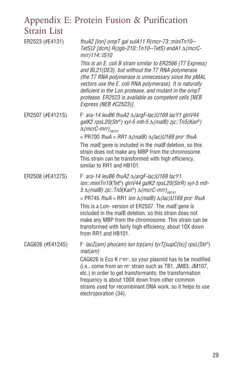
Appendix E: Protein Fusion & Purification Strain ListER2523 (#E4131) fhuA2 [lon] ompT gal sulA11 R(mcr-73::miniTn10--
TetS)2 [dcm] R(zgb-210::Tn10--TetS) endA1 Δ(mcrC-mrr)114::IS10
This is an E. coli B strain similar to ER2566 (T7 Express) and BL21(DE3), but without the T7 RNA polymerase (the T7 RNA polymerase is unnecessary since the pMAL vectors use the E. coli RNA polymerase). It is naturally deficient in the Lon protease, and mutant in the ompT protease. ER2523 is available as competent cells [NEB Express (NEB #C2523)].
ER2507 (#E4121S) F– ara-14 leuB6 fhuA2 Δ(argF-lac)U169 lacY1 glnV44 galK2 rpsL20(StrR) xyl-5 mtl-5 Δ(malB) zjc::Tn5(KanR) Δ(mcrC-mrr)HB101
= PR700 fhuA = RR1 Δ(malB) Δ(lac)U169 pro+ fhuA The malE gene is included in the malB deletion, so this
strain does not make any MBP from the chromosome. This strain can be transformed with high efficiency, similar to RR1 and HB101.
ER2508 (#E4127S) F– ara-14 leuB6 fhuA2 Δ(argF-lac)U169 lacY1 lon::miniTn10(TetR) glnV44 galK2 rpsL20(StrR) xyl-5 mtl-5 Δ(malB) zjc::Tn5(KanR) Δ(mcrC-mrr)HB101
= PR745 fhuA = RR1 lon Δ(malB) Δ(lac)U169 pro+ fhuA This is a Lon- version of ER2507. The malE gene is
included in the malB deletion, so this strain does not make any MBP from the chromosome. This strain can be transformed with fairly high efficiency, about 10X down from RR1 and HB101.
CAG626 (#E4124S) F– lacZ(am) pho(am) lon trp(am) tyrT[supC(ts)] rpsL(StrR) mal(am)
CAG626 is Eco K r+m+, so your plasmid has to be modified (i.e., come from an m+ strain such as TB1, JM83, JM107, etc.) in order to get transformants; the transformation frequency is about 100X down from other common strains used for recombinant DNA work, so it helps to use electroporation (34).
30
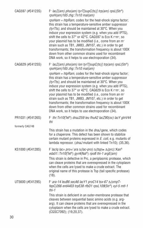
CAG597 (#E4123S) F– lacZ(am) pho(am) tyrT[supC(ts)] trp(am) rpsL(StrR) rpoH(am)165 zhg::Tn10 mal(am)
rpoHam = htpRam, codes for the heat-shock sigma factor; this strain has a temperature-sensitive amber suppressor (tyrTts), and should be maintained at 30°C. When you induce your expression system (e.g. when you add IPTG), shift the cells to 37° or 42°C. CAG597 is Eco K r+m+, so your plasmid has to be modified (i.e., come from an m+ strain such as TB1, JM83, JM107, etc.) in order to get transformants; the transformation frequency is about 100X down from other common strains used for recombinant DNA work, so it helps to use electroporation (34).
CAG629 (#E4125S) F– lacZ(am) pho(am) lon tyrT[supC(ts)] trp(am) rpsL(StrR) rpoH(am)165 zhg::Tn10 mal(am)
rpoHam = htpRam, codes for the heat-shock sigma factor; this strain has a temperature-sensitive amber suppressor (tyrTts), and should be maintained at 30°C. When you induce your expression system (e.g. when you add IPTG), shift the cells to 37° or 42°C. CAG629 is Eco K r+m+, so your plasmid has to be modified (i.e., come from an m+ strain such as TB1, JM83, JM107, etc.) in order to get transformants; the transformation frequency is about 100X down from other common strains used for recombinant DNA work, so it helps to use electroporation (34).
PR1031 (#E4126S) F– thr:Tn10(TetR) dnaJ259 leu fhuA2 lacZ90(oc) lacY glnV44 thi
formerly CAG748 This strain has a mutation in the dnaJ gene, which codes
for a chaperone. This defect has been shown to stabilize certain mutant proteins expressed in E. coli, e.g. mutants of lambda repressor. (dnaJ mutant with linked Tn10); (35,36).
KS1000 (#E4128S) F´lacIq lac+ pro+/ ara Δ(lac-pro) Δ(tsp)≡ Δ(prc)::KanR eda51::Tn10(TetR) gyrA(NalR) rpoB thi-1 argE(am)
This strain is defective in Prc, a periplasmic protease, which can cleave proteins that are overexpressed in the cytoplasm when the cells are lysed to make a crude extract. The original name of this protease is Tsp (tail specific protease) (18).
UT5600 (#E4129S) F– ara-14 leuB6 secA6 lacY1 proC14 tsx-67 Δ(ompT-
fepC)266 entA403 trpE38 rfbD1 rpsL109(StrR) xyl-5 mtl-1 thi-1
This strain is deficient in an outer-membrane protease that cleaves between sequential basic amino acids (e.g. arg-arg). It can cleave proteins that are overexpressed in the cytoplasm when the cells are lysed to make a crude extract. (CGSC7092); (19,20,37).
31
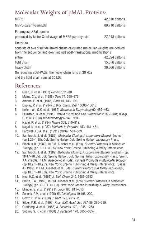
Molecular Weights of pMAL Proteins:MBP5 42,510 daltons
MBP5-paramyosinΔSal 69,710 daltons
ParamyosinΔSal domainproduced by factor Xa cleavage of MBP5-paramyosin 27,218 daltons
Factor Xaconsists of two disulfide linked chains calculated molecular weights are derived from the sequence, and don’t include post-translational modificationsentire 42,324 daltonslight chain 15,676 daltonsheavy chain 26,666 daltonsOn reducing SDS-PAGE, the heavy chain runs at 30 kDa and the light chain runs at 20 kDa
References:1. Guan, C. et al. (1987) Gene 67, 21–30.2. Maina, C.V. et al. (1988) Gene 74, 365–373.3. Amann, E. et al. (1985) Gene 40, 183–190.4. Duplay, P. et al. (1984) J. Biol. Chem. 259, 10606–10613.5. Kellerman, O.K. et al. (1982) Methods in Enzymology 90, 459–463.6. Lauritzen, C. et al.(1991) Protein Expression and Purification 2, 372–378; Takagi,
H. et al. (1988) Bio/technology 6, 948–950.7. Nagai, K. et al. (1984) Nature 309, 810–812.8. Nagai, K. et al. (1987) Methods in Enzymol. 153, 461–481.9. Bardwell J.C.A. et al. (1991) Cell 67, 581–589.10. Sambrook, J. et al. (1989). Molecular Cloning: A Laboratory Manual (2nd ed.)
(pp.1.25–1.28). Cold Spring Harbor:Cold Spring Harbor Laboratory Press.11. Bloch, K.D. (1989). In F.M. Ausebel et al. (Eds), Current Protocols in Molecular
Biology, (pp. 3.1.1–3.2.5). New York: Greene Publishing & Wiley-Interscience. 12. Sambrook, J. et al. (1989) Molecular Cloning: A Laboratory Manual (2nd ed.) (pp.
18.47–18.55). Cold Spring Harbor: Cold Spring Harbor Laboratory Press; Smith, J.A. (1989). In F.M. Ausebel et al. (Eds). Current Protocols in Molecular Biology (pp.10.2.1–10.2.7). New York: Greene Publishing & Wiley-Interscience; Sasse, J. (1989). In F.M. Ausebel, et al. (Eds) Current Protocols in Molecular Biology, (pp.10.6.1–10.6.3). New York: Greene Publishing & Wiley-Interscience.
13. Neu, H.C. et al. (1965) J. Biol. Chem. 240, 3685–3692.14. Smith, J.A. (1989). In F.M. Ausebel et al. (Eds) Current Protocols in Molecular
Biology, (pp. 10.1.1–10.1.3). New York: Greene Publishing & Wiley-Interscience.15. Ellinger, S. et al. (1991) Virology 180, 811–813.16. Schenk, P.M. et al. (1995) BioTechniques 19,196–200.17. Gentz, R. et al. (1988) J. Bact. 170, 2212–20.18. Silber, K.R. et al. (1992) Proc. Natl. Acad. Sci. USA 89, 295–299.19. Grodberg, J. et al. (1988) J. Bacteriol. 170, 1245–1253.20. Sugimura, K. et al. (1988) J. Bacteriol. 170, 3650–3654.
32
21. Nagai, K. et al. (1985) Proc Natl. Acad. Sci. USA 82, 7252–7255.22. Quinlan, R.A. et al. (1989) J. Cell Sci. 93, 71–83.23. Eaton, D. et al. (1986) Biochem. 25, 505–512.24. Wearne, S.J. (1990) FEBS Lett. 263, 23–26.25. Schilling, O. et al. (2011) Biol. Chem. 392, 1031–1037.26. Ellinger, S. et al.(1991) Virol. 180, 811–8113.27. DiScipio, R.G. et al. (1979) Biochemistry 18, 899.28. Rawlings, D.J. et al. (1992) J. Biol. Chem. 267, 3976–3982.29. Silhavy, T.J. et al. (1975) Proc. Natl. Acad. Sci. USA 72, 2120–2124.30. Spurlino, J.C. et al. (1991) J. Biol. Chem. 266, 5202–5219.31. Sharff, A.J. et al. (1992) Biochem. 31, 10657–10663.32. Miller, D.M. et al. (1983) J. Biol. Chem. 258, 13665–13672.33. Richarme, G. (1982) Biochem. Biophys. Res. Comm. 105, 476–481.34. Baker, T.A. et al. (1984) Proc. Natl. Acad. Sci. USA 81, 6779–6783.35. Straus, D.B. et al. (1988) Genes Dev. 2, 897–904.36. Reidhaar-Olson, J.F. et al. (1990) Biochem. 29, 7563–7571.37. Elish, M.E. et al. (1988) J. Gen. Microbiol. 134, 355–1364.
33
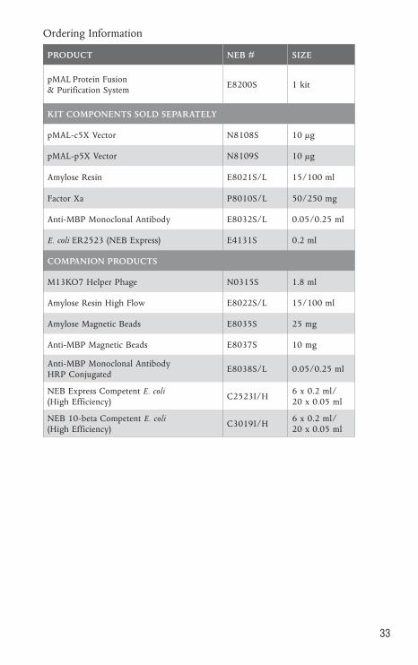
PRODUCT NEB # SIZE
pMAL Protein Fusion & Purification System
E8200S 1 kit
KIT COMPONENTS SOLD SEPARATELY
pMAL-c5X Vector N8108S 10 µg
pMAL-p5X Vector N8109S 10 µg
Amylose Resin E8021S/L 15/100 ml
Factor Xa P8010S/L 50/250 mg
Anti-MBP Monoclonal Antibody E8032S/L 0.05/0.25 ml
E. coli ER2523 (NEB Express) E4131S 0.2 ml
COMPANION PRODUCTS
M13KO7 Helper Phage N0315S 1.8 ml
Amylose Resin High Flow E8022S/L 15/100 ml
Amylose Magnetic Beads E8035S 25 mg
Anti-MBP Magnetic Beads E8037S 10 mg
Anti-MBP Monoclonal Antibody HRP Conjugated
E8038S/L 0.05/0.25 ml
NEB Express Competent E. coli (High Efficiency)
C2523I/H6 x 0.2 ml/ 20 x 0.05 ml
NEB 10-beta Competent E. coli (High Efficiency)
C3019I/H6 x 0.2 ml/ 20 x 0.05 ml
Ordering Information

DNA CLONING
DNA AMPLIFICATION & PCR
EPIGENETICS
RNA ANALYSIS
LIBRARY PREP FOR NEXT GEN SEQUENCING
PROTEIN EXPRESSION & ANALYSIS
CELLULAR ANALYSIS
USANew England Biolabs, Inc.240 County RoadIpswich, MA 01938-2723Telephone: (978) 927-5054Toll Free: (USA Orders) 1-800-632-5227Toll Free: (USA Tech) 1-800-632-7799Fax: (978) 921-1350e-mail: [email protected] www.neb.com
CANADANew England Biolabs, Ltd.Telephone: (905) 665-4632Toll Free: 1-800-387-1095Fax: (905) 665-4635Fax Toll Free: 1-800-563-3789e-mail: [email protected]
CHINANew England Biolabs (Beijing), Ltd.Telephone: 010-82378265/82378266Fax: 010-82378262e-mail: [email protected]
FRANCENew England Biolabs FranceFree Call: 0800-100-632Free Fax: 0800-100-610e-mail: [email protected]
GERMANY & AUSTRIANew England Biolabs GmbHTelephone: +49/(0)69/305 23140Free Call: 0800/246 5227 (Germany) Free Call: 00800/246 52277 (Austria)Fax: +49/(0)69/305 23149Free Fax: 0800/246 5229 (Germany) e-mail: [email protected]
JAPANNew England Biolabs Japan, Inc.Telephone: +81 (0)3 5669 6191Fax: +81 (0)3 5669 6192e-mail: [email protected]
SINGAPORENew England Biolabs Pte. Ltd.Telephone: +65 638 59623Fax: +65 638 59617e-mail: [email protected]
UNITED KINGDOMNew England Biolabs (UK) Ltd.Telephone: (01462) 420616Call Free: 0800 318486Fax: (01462) 421057Fax Free: 0800 435682e-mail: [email protected]