management of pathogens associated with storm water discharge
TRANSCRIPT

MANAGEMENT OF PATHOGENS ASSOCIATED WITH STORM WATER DISCHARGE:
METHODOLOGY FOR QUANTITATIVE MOLECULAR DETERMINATION OF VIRUSES, BACTERIA AND PROTOZOA
I
V
NTERIM REPORT PREPARED FOR DIVISION OF CALIFORNIA D
TRANSPORTATExecution Date: Contract N
Task Order No.:
eronica Rajal, Donald Thompson, BeveBelinda McSwain, and St
DEPARTMENT OF CIVIL & ENVIRONM
UNIVERSITY OF CALIFOR
July 2005
THE ENVIRONMENTAL EPARTMENT OF ION o. 43A0073
19
rly Kildare, Sangam Tiwari, efan Wuertz
ENTAL ENGINEERING NIA, DAVIS

TABLE OF CONTENTS
ACKNOWLEDGEMENTS .................................................................................... 3 LIST OF ACRONYMS AND ABBREVIATIONS................................................... 4 OBJECTIVES....................................................................................................... 5 EXECUTIVE SUMMARY...................................................................................... 6 1.0 INTRODUCTION .......................................................................................... 10 2.0 MATERIALS AND METHODS ..................................................................... 13 2.1 Sample sites and water collection.........................................................................................................13 2.2 Filtration and concentration of water samples ...................................................................................14 2.3 PP7 plaque assay ...................................................................................................................................15 2.4 Nucleic acid extraction..........................................................................................................................16 2.5 PP7 TaqMan system design and validation ........................................................................................16 2.6 PP7 probe and primer design...............................................................................................................17 2.7 Human viruses probes and primers design.........................................................................................17 2.8 TaqMan reactions..................................................................................................................................18 2.9 Calculation of PP7 virus recovery efficiency.......................................................................................19 2.10 Analytical sensitivity of the virus TaqMan PCR systems ................................................................20 2.11 Dilution approach................................................................................................................................20 2.12 Calculation of sample detection limits ...............................................................................................22 2.13 Calculation of virus and cell concentrations when detected ............................................................22 2.14 Quality Assurance and Quality Control Procedures........................................................................23 3.0 RESULTS..................................................................................................... 24 3.1 Analytical sensitivity of the virus TaqMan PCR systems ..................................................................24 3.2 Natural water samples ..........................................................................................................................25 3.3 PP7 recovery from spiked environmental water samples ..................................................................27 3.4 Detection of human adenovirus and enterovirus by TaqMan PCR..................................................32 3.5 Microbial source tracking using total and a subset of human Bacteroidales markers ....................33 3.6 Detection of other human pathogens including Cryptosporidium and Francisella...........................35 3.7 Survival of PP7, E. coli, and Bacteroidales fragilis nucleic acids in sampling containers................36 4.0 DISCUSSION ............................................................................................... 41 4.1 Measures of Water Quality: Choosing the Appropriate Indicator Organisms................................41 4.2 Detection of Pathogens..........................................................................................................................43 5.0 CONCLUSIONS ........................................................................................... 46 6.0 RECOMMENDATIONS ................................................................................ 48 7.0 REFERENCES ............................................................................................. 50
1

APPENDIX A: .................................................................................................... 53 DETECTION OF SALMONELLA SPP. IN WATER USING MAGNETIC CAPTURE HYBRIDIZATION COMBINED WITH REAL TIME PCR.................. 53 1.0 ABSTRACT ...........................................................................................................................................54 2.0 INTRODUCTION.................................................................................................................................55 3.0 METHODS ............................................................................................................................................57 4.0 RESULTS AND DISCUSSION............................................................................................................62 5.0 CONCLUSIONS....................................................................................................................................67 6.0 ACKNOWLEDGMENTS.....................................................................................................................67 7.0 REFERENCES......................................................................................................................................68 APPENDIX B: .................................................................................................... 70 QUALITY ASSURANCE / QUALITY CONTROL PROCEDURES..................... 70 1.0 Sampling Sites........................................................................................................................................70 2.0 Sampling Schedule ................................................................................................................................72 3.0 Sampling Event Preparation ................................................................................................................73 4.0 Protocol for Sample Processing Preparation ......................................................................................74 5.0 Sample Collection..................................................................................................................................78 6.0 Filtration and Processing of Samples...................................................................................................79 7.0 Plaque Assay for Recovery ...................................................................................................................87 8.0 Cleaning Tasks After Sample Processing............................................................................................88 9.0 TaqMan Analysis Procedures ..............................................................................................................90 10.0 Calculation of Sample Detection Limits for all Microbes of Interest..............................................97 APPENDIX C: .................................................................................................... 98 CALCULATION OF RECOVERIES FOR PP7 FROM PA AND TAQMAN ........ 98 1.0 Measurements from Plaque assay (PA)...............................................................................................98 2.0 Measurements from TaqMan...............................................................................................................99 3.0 Calculation of recoveries.....................................................................................................................100
2

ACKNOWLEDGEMENTS
The authors would like to thank the many organizations that made this study possible. Supporting agencies were the Environmental Division of California Department of Transportation; California Department of Health Services; Sacramento Storm Water Quality Partnership (SSQP) including the County of Sacramento and the Cities of Sacramento, Rancho Cordova, Galt, Folsom, Elk Grove, and Citrus Heights; the County of San Diego Watershed Protection Program; and Larry Walker Associates through their technical consulting services to the Calleguas Creek Watershed Management Plan.
We are indebted to the staff of the above organizations who helped in selecting study sites and contributed logistically. Steve Weisberg, John Griffith, and Eric Stein from Southern California Coastal Water Research Program (SCCWRP) provided many stimulating ideas and suggested several control sites for natural levels of indicators and pathogens in Southern California. Dr. Ed Schroeder (Professor Emeritus of Civil and Environmental Engineering, UC Davis) helped guide the project during the early stages. Dr. Christian Leutenegger (Lucy Whittier Molecular and Diagnostic Core Facility, UC Davis) was an invaluable resource for TaqMan design and analysis. Bill Sluis and Daryl Kehlet at UC Davis constructed the large filtration unit and field sampling equipment.
Additionally, the authors would like to thank the following undergraduate students for their help on this project: Mason Albrecht, Segolene de Batz, Julia Giessen, Erik Lobochefski, Eva Neugebauer, and Eva Siebel. Veronica Rajal was partially supported by a postdoctoral fellowship provided by the Fogarty International Center at the University of California Davis.
3

LIST OF ACRONYMS AND ABBREVIATIONS BS Large filtration system cDNA Complementary deoxyribonucleic acid Ct Threshold cycle DNA Deoxyribonucleic acid FBS Feed from large filtration system FSP Original PP7 amount spiked to the feed FSS Feed from small filtration system HFF Hollow fiber filtration MBS Membrane from large filtration system MMLV-RT Reverse transcriptase enzyme MPN Most Probable Number MW Molecular Weight PA Plaque assay PBS Permeate from large filtration system PCR Polymerase chain reaction pfu Plaque forming units PP7 A bacteriophage spiked as internal standard for viruses PSS Permeate from small filtration system RBS Retentate from large filtration system REBS Recirculated from large filtration system RNA Ribonucleic acid RSS Retentate from small filtration system RT-PCR Reverse transcriptase polymerase chain reaction S Solids SS Small filtration system TMDL Total Maximum Daily Load TQ TaqMan UNG Uracil-N-glycosylase enzyme vp Viral particles
4

OBJECTIVES
The long-term objectives of this study are to establish quantitative and reliable methodology and QA/QC protocols for (i) the detection of human pathogens in storm water samples and (ii) microbial source tracking to identify the contribution of human versus non-human fecal indicator bacteria to bacterial loads. The methodology can then be used to estimate the health risk associated with water contact and non-contact recreation and make science-based recommendations on the management of storm water releases. Goals addressed in the present report include the development of quantitative molecular methods based on PCR to analyze human viruses present at low concentrations in storm water and storm water-impacted locations, and the adaptation of hollow fiber ultrafiltration technology to large-volume reduction of water samples.
Accomplishment of the main objectives involved:
• Adaptation of a filtration method to concentrate pathogens from large volumes of water samples
• Characterization and optimization of the filtration systems’ performance
• Development and optimization of methodology for the quantitative PCR detection of human viruses: adenoviruses and enteroviruses
• Coupling of the filtration system with quantitative PCR detection
• Validation of methodology with field samples
• Development of an alternate approach to remove specific nucleic acids from extracts containing high concentrations of inhibitors
• Coupling of the filtration system with microbial source tracking based on detection of DNA sequences of Bacteroidales
5

EXECUTIVE SUMMARY
Conveyance of pathogens in storm water may have human health implications, especially when the flows impact recreational areas inland and along the coast of California. Traditional measures of bacterial contamination of water have relied upon the indicators total and fecal coliform, and more recently E. coli and Enterococcus spp. While such assays are popular due to low costs and simplicity of use, a growing body of research attests to the failure of these tests to accurately predict the true extent of pathogen contamination in natural waters and associated public health risks.
The reasons for the reduced value of traditional bacteria counts as predictors for
presence or absence of human disease-causing agents are manifold and include differential die off rates of pathogens and indicators, presence of protozoa like Cryptosporidium and Giardia, survival of coliforms in local nutrient-rich niches, and fecal sources of indicators that are non-human. Sensitive molecular methods have been sought by researchers to detect specific human pathogens associated with wastewater inputs and to distinguish among host-specific sources of fecal contamination, an approach termed microbial source tracking (MST). Such tests offer improved power of detection, and test results are often available within hours, rather than days or weeks. Polymerase chain reaction (PCR) may be used to specifically detect a wide variety of water-associated human pathogens, from the smallest viruses to larger bacteria and parasites. In addition, the U.S. EPA has tested PCR-based molecular assays for enterococci in an effort to reduce the time required to report indicator levels in natural waters.
While PCR is a sensitive and powerful tool, its diagnostic application to storm
water analysis can be complicated. Samples may contain compounds that inhibit PCR assays, leading to false-negative results. These substances may also lead to an underestimation of pathogen concentration when quantitative PCR assays (TaqMan) are used. With public health a key concern, knowledge of these limitations and how to interpret results is paramount.
Conventional and routine analyses of water quality are typically evaluated and
optimized for detection limits, recovery efficiencies, and matrix effects. Unfortunately, such fundamentals are often overlooked in research involving applications of molecular techniques like PCR to test for the presence of pathogens in water. An explicit measurement of these variables can provide a rational basis for declaring a body of water safe or endangered from a public health perspective. In the present study, several lines of research were conducted to help address these concerns.
Storm water was collected from various locations throughout California and from different sources (agricultural, urban, and highways) during dry- and wet-weather flows. The quality of water varied tremendously from site to site, and in some cases pushed the limits of PCR analysis to the extreme. Nonetheless, every effort was made to account for
6

environmental matrix effects on both molecular quantification using PCR and pathogen recovery during ultrafiltration.
A portable pathogen filtration system suitable for the concentration of viruses,
bacteria and protozoa in the field was adapted and tested extensively. The system reliably removed and concentrated viruses and bacteria from 100 liters of water. When the large-scale filtration system was combined with a smaller bench-top filtration unit, storm waters (and pathogens) were routinely concentrated by a factor of 1,000. This is important when considering that waterborne pathogens may be present in low numbers, and that PCR detection limits and recovery may suffer due to complex matrix effects of the water samples.
The overall performance of the filtration system(s) was tested by spiking a benign
virus, Pseudomonas aeruginosa bacteriophage PP7, into each water sample. Recovery of this virus was monitored with a simple culturing method at different stages during the filtration process, and then compared to recoveries obtained using quantitative (TaqMan) analysis. Knowledge of virus recovery efficiencies permitted the accurate calculation of individual pathogen-specific detection limits within the original storm water sample.
Since TaqMan (and PCR in general) require pure nucleic acid to be extracted
from water samples, the filtered concentrates were subjected to an improved extraction process that permitted larger fractions of the original samples to be analyzed. This step helped to lower detection levels for the tested viruses, human adenovirus and enterovirus. The TaqMan PCR reactions for all viruses - PP7, adenovirus, and enterovirus - were designed based on public DNA sequence databases and tested to ensure broad reactivity (for the adenovirus and enterovirus families) while retaining the required specificity to prevent false-positives. By including PP7 as a spike, the effects of TaqMan PCR inhibitors were assessed for each individual sample, and the necessary corrections made to the final determinations of detection limits in the original waters for both adenovirus and enterovirus.
Finally, the water samples collected and analyzed during this period varied greatly
in terms of physiochemical characteristics (pH, conductivity, turbidity, and total suspended solids). These measurements were compared to virus recovery to determine whether predictions could be made from bulk water quality parameters. No correlation was observed from these analyses. The concentrations of indicator bacteria (total and fecal coliforms) were quite variable and high in most samples (exceeding regulations), yet pathogens were found only infrequently at the stated detection limits.
A total of 56 samples from agricultural, urban and highway locations were
collected to validate the filtration method and its combination with quantitative PCR. Two sites, SDN in San Diego Co. and EFS in Los Angeles Co., served as natural background locations and were selected because they have been used in several other monitoring studies in California. During the course of the validation process, recoveries
7

of the spiked bacteriophage PP7 improved steadily from 24 ± 9% using the original procedure to 64 ± 9% after several modification steps. Inhibition of PCR by substances present in natural water samples accounted for variable detection limits. Nonetheless, detection limits were acceptable overall, ranging from 17 to 4,630 viral particles per 100 mL. When only detection limits for the improved filtration method and nucleic acid extraction procedure are considered, the detection limits were between 17 and 3,050 viral particles per 100 mL.
Viral pathogens, the primary targets of this study, were detected in 1 of 56
samples. Human adenovirus was detected once and enterovirus was not detected. In addition to viruses, several other pathogens were tested sporadically as quantitative assays became available: Cryptosporidium spp., Toxoplasma gondii, Francisella tularensis.
A separate approach was taken to explore the possibility of allaying inhibition by
selectively removing nucleic acids of specific target organisms from concentrated and extracted water samples, using magnetic capture hybridization (MCH). This method, which separates specific target DNA from other DNA and interfering compounds using biotin-labelled oligonucleotide probes and streptavidin coated magnetic beads, was evaluated using Salmonella enterica as the test pathogen. Hybrids were subjected to nucleic acid amplification, using both conventional and quantitative real-time (TaqMan) PCR. MCH-PCR increased the detection sensitivity 8 to 2,000-fold compared to the reaction system using only PCR. To determine the selectivity of MCH for target DNA (Salmonella), different amounts of non-target DNA (Escherichia coli) were added to the TaqMan reaction mixture. The highest non-target DNA concentration using only TaqMan interfered with the amplification, while MCH-TaqMan was unaffected. Consequently, a method based on the combination of MCH and quantitative real-time PCR (qPCR) was developed and evaluated. Average recovery of Salmonella enterica DNA was 31% using optimized buffers, washing solutions, and enzymatic digestion. A recovery function was established to calculate the real cell number based on the measured value. Further testing confirmed the suitability of this method for analysis of natural waters that contain extremely high concentrations of PCR inhibiting substances. Appendix A contains a detailed description of the methodology.
Microbial source tracking (MST) based on quantitative determination of the human Bacteroidales marker was explored in 17 water samples once the filtration method had been optimized. MST is an evolving methodology that offers the potential of characterizing the extent of human and non-human fecal contamination to the microbial quality of storm water. MST based on Bacteroidales was successfully incorporated into sample analysis by spiking a bacterial species, a benign Escherichia coli strain, into the 100-L water sample prior to filtration. The bacterial spike has the additional advantage that bacterial pathogens can be enumerated in nucleic acid extracts based on the recovery of the E. coli strain, similar to the use of bacteriophage PP7 as an internal standard for human viruses. Recovery and survival experiments showed that spiked E. coli correlated
8

well with spiked Bacteroidales fragilis cells. Preliminary data analysis suggested that many sources contribute to high indicator bacteria counts in storm water. It should be possible to determine the most likely source of fecal contamination in storm water receiving waters with improved microbial source tracking methods. Quantitative PCR assays for the delineation of Bacteroidales sequences from animal hosts are being developed in the present study and will lead to important recommendations for storm water management.
In conclusion, work performed in this task order has formed the basis of a robust
sampling, filtration, and TaqMan analysis scheme that accounts for variations in recovery and PCR inhibition in storm water samples. Procedures are in place to monitor for the presence of viruses and bacterial pathogens. The achieved sensitivity of analysis is sufficient to enable public health predictions as evidenced by the reported detection limits. Hollow fiber ultrafiltration also allowed detection of Cryptosporidium parasites, although the establishment of QA/QC procedures for their analysis was not the subject of this study. Future research and monitoring will extend testing to other environmentally relevant pathogens, such as Cryptosporidium spp., Francisella tularensis, Salmonella spp., Listeria monocytogenes, and Toxoplasma gondii. To further improve detection limits, the removal of PCR inhibitors during filtration and extraction must be investigated on a continuous basis. Likewise, during ultrafiltration solids that have become concentrated after the first filtration step are removed from the retentate as they would interfere with subsequent filtration and molecular analysis steps. These concentrated solids may have adhered pathogens and constitute another important fraction in water analysis that requires attention.
The developed methodology facilitates the detection of any pathogen in storm
water for which the relevant DNA or RNA sequences are known. Pathogen loads to receiving waters can be calculated and compared with bacterial indicator loads. The adaptation of microbial source tracking methodology, as explored by quantitative detection of Bacteroidales in this study, will further enhance the value of information obtained on the distribution of pathogens.
For all tested organisms, high detection limits may result in a non-detect although
target cells or viruses are actually present at lower concentrations. The risk associated with such uncertainty can be quantified and incorporated into microbial risk assessment. Reported detection limits in this study were acceptably low due to the large volume (100 L) of water samples and sufficient removal of inhibiting substances during clean-up.
9

1.0 INTRODUCTION The presence of human pathogens in recreational waters has been a concern of
municipalities for good reason. Disease-causing agents may have negative health implications for those individuals that come in contact with the water. In addition, knowledge that water is contaminated is useful to determine the extent of improvements in infrastructure or in treatment processes needed to ensure public safety.
Past and present efforts to determine the biological quality of water and source water have relied on testing of bacterial indicators (total, fecal coliforms, enterococci, and E. coli) to serve as markers of human pollution. Studies have demonstrated that such indicator concentrations may exceed monthly and daily thresholds for a majority of storm drains located in coastal areas of southern California (Noble et al., 2000; Schroeder et al., 2002). However, an increasing body of research casts doubt on the suitability of using indicators to assess the human health risks associated with these waters. Factors include the occurrence (Bernhard et al., 2000), survival (Monfort et al. 2000), and regrowth (Solo-Gabrielle, 2000) of bacteria in the environment. Additionally, bacterial indicators may have various sources (including various animals and soil), a fact which no longer ensures a direct relationship with human fecal material. Alternatively, enterovirus and adenovirus are human-specific and indicate that water has come into direct contact with human pollution (Noble et al., 2003). For indicator bacteria to be useful as a public health tool, increased levels should correlate to the presence of human-specific viruses. Unless the specific source of indicators (e.g. animal vs. human) is known, there is no relationship between levels of human viruses and indicators in various environmental waters (Ferguson et al., 1996; Hardina and Fujioka, 1991).
To evaluate the biological quality of natural waters, research has shifted from using indicator organisms to the detection and monitoring of specific human pathogens. Increasingly, methods utilizing PCR (the Polymerase Chain Reaction) have been in favor due to low detection limits and rapid analysis. PCR is a molecular method whereby very small amounts of nucleic acid from a selected pathogen may be amplified millions of times and easily identified and detected. Detection limits using molecular methods such as PCR may be lower when compared to conventional growth-based assays, and also have the advantage of increased specificity. Achieving low detection limits in any environmental pathogen assay is of paramount importance, especially in water samples where the presence of a single organism may result in human illness (Straub and Chandler, 2003).
10
While the merits of PCR are well documented, an often-overlooked problem is PCR inhibition. Successful PCR requires nucleic acid that is relatively free from inhibitors and interfering compounds, and extraction protocols often dictate the success or failure of the goals of a particular assay since inhibitors may be co-extracted and purified along with DNA. The list of known inhibitors of the PCR reaction is long and varied, and the concentration required to impede amplification is quite low for some compounds (Wilson, 1997). Samples from storm water and other natural waters contain

substances like humic acids, metal ions, and fats, which are potent inhibitors of PCR (Wilson, 1997; Burtscher and Wuertz, 2003). Methods to recover nucleic acids from these samples have been slow to develop and often result in the loss of material or they are ineffective at removing inhibitors (Harry et al., 1999). Obviously, the method of DNA purification must be carefully chosen with respect to sample type, and attention must be paid to the extraction and purification efficiencies of pathogen nucleic acid.
PCR analysis may be divided into two outcomes: qualitative and quantitative
(TaqMan). In the first case, the result is either positive or negative. Most PCR assays fall into this category, and such tests are relatively easy to perform for an experienced laboratory. With additional equipment and more sophisticated assays, extremely sensitive and accurate quantitative results are possible. Issues such as sample nucleic acid extraction and purification, recovery of pathogens from filtration processes, effects of PCR inhibition, and finally human health risk associated with pathogen concentration may all be elucidated by TaqMan PCR. All of these factors must be considered when assessing the degree of microbial contamination within a body of water. Poor pathogen recovery and inhibition of nucleic acid amplification can effectively increase detection limits of PCR above acceptable limits for human health (Loge et al., 2002).
Since pathogens may be present in low concentrations in storm water, efficient
filtration coupled with sensitive detection should ideally form the cornerstone of a TaqMan-based pathogen detection protocol. However, due to the complex physical and chemical properties of natural water, filtration and concentration techniques may be highly variable or ineffective at recovering pathogens. Additionally, available methodology to extract and purify nucleic acid from these sample types is limited to very small starting volumes. Therefore, an explicit measurement of pathogen recovery efficiency is crucial to any investigation of a water body where an assessment of human health risk is the desired end result.
Hollow fiber ultrafiltration (HFF) is an improved method whereby water or solutions are pumped through a cluster of long, tubular membranes with a very small pore size. Selection of the proper pore size allows for the removal and concentration of virus, bacteria, and parasites from upwards of 100 L of sample. Unlike other filtration systems, recovery of virus using HFF is unaffected by complex chemical constituents found in storm water since separation is based on size and not electrostatic properties.
Previous studies have demonstrated the suitability of using the harmless bacteriophage PP7 to mimic recovery of pathogenic human enterovirus from water using HFF (Oshima, 2001; Winona et al., 2001; Morales-Morales et al., 2003). In this study, each water sample was spiked with PP7 and virus recovery was calculated by conventional means (plaque assay) and also by TaqMan PCR. The nucleic acid was extracted and purified and subsequently tested using TaqMan for two pathogenic human viruses, adenovirus and enterovirus. As previously mentioned, these viruses are likely to be found in water contaminated by humans, regardless of bacterial indicator concentrations.
11

The present work focused on the detection of pathogenic microorganisms in natural waters. The detection of pathogens in the presence of extremely high concentrations of PCR inhibiting substances was also studied based on biotin-labelled oligonucleotide probes and streptavidin coated magnetic beads and the model pathogen Salmonella (see Appendix A). The integration of a subsequent sampling, filtration, and TaqMan analysis scheme was optimized and tested in preliminary samples of varying water quality for two medically important human virus families.
12

2.0 MATERIALS AND METHODS
2.1 Sample sites and water collection
Grab samples of water (Table 1) from various storm drains in California were collected in clean, rinsed, 20 L polypropylene carboys. The samples were filtered through three stainless-steel sieves (75, 53 and 38 µm) to remove solids. The turbidity, conductivity, pH and total suspended solids were measured according to Standard Methods (1998). A fraction of raw sample was analyzed for total and fecal coliforms according to Standard Methods, 20th edition (7), methods 9221B and 9221E. Table 1: Summary of locations and the origin of the runoff collection site Site ID Location County Runoff Origin B Broadway Sacramento Pump station: strictly highway DP Discovery Park Sacramento Pump station: strictly highway WD Road 96 Yolo Agricultural runoff in natural stream UC Drain at Ulatis Creek Solano Agricultural runoff in natural stream CAR Carquinez Solano Heavy industry sites, marsh COL Coliseum Alameda Urban with mixture of tidal water, marsh CWC Castro Valley Alameda Urban ORI Orinda Contra Costa Urban SDR San Diego River San Diego Urban ENC Chulas San Diego Urban with mixture of tidal water CHO Los Penasquitos San Diego Commercial and natural areas SLR San Luis River San Diego Commercial and natural areas SDN Fry Creek San Diego Natural MEN Mendota Fresno Pump station: strictly highway MAD Madera Fresno Siphon drain, roadway FO Fresno Fresno Pump station: strictly highway TRA PCH at Trancus Creek Los Angeles Urban runoff MAL PCH at Malibu Lagoon Los Angeles Urban runoff TPN PCH at Topanga Creek Los Angeles Urban runoff EFS Cattle Canyon Creek Los Angeles Natural
SMO PCH at West Channel Blvd Los Angeles Santa Monica Drain
The sites are located throughout California. Details are provided in Appendix B.
13

2.2 Filtration and concentration of water samples
In order to concentrate the initial grab samples down to approximately 100 mL, two filtration systems were designed and constructed based on previous studies (Oshima, 2001). Both utilized the 50,000 MW cutoff Microza filter (part AHP-2013, large, and AHP-1010, small) (see Figure 1).
Gauge Flow
meter
Filter
Valve
GaugePumpPermeate reservoir Sample
Figure 1. Diagram of hollow fiber ultrafiltration system The larger system (BS) concentrated the sample from as much as 100 liters down to
approximately 1.5 liters and was designed to be portable and used in the field or laboratory (Figure 2).
14
Figure 2. Portable large filtration system ready to be used in the field.

The raw samples were spiked with 100 µL of the bacteriophage PP7 (ATCC 15692-B2) (FSP) to a concentration of 105 – 106 pfu/mL and mixed by either recirculation through the Microza filter or an electric, mechanical mixer for 10 minutes. A 10 mL subsample of the Feed was taken (FBS). The water was filtered using a peristaltic pump (Watson-Marlow, Inc. Wilmington, MA) through the hollow fiber filter unit (Pall Corp., East Hills, New York) at an input pressure of 15-20 psi. Permeate was collected in a plastic carboy and the retentate was recirculated to the sample reservoir to the final hold up volume of the system, approximately 1.5 L. Ten milliliter subsamples of Permeate (PBS) and Retentate (RBS) were removed for subsequent analysis.
A solution of glycine/NaOH Tween 80 (pH 7.0) was added to the retentate and the volume was adjusted to 1.5 to 2 L. The final concentrations were 0.05 M for the glycine/NaOH and 0.1% for the Tween. The resultant solution was recirculated through the system (with no permeate) for 10 minutes in order to recover attached virus. Another elution step was performed by addition of 200 mL of 0.05 M glycine/NaOH (pH 7.0) to the filter, which was shaken for 15-20 minutes at ambient temperature and the liquid was recovered (Membrane, MBS). Subsamples from Recirculated (REBS) and Membrane (MBS) were also removed for analysis. Ten milliliter subsamples of REBS, MBS, and all other subsamples were immediately stored on ice and returned to the laboratory for further processing and analysis.
The REBS was combined with the MBS and the resultant solution spun at 1,000 × g for 10 minutes at 4oC to pellet solids. The supernatant was poured into the feed tank of the small filtration system (SS) and subsamples from feed (FSS) and from the solids (S) separated by centrifugation were taken for analysis. The filtration through the small system was performed identically to the large system until the volume was decreased to roughly 100 mL. Subsamples of final retentate (RSS) and permeate (PSS) were removed for analysis. After initial analyses showed that virus recovery was low for the small filtration system, a second elution step was added to the small filtration unit. After filtration was completed and the final retentate collected, 50 mL of a 0.05 M /NaOH, 0.1% Tween 80 solution (pH 7.0) was added to the Microza filter. The entire volume of liquid was manu-ally pumped through the filter at least fifteen times using 60 mL syringes attached to each end. The solution was then collected and added to the final retentate (RSS). This step greatly improved the overall PP7 recovery of the filtration system.
2.3 PP7 plaque assay
15
Serial ten-fold dilutions of each subsample were assayed for the bacteriophage PP7 (ATCC 15692-B2) using the host Pseudomonas aeruginosa (ATCC 15692) according to Morales-Morales et al. (2003). Each subsample was plated in triplicate. The permeate from each system (PBS and PSS) served as a negative control to ensure that the filtration system was functioning properly.

2.4 Nucleic acid extraction
From filtration subsamples. One hundred and forty microliters of subsamples (feed, retentate, permeate from both large and small-scale filtration units) were added to 560 µL of lysis buffer (Boom et al., 1990) and the solution was vortexed for 15 seconds. After 10 min. incubation at room temperature, the samples were either stored at -20°C or extracted immediately using the QIAamp Viral RNA kit (Qiagen, Valencia, CA) according to the manufacturer’s directions. Final eluted volumes were 80 µL. From concentrated water samples. In order to analyze a larger fraction of the original sample, 10 mL of FBS and RSS were added to a 200 ml conical plastic centrifuge bottle containing 40 mL of lysis buffer (Boom et al., 1990) and the solution was vortexed for 15 seconds. After 10 min. incubation at room temperature, the samples were either stored at -20°C or extracted immediately. For extraction, 40 mL of absolute ethanol was added and vortexed again for 15 sec. The resultant lysate was spun in a centrifuge for 10 min. at 5,000 × g to pellet solids. The entire supernatant was added to a QIAamp Maxi Spin column (Qiagen) using a vacuum manifold (Qiagen) under a suction of 800 mbar. The column was washed once with 5 mL buffer AW1 (Qiagen), followed by a washing step with 5 mL buffer AW2 (Qiagen). The column was placed into a sterile 50 mL collection tube, centrifuged 4,000 × g for 15 min., then incubated at 70°C for ten min. to remove traces of AW1 and AW2. Nucleic acid was eluted with 2 × 600 µL of ddH20 at 4,000 × g for 5 min.
2.5 PP7 TaqMan system design and validation
Real-time TaqMan polymerase chain reaction (PCR) systems for phage PP7 and a universal bacteria system were designed using Primer Express software (Applied Biosystems, Foster City, CA). Internal probes were labeled at the 5’ end with the reporter dye FAM (6-carboxy-fluorescein) and at the 3’ end with the quencher dye TAMRA (6-carboxytetramethyl-rhodamine). The 3’ ends of the probes were blocked with a phosphate group in order to prevent extension. Having reporter and quencher in close proximity results in suppression of reporter fluorescence of the intact probe by Förster-type energy transfer. The 5’-3’ exonuclease activity of Taq DNA polymerase digests the probe and releases the reporter from the vicinity of the quencher dye resulting in increased reporter fluorescence (Heid et al., 1996). Appearance of fluorescence intensity is directly related to the amount of input target DNA and can be detected with an automated fluorometer.
Amplification efficiency and linearity of amplification was tested using 10-fold diluted cDNA obtained from RNA preparations of PP7 phage cultures. A PCR reaction that amplifies the target sequence with 100 % efficiency (E) will double the amount of
16

PCR products with each cycle. The amount of PCR products (Cn) from C0, input target molecules, after n cycles could be calculated according to
Cn = C0 × (1+E)n (1)
Amplification efficiencies were therefore calculated according to the formula
)1log(
1E
s+
−= (2)
where s is the slope of the standard curve, therefore:
E = 10 1/-s - 1 (3)
2.6 PP7 probe and primer design
The TaqMan PCR system was designed on the replicase gene of PP7 (GenBank accession number NC_001628) using Primer Express (Applied Biosystems). The sequences are listed in Table 2. Serial ten-fold dilutions of PP7 RNA or cDNA were prepared in ddH20 and quantified by TaqMan to calculate the assay detection limit (ADL).
Table 2. PP7 oligonucleotides for TaqMan system.
Oligonucleotide Sequence (5’-3’) PP7R-247f GTTATGAACCAATGTGGCCGTTAT PP7R-320r CGGGATGCCTCTGAAAAAAG PP7R-323r AGGCGGGATGCCTGTGA PP7R-355r CGGAAAGCCAACGAGAAATAAG PP7R-366r TGGCCAAAAGTCGGAAAGC PP7R-274p 6-FAM-TCGGTGGTCAACGAGGAACTGGAAC-TAMRA
2.7 Human viruses probes and primers design
Real-time TaqMan PCR systems were designed against Adenovirus and Enterovirus using Primer Express software (Applied Biosystems, Foster City, CA). To increase the specificity of the Adenovirus PCR, three published TaqMan PCR systems from the literature were adapted and designed to target Adenovirus families A, B and C. An additional TaqMan PCR assay was designed to detect Adenovirus type 40 and 41. Each TaqMan PCR assay consisted of two primers and an internal, fluorescently-labeled TaqMan probe [5´ end, reporter dye FAM (6-carboxyflourescein); 3´ end, quencher dye
17

TAMRA (6-carboxytetramethylrhodamine)]. As a positive control on genomic DNA (gDNA) and complementary DNA (cDNA), we used a TaqMan PCR system targeting a conserved region of the Bacteria ssrRNA (16S rRNA).
2.8 TaqMan reactions
One-tube TaqMan RT-PCR. This procedure was used to assay the subsamples for calculation of individual recoveries of PP7 during filtration. Twenty-five microliters of reaction contained 10 mM Tris-HCl (pH 8.3), 50 mM KCl, 5 mM MgCl2, stabilized passive dye ROX (Applied Biosystems), 800 nM each of dATP, dCTP, dGTP and dTTP, 800 nM of the forward primer, 400 nM of each of four reverse primers, 80 nM of the TaqMan probe, 6 U MMLV-RT (Applied Biosystems), 1.25 U of AmpliTaq Gold DNA polymerase, and 10 uL of the nucleic acid. Cycling conditions were 30 min at 48°C, 10 min at 95°C, followed by 40 cycles at 95°C for 15 sec and 60°C for 1 min using an ABI Prism 7000 (Applied Biosystems). Ct values were calculated with a threshold was held set to 0.09 with a baseline of 3-15.
After completion of initial method development, one-tube PCR was also used to
detect overall PP7 recovery (from FBS and RSS large nucleic acid extracts). TaqMan was performed as described above.
Enterovirus was also detected using a one-tube TaqMan RT-PCR. Twenty-five
microliters of reaction contained the Applied Biosystems RT-PCR master mix as described above with 800 nM of forward primer, 1600 nM reverse primer, and 80nM TaqMan probe, all specific for enterovirus. Additionally, 100 ng of random hexamers were added to each reaction to aid reverse transcriptase. Two-tube TaqMan RT-PCR. Initially, this procedure was used to assay the final concentrated water for overall PP7 recovery. It involved two stages: 1) Reverse transcription to produce cDNA, and 2) Amplification-detection with TaqMan PCR.
Production of cDNA. Fifty microliters of RNA were added to 45 µL of the following reaction mixture (Invitrogen Superscript III): 1X RT buffer, 835 µM dNTPs, 5 mM MgCl2, 2 U RNase, 10 U SuperScript III, 15 ng of random hexamers. The total reaction volume was 100 µL. cDNA was synthesized by incubating the mixture at 50°C for 50 minutes, followed by another incubation step at 85°C for 5 minutes to inactivate the RT enzyme. TaqMan PCR for PP7. Each PCR reaction had a volume of 25 µL containing 10 uL of cDNA and 15 µL of commercially available PCR mastermix [TaqMan Universal PCR Mastermix (Applied Biosystems) with 10 mM Tris-HCl (pH 8.3), 50 mM KCl, 5 mM MgCl2, 2.5 mM deoxynucleotide triphosphates final concentations, 0.625 U AmpliTaq Gold DNA polymerase and 0.25 U AmpErase UNG per reaction, 800 nM each of dATP,
18

dCTP, dGTP and dTTP], 800 nM of the proper primer and 80 nM of the TaqMan specific probe. Cycling conditions were 2 min at 50°C and 10 min at 95°C, followed by 40 cycles at 95°C for 15 sec and 60°C for 1 min using an ABI Prism 7000 (Applied Biosystems). Taqman PCR for adenovirus, E. coli, Bacteroidales, and gDNA For adenovirus, Bacteroidales, and other gDNA virus detection, each twenty-five microliter PCR reaction contained 12.5 µL of commercially available TaqMan PCR mastermix (Eurogentec) with 400 nM each of forward and reverse primers and 80 nM probe for the respective TaqMan system.
An LD Taq kit (Applied Biosystems) was used to make a mastermix for the
detection of the E. coli spike. It contained 1x TaqMan buffer, 5 µM MgCL2, 200 µM each of dATP, dCTP, and dGTP, 400 µM dUTP, 1.25 units LD Taq, 0.9 µM forward primer, 0.3 µM reverse primer, and 0.2 µM probe for each reaction. The total volume of each reaction was twenty-five microliters with 10 microliters of template DNA.
Microbial source tracking was performed on a subset of samples. Total
Bacteroidales was detected with qPCR according to the procedure outlined by Dick and Field, 2004. Eurogentec 2x PCR Master Mix for probe assays was used with an optimized TaqMan probe concentration of 0.08 µM instead of the suggested 0.20 µM. Human Bacteroidales was detected according to the procedure outlined by Seurinck et al., 2005. A Eurogentec qPCR Mastermix for Sybr Green 1 was used with an optimized concentration of 0.1 µM for both forward and reverse primers. The cycle times were also adjusted to 2 min at 50°C and 10 min at 95°C, followed by 40 cycles at 95°C for 15 sec, 53°C for 45 sec, and 60°C for 1 min.
For all gDNA based TaqMan, 10 µl of the diluted gDNA sample was assayed in a
final reaction volume of 25 µl. The samples were placed in 96 well plates and amplified in an automated fluorometer (ABI PRISM 7700 Sequence Detection System, Applied Biosystems). AB’s standard amplification conditions were used: 2 min at 50°C, 10 min at 95°C, 40 cycles of 15 s at 95°C and 60 s at 60°C. Fluorescent signals were collected during the annealing temperature and Ct values calculated using a baseline values of 3-15 and a threshold of 0.04 (adenovirus), 0.18 (Bacteroidales), or 0.20 (E. coli).
2.9 Calculation of PP7 virus recovery efficiency
The partial viral recoveries for both large and small filtration systems, as well as the global recovery for the overall procedure, were determined using the following general equation:
100Reference
Sample (%)Recovery ×
= (4)
The variables for the specific calculations are presented in Table 3. Note that virus
19
recovery is calculated using PP7 as a surrogate for human viruses.
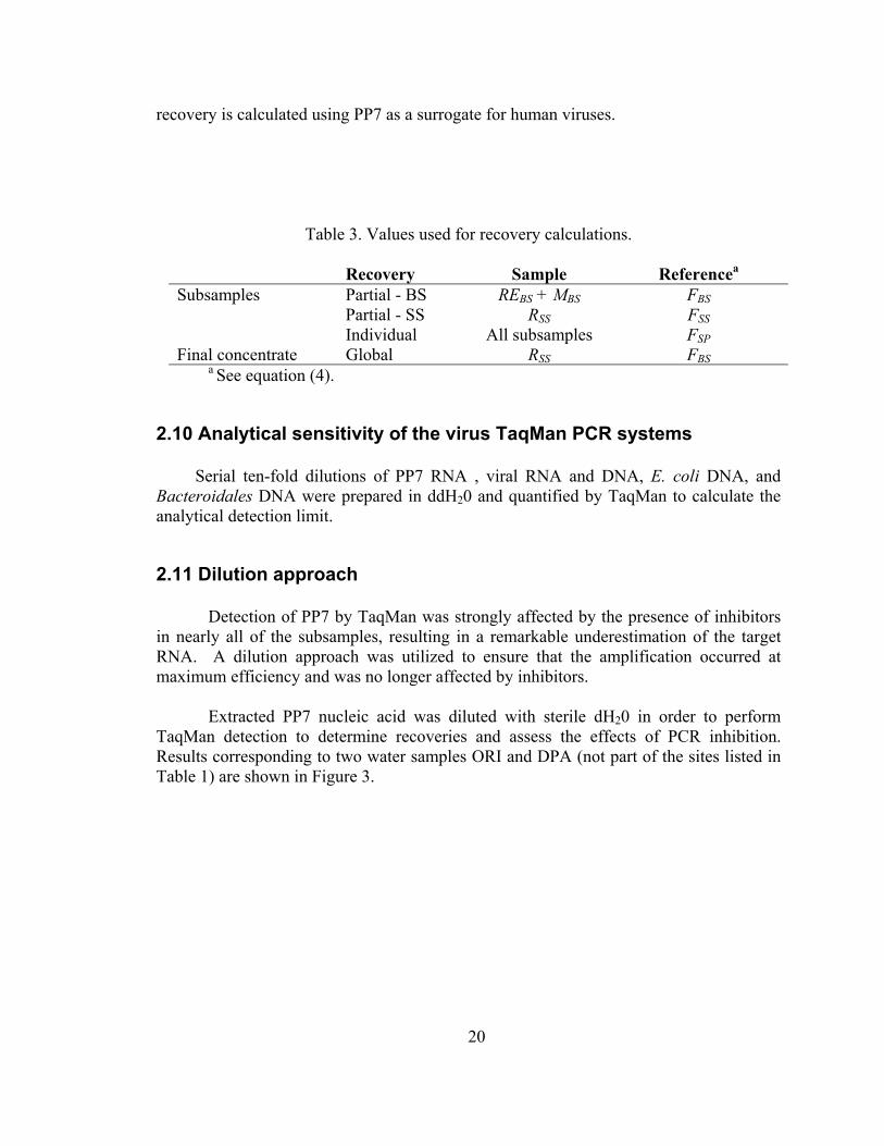
Table 3. Values used for recovery calculations.
Recovery Sample Referencea Subsamples Partial - BS REBS + MBS FBS Partial - SS RSS FSS Individual All subsamples FSP Final concentrate Global RSS FBS
a See equation (4).
2.10 Analytical sensitivity of the virus TaqMan PCR systems
Serial ten-fold dilutions of PP7 RNA , viral RNA and DNA, E. coli DNA, and Bacteroidales DNA were prepared in ddH20 and quantified by TaqMan to calculate the analytical detection limit.
2.11 Dilution approach
Detection of PP7 by TaqMan was strongly affected by the presence of inhibitors in nearly all of the subsamples, resulting in a remarkable underestimation of the target RNA. A dilution approach was utilized to ensure that the amplification occurred at maximum efficiency and was no longer affected by inhibitors.
Extracted PP7 nucleic acid was diluted with sterile dH20 in order to perform
TaqMan detection to determine recoveries and assess the effects of PCR inhibition. Results corresponding to two water samples ORI and DPA (not part of the sites listed in Table 1) are shown in Figure 3.
20

y = 3.4032x + 24.218R2 = 0.9868
10
15
20
25
30
35
40
45
0.0 0.5 1.0 1.5 2.0 2.5 3.0 3.5
Log [Dilution]
Ct
Sample ORI Sample DPA Control Lineal (DPA)
Figure 3. Dilution approach for the detection of PP7 by TaqMan.
A Ct value of 40 corresponds to a negative result by TaqMan. The first two points in Figure 3 for sample DPA are negative; further dilution of nucleic acid decreased the concentration of inhibitors and the detection signal was recovered (third point on line for DPA). Successive dilutions were assayed to the point where inhibitors did not affect the efficiency of amplification, as indicated by the linear range in Figure 3. Before that region, the detection is positive but any calculation based on those Ct values will underestimate the target number of PP7. Conversely, any calculation using Ct values from dilutions within the linear range will yield the same final target number. Ideally, the lowest dilution in this range should be used for the calculation in order to satisfy both linearity and sensitivity in terms of the detection limit. Alternatively, the y-intercept of the linear regression equation can be used as the theoretical Ct when there is no dilution (log 1 = 0).
In Figure 3, the ORI sample did not contain the high concentrations of inhibitors
present in DPA, as the undiluted reaction was positive (Ct = 30). However, the linear range was not observed until the third dilution, an indication that inhibitors still affected amplification.
When calculating the recovery of PP7 and E. coli, several dilutions were measured,
and an analysis of inhibition was performed as described. The lowest dilution within the linear range was also used to calculate the detection limit of viruses (using PP7 analysis) and Bacteroidales (using E. coli analysis).
21

2.12 Calculation of sample detection limits One-tube TaqMan RT-PCR. The sample detection limit (SDL) was calculated for each original volume of filtered water according to the following equation. This equation applies to all one-tube TaqMan reactions.
RV
VV
VI
VDS RF
ex
el
TSDL ×××= (5)
where SDL (pfu or vp/L) is the sample detection limit, D (pfu or vp) is the TaqMan analytical detection limit, I (mL diluted template/mL non-diluted template) is the dilution factor required to relieve TaqMan inhibition VT (mL diluted template) is the volume of nucleic acid template added to TaqMan reaction, Vel (mL eluted RNA or DNA) is the eluted volume from the extraction of the final concentrated sample, Vex (mL final sample) is the volume of concentrated final sample that was extracted, 10 mL in this study, VRF (mL final sample) is the volume of the final concentrated water, R is the overall filtration recovery, and VS (L) is the volume of the original water sample Two-tube TaqMan RT-PCR. The sample detection limit (SDL) was calculated for each original volume of filtered water according to the following equation. The number 0.5 was added to the denominator to account for a two-tube RT TaqMan reaction, during which the RNA is diluted to produce cDNA (50µL RNA/100 µL cDNA).
RV
VV
VI
VDS RF
ex
el
TSDL ×××=
5.0 (6)
2.13 Calculation of virus and cell concentrations when detected
When a positive signal was received from TaqMan, the concentration of the microorganism in the original water sample was calculated with equation 7. The concentration utilized the recovery of the appropriate surrogate (PP7 for viruses and E. coli for bacteria) in order to predict the amount of target lost during the filtration process.
RV
VV
VDilution
VTionConcentrat RF
ex
el
TS
×××= (7)
All values remain the same as the detection limit calculation but the analytical detection
22

limit is replaced by T, the viral particles or cells measured in the TaqMan reaction. Additionally, I, or the dilution factor of inhibition, is replaced by the dilution factor at which the target was detected.
2.14 Quality Assurance and Quality Control Procedures
All the above procedures are detailed in Appendix B.
23

3.0 RESULTS
3.1 Analytical sensitivity of the virus TaqMan PCR systems
RT-PCR for PP7 was performed with four different sets of reverse primers plus a combination of all primer sets to test the sensitivity and efficiency of amplification. While only three concentrations are shown in Table 4, linearity was observed over 7 orders of magnitude; therefore, the efficiency of amplification was constant for different target concentrations. Since the slopes of the straight lines obtained for the different sets of primers were similar, there was no significant difference in amplification efficiency for the primers. However, the combination of all 4 reverse primers in the RT reaction mix resulted in lower Ct values for each concentration of PP7 analyzed. Lower Ct values translate into an increase in sensitivity and a decrease in detection limit, two crucial factors for the detection of any pathogen present at low concentrations in environmental samples. This TaqMan system was used to construct a standard curve for the determination of the actual target number of a sample based on the measured Ct.
Table 4. Mean Ct values for different amounts of PP7 from one-tube TaqMan RT-PCR
Reverse primers PP7 phage
(pfu)
PP7-323r
PP7-355r
PP7-366r
PP7-320r All 4
primers 1.58 × 10⁶ 18.72 ±0.07 19.66 ±0.13 19.01 ±0.01 18.33 ±0.13 17.39 ±0.15 1.58 × 10⁴ 25.16 ±0.31 24.71 ±0.32 24.41 ±0.07 24.16 ±0.02 22.79 ±0.29 1.58 × 101 35.94 ±1.03 34.25 ±1.01 34.03 ±2.48 34.34 ±0.35 33.30 ±1.21 Efficiencyb 0.97 1.15 1.11 1.03 1.03
bAccording to equation (3)
The detection limit for PP7 by RT-PCR using the combination of all 4 reverse primers was 6 pfu. Further improvements in lowering the detection limit involved the addition of 600 ng of random hexamers to the one-tube RT-PCR mixture and detection using two-tube RT-PCR. Random hexamers had no appreciable effect on the Ct values, while changing to a two-tube RT-PCR reaction lowered the detection one order of magnitude, to 0.4 pfu for PP7. Consequently, the detection of human enterovirus was performed by two-tube TaqMan RT-PCR and the detection limit was 10 viral particles. Adenovirus was tested at both the RNA level (two-tube RT-PCR) and DNA level (TaqMan PCR) and the detection limit was 10 viral particles.
24

3.2 Natural water samples
Water samples were subjected to physicochemical and microbiological water quality analysis. It is important to note that the field samples varied considerably. The turbidity of all samples is presented in Figure 4, and conductivity is presented in Figure 5. Figure 6 presents the total and fecal coliform data in form of a bar chart. The range of conductivity was the largest, varying from less than 1 mS for some samples to more than 3,000 mS (Figure 5). In terms of microbial indicators, the levels of both total and fecal coliforms are remarkably high for some sites (Figure 6), clearly exceeding California state regulations for ambient water quality standards listed in Table 6. Of the 56 sites investigated, 28 sites exceeded the California standard for total coliforms (standard for a single measured sample), and 24 exceeded the California standard for fecal coliforms.
0
50
100
150
200
250
300
350
0 10 20 30 40 50 6
Sample
Turb
idity
(NTU
)
0
Figure 4: Range of turbidity of environmental samples
25

0
500
1000
1500
2000
2500
3000
3500
0 10 20 30 40 50 6
Sample
Con
duct
ivity
(mS)
0
Figure 5: Range of conductivity of environmental samples.
0
5
10
15
20
25
<500 500-1000 1000-5000 5000-10000 10000-100000
100000-1000000
<2000000
Coliforms (MPN/100 mL)
Num
ber S
ampl
es
Total ColiformFecal Coliform
Figure 6: Range of total and fecal coliforms present in environmental samples.
26

Table 6. Ambient water quality criteria for marine and fresh waters used for full contact recreation.
Indicator Organisms California Standard
(MPN/100 mL) Total Coliform
Single sample 10,000 Geometric meana 1,000
Fecal Coliform Single sample 400 Geometric meanb 200
a Geometric mean based on a minimum of 5 samples over a 30-day period.
.
3.3 PP7 recovery from spiked environmental water samples
The recovery of PP7 was determined in individual subsamples and in the final concentrated water according to the description in Materials and Methods. Filtration subsamples. The recoveries for PP7 at each individual step during the process were calculated using equation (4) and Table 3 (see Materials and Methods), taking as a reference the spiked PP7 amount (FSP). Table 7 presents recovery data from selected samples from Sacramento and Yolo Counties. The results show that there is a clear benefit, in terms of recovery, by performing the TWEEN recirculation step after concentration in the large filtration system. The recovery of PP7 for the subsample REBS is almost always larger than the recovery for RBS. Conversely, the subsample MBS does not have the same importance when the sample is relatively clean. Additionally, the information related to the permeate subsamples for both systems is useful as a quality control test of the integrity of the ultrafiltration membrane, since PP7 virus should not pass through the membrane and into the permeate.
Determinations of total number of spiked PP7 virus were compared using two
assays in order to evaluate infectivity and nucleic acid extraction methods after the sample was filtered / concentrated. The total virus count determined in the plaque assay (PA) should be lower than that determined by TaqMan (TQ) (TQ/PA >1), since not all detected viral particles will be infectious and multiple virus particles can result in one plaque forming unit. Additionally, the ratio of total virus by both methods could indicate whether the filtration process and/or sample constituents impact infectivity, or if nucleic acid extraction efficiencies are less than optimal. To test this assumption, deionized water was spiked and concentrated following the same procedure as the environmental samples to represent the best-case scenario for both PP7 infectivity and extraction success. The ratio TQ/PA was 8 and 12 for FBS and RSS, respectively. As expected, ratios are greater than one for both the feed and retentate. These subsamples represent two extremes in terms of concentrations of sediments, chemicals, and biological constituents that
27

contribute to reduced extraction and purification efficiencies. For the environmental water samples, ratios are variable (Table 7) and in many cases greater than one for FBS but usually lower than the unit for RSS. Besides extraction, the ratio may be affected by TaqMan detection (PCR inhibitors) and plaque assay (reduced infectivity).
Table 7. Recovery of PP7 from the spiked amount by Plaque Assay (PA) and TaqMan (TQ). Location: Sacramento and Yolo Counties.
RATIO
Recovery Dilution Recovery TQ/PADilution (%) (%)
B FBS 1:1,000 93.7 ND 10.9 0.07RBS 1:10,000 20.1 1:50 10.1 0.30
REBS 1:100,000 91.8 1:50 20.4 0.13MBS 1:10,000 0.6 ND 0.1 0.05PBS ND 0.0 ND 0.0
REBS + MBS 1:10,000 46.0 1:10 10.8 0.14S 1:100,000 3.3 1:1000 1.5 0.28
FSS 1:10,000 44.0 1:10 7.2 0.10RSS 1:100,000 23.6 1:100 21.1 0.54PSS ND 0.0 ND 0.0
UC FBS 1:1,000 54.5 ND 76.2 0.84RBS 1:10,000 7.6 1:10 6.9 0.55
REBS 1:10,000 39.8 1:10 22.6 0.34MBS 1:10,000 2.9 1:10 0.6 0.12PBS ND 0.0 ND 0.3
REBS + MBS 1:10,000 27.1 1:50 25.6 0.57S 1:10,000 0.5 1:100 0.1 0.12
FSS 1:10,000 39.4 1:10 20.6 0.31RSS 1:100,000 27.6 1:100 59.4 1.30PSS ND 0.0 ND 0.0
BY PLAQUE ASSAY (PA) BY TAQMAN (TQ)Sample
ND: non-diluted
A summary of the recoveries from all samples filtered using the original filtration system is presented in Table 8. The global recoveries were determined for RSS from the spiked PP7 amount, and the partial recoveries were calculated for each filtration system. The table presents data for the first set of samples only and reflects recoveries obtained with the original filtration system. It should be noted that the partial recoveries cannot be linearly combined to obtain the global one from FSP, since they are calculated using different references (see equation (4) and Table 3 in Materials and Methods). The objective of analyzing the partial recoveries was to assess each step in each filtration system separately; therefore, the initial and final subsamples involved in those processes were used for the calculation.
28

Table 8. Recovery of PP7 by plaque assay (PA) and TaqMan (TQ)
Recovery for PP7 Partial - BS Partial - SS Global from FSP Sample PA TQ PA TQ PA TQ (%) (%) (%) (%) (%) (%)
San Diego Co. LPE 35.6 141.9 86.1 ND 43.4 8.1 SMC 63.7 117.2 40.5 33.0 9.9 29.7 SDR 78.6 177.1 38.0 48.6 22.6 63.5 CH 194.3 50.4 40.9 41.8 9.5 37.6 SLR 73.6 3.4 93.4 17.7 66.4 30.6 Solano Co. CAR 196.4 68.9 61.2 54.9 11.3 1.3 Contra Costa Co. ORI 182.9 103.3 67.1 71.5 10.0 31.1 Alameda Co. CV 396.1 121.9 60.4 27.7 87.2 13.8 COL 339.7 122.9 80.9 24.5 126.3 6.2 Sacramento Co. B 98.7 188.5 53.7 292.8 23.6 21.1 DP 77.5 127.5 78.0 83.1 10.8 2.8 Yolo Co. WD 95.6 142.1 60.4 51.4 10.4 3.4 UC 78.2 30.4 70.0 288.8 27.6 59.4 Fresno Co. MAD 427.5 40.9 45.9 82.4 49.4 3.8 FO 465.5 113.8 79.2 31.6 73.2 1.4 ME 760.1 21.4 163.0 47.6 23.2 2.9
ND: not determined
It is important to remark that the results presented in these tables correspond to the first set of samples, and their evaluation permitted modifications to be made to the filtration systems to improve recoveries. For example, the recoveries obtained for the subsamples FBS were always lower than 100%, and in some occasions lower than the corresponding recovery for REBS subsample (see Table 7). Such a behavior can be explained by remembering that FBS was sampled after 10 minutes of recirculation (no permeation) of the spiked original water. During that recirculation period PP7 may have absorbed to the lines, plastic feed tank, and filtration membrane, but could later be recovered in the liquid phase (REBS) when the elution with glycine and tween was performed. Analysis of Table 8 showed that the partial recoveries for the large system (using FBS as reference) were usually larger than 100%, either by plaque assay or by TaqMan, reflecting again the problem of the viruses being attached to the lines, feed tank, and filter during the initial recirculation period, as explained before.
Attachment of the virus to the plastic feed tank was confirmed by sampling the
tanks’ internal surfaces with sterile wet cotton swabs (data not shown). Other researchers reported that the use of a blocking agent prior to filtration consisting of a solution of 5% calf serum (Oshima, 2001) reduced binding of virus to the filters. In the present study, addition of this step not only resulted in reduced PP7 recovery, but also was time-intensive and expensive. To reduce loss of PP7 during mixing using recirculation, the 50-
29

L plastic feed container was replaced with a stainless-steel 100-L vessel with motorized mixing impellers.
Evaluation of the recoveries from each filtration step (Table 7) revealed the
importance of the recirculation step, during which glycine and tween are passed through the membrane repeatedly. This step was not initially performed on the small filtration system, but an elution step was added after analyzing the first set of samples. Elution on the small system was performed using the same glycine and Tween solution and manual syringe pumping. These improvements increased overall virus recovery and stabilized FBS PP7 concentrations. The corresponding improvement of PP7 recovery with each change in the filtration procedure, as analyzed with TaqMan, is displayed in Figure 7. The mean recovery of all samples processed by each method is presented with the standard error; the improvement in recovery with the addition of mechanical mixing and elution of the small system was statistically significant.
0
10
20
30
40
50
60
70
80
Original Procedure Plus Mechanical Mixing Plus Elution of SS
% P
P7 R
ecov
ery
by T
aqM
an
Figure 7: Average PP7 recoveries from samples filtered using the original filtration procedure and two subsequent alterations: (1) a stainless steel tank with mechanical mixing and (2) elution of the small filter with glycine and Tween. Error bars refer to the standard error of the mean. A one-way ANOVA with α=0.05 showed a statistically significant difference between the original procedure and the improved method including mechanical mixing and elution.
Concentrated water samples. The nucleic acids of the concentrated water sample obtained after the two sequential ultrafiltrations were extracted according to the procedure described in Materials and Methods. An aliquot was analyzed by TaqMan, and the PP7 recovery was calculated using the spiked amount FBS (see equation (4) and Table 3).
30

Since the nucleic acids extracted by this procedure were used to detect enterovirus and adenovirus, the recovery values of PP7 require special attention. If the quantitative detection of viruses is positive it is possible to calculate the actual number of viruses present in the original water sample, assuming that the recovery for that specific virus is at least the value obtained for PP7. Conversely, if the detection of viruses is negative, the possibility of having a false negative should be considered. In that case, the recovery for PP7 together with the detection limit of the specific method allows the estimation of the upper limit for the detection of that specific pathogen (see Table 10).
Recoveries for PP7 for the final concentrated water sample are presented in Table
9. After improvements were made to the filtration system by adding mechanical mixing and elution of the small filtration membrane, PP7 recoveries improved drastically, which in turn lowered the enterovirus and adenovirus detection limits. Only samples filtered with this system are presented. The average PP7 recovery was 64 ± 9%.
Table 9. Final concentrated water recoveries for PP7 by TaqMan
Sample Group Sample % PP7 Recovery
Los Angeles -2 MAL 9.7 TRA 50.0 TPN 57.7 SMO 97.9 PCH 95.3 Fresno - 2 FO 2 75.7 MEN 2 82.0 MAD 2 53.3 San Diego - 3 SDN-3 65.0 SLR-3 68.1 SDR-3 37.3 CHO-3 77.8 ENC-3 44.1 Los Angeles -3 EFS 87.9 TRA 87.8 MAL 76.7 TPN 74.9 SMO 45.8 Bay Area - 3 CAR 53.0 ORI 82.1 CWC 44.8 COL 38.2
31

3.4 Detection of human adenovirus and enterovirus by TaqMan PCR
The convention in scientific literature is to report analytical PCR results for water samples as merely positive or negative, without regard to detection limits associated with the tests. Detection limits are important when considering that contact with contaminated water may pose a health risk if concentrations of pathogens reach a critical level. The factors that influenced detection limits in these studies were myriad, with some having more of an impact than others. Such factors included the volume of original sample, recovery efficiency, final volume of retentate (RSS), volume extracted (of nucleic acid), eluted nucleic acid volume, volume of nucleic acid added to PCR reaction, and inhibition of PCR. Of these, sample volume, recovery efficiency, and PCR inhibition were the most influential on detection limits. The pathogen results from the samples filtered with the improved filtration system are presented in Table 10 with their corresponding detection limits. The traditional measures of microbial water quality (total coliforms, fecal coliforms, and E. coli) are also presented for comparison.
Table 10. Microbial water quality and occurrence of adenovirus and enterovirus
Coliforms (MPN / 100 mL) Sample Group Sample
Total Fecal E. coli
Adenovirus (vp / 100 mL)
Enterovirus (vp / 100 mL)
Virus Detection Limit (vp / 100 mL)
Los Angeles -2 MAL-2 16000 80 63 Neg Neg 4637 TRA-2 1300 20 31 Neg Neg 141 TPN-2 2400 1100 663 Neg Neg 2203 SMO-2 50000 1700 934 Neg Neg 17 PCH-2 5000 80 63 Neg Neg 2344 Fresno - 2 FO-2 11100 11100 N.D.§ Neg Neg 719 MEN-2 28600 28600 N.D. Neg Neg 149 MAD-2 780000 2860 N.D. Neg Neg 402 San Diego - 3 SDN-3 30 <2 N.D. Neg Neg 1219 SLR-3 3000 300 N.D. Neg Neg 792 SDR-3 17000 1100 N.D. Neg Neg 3050 CHO-3 13000 1300 N.D. Neg Neg 1283 ENC-3 17000 170 N.D. Neg Neg 1306 Los Angeles -3 EFS-3 500 <20 <10 Neg Neg 96 TRA-3 2400 300 86 Neg Neg 781 MAL-3 500 20 <10 Neg Neg 1382 TPN-3 800 500 620 Neg Neg 670 SMO-3 2400 500 408 Neg Neg 875 Bay Area - 3 CAR-3 30000 230 230 Neg Neg 184 ORI-3 14000 500 500 13* Neg 116 CWC-3 30000 500 500 Neg Neg 186 COL-3 22000 800 300 Neg Neg 1500
* Adenovirus 40/41 § Not determined
For all samples filtered, including ones not shown, there was only one case of
positive detection of adenovirus 40/41. However, a careful analysis is necessary since there could be false negatives. The calculation of the detection limit incorporates an
32

inhibition factor, and it reflects the concentration at which viruses would positively be detected without any inhibition effects, based upon inhibition analysis of PP7. Therefore, even though it may be possible to detect viruses below this detection limit, the positive signal is expected to be affected by inhibitors. Alternatively, a negative signal does not indicate the absence of viruses in the sample. It is more accurate to say that if there were viruses in the water samples, then their concentrations were lower than the detection limit. The positive detection of adenovirus in sample ORI-3 occurred well below the calculated upper detection limit, which implies that the detection occurred during a range of inhibition for the TaqMan measurement. As such, the calculated concentration of adenovirus 40/41 in the sample may be an underestimation of the true value.
The detection limits varied widely from site to site. The highest detection levels
correspond to sites that were heavily contaminated with fuel, oil, or solids; and all these sites were direct runoff from freeways and roads. Such compounds interfere with the overall detection scheme in a complicated manner that cannot be predicted via the physiochemical measurements conducted herein. Further improvements in the clean up of nucleic acid are necessary to remove PCR inhibitors and interfering compounds from the dirtiest samples. The removal of inhibitors will lower detection limits and provide more consistent virus detection. An alternate approach to sensitive PCR amplification in heavily contaminated samples is presented in Appendix A.
3.5 Microbial source tracking using total and a subset of human Bacteroidales markers Surface water quality is strongly influenced by increasing anthropogenic activities, as natural waters receive a diversity of point and non-point source pollution. For the protection of human and ecosystem health, it is important to determine the abundance and diversity of human pathogens in these waters, as well as to identify the sources of fecal contamination.
Microbial source tracking (MST) is a method by which host-specific contributions of fecal contamination to water bodies can be determined. Its potential lies mainly in determining sources of non-point fecal pollution, which otherwise may be difficult to establish. Many drains represent a mixture of various non-point source inputs: human or animal fecal input due to runoff from agricultural lands receiving biosolids; combined sewer outflows; bovine feces from feedlots or other farming activities; site-specific wild life droppings contributed by birds, horses or elk; and feces from domestic pets like cats and dogs. In addition there may be non-storm water discharges to storm sewers. The present study attempted to explore MST based on two Bacteroidales Taqman assays available for total Bacteroidales (Dick and Field 2004) and a subset of Bacteroidales 16S rRNA sequences derived from human feces, the HF183 genetic marker (Seurinck et al. 2005).
Microbial source tracking was performed on seventeen samples taken after March 2005 using the optimized filtration procedure. To calculate target bacterial concentrations
33

and detection limits, a benign strain of E. coli was spiked together with PP7 into the water samples before filtration. The recovery and inhibition of E. coli were analyzed in the same manner as PP7, and these values were used for Bacteroidales calculations. Recoveries for individual samples are presented in Table 11. For the seventeen samples, global E. coli recovery averaged 67 ± 13%, which correlated well with the average PP7 recovery of 64 ± 9% for the same samples. However, it is important to note that the PP7 and E. coli recovery values were different for a particular sample, indicating that an appropriate surrogate is necessary to simulate the behavior of a target organism.
Table 11: E. coli and PP7 recoveries from samples upon which microbial source tracking was performed.
Sample Group Sample % E. coli Recovery % PP7 Recovery
Fresno - 2 FO 2 73.5 75.7 MEN 2 11.2 82.0 MAD 2 70.6 53.3 San Diego - 3 SDN-3 92.1 65.0 SLR-3 17.3 68.1 SDR-3 9.1 37.3 CHO-3 52.7 77.8 ENC-3 7.4 44.1 Los Angeles -3 EFS-3 92.4 87.9 TRA-3 83.4 87.8 MAL-3 86.3 76.7 TPN-3 16.5 74.9 SMO-3 45.2 45.8 Bay Area - 3 CAR-3 30.6 53.0 ORI-3 17.2 82.1 CWC-3 84.1 44.8 COL-3 25.1 38.2
The results from total Bacteroidales and the specific human Bacteroidales marker HF183 are presented in Table 12 with the concentration of cells detected and the calculated detection limit at which inhibition is not a concern. The table also lists the ratio of human HF183 Bacteroidales to total Bacteroidales. For the seventeen samples upon which microbial source tracking was performed, bacterial DNA sequences from the Bacteroidales group were detected in fifteen. This indicates that a majority of the samples reflected some non-point sources of fecal contamination. Of the fifteen samples containing Bacteroidales, human marker HF183 Bacteroidales was detected in fourteen samples. The percentage of human to total Bacteroidales ranged from 0.05% to 78.87% within the Fresno area alone. All Fresno sites were sources of highway runoff, indicating a high range of variability between similar sites within a close area. Assuming that the total Bacteroidales assay adequately accounts for the diversity of 16S rRNA sequences in that group, the MAD-2 sample (with
34

a ratio of 78.87%) is expected to reflect mostly Bacteroidales sequences of human fecal origin. For this site, the detected concentrations were well above the calculated detection limits indicating that the measured concentrations were truly quantitative. Therefore, a more in-depth study of non-point sources in the area would be interesting. All detected values below the detection limit may be an underestimation of the actual concentration, since they were detected in the range of inhibition effects. Likewise, all negative values do not indicate the absence of Bacteroidales but rather a concentration less than the detection limits. Finally, since the human Bacteroidales assay only targeted one known human marker HF 183, the ratio of human to total Bacteroidales is expected to be a minimum value. Table 12: Total Bacteroidales and human Bacteroidales concentrations and detection limits
Total Bacteroidales Human Bacteroidales* Sample Group Sample Measured Cells
(copy number/ 100 mL)
Detection Limit (copy number/ 100 mL)
Measured Cells (copy number/ 100 mL)
Detection Limit (copy number/ 100 mL)
Ratio Human/ Total (%)
Fresno - 2 FO 2 1151 1481 6 1851 0.51 MEN 2 74879 2188 36 2735 0.05 MAD 2 3314 606 2613 757 78.87 San Diego - 3 SDN-3 N.D.§ 1719 N.D. 2149 N.Aγ
SLR-3 1343 6237 N.D. 7796 N.A. SDR-3 N.D. 12554 N.D. 15693 N.A. CHO-3 2216 3786 298 4733 13.47 ENC-3 4363 15615 132 19519 3.02 Los Angeles -3 EFS-3 503 183 12 229 2.36 TRA-3 3280 329 27 412 0.82 MAL-3 12225 491 20 614 0.17 TPN-3 10496 1214 1916 1517 18.26 SMO-3 4730 355 78 444 1.65 Bay Area - 3 CAR-3 78064 636 2811 796 3.60 ORI-3 43581 1109 16529 1386 37.93 CWC-3 14197 198 1548 248 10.90 COL-3 99921 912 4649 1139 4.65 * using HF183 Marker § N.D. = not detected γ N.A. = not applicable
3.6 Detection of other human pathogens including Cryptosporidium and Francisella In addition to adenovirus group A, B, C, 40/41, and enterovirus, sixteen of the fifty-six samples were analyzed for the presence of other human pathogens, for which TaqMan qPCR assays had already been designed. These samples were all filtered using the original, non-optimized procedure. The TaqMan assays included Salmonella spp.,
35

Listeria monocytogenes, Francisella tularensis, Cryptosporidium spp., and Toxoplasma gondii. Four samples were positive for Cryptosporidium spp. using an unpublished assay specific for the genus (C. Leutenegger, personal information), and one was positive for Francisella tularensis. The concentrations of the detected pathogens are presented in Table 13.
A surrogate for protozoa was not spiked into the original sample, so appropriate recoveries for Cryptosporidium could not be calculated but concentrations were instead based on recovery of bacteriophage PP7. Considering that some samples were positive for Cryptosporidium and Francisella tularensis, future pathogen monitoring programs should include these organisms in addition to adenovirus and enterovirus assays.
Table 13: Detection of Cryptosporidium spp. and Francisella tularensis1
Sample
Cryptosporidium spp. (copy number / 100 mL)
Francisella tularensis (copy number/ 100 mL)
San Diego - 1 LPE SMC 15 SDR 64 CH SLR Bay Area - 1 CAR ORI CV COL Sacramento - 1 B 32 DP WD UC 19 Fresno - 1 MAD 145 FO MEN
1 No entry means the target organism was not detected. Detection limits are not calculated because the analyses were exploratory.
3.7 Survival of PP7, E. coli, and Bacteroidales fragilis nucleic acids in sampling containers
An experiment was conducted to determine whether viral and bacterial populations change significantly during holding times (often ranging from 3 to 20 hours). This study was conducted to determine if changes in the sampling protocol are necessary to avoid cell division or loss of the pathogens of interest.
36

Three samples (Sample # 1, Sample # 2, and Sample # 3) were studied. Each sample consisted of 18 liters of water that was collected in a clean 20-liter carboy. Sample # 1 was collected in a small drainage ditch in North Davis Creek. Sample # 2 was collected on the UC Davis campus from Putah Creek, just south of the Engineering III building. A third sample was used as a control and consisted of deionized water with 0.085% sodium chloride.
Negative control subsamples (10 mL from each sample) were taken previous to the addition of the spiked organisms in order to establish a baseline. Next, three different markers were spiked into each sample: 20 µL of PP7 (at a final concentration of ~ 109 vp/ml); 400 µL of Bacteroidales fragilis (at a final concentration of ~ 108 CFU/ml); and 400 µL of E. coli (at a final concentration of ~ 108 CFU/ml).
Each sample was shaken vigorously for 2-3 minutes after the addition of the spikes, and subsamples were taken and designated as the initial concentration (time 0). Subsequent subsamples were taken at varying time intervals (0 to 120 hrs).
As time permitted, the subsamples were immediately plated after collection for the enumeration of PP7 and E. coli on TSA and EMB plates, respectively. Additionally, 140 µL of each subsample was mixed with 560 µL of lysis buffer and stored at –200C for later extraction and TaqMan quantification. The same DNA/RNA extraction protocol (Qiagen QIAamp viral mini kit) was used for general sample analysis. Extractions were performed in triplicate, and quantification was performed in duplicate by TaqMan analysis.
Results from the EMB plates were difficult to analyze due to growth of bacteria other than E. coli. The E. coli colonies were unrecognizable among the bacterial colonies. However, the TSA plaque assays were successful. Using the data generated from plaque assays, which represents viable PP7, it was determined that:
• Sample # 1: The North Davis Creek sample showed viral viability loss of less than a
log within the first 24 hours of the experiment. A 2-log loss of viable viruses was observed after 45 hours.
• Sample # 2: The Putah Creek sample only had a slight loss within the first 45 hours, and still less than one order of magnitude decrease at hour 55.
• Sample # 3: Deionized water experienced much greater losses, with approximately a 3- log decrease within 55 hours.
Overall, the bacteriophage PP7 was able to retain its infectivity in the
environmental samples for at least 24 hours. Hence, samples can first be transported to the analytical laboratory before filtration and analysis if on site filtration is not feasible. Even more pertinent are the results of the TaqMan quantification of PP7, E. coli, and B. fragilis because the analysis of interest in this study was ultimately dependent on the survival of the nucleic acids in the sample. Significantly, bacteriophage PP7 demonstrated little to no loss in nucleic acids within the first 60 hrs (Figure 8).
37

10
100
0 20 40 60 80 100 120 140
Time (hrs)
Vira
l Par
ticle
Con
cent
ratio
n (1
04 v
p/10
0ml)
sample # 1 sample # 2 sample # 3
Figure 8: PP7 enumeration by TaqMan for three independent replicates over time
Each replicate showed some fluctuation of the total concentration of
bacteriophage over time. This result is to be expected considering the many steps that can affect extraction efficiency between each time period, plus errors in pipetting, as well as other issues. Overall, there is not a significant loss in viral nucleic acid stability with a holding time as long as 48-60 hours. Similarly, results for E. coli bacterial nucleic acid detection are consistent with those of bacteriophage PP7 (Figure 9). Bacteroides fragilis DNA was also detected without apparent loss of target (Figure 10).
38

1
10
100
1000
0 20 40 60 80 100 120 140
Time (hrs)
E. c
oli C
once
ntra
tion
(x10
4 CFU
/100
ml)
sample # 1 sample # 2 sample # 3
Figure 9: E. coli enumeration using TaqMan for three independent replicates over time
1
10
100
1000
0 20 40 60 80 100 120 140
Time (hrs)
Bac
tero
idal
es C
once
ntra
tion
(x 1
03 CFU
/100
ml)
sample # 1 sample # 2 sample # 3
Figure 10: B. fragilis enumeration using TaqMan for three independent replicates
39

Overall, it was concluded from this experiment that significant nucleic acid losses
of spiked marker organisms present in the water samples at the time of collection should not occur within the first 48-60 hours of collection. Assuming that target pathogens exhibit similar nucleic acid stability, these results suggest that it is possible to collect water samples in the field and then transport them back to the laboratory for processing (which may result in a holding time of up to 48 hours in some cases) without loss of surrogate. Hence the hollow fiber ultrafiltration technique is widely applicable for the analysis of large-volume field samples because processing may occur at a central location if no on-site filtration system is available.
40

4.0 DISCUSSION
The main goal of this study was to develop a molecular-based approach to detect and quantify pathogens associated with storm water, including viruses, bacteria, and protozoa. An ultrafiltration method was optimized to concentrate 100-L samples of water to ensure a representative grab sample for the reliable determination of pathogens present at low levels. In addition to the sample concentration method, quantitative PCR assays were developed and validated to detect several viruses, bacteria, and protozoa. When these organisms were not detected, a quantitative detection limit for each organism and sample was defined. Finally, quality assurance and control guidelines were written to ensure the utility of these methods by other laboratories and government agencies.
Overall, the validated approach to pathogen determinations described in this work is a significant outcome of the research study. In addition, the detection of viruses was also compared with traditional coliform measurements of water quality. Quantitative PCR was used to detect several bacterial pathogens and the parasite Cryptosporidium. Microbial source tracking based on fecal Bacteroidales was explored as a method to further classify indicator organisms and fecal contamination derived from human and non-human hosts. A comparison of results with similar studies provides a starting point to discuss the major issues and potential advantages for monitoring pathogens in natural water samples.
4.1 Measures of Water Quality: Choosing the Appropriate Indicator Organisms The microbial safety and associated health risk of water is typically monitored using indicator organisms, such as total and fecal coliform bacteria, enterocooci and E. coli. To safeguard public health, the State of California has adopted water quality standards for average and single measurements of coliforms in a recreational water body. Indeed, public waters are often closed when a grab sample indicates high coliform counts. In this way, indicator organisms have become a widely accepted and applied measure to estimate human health risk. However, the assumption that the concentration of indicator organisms is related to the concentration of pathogens in recreational waters has been challenged in recent years.
Use of current indicator organisms as a sole guide for the presence of pathogens has come under considerable scrutiny, and it is becoming apparent that a correlation between indicator organism concentrations and pathogen concentrations in recreational waters may not exist (Smith and Perdek, 2004 and Schroeder et al., 2002). Furthermore, there are related studies that depict a strong correlation between indicator organism concentrations and non-human fecal pollution, which casts doubt on their use in assessing the biological quality of water and the associated human health risks of contact with the affected water (Grant et al, 2001).
41

One of the goals of this research was to test a relationship between conventional total and fecal coliform counts of small, 100-mL grab samples and the molecular-based detection of human pathogens concentrated from 100-L grab samples. Of the 56 samples analyzed during this study, 50% exceeded the California standard for total coliforms, and 43% exceeded the California standard for fecal coliforms. However, based on the adenovirus and enterovirus PCR assays, adenovirus was detected in only one sample, ORI-3. ORI-3 also exceeded the limit for total coliforms (measuring 14,000 MPN/100 mL compared to a standard of 10,000 MPN/100 mL) as well as the California limit for fecal coliforms (500 MPN/100 mL compared to 400 MPN/100 mL). Taken together these data imply that there is no strong correlation between coliform counts and the detection of human viruses within the reported detection limits. There are precedents for these findings. For example, a large epidemiological study that examined the health risks along Mission Bay Park in San Diego did not find any correlation between indicator bacteria and illness contracted by beachgoers (results presented at National Beaches Conference in San Diego, 2004). The question beckons whether indicator counts are an appropriate measure of ambient water quality. If the direct molecular-based detection of pathogens is a better measure, which pathogens are appropriate indicators? In the current study, two complementary approaches to detect microbial contamination in storm water were investigated. The first approach involved the direct detection of human pathogens by quantitative PCR. All samples were analyzed for adenovirus and enterovirus, while a subset was additionally screened for other bacteria and protozoa. Adenovirus, Cryptosporidium, and Francisella were detected and quantified in a total of six different samples. However, the detection of one pathogen did not coincide with the detection of any other pathogen. Therefore, one pathogen cannot be used as an indicator for all pathogens, and future water samples would need to be screened for a suite of pathogens in order to directly detect and quantify these in water. The method developed in this study is highly suited for such an approach, and the pathogens causing the most incidence of illness in a particular area could be measured simultaneously using quantitative PCR. The benefits of this method include directly detecting the pathogens of interest and immediately obtaining a quantitative concentration or detection limit. The data obtained provide a maximum amount of specificity and correlation with human health risk. The second approach to detecting microbial contamination in storm water involved the quantification of Bacteroidales and microbial source tracking. This method (Bernhard and Field 2000) has been used in a number of studies, including the Mission Bay Bacterial Source Identification Study (Gruber et al. 2005). That investigation revealed that high indicator counts can be attributed to non-human sources like birds near beaches. Interestingly, no signal for human-derived 16S rRNA genes of Bacteroidales was found at storm drains. The Bacteroidales group consists of anaerobic, fecal bacteria, which are good indicators of fecal pollution in a body of water. In comparison to coliform counts, the molecular-based detection of Bacteroidales offers a precise method and specific target group, whereas a variety of microbial groups contribute to coliform counts. Both human and non-human sources release Bacteroidales and coliform-forming bacteria to water sources, hence microbial source tracking is a powerful tool to classify the types
42

of contamination. The genetic sequences of recently published human Bacteroidales strains have made it possible to quantify the fraction of total Bacteroidales that belong to that specific human marker. In contrast to earlier studies, the present report attempted to quantify host-specific Bacteroidales sequences in storm water using real time PCR. At the time the preliminary sample analysis was conducted, no quantitative PCR assays for non-human Bacteroidales sequences were available. For this reason, only the ratio of the human marker HF 183 (Seurinck et al. 2005) to total Bacteroidales (Dick and Field 2004) was determined. Both quantitative PCR assays are based on the pioneering work of Kate Field and co-workers. The HF 183 assay was developed by Willy Verstraate and co-workers based on the Field sequences. The ratios obtained in the present study are interesting because of their high variability. Although it is not possible to fully interpret these preliminary results in light of the fact that the true frequency of the HF 183 marker among human-specific Bacteroidales sequences is unknown, the high ratio of 0.79 at one Fresno site would suggest a high likelihood of human sources of indicator bacteria. The rapidly growing database of Bacteroidales sequences from a variety of animal sources will make it possible to design PCR primers and probes that quantify the contribution of Bacteroidales-fecal contamination by specific animals. This method has a huge potential for tracking the source of microbial contamination, and it is useful in indicating fecal contamination. However, the research area is new, and a correlation between human-derived Bacteroidales sequences and the presence of human pathogens has not yet been established. The approach developed in this task order makes such a comparison technically feasible.
4.2 Detection of Pathogens
One of the major benefits of the method developed in this study is that it provides quantitative information for each pathogen of interest, regardless of whether the pathogen was positively detected. By spiking the water samples with non-pathogenic model organisms, the loss and recovery of viral and bacterial organisms during the filtration process was calculated. The recovery of the spiked organisms and the analysis of PCR inhibition using these organisms are key parameters in the calculation of pathogen detection limits. Using this approach, a non-detect signal correlates with a numerical detection limit, which is defined as the minimum concentration at which a pathogen can be positively detected and analysis is not affected by inhibition. For the 22 samples that were concentrated with the optimized filtration procedure, the average recovery of PP7 was 64 ± 9%. Inhibition analysis showed that the samples were not inhibited at dilutions ranging from 1 to 1:100. Since all parameters in the detection limit calculation were relatively constant, including recovery rates, inhibition was the major factor controlling the viral detection limit. This is shown in Figure 11 with a plot of detection limit versus inhibition factor in which a strong linear correlation is evident. The plot also shows that as the inhibition factor increases, the corresponding detection limits become more scattered.
43

R2 = 0.8836
0
500
1000
1500
2000
2500
3000
3500
0 20 40 60 80 100 120
Inhibtion Factor
Dete
ctio
n Li
mit
(vp/
100
mL)
Figure 11: Detection limit versus inhibition factor for all samples concentrated with the optimized filtration procedure.
For the samples concentrated with the optimized procedure, the virus detection limits ranged from 17 to 3050 vp/100 mL, and the presence of PCR inhibitors was responsible for high detection limits. Given this correlation, more efficient removal of PCR inhibitors during nucleic acid extraction will lower detection limits and increase the ability to detect low concentrations of pathogens in environmental samples. Future method development should focus on inhibitor removal, since it controls the detection of any target organism using PCR. When a pathogen is positively detected, it is possible to calculate its concentration in the original water sample. In the present study, pathogens were detected in six samples: adenovirus 40/41 in one, Cryptosporidium in four, and Francisella tularensis in one.
A previous study attempted to develop a practical monitoring technique based on PCR that would indicate the probability of the presence of bacterial pathogens in waters (Yanko et al. 2004). By contrast, the present study focused on viruses but developed methodology to include bacteria as target organisms. The study by Yanko and colleagues involved a plethora of sampling locations and site types, including untreated surface water, urban runoff-influenced streams, and wastewater effluents. A large database was compiled with results from 200 samples collected from all parts of the country, with the majority of the sites being located in California. The work focused on larger volumes of water than might be typically seen in similar studies, with the largest water samples consisting of 10 liters. In contrast the present report used 100-L volumes. The procedure consisted of pressure filtration with glass filters (Whatman GF/F), which were overlaid with diatomaceous earth. The diatomaceous earth is thought to increase bacterial capture as well as extend the volume of water that can be processed through a single filter.
44

The researchers found that environmental inhibitors were interfering with the amplification efficiency of the target sequences, and that available treatment techniques were labor intensive and inefficient. As such, concentrates were allowed to grow in non-selective broth prior to extraction and PCR analysis. This method was found to be useful for the detection of a handful of pathogens in a series of collected surface water samples. The pathogens detected and their percentages of positive detection within the data set collected were: Aeromonas hydrophila (90%), Salmonella spp (72%), toxigenic coliforms (34%), and Yersinia enterocolitica (1.5%). Others have reported selective enrichment as an excellent way to recover bacterial pathogens present in biosolids at very low concentrations (Burtscher and Wuertz 2003). It should be noted, however, that as a consequence of bacterial growth, the bacterial densities are no longer representative of cell numbers in the original sample.
Yanko et al. (2004) also made an effort to optimize filtration of 1-liter samples with membrane dissolution and direct PCR detection (non-culture based). They found that the volume of water that could be concentrated from environmental samples was simply too low to permit target detection with this method. The detection frequency increased as the volume of the sample size increased. This result points to a need for an effective concentration procedure to detect most pathogens of concern in water, all of which are present at varying concentrations. Recovery studies were performed for all filtration schemes, but recovery was apparently not determined for the actual samples analyzed. Likewise, detection limits were not reported (Yanko et al. 2004).
The issues raised in the previous study were evaluated in the present investigation
and led to the selection of a hollow-fiber ultrafiltration system suitable for processing large water volumes. Both inhibition and surrogate recovery enter into the calculation of detection limits and non-detects are critically evaluated in light of the latter.
45

5.0 CONCLUSIONS
The concentration of large volumes of water is essential for the detection of pathogens that are present in very low concentrations since it ensures low detection limits and a high degree of confidence in the data obtained when pathogens are determined to be absent. Additionally, filtration of large volumes of water statistically improves the chances of finding pathogens when a grab sample is taken.
The ultrafiltration systems utilized in the current study were appropriate for the
efficient concentration of water samples. The methodology is simple, and the portable design offers the advantage of performing the first concentration step in the field, thus diminishing the risks and the costs of transportation and handling of large volumes of sample. However, results obtained in this study support the conclusion that water samples can be stored for at least 24 h and transported to a suitable laboratory for pathogen and MST analysis. This flexibility gives regulatory agencies and permittees several options when conducting monitoring efforts.
The recovery efficiencies for the filtration were variable and sample dependent.
The bacteriophage PP7 was used as a virus surrogate in order to determine the recoveries, and the detection was performed by plaque assay and by quantitative RT-PCR. Plaque assay is a simple method requiring no special preparation that can be used as a quality control measure to assess the filtration process. On the other hand, TaqMan recovery analysis requires a careful consideration of nucleic acid extraction efficiencies, detection limits, and PCR inhibition. However, the advantages of using quantitative RT-PCR are not only speed, sensitivity, and specificity of detection, but the versatility and potential to extend surrogate recovery to the final calculation of detected pathogen contamination in the original water samples. The recovery efficiency analysis was additionally helpful to identify pitfalls during the filtration steps and introduce some modifications in the design to overcome those problems. The 56 water samples tested were negative for adenovirus and enterovirus by quantitative real-time PCR except for one sample. Analysis of three crucial variables (sample volume, recovery, and PCR inhibition) allows for the quantitative assessment of the level of contamination in samples that otherwise would be classified as positive or negative by conventional PCR. Inhibition of PCR amplification due to the presence of storm water constituents continues to present problems in a limited number of samples. One approach to overcome such inhibition is the use of biotin-labelled oligonucleotide probes and streptavidin coated magnetic beads for the specific removal of target nucleic acids from inhibitors as opposed to the removal of inhibitors from nucleic acids. This method (presented in Appendix A) improved detection limits for the model pathogen Salmonella up to 2,000-fold and is recommended for samples containing high concentrations of inhibitory substances.
46
Microbial source tracking methodology based on differentiation of Bacteroidales 16S rRNA sequences can be conveniently combined with ultrafiltration and pathogen

analysis. Quantitative host-specific assays for humans and different animals including bovine, pig, gulls, cats and dogs should be developed to enhance the value of this approach.
47

6.0 RECOMMENDATIONS
Hollow fiber ultrafiltration of water yields a concentrated sample that may contain, in addition to the human adenovirus and enterovirus families studied in this report, other microorganisms including more viruses, bacteria, fungi and protozoa. Some of these microorganisms may be pathogens and represent a risk for public health. The final nucleic acid extract produced after filtration and extraction easily lends itself to the analysis of any number of additional target organisms and viruses. Therefore, a logical benefit of the developed technology is the extension of the quantitative detection by real time-PCR to other pathogens of interest. Procedures should be developed that explain and predict how constituents of water interfere with the overall detection system and how detection limits can be lowered in affected water samples. Appendix A lists a possible approach.
Positive results for fecal coliforms do not yield information about the sources, either human or animal, of the contamination, nor do they account for possible re-growth of coliforms in the environment. This issue is receiving attention by regulators seeking to assess public health risks. A wide range of alternative microbial and chemical indicators has been investigated for potential differentiation of fecal sources (Gilpin et al, 2003). This approach, called microbial source tracking, relies on genetic profiles of microorganisms using either cultured or uncultured target cells. In this report, the detection of Bacteroidales, a main constituent of the intestinal microflora, was explored as a way to complement information on the concentrations of viral pathogens. Like other MST techniques, the Bacteroidales method is continuously evolving. In the 6 months prior to completion of this report alone, many new sequences of uncultured strains from human and non-human hosts have been deposited in the public databases. This new information will greatly facilitate the development of host-specific quantitative PCR assays. It is recommended that MST based on Bacteroidales be included in the analysis of storm waters. The method is still evolving and requires further technical improvements such as development of additional host-specific real time PCR assays. The knowledge to be gained is far more helpful than traditional indicator counts alone when combined with real pathogen data, as evidenced by the results on human adenovirus and enterovirus. Apparent violations of state-wide standards can then be analyzed with respect to pathogen presence and likely sources of biological contamination. Common indicator tests and quantitative Bacteroidales tests must be compared side by side in natural waters that have received fecal contamination, waters that have not, and storm water. Survival studies of individual host-specific Bacteroidales markers are needed to further substantiate their use in determining sources of non-point pollution.
48
The use of quantitative microbial risk assessment (QMRA) techniques are supplementing and, according to Haas (2002), eventually will supplant, the use of indicator approaches in regulating the quality of drinking waters. However, before QMRA can be routinely used to assess the perils associated with storm water releases to human health, a better relationship between the occurrences of quantitative pathogen detects and outbreak of illness must be established. Currently, there is very little

information on pathogen levels in natural waters. In addition, risk assessment models are limited by the lack of dose-response information for many pathogens. Even if storm water is not as important as drinking water in terms of direct ingestion, its fate should be considered. Deposition and scour may play a significant role in water bodies in urban areas, especially after runoff events, when the counts of microorganisms are still high and the exposure is most likely to occur. On the other hand, identification and quantification of microbial sources will greatly benefit the management of storm water and help address concerns about its impact on public health. The technology to conduct monitoring of storm water based on quantification of pathogens and microbial sources alike is now available and should be applied to collect the data needed for QMRA in the context of storm water releases.
49

7.0 REFERENCES
Bernhard, A.E. and Field, K.G. 2000. Identification of nonpoint sources of fecal pollution in coastal waters by using host-specific 16S ribosomal DNA genetic markers from fecal anaerobes. Appl. Environ. Microbiol. 66, 1587-1594. Boom, R., Sol, C.J.A., Salimans, M.M.M., Jansen, C.L., Wertheim-van Dillen, P.M.E., and van der Noordaa, J. 1990 Rapid and simple method for purification of nucleic acids. Journal of Clinical Microbiology 28(3), 495-503. Boretti, J., Guenzburg, W. H., and H. Lutz 1999 Rapid FIV provirus quantification by PCR using the TaqMan® fluorogenic real time detection system. Journal of Virological Methods, 78, 105-116. Burtscher, C. and Wuertz, S. 2003 Evaluation of the use of PCR and reverse transcriptase PCR for detection of pathogenic bacteria in biosolids from anaerobic digestors and aerobic composters. Appl. Environ. Microbiol. 69, 4618-4627. Dick, L.K. and Field, K.G. 2004 Rapid estimation of numbers of fecal Bacteroidetes by use of a quantitative PCR assay for 16S rRNA genes. Appl. Environ. Microbiol. 70(9), 5695–5697 Ferguson, C.M., Coote, B.G. and Ashbolt, N. J. 1996 Relationships between indicators, pathogens and water quality in an estuarine system. Water Res. 30, 2045-2054. Gilpin, B., James, T., Nourozi, F., Saunders, D., Scholes, P. and Savill M. 2003. The use of chemical and molecular indicators for fecal source identification. Water Science and Technology, 47 (3), 39-43. Haas, C.N. 2002. Progress and data gaps in quantitative microbial risk assessment. Water Science and Technology 46 (11-12), 277-284. Hardina, C.M. and Fujioka, R.S. 1991. Soil: The environmental source of Escherichia coli and Enterococci in Hawaii’s Streams. Environ. Toxicol. Wat. Qual. 6, 185-195. Harry, M., Gambier, B., Bourezgui, Y. and Garnier-Sillam, E. 1999 Evaluation of purification procedures for DNA extracted from organic rich samples: Interference with humic substances. Analusis 27, 439-442. Heid, C.A., Stevens, J., Livak, K.J. and Williams, P.M., 1996. Real time quantitative PCR. Genome Res. 6, 986-994.
50
Klein, D., Leutenegger, C.M., Bahula, C., Gold, P., Hofmann-Lehmann, R., Salmons, B., Lutz, H. and Gunzburg, W.H. 2001 Influence of preassay and sequence variations on viral load determination by a multiplex real-time reverse transcriptase-polymerase chain

reaction for feline immunodeficiency virus. Journal of Acquired Immune Deficiency Syndrome 26(1), 8-20. Leutenegger, C.M., Klein, D., Hofmann-Lehmann, R., Mislin, Hummel, U., Böni, J., Grant, S.B., Sanders, B.F., Boehm, A.B., Redman, J.A., Kim J.H, Morse, R.D., Chu, A.K., Gouldin, M., McGee, C.D., Gardiner, N.A., Jones, B.H., Svejkovsky, J., Leipzig, G.V., and Brown, A. 2001. Generation of Enterococci Bacteria in Coastal Saltwater Marsh and Its Impact on Surf Zone Water Quality. Environmental Science and Technology, 35, 2407-2416. Gruber S.J., Kay, L.M., Kolb, R., and Henry K. 2005. Mission Bay bacterial source identification study. Stormwater May/June 2005. http://www.Stormh2o.com/sw_0505_ mission.html Leutenegger, C.M., Higgins, J., Matthews, T., Tarantal, A.F., Luciw, P., Pedersen, N.C., and North, T.W. 2001 Real-Time TaqMan PCR as a specific and more sensitive alternative to the branched-chain DNA assay for quantitation of simian immunodeficiency virus RNA. AIDS Research and Human Retroviruses 17(3), 243-251. Loge, F.J., Thompson, D.E. and Call, D.R. 2002 PCR detection of specific pathogens in water: a risk-based analysis. Environ. Sci. Tech. 36, 2754-2759. Monfort, P., Piclet, G. and Plusquellec, A. 2000. Listeria innocua and Salmonella panama in estuarine water and seawater: a comparative study. Water Res. 34, 983-989. Morales-Morales, H.A., Vidal G., Olszewski J., Rock C.M., Dasgupta D., Oshima K.H., and Smith G.B. 2003. Optimization of a reusable hollow-fiber ultrafilter for simultaneous concentration of enteric bacteria, protozoa, and viruses from water. Appl. Environ. Microbiol. 69, 4098-4102. Noble, R.T., Dorsey, J.H., Leecaster, M., Orozco-Borbon, V., Reid, D., Schiff, K., and Weisberg, S.B. 2000 A Regional Survey of the Microbiological Water Quality Along the Shoreline of the Southern California Bight. Environmental Monitoring and Assessment 64, 435-447. Noble, R.T., Allen, S.M., Blackwood, A.D., Chu, W., Jiang, S.C., Lovelace, G.L., Sobsey, M.D., Stewart, J.R., and Wait, D.A. 2003 Use of viral pathogens and indicators to differentiate between human and non-human fecal contamination in a microbial source tracking comparison study. Journal of Water and Health 1(4), 195-207. Oshima, K.H. 2001. Efficient and predictable recovery of viruses and Cryptosporidium parvum oocysts from water by ultrafiltration systems. Technical Completion Report, New Mexico Water Research Resources Institute, New Mexico State University. Schroeder, E.D., Stallard W.M., Thompson, D.E., Loge, F.J., Deshusses, M.A. and Cox,
51

H.H. 2002 Management of pathogens associated with storm drain discharge. Report for the Division of Environmental Analysis, California Department of Transportation. Seurinck, S., Defoirdt, T., Verstraete, W, and Siciliano, S.D. 2005 Detection and quantification of the human-specific HF183 Bacteroidales 16S rRNA genetic marker with real-time PCR for assessment of human faecal pollution in freshwater. Envr. Microbiol. 7(2), 249-259. Smith, J. and Perdek, J. 2004 Assessment and Management of Watershed Microbial Contaminants. Critical Reviews in Environmental Science and Technology, 34, 109-139. Solo-Gabrielle, H. M., M. A. Wolfert, T. R. Desmarais, and C. J. Palmer. 2000 Sources of Escherichia coli in a coastal subtropical environment. Appl. Environ. Microbiol. 66, 230-237. Standard methods for the examination of water and wastewater. Clesceri, L.S., Greenberg, A.E. and Eaton, A.D. Eds. American Public Health Association: Washington, DC, 1998. Straub, T.M. and Chandler, D.P. 2003 Towards a unified system for detecting waterborne pathogens. J. Microbiol. Methods. 53, 185-197. Winona, L.J., Ommani, A.W., Olszewski, J., Nuzzo, J.B. and Oshima, K.H. 2001. Efficient and predictable recovery of viruses from water by small scale ultrafiltration systems. Can. J. Microbiol. 47, 1033-1041. Yanko, W.A., De Leon, R., Rochelle, P.A., Chen, W. 2004. Development of practical methods to assess the presence of bacterial pathogens in water. In: Water Environment Research Foundation Report. IWA Publishing, United Kingdom.
52

APPENDIX A:
Detection of Salmonella spp. in Water Using Magnetic Capture
Hybridization Combined With PCR or Real-Time PCR
Publication in press, Journal of Water and Health
D.E. Thompson, V.B. Rajal, S. De Batz and S. Wuertz1
Department of Civil and Environmental Engineering, University of California, Davis,
One Shields Avenue, Davis, CA 95616, U.S.A.
Tel: +1530 7546407
Fax: +1 530 7527872
1 Corresponding author, E-mail: [email protected]
Short title: Detection of pathogens using MCH-qPCR.
53

1.0 ABSTRACT
Detection of pathogens in water samples requires specific and sensitive methods. Magnetic capture hybridization (MCH) separates specific target DNA from other DNA and interfering compounds. In this study, inhibitor removal using biotin-labelled oligonucleotide probes and streptavidin coated magnetic beads was evaluated using Salmonella as the test pathogen. Hybrids were subjected to nucleic acid amplification, using both conventional and quantitative real-time (TaqMan) PCR. PCR inhibitors commonly found in water were added in varying amounts to a fixed concentration of Salmonella DNA. MCH-PCR increased the detection sensitivity on the order of 8 to 2,000-fold compared to the reaction system using only PCR. To determine the selectivity of MCH for target DNA (Salmonella), different amounts of non-target DNA (Escherichia coli) were added to the TaqMan reaction mixture. The highest non-target DNA concentration using only TaqMan interfered with the amplification, while MCH-TaqMan was unaffected. A method based on the combination of MCH and quantitative real-time PCR (qPCR) was developed and evaluated. Average recovery of Salmonella DNA was 31% using optimized buffers, washing solutions, and enzymatic digestion. A recovery function was proposed in order to calculate the real cell number based on the measured value. Preliminary testing confirmed the suitability of this method for analysis of natural waters. Keywords Salmonella, PCR, inhibitors, water, magnetic beads
54

2.0 INTRODUCTION
PCR detection of pathogens has become increasingly popular as a method of identifying low numbers of organisms in a variety of samples. Detection limits using molecular methods such as PCR may be lower when compared to conventional growth-based assays, and also have the advantage of increased specificity. Achieving low detection limits in any environmental pathogen assay is of paramount importance, especially in water samples where the presence of a single organism may result in human illness (Straub & Chandler 2003).
Successful PCR requires nucleic acid that is free from inhibitors and interfering compounds, and extraction protocols often dictate the success or failure of the goals of a particular assay. Wastewater and environmental samples may be physically and chemically complex, and are often poorly characterized. The application of PCR presents issues of recovery efficiencies of the pathogens under study, and also awareness that the presence of inhibitors to enzymatic amplification in a reaction can increase detection levels above acceptable limits for human health with respect to recreational waters (Loge et al. 2002).
The list of known inhibitors of the PCR reaction is long and varied, and the concentration required to impede amplification is often quite low for some compounds (Wilson 1997). Samples from wastewater and water contain substances like humic acids, metal ions, and fats, which are potent inhibitors of PCR (Wilson 1997, Burtscher & Wuertz 2003). Methods to recover nucleic acids from these samples have been slow to develop and often result in the loss of material or are ineffective at removing compounds inhibitory to PCR (Harry et al. 1999). Clean-up methods include size-exclusion chromatography, electrophoresis, ion-exchange chromatography (Cullen & Hirsch 1998), and bispeptide nucleic acids (Chandler et al. 2000). The method of DNA purification must be carefully chosen with respect to the type of sample, and nucleic acid extraction protocols can also have an influence on the degree to which inhibitors are co-extracted and purified along with PCR template (Miller et al. 1999).
An alternative to conventional methods for purifying nucleic acid from inhibitors involves hybridization in solution with biotin-labelled oligonucleotide capture probes and magnetic beads coated with streptavidin. The strong affinity between biotin and streptavidin (KD = 10-15 M) permits the separation of hybrid from non-target nucleic acid, interfering compounds, and chemical species. This technique of combining magnetic capture hybridization with PCR (MCH-PCR) has been applied to pathogen detection in a wide variety of sample matrices, including plant material (Langrell & Barbara 2001), food (Chen & Griffiths 1998), air samples (Maher et al. 2001), clinical samples (Mangiapan et al. 1996), feces (Marsh et al. 2000), and bacteria in soil (Shapir et al. 2000). Many of these and other studies have demonstrated an improvement of PCR detection limits using the aforementioned beads.
55

To address the issues of efficiency, sensitivity, and reliability in purifying nucleic acids from environmental samples, the purposes of this study were to (i) examine the qualitative impact that a range of inhibitors at various concentrations has on the detection of Salmonella DNA using MCH-PCR, and (ii) combine the techniques of magnetic capture hybridization with quantitative, real-time PCR (MCH-qPCR) to assess the efficiency of capturing Salmonella DNA.
56

3.0 METHODS
3.1 Bacterial cultures Method development was performed using Salmonella enterica serovar
Typhimurium (ATCC 13311) as a model organism. Capture probe specificity to the invA gene was demonstrated using 10 strains of Salmonella enterica: serovar agona KS4, serovar give EI 1, serovar infantis FR, serovar infantis subsp. sensibel, serovar thompson, serovar Typhimurium, serovar Typhimurium b, serovar Typhimurium KS1, serovar Typhimurium KS2, serovar Typhimurium QB1. In addition, nine other strains were tested: S. bongori DSM 13772, S. cholerasuis subsp. arizonae DSM 9386, S. cholerasuis subsp. houtenae DSM 9221, S. cholerasuis subsp. salamae DSM 9220, S. isangi, S. livingstone, S. ohio KS3, S. rauhform QB2, and S. rissen. All strains were cultured overnight at 37ºC in 10 ml Luria-Bertani broth (Fisher Scientific). Cultures were centrifuged at 6,000 x g for 12 min, and resuspended in an appropriate volume of 1X TE buffer (50 mM Tris-HCl, 1 mM EDTA, pH 7.5).
3.2 Cell enumeration and DNA extraction
Washed cells were fixed by addition of 3 volumes of 4% paraformaldehyde in 1X PBS (130 mM NaCl, 10 mM Na2HPO4, pH 7.2) and stored at 4ºC for 2 h. Cells were washed by centrifuging at 6,000 x g for 12 min, resuspended in 1X TE buffer, and filtered through a 0.22-µm black polycarbonate filter. One hundred microliters of 1 µg/mL DAPI (4,6-Diamidino-2-phenylindole) was added to the filter surface, which was stained for 10 min at room temperature. Filters were mounted in Citifluor and viewed using a Zeiss Axioskop with a 63X oil objective and a DAPI filter set (Omega Optical, Brattleboro, VT). A minimum of 500 cells were counted in duplicate and the average was used to calculate the original cell concentration. DNA was released from cells by heating in a 100ºC water bath for 10 min and cooling on ice. Serial ten-fold dilutions were prepared using sterile double-distilled water.
Additionally, a comparison using mechanical and chemical lysis (FastDNA SPIN
Kit for Soil, Qbiogne, Inc. Carlsbad, CA) was performed according to manufacturer’s recommendations.
3.3 Primers and Probes used for PCR and MCH
All primers and probes used were based on previously published test systems with the exception of the MCH capturwe probe, which was designed using standard sequence analysis software (Table 1).
57

Table 1. Capture probe, PCR primers and internal probe used to detect invA gene in Salmonella Oligonucleotide designation Sequence (5’-3’) Position Reference
INVA-1 ACAGTGCTCGTTTACGACCTGAAT 104-127 Chiu, 1996
INVA-2 AGACGACTGGTACTGATCGATAAT 324-347 Chiu, 1996
INT-CAP† ATATCGTACTGGCGATATTGGTGTTTAT 205-242 This study
Styinva-JHO-2-left
TCGTCATTCCATTACCTACC 167-186 Hoorfar,
2000 Styinva-JHO-2-right
AAACGTTGAAAAACTGAGGA 234-285 Hoorfar,
2000
Target probe FAM-CTGGTTGATTTCCTGA TCGCA-TAMRAp
189-210 Hoorfar, 2000
† 5’ end labeled with biotin on a C6 spacer arm. 3.4 MCH-PCR
Three general steps are involved in the capture of DNA sequences and subsequent PCR amplification: (1) hybridization of target DNA with biotin-labeled probe(s), (2) binding of hybrid to streptavidin coated magnetic beads and separation of bead-hybrid complex from solution using a magnetic field, and (3) PCR amplification. Hybridization. The hybridization solution consisting of 200 µL of hybridization buffer, 1.5 pmole INT-CAP, and 20 µL of template DNA, was incubated at 50ºC overnight with gentle end-over-end mixing in a hybridization oven (Boekel Scientific, Feasterville, PA). Two different hybridization buffers were evaluated: 1X Binding and Washing Buffer (B&W) consisting of 5 mM Tris-HCl, pH 7.5, 0.5 mM EDTA, 1M NaCl (Dynal, Oslo, Norway) and 1X Hybridization Solution (Sigma, St. Louis, MO). Binding and separation. M-280 Streptavidin coated magnetic beads (Dynal) were washed according to the manufacturer's recommendations and resuspended in 1X TE buffer. Ten microliters of washed beads was added to the hybridization mixture, and incubated at 24ºC with gentle mixing for 1 h. Tubes were placed in a magnetic stand (MPC-S, Dynal) and washed twice according to the manufacturer's specifications. The beads that were hybridized using the B&W buffer were washed using the same buffer, and the others with a 1X PBS/ 0.1% BSA solution (Marsh et al. 2000). Beads were resuspended in 40 µL of dH20.
58

Conventional PCR. Five microliters of hybridized beads was amplified by conventional PCR using a 50 µL reaction volume consisting of 1X PCR buffer, 0.25 mM dNTP's, 1.5 mM MgCl2, 0.25 mM INVA-1, INVA-2, 1U Amplitaq Gold DNA polymerase (Applied Biosystems, Foster City, CA). The reaction was carried out using a GeneAmp 9700 thermocycler (Applied Biosystems) with the following profile: initial denaturation 10 min at 95ºC, then 35 cycles of 95ºC for 20 s, 55ºC for 30 s, 72ºC for 30 s, and a final end extension step of 72ºC for 7 min. 3.5 Effect of inhibitors on conventional PCR and MCH-PCR detection of Salmonella
The following compounds and ions were used to assess their impact on PCR: humic acid (Sigma), 1.5 µg/µL, 1.0 µg/µL, 200 ng/µL, 20 ng/µL, 2 ng/µL, 1 ng/µL, 0.2 ng/µL; Fe3+, 740 µM, 74 µM, 37 µM, 3.7 µM, 370 nM, 37 nM; Ca2+, 20 mM, 2 mM, 1 mM, 200 µM, 100 µM, 20 µM, 2 µM; triglycerides (Sigma), 300 µg/µL, 200 µg/µL, 140 µg/µL, 100 µg/µL, 40 µg/µL, 20 µg/µL; and aluminum (Cat-floc TL, Calgon, Pasadena, TX), 0.12 µg/µL, 1.2 µg/µL, 12 µg/µL, 120 µg/µL, and 440 µg/µL. Stock solutions were prepared for each compound using sterile, double distilled water. To determine the minimum inhibitory concentration for conventional PCR, Salmonella DNA from 2,000 cells was mixed with varying concentrations of inhibitory compounds in the PCR reaction. The PCR products were analyzed on a microcapillary electrophoresis chip (Bioanalyzer 2100, Agilent, Palo Alto, CA). To determine the inhibitor removal capacity of the magnetic beads, increasing concentrations of each compound were added to the hybridization mixture along with 20 µL Salmonella DNA. The hybridization and bead binding was performed as described previously. Five microliters of eluted DNA was amplified by conventional PCR and analyzed using microcapillary electrophoresis. 3.6 MCH-qPCR
Five serial ten-fold dilutions of nucleic acid from Salmonella cells were used to generate the standard curve for all MCH-qPCR determinations. Each dilution point in the standard curve was done in triplicate. Enumeration of cells and extraction of DNA from Salmonella was performed as described above. Twenty microliters of DNA from the same dilutions used to generate the standard curve was added to the MCH tubes and the bead procedure was followed as described above. Five microliters of hybridized beads was added to a 45-µL reaction volume containing 1X TaqMan Master Mix, 900 nM Styinva-JHO-2-left and Styinva-JHO-2-right, and 200 nM target probe. Amplification was performed on a GeneAmp 5700 Sequence Detection System (Applied Biosystems) using the following thermocycle profile: 50ºC for 2 min, 95ºC for 10 min, then 40 cycles of 95ºC for 15 s, and 60ºC for 1 min. Detection and quantification of amplified DNA was determined using SDS software provided by the manufacturer (Version 1.3 Applied Biosystems). All MCH-qPCR reactions were performed in triplicate. Positive controls included 5 µL of DNA from each dilution to ensure accuracy of cell number added to MCH reaction. Negative controls were included in each assay.
59

3.7 Restriction digest of Salmonella DNA
Short fragments of the invA gene were generated by incubating 20 µL of Salmonella DNA with 10 units of the restriction enzyme Hph 1 in 80 µL of NEBuffer 4 (50 mM potassium acetate, 20 mM Tris-acetate, 10 mM magnesium acetate, 1 mM dithiothreitol, pH 7.9). Digestion was performed at 37°C for 1 h, and the enzyme inactivated by heating at 65°C for 20 min. Duplicate reactions without enzyme were included in each experiment to compare MCH efficiencies using long and short fragments of DNA.
3.8 Effect of non-target DNA on qPCR and MCH-qPCR
Escherichia coli (ATCC 15597) cells were cultured, harvested, counted, and the nucleic acid extracted using the methods outlined above. DNA from Salmonella (target) was mixed with E. coli (non-target) in the following cellular ratios: 1:100, 1:1,000, 1: 10,000, 1: 50,000. For Salmonella, DNA from a total of 250 cells was included in each reaction. For comparison, appropriate volumes of template were included in two reactions: MCH-qPCR and qPCR (TaqMan). 3.9 Recovery function
To check the quality of results and the nature of the systematic deviation following MCH-qPCR, the data were analyzed employing a statistical procedure. A recovery function, relating the original (x) and the measured (from MCH-qPCR) cell numbers (xm), was established:
xbax mmm += (1)
where am and bm are the origin ordinate and the slope, respectively.
The process standard deviation of the calibration function (Sxo) was calculated according to
bS
S yxo = (2)
where Sy is the residual standard deviation
2
)ˆ(1
2
−
−=
∑=
N
yyS
N
iii
y (3)
and is the calculated cell number for the standard curve, defined by iy
ii xbay +=ˆ (4)
60

with N the number of samples analyzed, yi the cell number for each sample, and a (intercept) and b (slope) are the parameters for the calibration curve.
The standard deviation of the recovery function Sym was calculated from the equation
2
)]([1
2
−
+−=
∑=
N
xbamxS
N
iimmi
ym (5)
The process standard deviation of the calibration function (Sxo) and the standard
deviation of the recovery function for the MCH-qPCR (Sym) were tested for significant differences by defining the ratio
2
=
xo
ym
SS
TV (6)
using the F-test: if TV > F(f1 = f2 = N – 2, P = 99%), then a significant difference between the standard deviations exists and it is necessary to find the cause of the high imprecision or to change the recovery function (Funk et al. 1995).
3.10 Application of MCH-qPCR to a water sample
A local river was selected to test the proposed method. This location was chosen
because the water was representative of turbid environmental samples likely to contain high concentrations of inhibitors. Fifty milliliters of sample was collected and centrifuged at 5,000 x g for 10 min to concentrate inhibitors and sediment. The pellet was resuspended in 500 µL of 1X TE buffer and mixed with 1 x 10⁶ Salmonella cells. An additional control consisted of deionized water spiked with Salmonella cells. Nucleic acid was extracted and enzymatically digested as described above. An unspiked control was analyzed to ensure the absence of any indigenous Salmonella. Both control and river sample were subjected to MCH-qPCR and qPCR as described above. All samples were analyzed in duplicate.
61

4.0 RESULTS AND DISCUSSION
4.1 DNA extraction
Extraction of Salmonella DNA was evaluated comparing a simple heat extraction to bead-beating. The results from bead-beating were variable and were at least 50 % lower than for heat treatment. 4.2 Specificity of capture probe
The biotin-labelled capture probe, INT-CAP, was tested against 19 strains of Salmonella by MCH-qPCR. All strains except two tested positive. DNA from S. agona and S. cholerasuis subsp. houtenae, failed to hybridize (no cells detected) and also was not amplified when added directly to a qPCR reaction. These two strains are commonly isolated from animals and cold blooded, respectively (Brenner et al. 2000). Nearly all Salmonella strains represent a risk for human health, but the virulence varies depending on the strain and host characteristics. 4.3 Effects of inhibitors on MCH-PCR
PCR inhibitory compounds known to be common to environmental and wastewater were spiked into PCR reactions containing constant concentrations of Salmonella DNA. Based on electrophoresis results, a minimum concentration was established for each compound that resulted in PCR inhibition in a standard reaction assay. For comparison, a duplicate assay was performed with the compounds added to the hybridization mixture along with Salmonella DNA, then subjected to MCH-PCR. Inhibitory concentrations were compared for the two assays.
For each compound tested, the MCH-PCR method was quite effective at
removing DNA from the tested inhibitory constituents (Table 2). MCH was an efficient removal mechanism for humic acid in particular. Humic acids are ubiquitous in the environment; they comprise a very large, complex group of compounds, which have been shown to exert a variety of deleterious effects on amplification (Jacobsen 1995). Concentrations inhibiting PCR are dependent not only upon the source and purity, but also on the DNA polymerase used (Tebbe & Vahjen 1993). The minimum inhibitory concentration (MIC) for humic acid was increased 3 orders of magnitude by employing the MCH procedure.
To allow a comparison between MICs in a standard PCR and the MCH-PCR, it
was necessary to spike the contaminant into the hybridization mixture. The compounds were assumed to have been removed following the washing procedure and, therefore, their concentrations were reduced in the PCR reaction. Alternatively, the compounds
62

could have exerted inhibitory effects on the hybridization of Salmonella DNA to the labeled probes, or upon the binding of hybrids to the beads. The mechanism of inhibition in either PCR or MCH-PCR was not the objective of this study. Table 2. Comparison of PCR inhibition in two different reaction systems
Minimum inhibitory concentration in
PCR reaction Compound
PCR MCH-PCR
Increase in PCR sensitivity with
MCH
Humic acid > 0.20 ng/µL > 200 ng/µL 1,000-fold Calcium > 20 µM > 20 mM 1,000-fold Iron > 370 nM > 740 µM 2,000-fold Lipids > 40 µg/µL > 300 µg/µL 7.5-fold Aluminum > 1.2 µg/µL > 120 µg/µL 100-fold
4.4 Restriction digest of Salmonella
Hybridization of short fragments may be more efficient when binding hybrids to the beads due to minimization of steric effects. For this reason, enzymatic restriction of the invA gene was used to cleave the DNA near the region of capture and amplification. The results indicate that using shorter lengths of DNA for hybridization translates into higher recoveries, but only when the concentration of DNA is relatively high. Figure 1 shows the recovery of DNA after MCH-qPCR for both digested and undigested samples. For concentrations greater than 100 cells/µL, digestion of the DNA had a positive effect on the recovery of Salmonella in the range tested. 4.5 Effect of non-target DNA on qPCR
The ability of MCH to recover target DNA from varying concentrations of non-target DNA was evaluated using qPCR. The results are presented in Table 3. Using only qPCR, detection was reduced by one-fifth when the ratio of target to non-target was 1:50,000. For the concentrations tested, the recovery of Salmonella DNA using MCH-qPCR ranged from 42.3% to 48%, and was unaffected by high background levels of non-target DNA. Clearly, MCH can be an effective mechanism for isolating and removing target nucleic acid from matrices that may contain high amounts of interfering DNA. Such a complex matrix can be found in water samples, where pathogen concentrations may be quite low compared with relatively high levels of prokaryotic and eukaryotic organisms.
63

Table 3. Effects of non-target DNA on quantitative TaqMan PCR assay in two reaction systems
Target: Non-target
ratio†
qPCR‡ MCH -qPCR ‡
1: 100 270.55 ± 7.84 116.55 ± 26.77 1: 1,000 315.31 ± 33.05 105.83 ± 13.70 1: 10,000 275.42 ± 20.40 114.18 ± 17.20 1: 50,000 47.96 ± 1.95 120.15 ± 12.29
† 1 :100 represents 250 cells of Salmonella (target) to 25,000 cells of E. coli (non-target). ‡ Calculated mean cell number of target detected.
4.6 MCH-qPCR recovery function
The results obtained from MCH-qPCR for serial ten-fold dilutions and expressed as cell numbers, revealed a systematic constant deviation for the undigested samples, independent of the template concentration, which resulted in a parallel displacement of the recovery curve in relation to the calibration curve (Figure 1). That displacement was greater for the sample that was hybridized and washed with B&W Buffer (Buffer 1), giving low recoveries of DNA between 2 and 5%. In contrast, when using Hybridization Buffer and PBS/BSA solution for washing (Buffer 2), recoveries increased to 20-30%. The results for the digested sample (Buffer 2) demonstrated a proportional systematic deviation, which was dependent on the concentration of the cells. In this case the recoveries varied from 12% for the lower cell numbers to 50% for the higher cell levels. The parameters for all the curves and the standard deviations are presented in Table 4. Table 4. Parameters related to standard and recovery function
Parameters
Recovery Function a b r2 Sy Sxo TV
Equations used
Calibration curve -0.139 0.996 0.991 0.145 0.145 ---- 2-4
Buffer 1, Non-Digested -1.035 0.867 0.9786 0.202 1.0156 1, 5, 6
Buffer 2, Digested -1.183 1.205 0.9912 0.124 0.7339 1, 5, 6
Buffer 2, Non-Digested -0.964 1.042 0.9559 0.245 2.857 1, 5, 6
64

The precision of the analytical procedure was checked by comparing TV with F = 3.89. For each recovery function, since TV < F, then the deviation of the standard procedure and the residual standard of the recovery function were not significantly different. This analysis validates the use of the recovery function to calculate the real cell number from the measured or experimental values obtained using the MCH-qPCR procedure. When the recovery of DNA from Salmonella cells added to the MCH-qPCR is not 100%, application of the recovery function can be used to derive the actual cell number. The detection limit using MCH-qPCR for undigested samples was 30 cells with Buffer 1, approximately 5 cells with Buffer 2, and for qPCR alone fewer than 5 cells. It should be noted that a sample containing lower numbers of Salmonella may lead to a false negative result. In this case an enrichment culture or other steps to increase cell concentration would be necessary (Burtscher & Wuertz 2003) before making a confirmatory analysis using MCH-qPCR. 4.7 Application of MCH-qPCR to water samples
While the application of fluorometric qPCR assays (e.g., TaqMan) to environmental samples is an attractive prospect, the difficulty lies in the presence of interfering compounds in the template. Humic acids and fulvic acids have been shown to either autofluoresce or quench fluorescence in such assays (Stults et al. 2001), which can lead to over-estimation or underestimation of target in the final analysis. Table 2 lists other compounds that can influence the enzymatic amplification of target DNA. Additionally, as was shown before (Table 3), the presence of foreign DNA in the PCR reaction can negatively affect quantitative detection. The methodology proposed herein, MCH-PCR and MCH-qPCR, is an effective means of separating these substances and others from nucleic acids ensuring more accurate and reproducible results. The newly developed method was tested on a spiked water sample that was concentrated 100-fold in order to ensure high levels of inhibitors and contaminants. The inhibitory effects of this matrix on amplification were studied by comparing the effectiveness of qPCR and MCH-qPCR using digested nucleic acid as spiked template (Table 5).
Approximately 1,000 cells were added to each reaction. The water control demonstrates the optimal detection scenario for both qPCR and MCH-qPCR. The recovery of Salmonella DNA using MCH-PCR for both the control and river samples was nearly identical, and close to the optimal recovery of 50% when employing the correct buffers and enzymatic digestion. Therefore, the beads removed DNA from inhibitory constituents in the original river water and detection by MCH-qPCR was not affected. However, with qPCR, the cell number for the river sample was approximately half of the expected value compared to the control; a reduction in detection that may be attributed to the presence of inhibitory compounds in the digested nucleic acid template. Even when detection by qPCR is possible, the actual cell number in the sample remains unknown since the effect of inhibitors is not quantified. Conversely, MCH-qPCR combined with the recovery function provides the tools to both remove inhibitory compounds from nucleic acids and calculate actual sample cell concentrations.
65

0
1
2
3
4
5
6
0 1 2 3 4 5 6
Log [Theoretical cell number]
Log
[Mea
sure
d ce
ll nu
mbe
r]
Standard curve (Y vs. X) Buffer 1, Non-Digested
Buffer 2, Digested Buffer 2, Non-Digested
Figure 1. Detection after MCH-qPCR. Buffer 1: Hybridization and washing with B&W Buffer, Buffer 2: Hybridization Buffer and PBS/BSA solution for washing.
Table 5. Application of MCH-qPCR to digested water sample
Sample§ qPCR MCH-qPCR % Recovery
Control - dH20 1,025 ± 113 496 ± 23 48
River water 573 ± 31 477 ± 86 47
§ Spiked with 1,000 Salmonella cells
66

5.0 CONCLUSIONS
Quantitative measurements made possible by real-time PCR are a valuable tool when assessing the effects that various changes in hybridization conditions have on magnetic capture hybridization (MCH). The addition of the beads in the qPCR reaction introduces some of the difficulties associated with manipulation of heterogeneous systems. However, these are largely offset by the advantage that beads offer in terms of reducing the detection limit by removal of PCR inhibitors and non-target nucleic acid. Further work must address the optimization of template recoveries using MCH.
The procedures outlined herein, MCH-PCR and MCH-qPCR, have been demonstrated to be useful for the detection of Salmonella in water samples containing high levels of PCR inhibitors. The effects of PCR inhibition can be mitigated by the use of beads, and actual sample cell concentrations can be determined by applying a recovery function. The proper choices of buffers for washing and hybridization, and the enzymatic digestion of target DNA prior to hybridization, were found to dramatically improve the capture and quantitative detection of specific nucleic acid.
6.0 ACKNOWLEDGMENTS
We thank Martina Hausner (Technical University of Munich) for providing the Salmonella strains. This work has been partially supported by a Fellowship from the Fogarty Center at the University of California Davis to V.B.R. and by the California Department of Transportation through its Storm water Management Program under contract number 43A0073.
67

7.0 REFERENCES
Brenner, F.W., Villar, R.G., Angulo, F.J., Tauxe, R. & Swaminathan, B. 2000 Salmonella nomenclature. Guest commentary. J. Clin. Microbiol. 38, 2465-2467. Burtscher, C. & Wuertz, S. 2003 Evaluation of the use of PCR and reverse transcriptase PCR for detection of pathogenic bacteria in biosolids from anaerobic digestors and aerobic composters. Appl. Environ. Microbiol. 69, 4618-4627. Chandler, D.P., Cebula, S.J., Schuck, B.L., Weaver, D.W., Anderson, K.K., Egholm, M. & Brockman, F.J. 2000 Affinity purification of DNA and RNA from environmental samples with peptide nucleic acid clamps. Appl. Environ. Microbiol. 66, 3438-3445. Chen, J.R. & Griffiths, M. 1998 Detection of verotoxigenic Escherichia coli by magnetic capture hybridization PCR. Appl. Environ. Microbiol. 64, 147-152. Cullen, D. W. & Hirsch, P.R. 1998 Simple and rapid method for direct extraction of microbial DNA from soil for PCR. Soil Biol. Biochem. 30, 983-993. Funk, W., Dammann, V. & Donnevert, G. 1995 Quality assurance in analytical chemistry. New York, VCH Publishers, New York, NY (USA). Harry, M., Gambier, B., Bourezgui, Y. & Garnier-Sillam, E. 1999 Evaluation of purification procedures for DNA extracted from organic rich samples: Interference with humic substances. Analusis 27, 439-442. Jacobsen, C. S. 1995 Microscale detection of specific bacterial DNA in soil with a magnetic capture-hybridization and PCR amplification assay. Appl. Environ. Microbiol. 61, 3347-3352. Langrell, S.R.H. & Barbara, D. J. 2001 Magnetic capture hybridisation for improved PCR detection of Nectria galligena from lignified apple extracts. Plant Molecular Biology Reporter. 19, 5-11. Loge, F. J., Thompson, D. E. & Call, D. R. 2002 PCR detection of specific pathogens in water: a risk-based analysis. Environ. Sci. Tech. 36, 2754-2759. Maher, N., Dillon, H.K., Vermund, S.H. & Unnasch, T.R. 2001 Magnetic bead capture eliminates PCR inhibitors in samples collected from the airborne environment, permitting detection of Pneumacystis carinii DNA. Appl. Environ. Microbiol. 67, 449-452. Mangiapan, G., Vorkuka, M., Shouls, L., Cadranel, J., Lecossier, D., vanEmbden, J. & Hance, A.J. 1996 Sequence capture-PCR improves detection of mycobacterial DNA in
68

clinical specimens. J. Clin. Microbiol. 34, 1209-1215. Marsh, I., Whittington, R. & Millar, D. 2000 Quality control and optimized procedure of hybridization capture-PCR for the identification of Mycobacterium avium subsp paratuberculosis in faeces. Mol. Cell. Probes. 14, 219-232. Miller, D.N., Bryant, J.E., Madsen, E.L. & Ghiorse, W. C. 1999 Evaluation and optimization of DNA extraction and purification procedures for soil and sediment samples. Appl. Environ. Microbiol. 65, 4715-4724. Shapir, N., Goux, S., Mandelbaum, R.T. & Pussemier, L. 2000 The potential of soil microorganisms to mineralize atrazine as predicted by MCH-PCR followed by nested PCR. Can. J. Microbiol. 46, 425-432. Straub, T. M. & Chandler, D. P. 2003 Towards a unified system for detecting waterborne pathogens. J. Microbiol. Methods. 53, 185-197. Stults, J. R., Snoeyenbos-West, O., Methe, B., Lovley, D. R. & Chandler, D. P. 2001 Application of the 5' flourogenic exonuclease assay (TaqMan) for quantitative ribosomal DNA and rRNA analysis in sediments. Appl. Environ. Microbiol. 67, 2781-2789. Tebbe, C. C. & Vahjen, W. 1993 Interference of humic acids and DNA extracted directly from soil in detection and transformation of recombinant DNA from bacteria and a yeast. Appl. Environ. Microbiol. 59, 2657-2665. Wilson, I. G. 1997 Inhibition and facilitation of nucleic acid amplification. Appl. Environ. Microbiol. 63, 3741-3751.
69

APPENDIX B:
QUALITY ASSURANCE / QUALITY CONTROL PROCEDURES
1.0 Sampling Sites
A variety of sampling sites were chosen for this study: four sites receiving runoff from strictly highway activities, two sites receiving runoff from strictly agricultural activities, three sites receiving runoff from industrial zones, eight sites receiving runoff from urban areas, two sites receiving natural runoff, and one site receiving urban runoff with tidal influence (Table 1). The sites are located throughout California, and directions to each site are provided in the table on the following page (Table 2).
Table 1: Summary of locations and the origin of the runoff collection site Site ID Location County Runoff Origin B Broadway Sacramento Pump station: strictly highway DP Discovery Park Sacramento Pump station: strictly highway
WD Road 96 Yolo Agricultural runoff in natural stream
UC Drain at Ulatis Creek Solano Agricultural runoff in natural stream
CAR Carquinez Solano Heavy industry sites, marsh
COL Coliseum Alameda Urban with mixture of tidal water, marsh
CWC Castro Valley Alameda Urban ORI Orinda Contra Costa Urban SDR San Diego River San Diego Urban ENC Chulas San Diego Urban with mixture of tidal water CHO Los Penasquitos San Diego Commercial and natural areas SLR San Luis River San Diego Commercial and natural areas SDN Fry Creek San Diego Natural MEN Mendota Fresno Pump station: strictly highway MAD Madera Fresno Siphon drain, roadway FO Fresno Fresno Pump station: strictly highway TRA PCH at Trancus Creek Los Angeles Urban runoff MAL PCH at Malibu Lagoon Los Angeles Urban runoff TPN PCH at Topanga Creek Los Angeles Urban runoff EFS Los Angeles Natural SMO PCH at West Channel Blvd Los Angeles Santa Monica Drain
Cattle Canyon Creek
70

Table 2: Directions to each site Site ID Directions B Off Broadway in Sacramento, under the interchange of I-5, I-80, and US-50 DP In Discovery Park off of Richards Boulevard, next to I-80 WD Off of Hwy 113 North, on Road 96 UC Off of Hwy 113 South at Ulatis Creek CAR Off of 680 South in Industrial Park COL At the main entrance of the of the Coliseum CWC At the intersection of E. Castro Valley Blvd and Crow Canyon Rd in HaywardORI Off of 688 South at the intersection of Orinda Way and Santa Maria SDR Off of I-8 on Fashion Valley Rd ENC Off of I-5 on Encinitas Blvd and 5th St CHO On off ramp going from Hwy 15 to I-5 SLR Off of I-5 on Harbor Dr SDR In the San Luis Rey Watershed, take I-5 to Hwy 76 to road S6 N to Fry Creek MEN Siphon drain in Mendota MAD Pump station of Madera FO Pump station of Fresno TRA Intersection of PCH and Trancus Creek MAL Intersection of PCH and Malibu Lagoon TPN Intersection of PCH and Topanga Creek
EFS East Fork of San Gabriel River in the Los Angeles National Forest at cross-section with Cattle Canyon Creek
SMO Intersection of PCH and West Channel Blvd in Santa Monica
71

2.0 Sampling Schedule
For this study, the sampling schedule was composed of sampling at each of the
sites listed every two months. In an effort to provide information concerning the possible fluctuation in microbial presence with differing weather conditions, both dry weather samples and wet weather samples were collected. Due to the considerable geographical distance from UC Davis, wet weather-sampling events are difficult to capture at some of the sites. However, all efforts were made to include at least two wet weather-sampling events during the wet season.
A wet weather sample is defined as a sample that is collected after a target storm event. Here a target storm event is defined as a storm that produces at least 0.5 inches of precipitation. Dry weather samples were collected in the absence of a target storm event for more than three weeks.
72

3.0 Sampling Event Preparation
3.1 General Guidelines
1- Contact laboratories to make arrangements for testing of bacterial indicators: total and fecal coliforms and Escherichia coli. If testing will be done in-house, ensure that the appropriate media have been prepared.
2- Rent a suitable vehicle two days prior to the trip.
3- Verify that the filters and carboys are clean and disinfected, and the items below
(see check list) are ready. 3.2 Sampling Checklist Checklist Small cooler with ice Flashlight Carboys (5/site) Buckets, and “special” bucket Funnel Maps: Thomas Guide Small containers for indicator sample (2 for each sample) Gloves First aid kit Waste bags or container Turbidity Meter Conductivity/pH/Thermo Meter Labeling tape Sharpie (multiple) Hand sanitizer and lotion Sunscreen Water boots Rain gear Gate keys Cell phone Instructions folder Lab book
73

4.0 Protocol for Sample Processing Preparation
4.1 One Week Prior to Sampling (To be performed within the week prior to a sampling event)
1. Make tripticase soy agar (TSA) plates (at least 15 each per sample to process). 2. Make nutrient broth agar tubes (at least 15 each per sample to process). 3. Make dilution water tubes (at least 10 per sample to process). 4. Make the following solutions: glycine 1, glycine 2, disinfectant, storage solution
and cleaning solution. 5. Make lysis buffer. 1. TSA Plates
- Determine total volume needed by estimating 25mL / plate. - To a large beaker with stir bar, add the correct mass of TSA to
nanopure water. The ratio is 0.04g TSA / mL water. - Heat on hotplate with vigorous stirring until boiling. Boil for one
minute. - Transfer to a flask that holds a volume twice that of the TSA solution. - Cap tightly with foil and autoclave on liquid cycle for 18 min. - After autoclaving, cool until you can hold your palm to the flask for at
least 5 seconds. - Disinfect area with bleach or ethanol, and pour media aseptically to
plates. - Allow plates to solidify before storing at 4oC. - Label package with date prepared and name of preparer.
2. Small Tubes with Nutrient Agar
- Prepare the same number of tubes as TSA plates. Each tube will require 2.5mL.
- Weigh out nutrient broth (0.008g / mL nanopure), and Noble agar (1.5%)
- Stir and heat to boiling. Remove from heat and add 2.5mL to screw cap tubes using a 10 mL sterile pipette.
- Cap loosely, and autoclave 18 min. - Store at 4oC, and heat until liquefied before use.
3. Dilution water tubes
- Prepare 1 liter of dilution water by adding 5 mL MgCl and 1.25 mL phosphate stock solutions to 1 liter of nanopure water. Use a 1L volumetric flask (per Standard Methods).
- Using a 5 mL sterile pipette, dispense 4.5 mL into each test tube. Cap with sterile plastic caps.
- Autoclave 18 min, and store at room temperature until needed.
74

4. Preparation of 10 % Tween, Glycine 1 and Glycine 2
A. 10 % Tween: For each 100mL, add: 1. 90mL of Nano-H20 2. 10mL of Tween 80 3. Label, date, and store at room temperature
B. Glycine 1 (0.5M, 1 % Tween 80):
For each 1L made, add: 1. ~850mL of Nano-H20 (see step five for details) 2. 37.5 g. of Ultrapure Glycine 3. 100mL of 10% Tween 4. Mix and adjust pH to 7.0 with NaOH 5. Bring pH 7.0 solution up to 1L with Nanopure water 6. Label, date, and store at 4oC
C. Glycine 2 (0.05M): For each 1L made, add: 1. ~950mL of Nano-H20 (see step five for details) 2. 3.75g of Ultrapure Glycine 3. Mix and pH to 7.0 with NaOH 1. Bring pH 7.0 solution up to 1L with Nanopure water 4. CAUTION: Dilute Solution, pH changes quickly!! 5. Label, date, and store at 4oC
D. Disinfectant
For 15 L of disinfectant, add: 1. 18.75 ml NaOH (5 N) 2. 75 ml of Clorox bleach 3. Bring to volume of 15 L with Nanopure water 4. Store at room temperature
E. Storage Solution
For 3 L of storage solution, add: 1. 3.75 ml NaOH (5 N) 2. Bring to volume of 3 L with Nanopure water 3. Store at room temperature
F. Cleaning Solution For 20 L of cleaning solution, add: 1. 400 ml of NaoH (5 N) 2. 100 ml of Clorox bleach 3. Bring to volume of 20 L with Nanopure water 4. Store at room temperature
75

5. Preparation of Lysis Buffer
Ingredient Amount Needed Guanidine Isothiocyanate (GuSCN) 480 grams 0.1 M Tris HCL ( @pH 6.4 ) 400 ml 0.2 M EDTA 88 ml Triton X-100 10.4 grams Poly (A) RNA ~ 10 mg/ml* *See detail below **Note: Use only nuclease free water and very clean glassware that has been thoroughly rinsed with nanopure water.
1. Dilute 1.0 M Tris-HCl 1:10 with nuclease free water. Pour into a 2-liter beaker,
and place a clean stir bar into it. Place solution onto a stir plate, and allow the solution to mix while testing the pH. Slowly add 0.6 M HCl with a sterile dropper until the solution has reached a pH of 6.4.
2. Move the beaker to a stir plate that has a heating option. Meanwhile, weigh out the GuSCN.
3. Add the GuSCN to the Tris HCl solution. This will create an endothermic reaction, making the solution very cold. Turn the heat up to about medium, and allow the solution to stir while slowly heating until the solids have completely dissolved into the solution. Turn off heat, but continue to stir.
4. Dilute 0.5 M EDTA to 0.2 M EDTA (@ pH 8). Add the final diluted EDTA to the Tris HCl/GuSCN solution on the stir plate.
5. Add 10.4 grams of Triton X-100 to the mixture on the stir plate. Allow solids to completely dissolve. If the liquid form of Triton X-100 is used, add the appropriate amount based on the formula density, using a sterile 10 ml pipette. Flush the pipette with the solution in an effort to remove as much of the very viscous Triton X-100 as possible.
6. Measure the volume of the final solution once it has completely mixed. 7. Calculate the amount of Poly (A) RNA needed based on the final volume of the
mixture. Carefully pour the solution back into the 2-liter beaker, and place back onto the stir plate. Weigh the accurate amount of Poly (A) RNA, and add it to the solution.
8. Mix well, and dispense into 50-ml sterile centrifuge tubes. Add 40 ml to each tube while the solution is continuously mixing.
9. Label, date, and store at 4oC.
76

4.2 One Day Prior to Sampling (To be performed one day prior to sample processing)
1. Label TSA plates as follows: - Sample name, subsample name, dilution
[ex: TPC F-BS (-2)]
Each sample will require triplicate plates for each of the following subsamples:
Feed of the big system (F-BS) – dilution (10-2) Feed of the big system (F-BS) – dilution (10-3) Retentate of the small system (R-SS) – dilution (10-5) Retentate of the small system (R-SS) – dilution (10-6) Each sample will require only one plate for each of the following subsamples: Permeate of the big system (P-BS) – dilution (ND) Permeate of the small system (P-SS) – dilution (ND)
2. Fill 1.5-ml micro-centrifuge tubes with 560 µl of AVL lysis buffer (5 tubes per sample to be processed).
- Label each set with the following (one set per sample): Sample name, sample date, subsample name, “extraction prep”
[ex: TPC-November Re-BS Extraction prep.]
Each set will require the following subsample extraction preparations labeled: Feed of the big system (F-BS) Recirculate of the big system (Re-BS) Membrane of the big system (M-BS) Feed of the small system (F-SS) Retentate of the small system (R-SS)
3. Label 10-ml subsample tubes - Label each set with the following (one set per sample):
Sample name, sample date, subsample name [ex: TPC M-BS 3/17/05]
Each set will require the tubes for the following subsamples labeled: Feed of the big system (F-BS) Permeate of the big system (P-BS) Retentate of the big system (R-BS) Recirculate of the big system (Re-BS) Membrane of the big system (M-BS) Feed of the small system (F-SS) Permeate of the small system (P-SS)
77
Retentate of the small system (R-SS)

5.0 Sample Collection
5.1 Water Collection
Clean plastic buckets are to be used to collect all samples. Samples are then to be poured into clean 20-liter plastic carboys. At least 100 liters of water are to be collected at each site for filtration purposes. Both buckets and carboys must be cleaned and disinfected prior to leaving for a sampling trip.
All carboys should be labeled with the following information: location, date and time collected. All labels will be located on the side of the carboy, and not on the lid. Carboys should be labeled at the time of sample collection to avoid possible sample switches.
Collect 100 mL of water sample with a sterile plastic indicator collection bottles containing a tablet of sodium thiosulfate, by carefully submersing the rim of the bottle mid stream. Label these indicator collection bottles with the same information as is listed above. Place these samples on ice, and analyze within six hours of collection.
At all times during sample collection, latex gloves must be worn.
5.2 Water Quality Parameters Additional lumped water quality parameters are to be collected for use as needed within the study, and as a reference in comparison to historical data. The additional parameters to be collected include pH, turbidity, conductivity, and water temperature.
78

6.0 Filtration and Processing of Samples
6.1 Lab Filtration and Processing of Samples with the Large System
F: filter LI: line P: pump FT: Feed Tank PT: permeate tank PP: permeate port RV: recirculation valve 1) Rinse F with 20 L of nanopure water (they were stored with 0.025% NaOH). Make
sure that both the drain and the flow valves are closed before filling tank with the rinse water. Connect the feed line of tank to the pump. Do not connect the return line to the feed tank, instead route it to waste. Open PP, and allow a line to run to waste also.
2) Turn P on at 240 rpm. Run rinse water to waste with RV open so that the recirculate
line alone is rinsing for 10 L. Then close the RV slowly to achieve an inlet pressure of 15-17 psi, and allow the rinse water to go to waste through the recirculated line and the permeate line simultaneously.
3) Pump until dry. Drain all from permeate and retentate. All the LI and F should be
completely empty. 4) Connect the feed line of tank to the pump. Also, connect return line to feed tank. 5) Using sieves and funnel fill the tank to the 100 L mark inside the tank, again making
sure that the tank’s flow valve and drain valves are closed. Take small water sample ~ 1.5 L for suspended solids (Standard methods were used to analyze for suspended solids). (Raw, see description below)
6) Spike with 100 µL of bacteriophage PP7 (and 400 µL of E. coli spike, if applicable). 7) Mix well with the mixer at full speed for 10 mins. Take a subsample (Feed, see
description below). 8) Place 10 ml of Feed on lysis buffer for extraction. See “Large-Scale DNA/RNA
Extraction” steps 3-6 in “TaqMan Analysis Procedures” section. 9) Open PP, check that permeate line goes to PT and that RV is opened. Open the flow
valve on the feed tank. Turn P on at 240 rpm. Close RV slowly to achieve an inlet pressure of 15-17 psi.
79

10) When the PT fills up, take a sample from PT (Permeate, see description below). Run
a line to waste and discard the remaining permeate. 11) Filter until the retentate in the feed tank is approximately near the black line in the
bottom of the tank. (This is important so that the volume of collected retentate will be about 1 L. Turn P off.
12) Drain permeate port first and discard. 13) Collect all the retentate (recovery will strongly depend on it). Open all drains on the
FT and filter cart. Make sure that all LI is empty. Retentate volume should be around 1 L. Measure the volume, and record in field log. Place retentate in a clean beaker with a clean stir bar, and place on a stir plate for 2 mins. Take a sample while still on the stir plate. (Retentate, see description below).
14) Add Glycine 1 bottle to retentate. Bring the volume to 1.5 L with nanopure water.
Make sure the tank drain valve is again closed, as well as the flow valve. Carefully pour retentate mixture back into FT. Avoid splashing or bubbling.
15) Close PP and open RV. Open the tank flow valve, and turn P on at 130 rpm.
Recirculate for 10 minutes to elute the membrane. Turn P off. Disconnect feed and drain perfectly all LI (recovery will strongly depend on this). Measure and record volume in field log. Place recirculte in a clean beaker with a clean stir bar, and place on a stir plate for 2 mins. Take a sample while still on the stir plate. (Recirculated, see description below). Place the rest, covered, aside for later filtration in the small system.
16) Drain permeate if necessary (due to little pressure some liquid can cross the
membrane during recirculation). 17) Disconnect F and take it out for another elution and later cleaning. Replace F with
dummy steel tube to clean the LI. 18) To elute the F add Glycine 2 bottle. Bubbles will be slowly displaced while the liquid
fills the fibers. Cap end of F, and put the F in the shaker, making sure that it will not slide and shake at maximum speed for 20 minutes at room temperature. Completely drain the liquid and register the volume. Place “membrane” in a clean beaker with a clean stir bar, and place on a stir plate for 2 mins. Take a sample while still on stir plate. (Membrane, see description below).
19) Add the rest to the recirculated sample that was set aside. This will be used in the
small filtration system. 20) Add Storage bottle to F for storage until cleaning.
80

21) To disinfect add approximately 3 L of Disinfectant to FT. Connect the dummy bar in place of the filter. Connect return line to feed tank, and open PP with a return line to FT also. Check that the RV is opened. Turn P on at maximum speed and recirculate for 10 minutes.
22) Spray the FT with 10% bleach solution, and wash well with a brush. Rinse the FT
with DI water thoroughly. The system is now ready for processing the next sample. 6.2 Bottle Definitions Glycine 1: 165 mL of 0.5 M glycine/NaOH, pH 7.0 + 20 mL of 10% Tween 80. It is added to the retentate to obtain (in 1.5 L volume) a final concentration of 0.05 M glycine/NaOH and 0.1% Tween 80. Glycine 2: 200 mL of 0.05 M glycine/NaOH, pH 7.0 6.3 Description of Filtration Subsamples Raw: From original water sample, after sieving, and before any processing with the pump. Feed Big System: From feed tank, after addition of 100 L of sample, all appropriate spikes, and the completion of the mixing step for 10 mins. Permeate Big System: Collected from the permeate tank after at least 45 L has accumulated. Retentate Big System: The retentate after completing the filtration of the whole volume of sample. Subsample should be taken after stirring for 2 mins on a stir plate. Recirculate Big System: Retentate with the addition of glycine and Tween 80, after recirculation step. Subsample should be taken after stirring for 2 mins on a stir plate. Membrane Big System: The liquid from eluting membrane with glycine, after shaking. Subsample should be taken after stirring for 2 mins on a stir plate. Feed Small System: The supernatent that remains from the centrifugation (at 2000 rpm for 5 mins) of the mixture of the recirculate and the membrane from the big system. Subsample should be taken after stirring for 2 mins on a stir plate. Permeate Small System: Collected from the permeate tank after the entire volume of the sample has been filtered. Retentate Small System: The mixture of the retentate remaining in the small system after filtration, and the volume that results from the elution of the small filter. Subsample should be taken after stirring for 2 mins on a stir plate.
81

6.4 Subsampling Procedure The subsample will consist of 10 mL. To remove the subsample, use a sterile pipette each time. Place subsample in a sterile plastic tube, tighten the cap and label the tube with a water-proof sharpie. The label should contain the following information: sample location, type of subsample, small or big system, and date. Example: LP (Las Posas), Feed, BS or SS, 2/10/04. 6.5 Large Filtration System Check List: Sample name Filter serial # Date Rinse filter Sieve 100 L of sample Take 1-2 L "raw" sample Spike 100 µL of PP7 & 400 µL of E. coli Mix in feed tank for 10 minutes Take Feed sample (Add to Lysis Buffer) Take Feed sample (Subsample) Filter total volume Take Permeate subsample Collect retentate from all lines and tank Record retentate volume Mix well, and take Retentate subsample Add Glycine 1 to retentate (165 ml) Bring volume of retentate to 1.5 L Recirculate 10 min Record recirculate volume Mix well, and take a Recirculated sample Add Glycine 2 bottle to filter (200 ml) Shake filter for 20 minutes Drain the liquid and register the volume Take a Membrane sample Add Storage 1 bottle to filter (200 ml) Label filter "dirty" with date
82

6.6 Filtration and Processing of Samples with Small System See above Large System Filtration for abbreviation definitions 1) Divide the recirculate and membrane mixture between four large centrifuge tubes.
Centrifuge at 2000 rpm for 5 minutes. 2) Pour supernatant from all four centrifuge tubes into a clean beaker, with a clean stir
bar. Leave enough supernatant to allow efficient removal of solids from the centrifuge tubes.
3) Remove solids. Measure and record volume in the field log. Place solids in a
container for storage at –20oC. Label the container with the following:
Sample Name, Solids – SS, date collected, and the volume collected. 4) Place beaker with supernatant on a stir plate, and stir for two mins. Collect
subsample while still stirring (Feed - SS). 5) Pour feed into the FT of the small system. Make sure that the tank drain line in
closed, that the RV is open, and that the permeate line is connected to the PP on the small filter and is draining to a PT. Recirculation line should feed into top of feed tank.
6) Turn on pump, and press Up arrow until it registers a reading of 10. 7) Make sure that the feed is recirculating properly, and then close RV slowly until an
inlet pressure of about 18 psi. 8) Allow to filter until retentate is at proper volume (this comes with experience). 9) Collect a permeate subsample. (Permeate). 10) Completely drain all retentate from all lines and from feed drain valve. (Recovery
will strongly depend on this). Place in a clean beaker and set aside. 11) Remove filter, and place rubber fittings at the end. Fill a 60 CC syringe with 50 ml
of a 1:10 dilution of glycine 1 solution (see large system filtration notes). Dilution should be made with nanopure water.
12) Insert the filled syringe into the end of the rubber fitting on the filter. Insert an
empty syringe in the rubber fitting on the opposite end of the filter. Force liquid out of the syringe, through the filter, and allow it to fill the empty syringe. Continue this step, turning the filter upside down each time, for about 10 cycles. (This is ambiguous, but will be clearer when it is demonstrated).
83

13) Collect all liquid from the syringes and the filter. Use one of the syringes to force
any of the remaining liquid from the filter. 14) Combine the eluted liquid and the retentate liquid. Measure and record the volume
in the processing log. Place the mixture in a clean beaker, with a clean stir bar. Place on stir plate for 2 mins and collect a subsample (Retentate).
15) Place 10 ml of Retentate on lysis buffer for extraction. See “Large-Scale
DNA/RNA Extraction” steps 3-6 in “TaqMan Analysis Procedures” section. 16) Place remaining liquid into a container for storage at –20oC. Label the container
with the following: Sample Name, Retentate – SS, date collected, and the volume collected.
17) Clean filter, along with the system, by pouring about 2 L of cleaning solution to
feed tank (4g/L NaOH with 7.5 ml/L of bleach). Recirculate with the RV open at a reading of 30. Make sure that lid to feed tank is secured properly for this step! Run for about 20 mins or longer if possible.
18) Drain all liquid and rinse the system well with DI water. It is now clean for the
next processing. 19) Rinse the filter well with DI water. The filter can be directly attached to the DI
faucet with a hose and allowed to run clean for 10 mins or more. Flush the permeate with DI water also.
20) Fill the filter with storage solution (50 ml) until next use.
84

6.7 Small Filtration System Check List Sample name Filter serial # Date Combine (Re+M) Centrifuge @ 2000 rpm for 5 mins. Remove solids, record volume, and store Mix (Re+M) very well on stir plate Take a feed sample Filter entire sample to about 70-100 ml Take permeate sample Elute filter with 10% glycine 1 solution Combine retentate and elution solution = retentate Record total retentate volume Mix well, and take retentate subsample Mix well, and take retentate sample (place on lysis buffer) Clean system Label filter " clean" and date Add 50 mls of storage solution to filter
Use checklist below to assure all subsamples were collected and analyzed with plaque assay and placed on lysis buffer for later extraction and TaqMan analysis.
85

6.8 Sample Processing Checklist
Subsamples Collected
Plaque Assays
Subsample on Lysis Buffer
Feed - BS Retentate - BS N/A N/A Recirculate - BS N/A Membrane - BS N/A Permeate - BS N/A Feed - SS N/A Retentate - SS Permeate - SS N/A
Big System Feed on Lysis Buffer (10 ml)? Small System Retentate on Lysis Buffer (10 ml)?
(Fill in dates when each item is completed and initial)
86

7.0 Plaque Assay for Recovery
1. Vortex all subsamples well before making dilutions. 2. Prepare all dilutions with 0.5 ml of sample with 4.5 ml of dilution water. Vortex
well and make dilutions according to the subsample. Dilutions needed: ~ Feed - BS : dilute to 10-3 ~ Retentate – SS : dilute to 10-6
~ Permeate – BS : non dilute ~ Permeate – BS : non dilute
3. Vortex well in between each dilution and use clean pipette tip each time. 4. Uncap vial of warm, liquefied top agar and add 4-5 drops of Pseudomonas
aeruginosa cultured “host” broth. 5. Quickly add 1 ml of sample dilution.
6. Vortex well (very important).
7. Pour onto a prepared, sterile TSA plate and swirl slightly to cover entire plate
evenly. Note: this process must be done fairly quickly to avoid the formation of gel in the tube, but needs to be done properly to obtain accurate results.
8. After the plates have solidified, tape all plates from one sample together and turn upside down. Label the tape with the sample name, and the time that it is being placed into the incubator.
9. Incubate for 6-8 hours.
10. After incubation count the dilutions that result in colony counts in the range of 20-
200. Record data on hard copy (see Sample record Sheet below) as well as in the excel spreadsheet.
Sample record sheet
Date:
Sample:
Subset Dilution # Counts BS-F BS-P SS-P SS-R
87

8.0 Cleaning Tasks After Sample Processing
8.1 General
1. Dishes 2. Clean and organize room that is used for filtration. Restock any used up supplies
(paper towels, pipettes, centrifuge tubes, etc). Take large trash bags to the dumpster (Autoclave trash that requires disinfection prior to disposal).
3. Count plaque assay plates. Make sure to record the sample name, subsample
name, dilution, and all three final counts on the plate count form. File the plate count form in the “Sample Processing” folder, and record the final plate counts into the proper folder on the computer.
4. Autoclave media trash (plates, dilution tubes, etc). Place cooled autoclave bags
into black garbage bags and take to the dumpster.
5. Clean carboys. Scrub inside and out of each carboy (and its lid) with Alconox soap and hot water. Rinse very well with tap water. Follow by rinsing three times with deionized water. Allow carboys to dry before replacing lids.
6. Clean all filters. See “Cleaning and Disinfection of Microza Filters” form.
7. Clean small system and small filters.
8. Organize supply room for the next sampling trip. This includes stacking all clean,
capped carboys in the same area in addition to those items listed on the “Sampling Checklist (processed in the lab)” form. Make sure all items on the list are neatly placed in boxes and organized in a manner that will be useful for quickly packing them for a trip, but still making them accessible in the field.
8.2 Cleaning and Disinfection of Microza Filters
1. Secure filter to stand and attach recirculation line. Route hose back to feed container.
2. Pour 2-3 L of cleaning solution (4 g/L NaOH + 7.5 ml/L free chlorine) into 50 L
feed container. Turn on pump to max.
3. Pulse liquid through the lines and the filter. Stop pump, unhook feed line, and drain filter and lines.
4. Reconnect all lines and filter, and repeat steps 2-4 several times.
88

5. Run approximately 1.5 L of cleaning solution through lines and filter continuously for about 20 minutes.
6. Stop pump, unhook feed line, and drain filter and lines. 7. Fill feed tank with about 50 L of DI water. Repeat steps 2-6, this time with the DI
water instead of the NaOH. Check flux of the filter. Flux must be at least 368 L / h. (9.7 sec per liter). See full explanation in step # 10.
8. Drain water from all lines. If flux is acceptable, remove filter and fill with 200 ml
of storage solution. If not, continue with steps 9-11.
9. Fill feed container with 2-3 L of citric acid solution (20 g/L citric acid). Turn on pump to max, no permeate. Tilt feed container to reduce air being sucked.
10. Run for at least 30 minutes, or as long as it takes to secure the proper flux (368
L/hr – see step 10 for approximation). While this is running, fill the 100 L feed tank with 60-70 L DI water.
11. Once proper flux has been achieved, rinse filter and record flux as follows:
- Attach 100 L feed tank to pump. The feed tank should have at least 60 L of DI water in it at this point.
- Mount filter to stand and attach lines to recirculation and permeate lines. Make sure both lines drain to sink.
- Turn on pump to 190 rpm, close valve to achieve an input pressure of 15 psi.
- After lines fill and equilibrate, place permeate line in a large graduated cylinder and measure time needed to pump 1 liter. Repeat.
- Calculate flux in L/ hour. Flux must be at least 368 L / h. (9.7 sec per liter).
- If flux is acceptable, test the pH of the permeate. pH should be in the range of 5.5 – 7.0. Remove filter, drain all water, and fill with 200 mL 0.025% NaOH. Cap and store at 4°C.
- If flux is unacceptable, repeat steps 5-7.
89

9.0 TaqMan Analysis Procedures
9.1 Large Scale DNA/RNA Extraction
1. Label a large centrifuge tube with sample name and subsample name. 2. Turn on Bambino incubator to allow it to warm to 70oC. The incubator will be
used later.
Steps 3-6 were done previously (See “Lab Filtration and Processing of Samples with Large System” and “Lab Filtration and Processing of Sample with Small System”).
3. Add 40 ml of lysis buffer (1:4 ratio of sample:buffer) to the centrifuge tube. Make sure that the solids in the lysis buffer (which form due to refrigeration) are completely dissolved.
4. Vortex sample that you wish to extract (either F-BS or R-SS).
5. While sample is still well mixed and using a sterile pipette, transfer 10 ml of the
sample into the centrifuge tube.
6. Pulse vortex 15 times, and let sit for 10 minutes.
Allow sample previously frozen in lysis buffer to thaw at room temperature. Continue with steps 7-27.
7. Add 40 ml of 100% ethanol to the centrifuge tube that contains the sample and the
lysis buffer. Make sure to use either a sterile pipette or a sterile plastic tube to measure the ethanol. (1:1 ratio of ethanol:buffer).
8. Pulse vortex 15 times.
9. Centrifuge for 5 minutes at 4000 rpm.
10. Label all three pieces of a large extraction tube from the Qiagen QIAampDNA
Blood Kit. Label with sample name and subsample name.
11. Place the spin column into an opening in a vacuum manifold. Replace the lids on the correctly labeled catch tube, and set aside. The vacuum apparatus should be very clean and sterile before use. Cap all openings of the vacuum manifold.
12. Apply vacuum while slowly pouring centrifuged mixture into it. DO NOT pour
90

the pellet into the spin column.
13. Add 5 ml of AW1 wash buffer from the Qiagen kit, and slowly vacuum through. Apply evenly over filter. Use a sterile pipette when applying the wash buffer to each extraction column.
14. Add 5 ml of AW2 wash buffer from the Qiagen kit, and vacuum through. Use the
same precautions as in step # 13. 15. Place the spin column in its appropriate catch tube, and centrifuge for 15 minutes
at 4000 rpm.
16. Remove the lid, and place spin column on top of upside-down lid. Place both in 70oC Bambino incubator for 5 minutes to evaporate excess ethanol. Discard catch tube.
17. Perform “smell test” for ethanol on each spin column. Place columns back into
incubator until all ethanol smell is gone.
18. Upon validation of the absence of ethanol in the column, place the spin column into new catch tube with a new lid. Make sure to label both correctly.
19. Layer filter evenly with 600 µl of Genemate DEPC treated water (nuclease free).
Use a clean pipette tip for each spin column. Let sit for 5 minutes.
20. Centrifuge 5 minutes at 4000 rpm.
21. Add another 600 µl of Genemate DEPC treated water and let sit again for 5 minutes.
22. Centrifuge again for 5 minutes at 4000 rpm.
23. Measure the volume of the extract, and place into a labeled 1.5 ml centrifuge tube.
Record volume in appropriate places.
24. For feed samples freeze as is.
25. For Retentate samples: a. Make 2 lots of cDNA using 50 µl each of retentate extract. b. Divide remaining retentate into two 1.5 ml tubes for storage.
26. Store samples at –20oC.
91

9.2 General Guidelines for all Samples Analyzed using TaqMan
1. Thaw and vortex samples to be analyzed. Extract nucleic acid using the Qiagen QiaAmp Viral RNA Mini Kit (small extraction) according to manufacturer’s instructions or the large extraction protocol outlined previously.
2. Prepare the appropriate dilutions of sample RNA using RNase, DNase free
molecular grade water.
3. Determine the total number of reactions needed (all dilutions plus a negative control) and prepare a master mix appropriate for the microbe of interest. The negative control should be made with the same water used to make the master mix.
4. Load a 96 well plate with reactions and cover plate with an optical adhesive
cover.
5. Run the appropriate thermocycling profile for the microbe of interest.
6. Use Ct values to calculate total concentration of the microbe of interest per reaction by applying the appropriate standard curve (CFU or PFU). Calculate the corresponding concentration of the microbe of interest in the sample volume added to the TaqMan reaction. Apply the appropriate equation to determine the original concentration of the microbe in the environmental water sample.
7. If no target is detected in a TaqMan reaction, determine the detection limit for the
microbe of interest.
92

9.3 Recovery of Viral Pathogens Using PP7 Surrogate Reaction Mixture (Applied Biosystems):
Concentration Volume per Reaction Stock Taqman (µL)
One Step RT-PCR Master Mix 2 X 1 X 12.5 MuLV / RNase Inh. 40 X 1 X 0.625
Forward primer 40 µM 800 nM 0.5 Reverse primer 1 40 µM 400 nM 0.25 Reverse primer 2 40 µM 400 nM 0.25 Reverse primer 3 40 µM 400 nM 0.25 Reverse primer 4 40 µM 400 nM 0.25
Probe 10 µM 80 nM 0.2 H2O 0.175
RNA sample 10
Run the following thermocycling profile: 48°C for 30 min., 10 min. at 95°C, and 40 cycles of 15 s at 95°C and 60°C for 1 minute. Analyze the samples using a threshhold value of 0.09 with a set baseline of 6-15. Calculate viral recovery of PP7 (%) according to the following equation:
100Retentate System Smallin PP7
Feed System Bigin PP7 (%)Recovery ×
= Eq. 1
9.4 Enumeration of Adenovirus Using TaqMan Reaction Mixture (Eurogentec):
Concentration Volume per Reaction Stock TaqMan (µL)
TaqMan Universal Mastermis 2 X 1 X 12.5 Forward primer 100 µM 400 nM 0.5 Reverse primers 100 µM 400 nM 0.5
Probe 100 µM 80 nM 0.2 H2O 1.3
RNA sample 10
93

Run the following thermocycling profile: 2 min at 50°C, 10 min at 95°C, and 40 cycles of 15 s at 95°C and 60°C for 1 minute. Analyze the samples using a threshold value of 0.05 with a set baseline of 6-15. 9.5 Enumeration of Enterovirus Using TaqMan Reaction Mixture (Applied Biosystems):
With Hexamers
Concentration Volume per Reaction Stock TaqMan (µL)
RT-Master Mix 2 X 1 X 12.5 MuLV / RNase Inh. 40 X 1 X 0.625
Forward primer 11.8 µM 800 nM Reverse primer 23.5 µM 1600 nM
Probe 1.2 µM 80 nM 1.7
Random Hexamers 750 ng/uL 100 ng/rxn 0.133 H2O 0.042
RNA sample 10 Run the following thermocycling profile: 48°C for 30 min., 10 min. at 95°C, and 40 cycles of 15 s at 95°C and 60°C for 1 minute. Analyze the samples using a threshold value of 0.09 with a baseline of 6-15.
94

9.6 Recovery of Bacterial Pathogens and Bacteroidales Using E. coli surrogate Reaction Mixture (Applied Biosystems):
Concentration Volume per Reaction Stock TaqMan µL
TaqMan Buffer 10 X 1 X 2.5
MgCl2 25 µM 5 µM 5 DATP 10 µM 200 µM 0.5 DCTP 10 µM 200 µM 0.5 DGTP 10 µM 200 µM 0.5 DUTP 20 µM 400 µM 0.5
LD Taq (Low DNA-Purifed Taq) 5 X 1.25 X 0.125 Forward Primer (784F) 10 µM 0.9 µM 2.25 Reverse Primer (866R) 10 µM 0.3 µM 0.75
Probe (EC807) 10 µM 0.2 µM 0.5 Water 1.875
DNA Sample 10 Run the following thermocycling profile: 50°C for 2 min., 10 min. at 95°C, and 40 cycles of 15 s at 95°C and 60°C for 1 minute. Analyze the samples using a threshold of 0.20 with a set baseline of 6-15. Calculate bacterial recovery of E. coli (%) according to the following equation:
100Retentate System Smallin coli E.
Feed System Bigin coli E. (%)Recovery ×
= Eq. 2
95

9.7 Microbial Source Tracking with Bacteroidales Total Bacteroidales Reaction Mixture (Eurogentec)
Concentration Volume per Reaction Stock TaqMan µL
Universal PCR Mastermix 2 X 1 X 12.5 Forward Primer (Bac1-F) 100 µM 400 nM 0.5 Reverse Primer (Bac2-R) 100 µM 400 nM 0.5
Probe (Bac-P) 100 µM 80 nM 0.2 Water 1.3
DNA Sample 10 Run the following thermocycling profile: 50oC for 2 mins, 95oC for 10 mins, and 40 cycles of 15 s at 95°C and 60°C for 1 minute. Human-Specific Bacteroidales Reaction Mixture (Applied Biosystems)
Concentration Volume per Reaction Stock TaqMan µL
SYBR Green 1 Plus Master Mix 2 X 1 X 12.5
Forward Primer (HF183F) 10 µM 0.1 µM 0.25 Reverse Primer (SYBR
708R) 10 µM 0.1 µM 0.25 Water 2
DNA Sample 10 Run the following thermocycling profile: 50oC for 2 mins, 95oC for 10 mins, and 40 repetitions of 95oC for 15 seconds, 53oC for 45 sec, and 60oC for 1 min.
96

10.0 Calculation of Sample Detection Limits for all Microbes of Interest The sample detection limit (SDL) was calculated for each original volume of filtered water according to the following equation. This equation applies to all one-tube TaqMan reactions.
RV
VV
VI
VDS RF
ex
el
TSDL ×××= Eq. 3
where, SDL (pfu/L) is the sample detection limit for PP7, D (pfu) is the TaqMan analytical detection limit, I is the dilution factor required to relieve TaqMan inhibition VT (mL) is the volume of nucleic acid template added to TaqMan reaction, Vel (mL eluted RNA) is the eluted volume from the extraction of the final concentrated sample, Vex (mL final sample) is the volume of concentrated final sample that was extracted, 10 mL in this study, VRF (mL final sample) is the volume of the final concentrated water, R is the overall filtration recovery, and VS (L) is the volume of the original water sample
97

APPENDIX C:
CALCULATION OF RECOVERIES FOR PP7 FROM
PLAQUE ASSAY AND TAQMAN
The purpose of this section is to detail the procedure followed for the calculations of PP7 recoveries from Plaque Assay and TaqMan for the samples. The results of these calculations are presented in several Tables, as indicated below.
The original water sample was subjected to the concentration process described in Materials and Methods, and subsamples from each step of the filtration were removed and analyzed according to the methodology described.
1.0 Measurements from Plaque assay (PA)
Serial ten-fold dilutions of the subsamples were assayed for plaque formation in triplicate. The plaques with counts between 20 to 300 plaque forming units were selected for the calculations. The average of the counts (pfu/mL) for the three replicates was calculated:
∑=
=3
1iPiAverage (C-1)
with Pi (pfu/mL) the individual count for the replicate i. The titer (pfu/mL) was then determined taking into account the dilution factor (D):
DAverageTiter ×= (C-2) and the total number of viral particles (pfu) in the total volume V (mL) of that subsample was:
VTiterTotal ×= (C-3)
98

2.0 Measurements from TaqMan
Several dilutions from the nucleic acid extracted from the subsamples were assayed by TaqMan and the Ct were obtained for the two replicates. The dilution (DT) giving the lower Ct without inhibition (see Dilution approach in Material and Methods) was selected for the calculations. The mean Ct (Ctm) was:
∑=
=2
1iim CtCt (C-4)
The corresponding viral particle number N (pfu) was determined using the
standard curve:
−
−
= 2503.3815.36
10mCt
N (C-5)
and the total viral particles (pfu) were calculated considering the dilution factor for TaqMan:
TDNCalculated ×= (C-6)
The titer is a function of the volumes of sample used for the nucleic acid extraction (Vs), the total eluted volume of extracted RNA (Vel), and the volume fraction of the RNA eluate added to the TaqMan reaction mixture (Vtq) (see Materials and Methods):
mLmL
mLCalculated
VV
VCalculatedmLpfuTiter
s
el
tq 14.0080.0
010.0)/( ×=×= (C-7)
and the total number of viral particles (pfu) in the total volume V (mL) of that sample was:
VTiterTotal ×= (C-8)
99

100
3.0 Calculation of recoveries
3.1 For filtration subsamples The total numbers obtained for plaque assay from equation (C-3) and TaqMan from equation (C-8) were replaced in equation (4), according to Table 3, to calculate the recoveries.
As an example, the recoveries, for Taqman (similar treatment for plaque assay), for the sample SMC will be:
100×
+=
BS
BSBS
FMRE
- BS (%) RecoveryPartial (C-9)
100×
=
SS
SS
FR
(%) SS-Recovery Partial (C-10)
The global recoveries for the entire process were calculated from:
100×
=
SP
SS
FR
(%)Recovery Global (C-11)
Additionally, and with the purpose of monitoring the different filtrations steps, the
Individual Recovery of each particular subsample (fraction of viruses present in each subsample) referred to the spiked amount was calculated, i.e. for the MBS subsample:
100×
=
SP
BSBS F
M (%)Recovery M (C-10)
3.2 For the concentrated water sample The final retentate obtained after the two consecutive ultrafiltration processes was assayed only by TaqMan (note that the nucleic acid extraction procedure for this sample differs from the subsamples) and the global recovery was calculated as follows:
100×
=
SP
F
FR
(%) eConcentrat Final forRecovery Global (C-11)