management of craniofacial injuries - cpmundi.org · 3 so this small manual and lecture is...
TRANSCRIPT

Management of Craniofacial injuries
Plastic and Reconstructive Surgery Cirujanos PlástiKos Mundi

2
Cranio-Facial Trauma
1. Introduction Cranio-facial trauma is as old as the human race. What has changed over the last years is the speed in which many of these traumas occurred, the way in which they are explored (CT scans), and the way they are treated with the extensive use of miniplates and screws .
Fractures and traumas over the facial skeleton since recent times has been always produced, more or less, at low speed. It means that most of the fractures, despite its importance and consecuences, results in more or less big fragments. These big fragments are much easily brought together and stabilized, even which wire, than the very small fragments that are produced in high speed traumas. The result of a high speed trauma are great comminuted fractures involving single or various facial bones, soft tissues injuries in the form of open wounds and/or loss of tissues, and associated injuries in other regions. So when facing a cranio-facial trauma, bone fractures of facial skeleton nor the open the wounds, are the first problem we must take care for.
As has been said before, during the last decades CT scans has made diagnosis of fractures very easy and accurate, craniofacial techniques has provided access to the facial skeleton through aesthetically acceptable incisions, plates and screws has given long term stability and better bone healing, and better knowledge of local flaps has allow immediate sot tissue repair, but …
… you must have a CT scanner, surgical knowledge on cranio-facial techniques and miniplates and screws at hand !!!
The reality in undeveloped areas is that the only thing you
will surely have is your own skill and experience.
It means that one must relay on the old principles of surgery as, anatomical exploration, physical exam, clinical evaluation, simple X-ray films, etc., etc. to do a consistent diagnosis. Finally, the way to get a good result depends on your skill, experience and ability to stabilize the bones fragments in the more reliable and fixed position (even with the use of bone grafts) with the minimum technology.

3
So this small manual and lecture is addressed to the practice of management of craniofacial trauma in undeveloped conditions more than to be the last word in cranio-facial trauma with the full technological advances and materials that fully developed areas and hospitals could offer to day.
2. Evaluation and Management Between 50 to 70% of patients that had suffer a cranio-facial trauma have other associated injuries that could be even more important than the fractures themselves. So, evaluation began with a coordinated and systematic evaluation of all areas, local or general, in order to detect the priorities in management.
Management of Cranio-facial trauma begins with a coordinated
and systematic evaluation of all areas, involved or not.
Regional problems associated with craniofacial trauma may include a vast forms of damage everywhere, of course a vital damage must be first detected and treated consequently. A systematic evaluation is so the most important principle when facing a craniofacial trauma and it must done properly and with certain mandatory priorities with revision and management of :
1. Cervical spine injuries : Over 10% of patients with cranio-facial trauma will have some type of cervical spine injury, so until proven otherwise all the patients must be considered to have this type of injury and treated in consecuence, in order to proceed immediatly to treat the vital compromises.
2. Airway compromise : Airway is always the first problem to be evaluated. Fractured teeth, fractured segments of the maxilla or mandible, displaced midfacial fractures, fracture of the larynge, blood or even foreign bodies, may interfere or occlude the upper airway tract, so immediate attention must be addressed to ensure an open airway flow. It may be obtain, if necessary with a careful endotracheal intubation or tracheotomy.
3. Hemorraghe : Is always the second problem to be treated immediatily after the airway flow has been secured. Open or even closed injuries may cause considerable blood loss. Frequently these hemorraghes come from the nose or posterior pharynx. Posterior nasal packing and reduction of facial fractures are the best ways to reduce or stop bleeding. Ligation of the external carotid artery is rarely needed.
4. Open or closed head injury : High speed or high energy facial trauma usually causes a varying degree of head damage. This is best evaluated using the Glasgow coma scale. Special attention must be paid to the possibility of intracranial hemorraghes that may cause subdural hematomas.
5. Thoracic, Abdominal and Orthopedic injuries : Management of these injuries is done after the other considerations and coordinated with the management of the facial fractures.

4
3. Local exploration As X-Ray films of excellent quality may be difficult to obtain and CT scans are the most of the times not available, an accurate history and physical examination will be the basis for a proper diagnosis. Dental models and recent photos are an excellent good help but almost utopic in undeveloped areas. The way in which the trauma has happen and details about the direction of the stroke, speed, energy and situation of the head of the patient provides very useful information about what we could expect. Physical exploration by means of palpation and mobility of fragments, local numbness or anesthesia, occlusal alterations, functionality of mobile bones as the mandible, and functionality of mobile organs as the eyes and the visual acuity itself, will address you to o proper diagnosis. So proceed systematically.
• Careful exploration of every laceration and open wound. • Careful exploration of bleeding or leakages from nose or ears. • Exploration of the mobility of the mandible in rest position, while opening
and when fully open, searching for asymmetries, open or cross bite. • Palpation of all bony prominences looking for asymmetries, crepitances and
pain. • Neurological exploration : search for numbness, hyposthesia or anesthesia
in nose, forehead, maxillary areas, and chin. Explore the different movements and expressions of the face (lids, brows, mouth, etc)
• Exploration of eye mobility and vision.
X-Ray exploration depends on the technology available. CT-scans provide for the best images and can precisely outline and diagnose fracture patterns of the cranio-facial skeleton. But is unusual to have an Computer tomography so we must relay on conventional radiographies.
Fig. 1.- CT-scan showing 90º displaced fracture of the left condyle When exploring possible fractures of the face the best image, or at least, that which could give you more information for a single X-Ray is the Waters Reverse type. In this film you could study the orbital rims, the maxillary and frontal sinuses, de angles,

5
ramus and condilar processes of the mandible, the malar bone, cygomatic arches, the frontal bones and the nose, thus giving you an excellent starting point when exploring a suspected fracture of the craniofacial skeleton.
Fig. 2- Waters Reverse position
4. Management of wounds and open fractures Combination of fractures with open wounds and/or tissue loss is a great challenge that requires not only skill but knowledge of local flaps and experience to coordinate the whole reconstruction as a functional and aesthetic unit. Before anything is done careful planning of how to treat the soft tissue defects must be planned, and then proceed to reduce and fix the fractures and finally the soft tissues are repaired or reconstructed. The most of the times, soft tissue defects could be repaired by surgical debridation and suture. Conservative debridation is first done, revision of fractures, reduction and fixation (rigid when possible) is carried before proceed to reconstruct the soft tissue loss in the way was previously planned. Fig. 3.

6
In some special cases, as partial or complete avulsion of the scalp with the periosteum, multiple trephines reaching the diploe must be done (the trephines must be seen bleeding) then changes of dressing at least two to three times a day with wet gauzes will allow the buds of granulating tissue to emerge from the diploe and gradually cover the full extension of the defect. This defect could then be covered with split thickness grafts.
Fig. 4.- Complete avulsion of the scalp and left ear There are situations I which the loss of soft tissue could be repaired at the same time only by using local flaps. The immediate cover will allow better bone healing, better functional recovering and better cosmetic result, as secondary procedures to restore the normal anatomy tends to be much more difficult.
Fig. 5.- Loss of 2/3rds of the lip by a dog biteand reconstruction with a cheek flap
5. Principles of bone healing The most important factor for a bone to heal is stability, of course bone to bone contact and good local conditions are important, but without stability, the bone undercome a process of resorption, that finish with loss of mass, strength and volume. In the face (composed by membranous bone), the process of healing occurred by fibrous union alone. The fracture sites first form a callus consisting

7
partly of cartilage, and four weeks later some stability appeared, and despite there is evidence of bony union, the fracture site remains radiographically translucent. It does not mean that fracture consolidation has not been obtained, it only means that deposits of minerals in the fractured site of a membranous bone are such that radiolucency persists. However, if there is not good contact between the fractured bones the healing would fail. Is for that than open reduction techniques with anatomical repositioning of the fragments and rigid fixation, would lead to a better healing of the bones. In certain circumstances, when the loss of bone has been important, bone grafts may be required to stabilize the craniofacial skeleton. Loose segments would tend to resorption altering the final result, both functional and aesthetically. In summary, good bone healing requires :
• Perfect reduction of the fragments to its anatomic position • Rigid fixation of the fragments • Get good soft tissue cover • Allow enough time for the bone to heal.
Fig. 6.- Orthopantomography of the mandible showing the reduced fragments and rigid fixation. Previously a wire was use at the right body to reduce the right fracture
6. Techniques and elements for bone fixation Rigid fixation of fractured bones miniplates and screws is actually considered the best way to achieve total stability. Steel plates, plain, concentric or excentric ones had been used in the mandible, but to day the best material for craniofacial fractures continue to be titanium miniplates. But this equipment of multiple plates of multiple forms with a lot of screws of different sizes and diameters are rarely available in undeveloped areas, so we must prepared to give appropriate stability only with the employment of wire and IMF (Inter Maxillary fixation). Fig. 8

8
a) Concentric compression plate b) Miniplates c) Intermaxillary fixation When lacking rigid fixation plates for osteosynthesis in fractures that involves the dentition (maxillary or mandible), some type of Intermaxillary Fixation is mandatory. It could be done using metallic arcs as those of Erich arcs, that are modelled to the shape of the dental arcade and fixed to the teeth with wire, after this arcs are fixed, Intermaxillary fixation could be carried on with wire or rubber bands (If you do not have them, just cut small sections of the “fingers” of a surgical glove and you will obtain some sort of “domestic” rubber bands).
Fig 9.- Erich arcs already modelled to the dental arcade
Even more if we lack these types of arcs the technique of Ivy could be employed to get the desired Intermaxillary fixation. In that case you use a piece of 6 to 8 cms of soft and thin wire (otherwise you will injure the teeth, broke them or cause them to extrude) and proceed as in the pictures
Fig. 10.- a) Technique for Ivy b) Multiple Ivy ligatures c) Clinical view of Ivy Ligatures on teeth and Intermaxillary fixation ligatures in place As has been said IMF could be done with wire or elastic bands , but depending of what we desired to get we will use one or another. Generally is best to use :
• Elastic bands : For the immediate IMF after the fractures had been reduced and fixed. The elastic traction would allow the segments (specially when closed reduction has been done) to be brought together progressively (or when IMF is the previous step for rigid fixation). Another advantage is that in case of vomits are easier to be cut.The negative point is that elastic forces exert some type of continuous traction over the teeth that could result in displacement or even extrusion.
• Wire : Is more reliable than rubber bands and do not exert “orthodontic” forces over the teeth, but lacks the continuous and progressive force of

9
traction. We use it when the fracture is completely stable or to replace the elastic bands when the effect of traction had succeed .
Rigid fixation is in the most of the cases, specially in the midface, the first option to be considered as it gives proper stability and thus better bone healing and more reliability in long term evolution. The basis for using rigid fixation is to get the more precise and anatomic reduction (even employing wires or bone grafts) previous to screw the plates if not, small errors between the fragments will finish in a distorted bone, face or an impossible occlusion.
Fig. 11.- Heavy lines indicates the buttress support system,
Vertical and horizontal, of the face, that must be maintained after proper reduction of the fractures.
However despite the type of osteosynthesis, the first goal when reducing and stabilizing bone fractures of the face is maintain or restore the main buttress support system of the facial skeleton. If these could be maintained, adequate projection of bones and proportions will be achieved and the resulting face will be the same or very, very similar to that previous to the traumatism. As rigid fixation will be rarely employed in undeveloped areas because its disposability, only few tips about it will be pointed out. The guidelines for a proper open reduction and fixation (rigid or not) are now detailed :
1. All the fractures must be exposed clearly in order to have a precise idea about the fracture itself, the structures involved, the fragments (viable for use or not) and the needing of bone grafts.
2. Intermaxillary fixation must be done first, carrying maxillary and mandible segments together in its previous occlusion.
3. Start reducing and fixing (temporarily or not) all the fractured pieces concentrically, it means from the outer stable and non fractured bones (maxillary segments already fixed by IMF could be considered as “non fractured bones” if they are aligned) to next fractured segment. Just consider that you are “mounting” a puzzle from the borders.
4. Consider the use of bone grafts when bone is lost over the main buttress of the face (horizontal or vertical).
5. When every piece of bone is in its proper position, proceed to fix them with the plates you have (or wire if you do not have plates). At least 2 screws

10
must be in the solid non fractured bone to give stability to the fractured piece.
6. Check your osteosynthesis and relative position before proceed to another bone fragment until all fragments had been brought together in its original position
7. Frontal sinus fractures The frontal sinuses are two asymmetric compartments separated or not by a septum in the middle, located in the frontal bone just in the upper a medial part of the orbits and both have an anterior wall (supraorbital rim) and a posterior wall (anterior cranial fossae). Both sinus drain into the nasal cavity through the nasofrontal ducts. Fractures may occur as a result of direct trauma or associated with middle third traumatisms (Lefort III), nasoethmoidal fractures or even cranial vault fractures. The fracture could involve the outer table only or both the outer and inner table including or not the nasofrontal duct. Late complications of frontal sinus fractures improperly treated include acute and chronic sinusitis, mucocele, mucopyocele, osteomyelitis, meningitis and brain abscess.
Fig. 12.- a) Right comminute frontal sinus b) Bilateral comminute frontal sinus fracture fracture
Two are the main goals of treatment for frontal sinus fractures : • Recreate the normal contour of the forehead • Recreate a safe sinus
Trough a coronal incision a good anatomic reduction, employing bone grafts if necessary, could be achieved. If there are doubts about the integrity of the posterior wall or the nasofrontal duct, is better to open completely the sinus, remove all the rests of the mucosa, obliterate the nasofrontal duct and reconstruct it. If fractures are comminute and the posterior wall and/or nasofrontal duct has been affected, is better to proceed to the cranialization of the sinus. This procedure includes complete
11
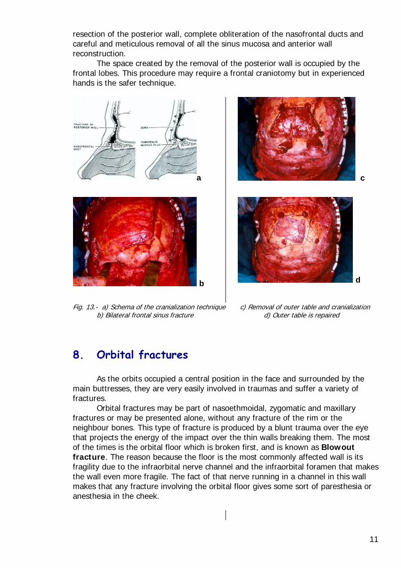
resection of the posterior wall, complete obliteration of the nasofrontal ducts and careful and meticulous removal of all the sinus mucosa and anterior wall reconstruction. The space created by the removal of the posterior wall is occupied by the frontal lobes. This procedure may require a frontal craniotomy but in experienced hands is the safer technique.
a
b
c
d
Fig. 13.- a) Schema of the cranialization technique c) Removal of outer table and cranialization b) Bilateral frontal sinus fracture d) Outer table is repaired
8. Orbital fractures As the orbits occupied a central position in the face and surrounded by the main buttresses, they are very easily involved in traumas and suffer a variety of fractures. Orbital fractures may be part of nasoethmoidal, zygomatic and maxillary fractures or may be presented alone, without any fracture of the rim or the neighbour bones. This type of fracture is produced by a blunt trauma over the eye that projects the energy of the impact over the thin walls breaking them. The most of the times is the orbital floor which is broken first, and is known as Blowout fracture. The reason because the floor is the most commonly affected wall is its fragility due to the infraorbital nerve channel and the infraorbital foramen that makes the wall even more fragile. The fact of that nerve running in a channel in this wall makes that any fracture involving the orbital floor gives some sort of paresthesia or anesthesia in the cheek.

12
fig. 14.- a) Mechanism of blowout fracture b) Fat entrapment in the fractured floor
Blowout fractures or simple fractures involving the orbital floor, gives certain signs and symptoms. Eye mobility must be checked because the possibility of fat entrapment between the fragments or just trapped in the maxillary sinus. Numbness or anesthesia indicates damage in the floor of that orbit. Crepitances and emphysema talks about damage of the ethmoidal wall. Palpebral and orbital swelling combined with enophthalmus means displaced fractures that enlarge the orbit. Of course, diplopia and visual acuity must be explored.
a
b
Fig. 15.- a) Blowout fracture ;no swelling b) Fat and muscle entrapment at and no apparent distortion. orbital floor fix the left eye down.
Medial canthal ligament disruption is a possibility in fractures of the naso-ethmoidal complex involving the orbit. Great care must be taken to explore this situation and to solve it, as if it remains untreated, a lateral displacement of the eye will be produced with distortion of the eyes and face. Though is rare in this types of injuries, section of the lacrimal system must be taken in consideration However the goals of the treatment is to restore the previous anatomic shape, more important, the previous orbital volume (in order to avoid enophthalmus) and even more, reconstruct the lost or damaged structures as the canthal ligament or lacrimal duct and restore the orbital content to its original position.

13
The surgical approach to the orbit usually involves a subcilliary incision. A skin muscle flap is raised until the orbital rim is reached, then continue subperiostically to expose the different fragments of the rim itself or the floor. Subperiosteal removal of the lateral canthal ligament enlarges the access to the lateral orbit. However, the intact margins of the orbit serves as a guide for proper reduction and osteosynthesis of the fragments. Grafts must be employed if there are gaps in the orbital walls and medial canthopexy or reduction and osteosynthesis of the fragment including de medial canthus ligament, must be done.
Fig. 16.- a) Donor site from the back of b) Ear cartilage graft covering the the ear for a cartilage graft the orbital fractured floor. If the superior orbital rim is affected a classic eyelid incision is the best way to reach the injured area. If the fracture is great and comminute, coronal incision must be considered. Minor and temporary complications associated to orbital fractures and/or its treatment are, diplopia, scleral show and ectropion. Time, massage and eyelid exercises usually solve these problems. Of course complications associated with unsuccessful surgeries or untreated fractures, includes, enophthalmus, diplopia, canthal ligament displacement, eye entrapment, severe ectropion, lacrimal obstruction and even dystopia.
9. Fractures of the Zygoma The zygoma, because its prominent position in the face, makes it subject to blunt trauma. As a prominence itself has a very important role in the aesthetics of the face and as part of the orbit (lateral and inferior walls) play also an important role maintaining the eye in its proper position. Fractures of the zygoma frequently have swelling, periorbital ecchymosis and numbness in the infraorbital nerve territory (ipsilateral anterior cheek, nose upper lip and teeth) .It is not rare to have limited mouth opening because the coronoid apophisis stops against the displaced zygomatic arch or body or just because pain in the fractured area. The zygoma displacement and luxation may be quite evident in the first hours, later, local swelling could hide this situation. Palpation at the inferior

14
orbital rim usually denotes the fracture. Depending on the displacement various types of deformities could be found and because being part of the orbit, ophthalmologic exploration must be done (diplopia, entrapment and enophthalmus). X-ray signs are more obvious the most of the times, specially opacification of the maxillary sinus and steps in the orbital rim. Waters projection is the best X-ray for exploring the zygoma.
Fig. 17.- a) Infraorbital nerve channel b) Typical signs of a zygoma in the orbital floor. lateral displacement Indications for surgery include aesthetic and functional restoration of the orbit and orbital contents and may be approached by a variety of incisions from the most commonly used, the subciliary approach, to small lateral brow incisions to reach the frontal apophisis and temporal or buccal sulcus incisions to reach the zygomaticomaxillary junction. After all the fractures have been exposed and soft tissue freed, reduction of the zygoma must be done by gently but energetic pull from the temporal or buccal approach.
a) Fig. 18.- a & b :Temporal and oral approach for close reduction of the zygoma
b
Fractures of the zygoma, however, is best treated by rigid fixation. If only wire is at hand, at least three osteosynthesis must be done at the inferior orbital rim, the zygomaticofrontal suture and the zygomaticomaxillary buttress, if not, the pulling of the strong masseter muscle from the mandible would displaced the zygoma downward after some weeks, and late enophthalmus and diplopia may appeared.

15
Fig. 19.-a) Wire osteosynthesis at the inferior b) Wire osteosynthesis at the zygomatico-orbital rim. Frontal suture.
Zygomatic arch must be always explored in the presence of zygoma fractures. It only requires open reduction and fixation if it is severely comminuted or is so displaced that no closed reduction could achieve its reposition. Then, preauricular and temporal approach is used to expose, reduce and fix the arch.
10. Fractures of the Nose The nose is composed by the nasal bones, the frontal apophisis of the maxillae, the septal cartilage and its extension to the perpendicular plate of the ethmoid and vomer. Its prominent and central position in the face makes the nose an easy subject for trauma, and the thin bones, extremely fragile. Is the most frequent facial fracture. As a whole three-dimensional complex when the nose is fractured and displaced, all the parts become more or less broken and displaced. Epistaxis and nasal deformity are evident from the very first moment, later the great swelling could make the deformity less evident. In the same way, close reduction (open reduction is rarely used) must be performed in the first hours, if not, a delay of 5 to 7 days is preferable, to allow the swelling disappear and be more accurate when performing the close reduction. The septum must be checked, reduced and maintained in place with a symmetrical nasal packaging. After that, an external nasal splint is applied to immobilize the fractured bones during 10 to 14 days.
Fig. 20.-a) Lateral displacement of the nose b) After close reduction

16
Open fractures of the nose are not very uncommon, and if the wound is large enough it could be used for an open reduction of the fragments. Of course careful attention must be paid when repairing all the inner layers, in order to avoid circular scars or internal bridles that would result in a functional and/or aesthetic outcome. However if careful and meticulous reduction and reconstruction has been carried out, the final result is excellent.
Fig. 21.- Open fracture of the bones, cartilages a nasal mucosa of the nose. Right after complete reconstruction of all the layers, reduction of the fracture and suture of the skin.
11. Fractures of the Naso-Orbital-Ethmoidal complex The intricate anatomy and the thinness of the bones of the naso-orbital-ethmoidal complex, make the proper reconstruction very difficult and complicate. The existence of the lacrimal system and the medial canthus, make the matter worst. This complex contributes to the medial orbital walls as well as the support to the nasal pyramid. Due to its close relation with the frontal sinus and the anterior cranial fosse up to 25 to 50 % of this fractures have dural leak, leaks that in approximately 95 % that will seal alone within 2 weeks. Despite its position, the lacrimal system as well as the medial canthus are rarely transected. Damage of these structures usually causes them to be detached. The Naso-Orbital-Ethmoidal complex (NAOC) may present a great variety of forms, from simple linear fractures, to the more complicate fractures that detached the lateral medial canthus, to the more complex of all fractures, the complete fracture and dislocation of the whole NAOC. It means four fractures in a time, fracture of the frontal processes of the maxilla at the glabella, the inferior orbital rim, medial orbital wall and nasal bones. This “central fragment”, when fully fractured and free is very unstable making its management difficult and the prognosis poor, specially when only wire is at hand for osteosynthesis. In absence of CT-scan, X-ray films as Waters projection combined with a meticulous palpation, gives the keys for the diagnosis. Local incisions and coronal approach are the common practice for open reduction and internal fixation. However, and specially in these cases, rigid fixation is the first choice (and the unique) if acceptable long term result are expected.

17
Fig 22.- a) Complete comminute open fracture b) Complete reduction and rigid fixation of the naso-orbital-ethmoidal complex. of every fragment. Before proceeding to repair medial canthus avulsions, the lacrimal system must be checked. If that system has not been sectioned or lacerated, monocanalicular intubation has proved to be quite effective. The silicone tube must be sutured to the surrounding tissue in the eyelid and must be threaded into the proximal canaliculus to the lacrimal sac (3 to 4 cms). If the lacrimal system seems to be severely damage a DacrioCistoRhinostomy with a polyethilen tube may be done, suturing the proximal end in position close to the lacrimal lake and the distal end inside the nose cavity through the fracture or a burr-hole. But in general lines, Dacriocistorhinostomy could reasonable be done 4 to 6 months after trauma surgery.
Fig. 23.- Dacriocystorhinosthomy with a polyethylene tube Management of canthal ligaments dislocation is the basis in the treatment of NAOC fractures. When performing open reduction and internal fixation of the fragments, detaching the canthal tendon from bone fragments during dissection, increases the likelihood of telecanthus. So meticulous bony reduction with overcorrection of the intercanthal distance and the use of bone grafts, provides the basis for the best long term results. In consequence the medial transnasal canthopexie is the Key point to get stable and acceptable aesthetic results.
18
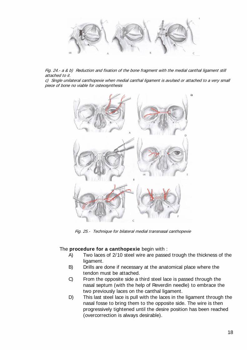
Fig. 24.- a & b) Reduction and fixation of the bone fragment with the medial canthal ligament still attached to it. c) Single unilateral canthopexie when medial canthal ligament is avulsed or attached to a very small piece of bone no viable for osteosynthesis
Fig. 25.- Technique for bilateral medial transnasal canthopexie The procedure for a canthopexie begin with :
A) Two laces of 2/10 steel wire are passed trough the thickness of the ligament.
B) Drills are done if necessary at the anatomical place where the tendon must be attached.
C) From the opposite side a third steel lace is passed through the nasal septum (with the help of Reverdin needle) to embrace the two previously laces on the canthal ligament.
D) This last steel lace is pull with the laces in the ligament through the nasal fosse to bring them to the opposite side. The wire is then progressively tightened until the desire position has been reached (overcorrection is always desirable).

19
E) Suture on the healthy side is done through the healthy ligament, if there are any doubt because of fissures o the size of the hole you have done, a graft could be use to retain the steel lace in place.
F) In case of bilateral canthopexie due to complete or bilateral comminute fracture of the NAOC, wires are passed transnasally from one side to the other and tightened over bone grafts in order to maintain tension and not slip away through the hole to the nose.
Finally, despite adequate reduction of the NAOC the thin bones of the nose cannot be reconstructed and skeletal support to the nasal dorsum is usually lost, for this reason a cantilever nasal dorsal bone graft must be fashioned. Late complications after treatment of NAOC fractures are recurrent telecanthus, epicanthal folds, epiphora and darcryocystitis.
12. Fractures of Maxilla The maxillas are the main bones of the face and particularly important in maintaining the shape and structure basically by a system of “pillars” or buttresses. There are three of this “pillars” in each Maxilla that must be preserved or reconstructed when a cranio-facial fractured is treated. One is the medial Pillar or nasomaxillary buttress, other is the lateral “pillar” or zygomaticomaxillary buttress and the third is the posterior “pillar” or pterigomaxillary buttress.
Naso maxillary Zygomatico maxillary Pterigomaxillary Fig. 26.- The three pillars of the maxilla
There are three types of fracture patterns that are usually seen in maxillary traumas, and these are those described by the french Surgeon Rene LeFort. He made dissection of cadavers of imprisoned man that (after a natural death) were hit or simulated big traumatism. He discover that with high frequency, three types of fracture patterns were reproduced in the craniofacial skeleton, the :
• Lefort I type is a monoblock fracture in which the line of fracture is just above the alveolus of the maxilla, thus separating the dentition block from the naso-orbital-ethmoidal complex.
• LeFort II type has a pyramidal shape and comprises both maxillas with the line of fracture extending from the pterigomaxillary junction up within the zygomaticomaxillary suture, the inner medial part of the orbit up to the radix of the nose along with the naso-orbital ethmoidal complex.

20
• LeFort III type is a true facial disjunction from the cranium at the level of the cranial base.
Fig. 27.- A) LeFort I B) LeFort II C) LeFort III These fractures where described from low energy impacts at low speed. Actually, as the more common origin of fractures are from traffic accidents, great energy impacts are the main origin of cranio-facial injuries, and this means that these pure types of Lefort descriptions are rarely presented alone. These fractures usually occur as segmental fractures involving other areas of the facial skeleton. Exploration includes X-ray films like Waters and Lateral views, in which the presence of bilateral air-fluid levels are highly suggestive of this type of fracture. CT-scans when possible are definitive. However, physical exploration is conclusive. Many times the face is not severely injured, and the orbits in particular, and the face in general could not be much swollen with haematomas or ecchymosis, but the general aspect is that “something has happen in that face”. Some times the aspect is that of an elongated face (previous patients pictures would be a great help). Mal occlusion is another common finding (open anterior bite because the impactation of the upper maxilla, the weakest part, over the cranial base. When palpation is made several degrees of instability may be found by moving forward or downward the maxilla.
The goals of treatment in LeFort fractures are to re-establish the mid-facial height, width, projection and occlusion.

21
Fig. 28.- a) Lefort I and II with left zygoma comminute fracture.
b) Open reduction and rigid fixation of all the fragments, width and height is maintain
The approach for the maxilla varies depending on the extent and type of fractures. For a Lefort I type, a buccal sulcus incision is enough, but if more fractures are associated or a Lefort II is present subciliary incisions must be made. Fractures involving the naso-orbital-ethmoidal complex, the zygoma or Lefort III fractures may require a coronal incision as well. A proper anatomic reposition of the maxilla will depend on the integrity of the mandible, as her ramus and condyles will asses in maintaining the posterior high. If the mandible presents one or various fractured segments, one must begin with an open reduction and fixation of the mandible and then proceed to the maxilla. In the absence of rigid fixation, reduction and stabilization must be performed with steel wire and the maxillae must be stabilised by intermaxillary fixation and craniofacial suspension, even in the case of Lefort I. Craniofacial suspension consist in support the maxilla from the cranium. It is done through certain holes or screws drilled at the glabella, nasal and/or zygomatic apophisis of the frontal bone, and/or inferior orbital rim in case of Lefort II fracture. From these attachments, long laces of steel wire are passed through the soft tissues to the maxillary arcade and tightened around the metallic bar over the superior arcade. When tighten the different steel laces, great care must be taken not to leave them loose nor too tight, as an excessively compressive suspension could result in partial resorption of the middle third and loss of height in that area.
Fig. 29.- a) Technique for close reduction b) Various types of midfacial suspension and suspension of a LeFort II fracture

22
Re-establishing the original dimensions of the face in height and width (see fig. 11) is necessary to achieve acceptable aesthetics results. Meticulous reduction, fixation and suspension are mandatory to maintain threes aesthetic and functional results to last.
Fig. 30.- a) Lefort I,III and Naso,orbital,ethmoid b) 1 year after open reduction and rigid Fracture. fixation with bilateral canthopexie.
13. Fractures of Mandible The mandible is a bone that contains the lower dental arcade and forms the lower third of the face. Because of its prominence is frequently subject to trauma, and because its semicircular shape, the mandibular arch when hit usually brakes in two or more places. The mandible is an extremely strong bone in some places (the symphysis) and week in others (the subcondylar region). Another marked difference with the rest of the other facial bones is dentition. Treatments will differ from a child fractured mandible, to an edentulous one. Another important difference between this bone and the others is the amount of strong and multidirectional muscles attached to it and its mobility. There a great variety of X-Ray views for radiological exploration of the mandible as the Fronto-occipital position (external frontal profile of body and ramus), Lateral (lower profile of one side body an angle), Verticosubmental position (inferior profile of the body and ramus), Oblique Lateral views (symphysys, angle and lower part of the ramus), Oblique ant.-post. fronto-occipital and lateral transcranial views (condilar neck an condole). Of course the best exploration is CT-scann, specially for Temporomandibular joint (TMJ) traumas.

23
Fig. 31.- a) Oblique Lateral view b) Oblique anteroposterior fronto- Occipital view In all instances history and physical examination are important, but in the case of mandibular fractures are imperative. History about the mechanism, direction and point of impactation, will tell us a lot about the places and types of fractures, and the possible fractures associated with (impact in the frontal aspect of the symphysis usually produces fractures in both condyles). Anatomical knowledge of the muscles and its insertions on the mandible are definitive when considering a fracture in terms of “Stable” or “Instable”. These terms make reference to the type and direction of the line of fracture. Stable fractures would be those fractures “reduced naturally by themselves” because the muscles pull the fractured segment against the non fractured one.
Fig. 32.- a) Pulling of the Myloyoid m. b) Pulling of the Geniohyoid and on a lateral body fragment. Digastric on a symphysis fragment.
Fig. 33.- a) Pulling of the Masseter m. b) Pulling of the masseter m. in a stable In stable and instable angle fracture. and instable body fracture.
Unstable fractures are those in which the muscles pull the segment out of contact with the rest of the bones (see figs. 32, 33 and 34).

24
Thus is very important to know the line of fracture in a mandible, its direction and location in order to determine if a fracture y stable or not. When non stable fractures are present great attention must be made to perfect anatomic reduction. Constant checking of occlusion must be done while reducing the segments, if not close contact will be get in one point while a gap is developing in other site.
Fig. 34.- Medial-forward displacement of Fig. 35.-a) Correct place and management the condyle by the pulling of the lateral Pterygoid m. c) wrong ostheosyntesis. of a body fracture Generally, mandibular fractures could be treated by close reduction and Intermaxillary fixation (6 weeks for body and ramus fractures, and 3 weeks for condylar ). Though wires is the best option in many cases, elastic bands may be necessary, specially if progressive reduction of severely displaced segments is desired. This is the case of fractured displaced condyles (less than 90º) in which the condyles are pull medially and forward by the lateral pterygoid muscle. The mandible, pulled upward at the angle and downward in the symphysis, develops an anterior open bite. In a case like this, close reduction and reposition of the condyles could be achieved by wiring arch bars in each of the dental arcades and locate elastic bands only in the section of front teeth and canines. Open reduction for condylar fractures is rarely used and is preserved to those condylar heads displaced out of the condylar fossa or with a displacement of more than 90º. Is a complicated operation that must be carried only by skilled surgeons. Mandibular fractures in children has its own tips, because the mandible at this age is not an static bone but a dynamic growing entity with a mixed dentition. These two facts makes its management a very delicate matter as injure to the functional matrix may be the cause of a subsequent growth disturbances and the mixed dentition may limitate the method for fixation.
25
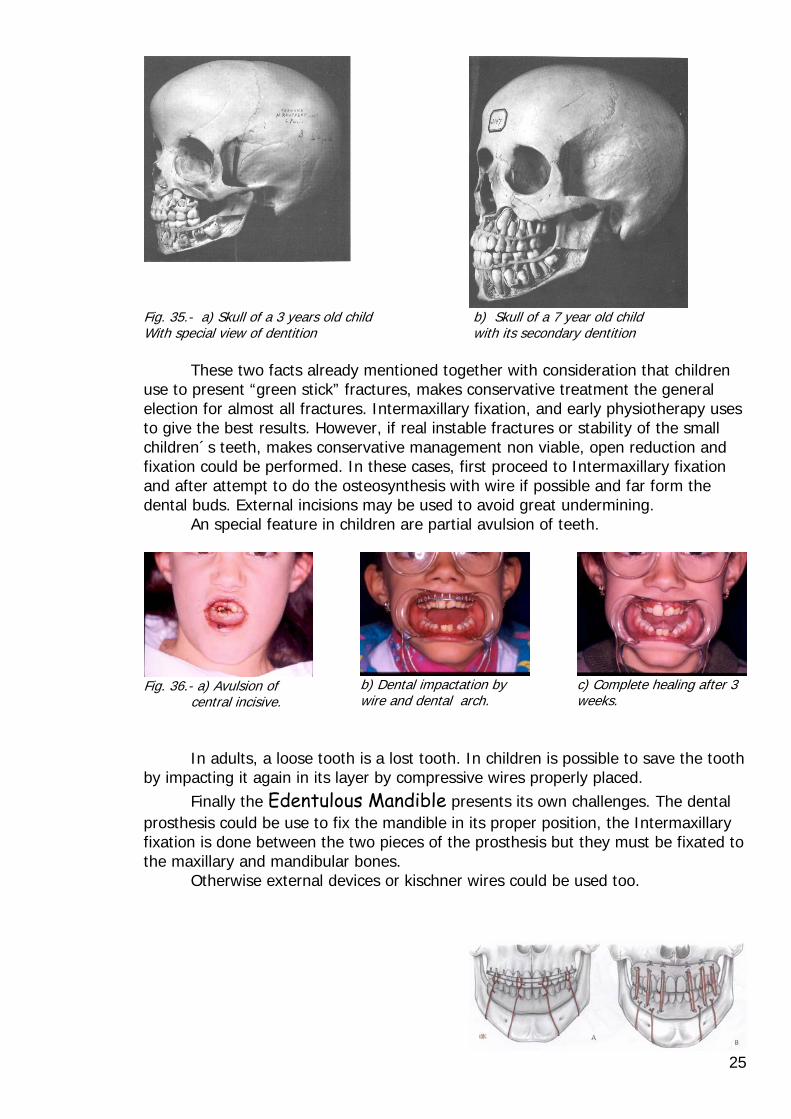
Fig. 35.- a) Skull of a 3 years old child b) Skull of a 7 year old child With special view of dentition with its secondary dentition These two facts already mentioned together with consideration that children use to present “green stick” fractures, makes conservative treatment the general election for almost all fractures. Intermaxillary fixation, and early physiotherapy uses to give the best results. However, if real instable fractures or stability of the small children´s teeth, makes conservative management non viable, open reduction and fixation could be performed. In these cases, first proceed to Intermaxillary fixation and after attempt to do the osteosynthesis with wire if possible and far form the dental buds. External incisions may be used to avoid great undermining. An special feature in children are partial avulsion of teeth.
Fig. 36.- a) Avulsion of central incisive.
b) Dental impactation by wire and dental arch.
c) Complete healing after 3 weeks.
In adults, a loose tooth is a lost tooth. In children is possible to save the tooth by impacting it again in its layer by compressive wires properly placed. Finally the Edentulous Mandible presents its own challenges. The dental prosthesis could be use to fix the mandible in its proper position, the Intermaxillary fixation is done between the two pieces of the prosthesis but they must be fixated to the maxillary and mandibular bones. Otherwise external devices or kischner wires could be used too.

26
14. Summary The evolution of techniques for diagnosis, exploration, surgical access to the fractured bones and rigid internal fixation has improved the aesthetic and functional results after injuries and fractures of the cranio-facial skeleton. However in undeveloped areas, certain technological devices are not at hand but the leading points for a successful treatment are the same :
• Careful and meticulous exploration. • Skill and excellent anatomical knowledge. • Earlier and combined treatment of injuries a fractures. • Open reduction and detailed fixation (rigid when possible).
Reducing and reconstructing facial fractured bones is like mounting a “puzzle”. There are bigger puzzles o smaller, with many or few pieces, but all require its own doses of patient for a proper result.

27