craniofacial and dental manifestations of proteus syndrome… · 2014-06-26 · craniofacial and...
TRANSCRIPT

233
Craniofacial and Dental Manifestations of Proteus Syndrome:A Case Report
K.B. BECKTOR, D.D.S.J.P. BECKTOR, D.D.S.
P.S. KARNES, M.D.E.E. KELLER, D.D.S., M.S.D.
The Proteus syndrome is a rare congenital hamartomatous condition that ischaracterized by a wide range of malformations, sometimes involving the face.Common manifestations include partial gigantism, congenital lipomas, andplantar hyperplasia. In this report we describe the craniofacial clinicopatho-logical development in a girl with Proteus syndrome from age 6 to 20 years.The patient had pronounced hemifacial hypertrophy, exostoses in the left pa-rietal region, and enlargement of the inferior alveolar nerve and mandibularcanal in the affected region. The dental development of the affected left man-dible and maxilla was characterized by extremely premature development anderuption of the primary and permanent teeth and by pronounced idiopathicroot resorptions. The multidisciplinary management of the patient and thetreatment outcome is reported. A review of the Proteus patients in the literaturewho exhibited manifestation in the craniofacial region is presented.
KEY WORDS: hemifacial hypertrophy, precocious dental development and erup-tion, Proteus syndrome
Proteus syndrome is a complex hamartomatous conditioncharacterized by partial gigantism of the hands, feet, or both;plantar hyperplasia; hemangiomas; lipomas; lymphangiomas;varicosities; verrucous epidermal nevi; macrocephaly; cranialexostosis; and asymmetry of the limbs because of long boneovergrowth. The first description of Proteus syndrome is attri-buted to Cohen and Hayden (1979), who reported two cases.Unaware of the earlier report, Wiedemann et al. (1983) de-scribed four children with similar findings. The syndrome wasgiven the name Proteus, which refers to the mythical Greekgod who was capable of changing his bodily shape.
This report describes a female patient with a localized form ofProteus syndrome and compares her with other patients with Pro-teus syndrome and with patients with hemifacial hypertrophy.
Etiology
The cause of Proteus syndrome is unclear. Its progressivenature and multisystem involvement suggest a genetic cause.
Dr. K.B. Becktor is a Visiting Clinician, Division of Orthodontics, Dental Spe-cialties, Mayo Clinic, Rochester, Minnesota, and a PhD Student in the Departmentof Orthodontics, School of Dentistry, University of Copenhagen, Denmark. Dr. J.P.Becktor is a Visiting Clinician, Department of Surgery, Division of Oral and Max-illofacial Surgery, Mayo Clinic, and Senior Registrar at the Department of Oral andMaxillofacial Surgery, Lanssjukhuset, Halmstad, Sweden. Dr. Karnes is a Consul-tant, Department of Genetics and Assistant Professor, Mayo Medical School. Dr.Keller is a Consultant, Department of General Surgery, Division of Oral and Max-illofacial Surgery, Mayo Clinic, and Professor, Mayo Medical School.
Submitted October 2000; Accepted March 2001.Reprint requests: Dr. Eugene Keller, Division of Oral and Maxillofacial Sur-
gery, Mayo Clinic, 200 First Street SW, Rochester, MN 55905.
Long thought to be sporadic in occurrence, it may be due toa lethal mutation that survives only in a mosaic form. It hasbeen postulated that an acquired somatic mutation may affectreceptors of tissue growth factors or fibroblast growth factorsin specific parts of the body in this condition (Hall, 1988;Rizzo et al., 1993; Tattelbaum and Dufresne, 1995). The extentof involvement is variable, but diffuse involvement of the en-tire body or an entire organ system is not characteristic. Thereis no statistical difference in sex predilection (Gorlin, 1984).The criteria for the diagnosis of Proteus syndrome are dividedinto general and specific by Biesecker et al. (1999).
PATIENT HISTORY
The patient was seen for consultation and treatment at theMayo Clinic at age 13 years.
Previous Investigations and Treatment
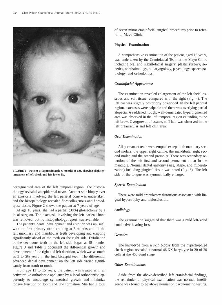
The propositus was a Caucasian girl, the third child ofhealthy parents. One male and one female sibling were healthy.She was born at term weighing 7 pounds 4 ounces after anormal pregnancy. In the neonatal period, the maternal grand-mother noted mild facial asymmetry, but it was not apparentto other family members. At age 6 months, progressive en-largement of the left cheek, lower lip and tongue was observed(Fig. 1). The lower lip enlarged rapidly, and at age 4 years shehad a lip reduction procedure performed by a local surgeon.The histopathology revealed angioma in association with fi-broadipose hamartoma. A skin biopsy was taken from a hy-
234 Cleft Palate–Craniofacial Journal, March 2002, Vol. 39 No. 2
FIGURE 1 Patient at approximately 6 months of age, showing slight en-largement of left cheek and left lower lip.
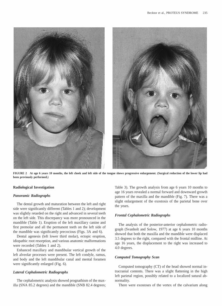
perpigmented area of the left temporal region. The histopa-thology revealed an epidermal nevus. Another skin biopsy overan exostosis involving the left parietal bone was undertaken,and the histopathology revealed fibrocollagenous and fibroad-ipose tissue. Figure 2 shows the patient at 7 years of age.
At age 10 years, she had a partial (30%) glossectomy by alocal surgeon. The exostosis involving the left parietal bonewas removed, but no histopathology report was available.
The patient’s dental development and eruption was unusual,with the first primary tooth erupting at 3 months and all theleft maxillary and mandibular teeth developing and eruptingsignificantly ahead of the teeth on the right side. Exfoliationof the deciduous teeth on the left side began at 18 months.Figure 3 and Table 1 document the differential growth anddevelopment of the right and left dentition, which was as muchas 5 to 5½ years in the first bicuspid teeth. The differentialadvanced dental development on the left side varied signifi-cantly from tooth to tooth.
From age 13 to 15 years, the patient was treated with anactivatorlike orthodontic appliance by a local orthodontist, ap-parently to encourage symmetrical growth and minimizetongue function on tooth and jaw formation. She had a total
of seven minor craniofacial surgical procedures prior to refer-ral to Mayo Clinic.
Physical Examination
A comprehensive examination of the patient, aged 13 years,was undertaken by the Craniofacial Team at the Mayo Clinicincluding oral and maxillofacial surgery, plastic surgery, ge-netics, ophthalmology, otolaryngology, psychology, speech pa-thology, and orthodontics.
Craniofacial Appearance
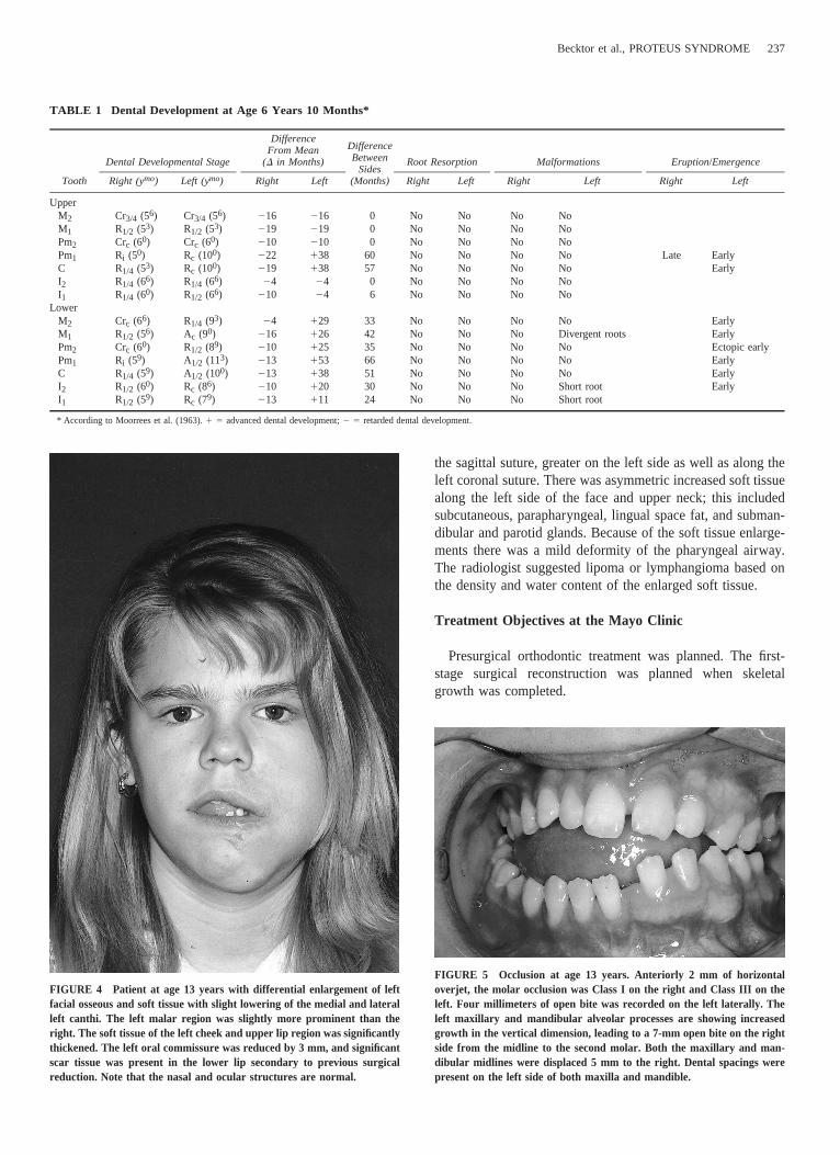
The examination revealed enlargement of the left facial os-seous and soft tissue, compared with the right (Fig. 4). Theleft ear was slightly posteriorly positioned. In the left parietalregion, exostoses were palpable and there was overlying partialalopecia. A reddened, rough, well-demarcated hyperpigmentedarea was observed in the left temporal region extending to theleft brow. Overgrowth of coarse, stiff hair was observed in theleft preauricular and left chin area.
Oral Examination
All permanent teeth were erupted except both maxillary sec-ond molars, the upper right canine, the mandibular right sec-ond molar, and the second premolar. There was secondary re-tention of the left first and second permanent molar in themandible. Normal dental anatomy (size, shape, and minerali-zation) including gingival tissue was noted (Fig. 5). The leftside of the tongue was symmetrically enlarged.
Speech Examination
There were mild articulatory distortions associated with lin-gual hypertrophy and malocclusion.
Audiology
The examination suggested that there was a mild left-sidedconductive hearing loss.
Genetics
The karyotype from a skin biopsy from the hypertrophiedcheek region revealed a normal 46,XX karyotype in 20 of 20cells at the 450-band stage.
Other Examinations
Aside from the above-described left craniofacial findings,the remainder of physical examination was normal. Intelli-gence was found to be above normal on psychometric testing.
Becktor et al., PROTEUS SYNDROME 235
FIGURE 2 At age 6 years 10 months, the left cheek and left side of the tongue shows progressive enlargement. (Surgical reduction of the lower lip hadbeen previously performed.)
Radiological Investigation
Panoramic Radiographs
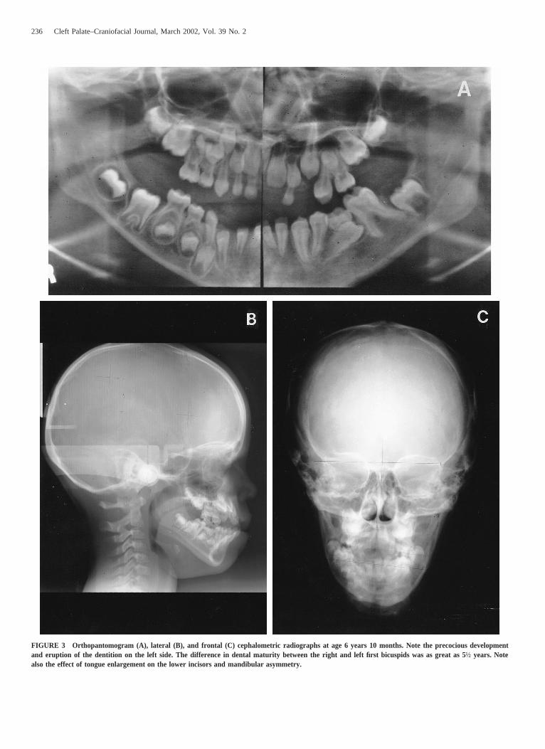
The dental growth and maturation between the left and rightside were significantly different (Tables 1 and 2); developmentwas slightly retarded on the right and advanced in several teethon the left side. This discrepancy was more pronounced in themandible (Table 1). Eruption of the left maxillary canine andfirst premolar and all the permanent teeth on the left side ofthe mandible was significantly precocious (Figs. 3A and 6).
Dental agenesis (left lower third molar), ectopic eruption,idiopathic root resorption, and various anatomic malformationswere recorded (Tables 1 and 2).
Enhanced maxillary and mandibular vertical growth of theleft alveolar processes were present. The left condyle, ramus,and body and the left mandibular canal and mental foramenwere significantly enlarged (Fig. 6).
Lateral Cephalometric Radiographs
The cephalometric analysis showed prognathism of the max-illa (SNA 85.2 degrees) and the mandible (SNB 82.4 degrees;
Table 3). The growth analysis from age 6 years 10 months toage 16 years revealed a normal forward and downward growthpattern of the maxilla and the mandible (Fig. 7). There was aslight enlargement of the exostosis of the parietal bone overthe years.
Frontal Cephalometric Radiographs
The analysis of the posterior-anterior cephalometric radio-graph (Svanholt and Solow, 1977) at age 6 years 10 monthsshowed that both the maxilla and the mandible were displaced3.5 degrees to the right, compared with the frontal midline. Atage 16 years, the displacement to the right was increased to4.0 degrees.
Computed Tomography Scan
Computed tomography (CT) of the head showed normal in-tracranial contents. There was a slight flattening in the highleft parietal region, possibly related to a localized sutural ab-normality.
There were exostoses of the vertex of the calvarium along
236 Cleft Palate–Craniofacial Journal, March 2002, Vol. 39 No. 2
FIGURE 3 Orthopantomogram (A), lateral (B), and frontal (C) cephalometric radiographs at age 6 years 10 months. Note the precocious developmentand eruption of the dentition on the left side. The difference in dental maturity between the right and left first bicuspids was as great as 5½ years. Notealso the effect of tongue enlargement on the lower incisors and mandibular asymmetry.
Becktor et al., PROTEUS SYNDROME 237
TABLE 1 Dental Development at Age 6 Years 10 Months*
Tooth
Dental Developmental Stage
Right (ymo) Left (ymo)
DifferenceFrom Mean
(D in Months)
Right Left
DifferenceBetween
Sides(Months)
Root Resorption
Right Left
Malformations
Right Left
Eruption/Emergence
Right Left
UpperM2M1Pm2
Cr3/4 (56)R1/2 (53)Crc (60)
Cr3/4 (56)R1/2 (53)Crc (60)
216219210
216219210
000
NoNoNo
NoNoNo
NoNoNo
NoNoNo
Pm1CI2I1
Ri (50)R1/4 (53)R1/4 (66)R1/4 (60)
Rc (100)Rc (100)R1/4 (66)R1/2 (66)
22221924
210
1381382424
605706
NoNoNoNo
NoNoNoNo
NoNoNoNo
NoNoNoNo
Late EarlyEarly
LowerM2M1Pm2
Crc (66)R1/2 (56)Crc (60)
R1/4 (93)Ac (90)R1/2 (89)
24216210
129126125
334235
NoNoNo
NoNoNo
NoNoNo
NoDivergent rootsNo
EarlyEarlyEctopic early
Pm1CI2I1
Ri (59)R1/4 (59)R1/2 (60)R1/2 (59)
A1/2 (113)A1/2 (100)Rc (86)Rc (79)
213213210213
153138120111
66513024
NoNoNoNo
NoNoNoNo
NoNoNoNo
NoNoShort rootShort root
EarlyEarlyEarly
* According to Moorrees et al. (1963). 1 5 advanced dental development; 2 5 retarded dental development.
FIGURE 4 Patient at age 13 years with differential enlargement of leftfacial osseous and soft tissue with slight lowering of the medial and lateralleft canthi. The left malar region was slightly more prominent than theright. The soft tissue of the left cheek and upper lip region was significantlythickened. The left oral commissure was reduced by 3 mm, and significantscar tissue was present in the lower lip secondary to previous surgicalreduction. Note that the nasal and ocular structures are normal.
FIGURE 5 Occlusion at age 13 years. Anteriorly 2 mm of horizontaloverjet, the molar occlusion was Class I on the right and Class III on theleft. Four millimeters of open bite was recorded on the left laterally. Theleft maxillary and mandibular alveolar processes are showing increasedgrowth in the vertical dimension, leading to a 7-mm open bite on the rightside from the midline to the second molar. Both the maxillary and man-dibular midlines were displaced 5 mm to the right. Dental spacings werepresent on the left side of both maxilla and mandible.
the sagittal suture, greater on the left side as well as along theleft coronal suture. There was asymmetric increased soft tissuealong the left side of the face and upper neck; this includedsubcutaneous, parapharyngeal, lingual space fat, and subman-dibular and parotid glands. Because of the soft tissue enlarge-ments there was a mild deformity of the pharyngeal airway.The radiologist suggested lipoma or lymphangioma based onthe density and water content of the enlarged soft tissue.
Treatment Objectives at the Mayo Clinic
Presurgical orthodontic treatment was planned. The first-stage surgical reconstruction was planned when skeletalgrowth was completed.

238 Cleft Palate–Craniofacial Journal, March 2002, Vol. 39 No. 2
TABLE 2 Dental Development at Age 13 Years 5 Months*
Tooth
Dental Developmental Stage
Right (ymo) Left (ymo)
DifferenceFrom Mean
(D in Months)
Right Left
Differ-ence
BetweenSides
(Months)
Root Resorption
Right Left
Malformations
Right Left
Eruption
Right Left
UpperM2M1Pm2
Rc (113)CompletedR3/4 (100)
Rc (113)CompletedCompleted
226—
241
226——
0——
NoNoNo
NoNoNo
NoTaurodontNo
TaurodontTaurodontNo
Secondary retention
Pm1CI2I1
Rc (100)A1/2CompletedCompleted
CompletedCompletedCompletedCompleted
241241——
————
————
NoNoNoNo
No111NoNo
NoNoInvaginationNo
NoNoInvaginationNo
LowerM2M1Pm2
Rc (113)CompletedR3/4 (100)
CompletedCompletedCompleted
226—
241
———
———
NoNoNo
11111
TaurodontNoNo
TaurodontNoNo
Secondary retentionSecondary retention
Pm1CI2I1
Rc (100)CompletedCompletedCompleted
CompletedCompletedCompletedCompleted
241———
————
————
NoNoNoNo
11No11
NoNoNoNo
NoNoNoNo
* According to Moorrees et al., (1963). 2 5 designates retarded dental development.
Orthodontic Management
From age 13 to 15 years, the patient wore a removable oraltongue crib appliance to attempt to reduce the effect of mac-roglossia on the erupting dentition.
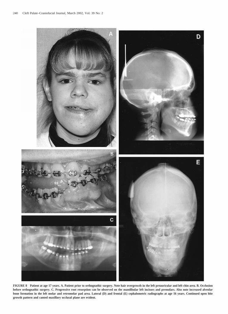
Comprehensive orthodontic treatment was initiated at age16 years. Three permanent teeth were removed because of ad-vanced root resorption, secondary retention, or both. The or-thodontic treatment was performed on right- and left-side seg-mental arches in the mandible (Fig. 8A through 8E).
Postorthognathic surgery anterior nocturnal vertical elasticwere terminated at age 17 years, and a maxillary Hawley re-tainer and a mandibular overlay retainer with posterior tonguerestraining loops was initiated.
Progressive root resorption in connection with the ortho-dontic treatment was observed on the maxillary left canine andincisors and premolars in the left side of the mandible (Fig.8C).
Surgical Management at Mayo Clinic
Liposuction of the left cheek was performed at age 14 years.At age 17 years, skeletal growth was complete and orthog-nathic surgery was performed under general anesthesia.
Mandible
A right ramus sagittal osteotomy was performed to advanceand rotate the right mandible. The symphysis was exposed bydegloving from right to left first molar. The mental nerve wastranspositioned and preserved bilaterally utilizing burr tech-nique. A horizontal osteotomy was placed 20 mm superior tothe mandibular inferior border in the anterior region and 10mm above the mandibular inferior border in the region of thefirst molars. The tooth-bearing segment of the mandible wasosteotomized at the midline in 5 mm of interdental space that
had been created by orthodontic treatment. The osteotomy wasfollowed by removal of the 5-mm interdental bone to allowrotation of the right segment of the mandible anteriorly andmedially. The left segment was rotated inferiorly. The twotooth-bearing segments were repositioned and secured with aprefabricated interocclusal-lingual splint. The inferior bordersegment of the mandible was then advanced 8 mm. The leftside of the posterior inferior border was reduced vertically 5mm. The inferior border segment was then secured by trans-osseous wires. The mental nerves were repositioned.
Maxilla
A Le Fort I osteotomy was accomplished at the level of thefloor of the nose from the pyriform aperture to the pterygoidplates.
Intermaxillary fixation was accomplished. Two blocks, 10by 20 by 20 mm, of corticocancellous bone were obtainedfrom the superior medial iliac crest. The bone blocks werefashioned and fit on the right side to accomplish a downwardmovement of 6 mm in the molar region and 5 mm at thepyriform aperture. On the left side, there was no vertical move-ment in the posterior region. The maxilla was secured in thisposition by miniplates and titanium screws.
Soft Tissue
A wedge of the interfering buccal mucosa on the left side,30 mm in length and 20 mm in width, was removed. The fatpad of Bichat of the left cheek was also removed, through thesame buccal incision, after blunt dissection exposure throughthe buccinator muscle posteriorly.
Postoperative Management
Anterior intermaxillary elastics were applied to control oc-clusion postoperatively. The postoperative period was unevent-
Becktor et al., PROTEUS SYNDROME 239
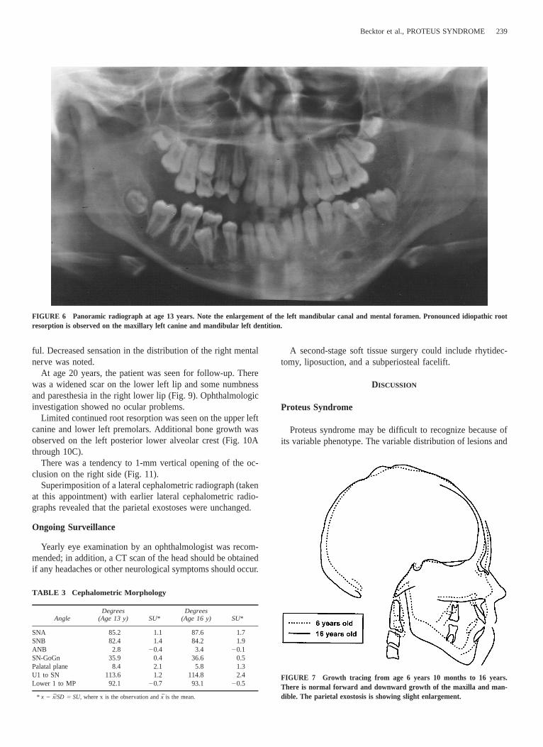
FIGURE 6 Panoramic radiograph at age 13 years. Note the enlargement of the left mandibular canal and mental foramen. Pronounced idiopathic rootresorption is observed on the maxillary left canine and mandibular left dentition.
TABLE 3 Cephalometric Morphology
AngleDegrees
(Age 13 y) SU*Degrees
(Age 16 y) SU*
SNASNBANBSN-GoGnPalatal planeU1 to SNLower 1 to MP
85.282.42.8
35.98.4
113.692.1
1.11.4
20.40.42.11.2
20.7
87.684.23.4
36.65.8
114.893.1
1.71.9
20.10.51.32.4
20.5
* x 2 x/SD 5 SU, where x is the observation and x is the mean.
FIGURE 7 Growth tracing from age 6 years 10 months to 16 years.There is normal forward and downward growth of the maxilla and man-dible. The parietal exostosis is showing slight enlargement.
ful. Decreased sensation in the distribution of the right mentalnerve was noted.
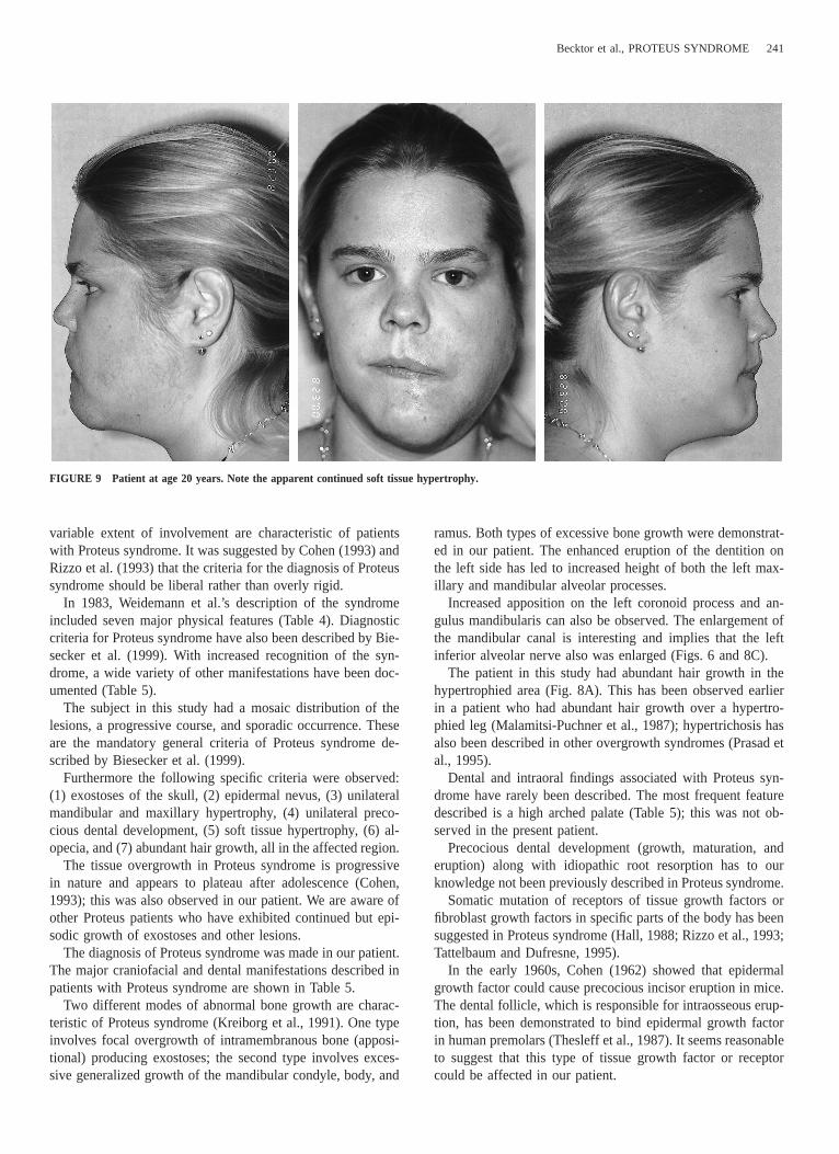
At age 20 years, the patient was seen for follow-up. Therewas a widened scar on the lower left lip and some numbnessand paresthesia in the right lower lip (Fig. 9). Ophthalmologicinvestigation showed no ocular problems.
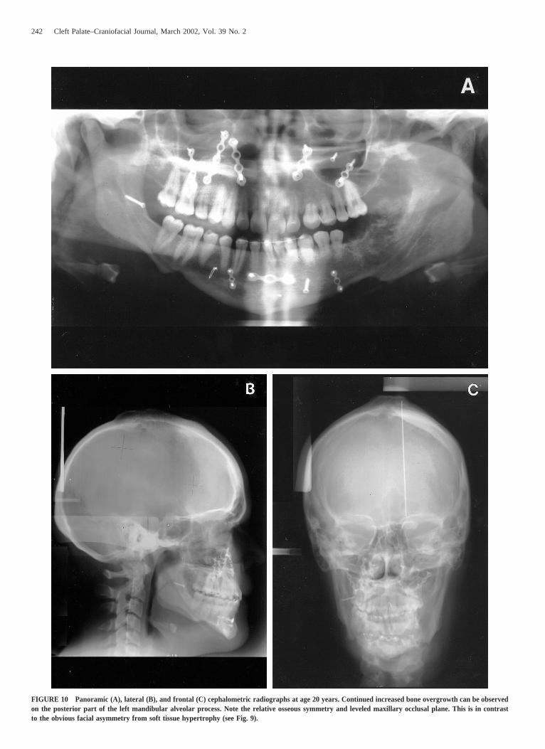
Limited continued root resorption was seen on the upper leftcanine and lower left premolars. Additional bone growth wasobserved on the left posterior lower alveolar crest (Fig. 10Athrough 10C).
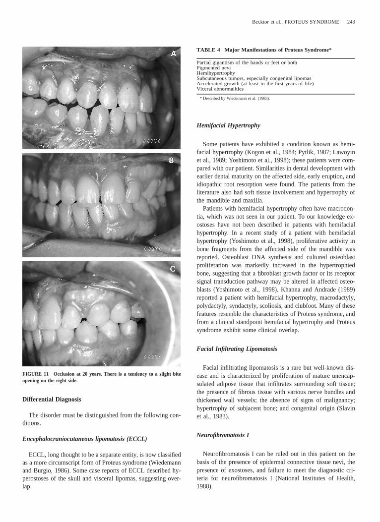
There was a tendency to 1-mm vertical opening of the oc-clusion on the right side (Fig. 11).
Superimposition of a lateral cephalometric radiograph (takenat this appointment) with earlier lateral cephalometric radio-graphs revealed that the parietal exostoses were unchanged.
Ongoing Surveillance
Yearly eye examination by an ophthalmologist was recom-mended; in addition, a CT scan of the head should be obtainedif any headaches or other neurological symptoms should occur.
A second-stage soft tissue surgery could include rhytidec-tomy, liposuction, and a subperiosteal facelift.
DISCUSSION
Proteus Syndrome
Proteus syndrome may be difficult to recognize because ofits variable phenotype. The variable distribution of lesions and
240 Cleft Palate–Craniofacial Journal, March 2002, Vol. 39 No. 2
FIGURE 8 Patient at age 17 years. A. Patient prior to orthognathic surgery. Note hair overgrowth in the left preauricular and left chin area. B. Occlusionbefore orthognathic surgery. C. Progressive root resorption can be observed on the mandibular left incisors and premolars. Also note increased alveolarbone formation in the left molar and retromolar pad area. Lateral (D) and frontal (E) cephalometric radiographs at age 16 years. Continued open bitegrowth pattern and canted maxillary occlusal plane are evident.
Becktor et al., PROTEUS SYNDROME 241
FIGURE 9 Patient at age 20 years. Note the apparent continued soft tissue hypertrophy.
variable extent of involvement are characteristic of patientswith Proteus syndrome. It was suggested by Cohen (1993) andRizzo et al. (1993) that the criteria for the diagnosis of Proteussyndrome should be liberal rather than overly rigid.
In 1983, Weidemann et al.’s description of the syndromeincluded seven major physical features (Table 4). Diagnosticcriteria for Proteus syndrome have also been described by Bie-secker et al. (1999). With increased recognition of the syn-drome, a wide variety of other manifestations have been doc-umented (Table 5).
The subject in this study had a mosaic distribution of thelesions, a progressive course, and sporadic occurrence. Theseare the mandatory general criteria of Proteus syndrome de-scribed by Biesecker et al. (1999).
Furthermore the following specific criteria were observed:(1) exostoses of the skull, (2) epidermal nevus, (3) unilateralmandibular and maxillary hypertrophy, (4) unilateral preco-cious dental development, (5) soft tissue hypertrophy, (6) al-opecia, and (7) abundant hair growth, all in the affected region.
The tissue overgrowth in Proteus syndrome is progressivein nature and appears to plateau after adolescence (Cohen,1993); this was also observed in our patient. We are aware ofother Proteus patients who have exhibited continued but epi-sodic growth of exostoses and other lesions.
The diagnosis of Proteus syndrome was made in our patient.The major craniofacial and dental manifestations described inpatients with Proteus syndrome are shown in Table 5.
Two different modes of abnormal bone growth are charac-teristic of Proteus syndrome (Kreiborg et al., 1991). One typeinvolves focal overgrowth of intramembranous bone (apposi-tional) producing exostoses; the second type involves exces-sive generalized growth of the mandibular condyle, body, and
ramus. Both types of excessive bone growth were demonstrat-ed in our patient. The enhanced eruption of the dentition onthe left side has led to increased height of both the left max-illary and mandibular alveolar processes.
Increased apposition on the left coronoid process and an-gulus mandibularis can also be observed. The enlargement ofthe mandibular canal is interesting and implies that the leftinferior alveolar nerve also was enlarged (Figs. 6 and 8C).
The patient in this study had abundant hair growth in thehypertrophied area (Fig. 8A). This has been observed earlierin a patient who had abundant hair growth over a hypertro-phied leg (Malamitsi-Puchner et al., 1987); hypertrichosis hasalso been described in other overgrowth syndromes (Prasad etal., 1995).
Dental and intraoral findings associated with Proteus syn-drome have rarely been described. The most frequent featuredescribed is a high arched palate (Table 5); this was not ob-served in the present patient.
Precocious dental development (growth, maturation, anderuption) along with idiopathic root resorption has to ourknowledge not been previously described in Proteus syndrome.
Somatic mutation of receptors of tissue growth factors orfibroblast growth factors in specific parts of the body has beensuggested in Proteus syndrome (Hall, 1988; Rizzo et al., 1993;Tattelbaum and Dufresne, 1995).
In the early 1960s, Cohen (1962) showed that epidermalgrowth factor could cause precocious incisor eruption in mice.The dental follicle, which is responsible for intraosseous erup-tion, has been demonstrated to bind epidermal growth factorin human premolars (Thesleff et al., 1987). It seems reasonableto suggest that this type of tissue growth factor or receptorcould be affected in our patient.
242 Cleft Palate–Craniofacial Journal, March 2002, Vol. 39 No. 2
FIGURE 10 Panoramic (A), lateral (B), and frontal (C) cephalometric radiographs at age 20 years. Continued increased bone overgrowth can be observedon the posterior part of the left mandibular alveolar process. Note the relative osseous symmetry and leveled maxillary occlusal plane. This is in contrastto the obvious facial asymmetry from soft tissue hypertrophy (see Fig. 9).
Becktor et al., PROTEUS SYNDROME 243
FIGURE 11 Occlusion at 20 years. There is a tendency to a slight biteopening on the right side.
TABLE 4 Major Manifestations of Proteus Syndrome*
Partial gigantism of the hands or feet or bothPigmented neviHemihypertrophySubcutaneous tumors, especially congenital lipomasAccelerated growth (at least in the first years of life)Viceral abnormalities
* Described by Wiedemann et al. (1983).
Differential Diagnosis
The disorder must be distinguished from the following con-ditions.
Encephalocraniocutaneous lipomatosis (ECCL)
ECCL, long thought to be a separate entity, is now classifiedas a more circumscript form of Proteus syndrome (Wiedemannand Burgio, 1986). Some case reports of ECCL described hy-perostoses of the skull and visceral lipomas, suggesting over-lap.
Hemifacial Hypertrophy
Some patients have exhibited a condition known as hemi-facial hypertrophy (Kogon et al., 1984; Pytlik, 1987; Lawoyinet al., 1989; Yoshimoto et al., 1998); these patients were com-pared with our patient. Similarities in dental development withearlier dental maturity on the affected side, early eruption, andidiopathic root resorption were found. The patients from theliterature also had soft tissue involvement and hypertrophy ofthe mandible and maxilla.
Patients with hemifacial hypertrophy often have macrodon-tia, which was not seen in our patient. To our knowledge ex-ostoses have not been described in patients with hemifacialhypertrophy. In a recent study of a patient with hemifacialhypertrophy (Yoshimoto et al., 1998), proliferative activity inbone fragments from the affected side of the mandible wasreported. Osteoblast DNA synthesis and cultured osteoblastproliferation was markedly increased in the hypertrophiedbone, suggesting that a fibroblast growth factor or its receptorsignal transduction pathway may be altered in affected osteo-blasts (Yoshimoto et al., 1998). Khanna and Andrade (1989)reported a patient with hemifacial hypertrophy, macrodactyly,polydactyly, syndactyly, scoliosis, and clubfoot. Many of thesefeatures resemble the characteristics of Proteus syndrome, andfrom a clinical standpoint hemifacial hypertrophy and Proteussyndrome exhibit some clinical overlap.
Facial Infiltrating Lipomatosis
Facial infiltrating lipomatosis is a rare but well-known dis-ease and is characterized by proliferation of mature unencap-sulated adipose tissue that infiltrates surrounding soft tissue;the presence of fibrous tissue with various nerve bundles andthickened wall vessels; the absence of signs of malignancy;hypertrophy of subjacent bone; and congenital origin (Slavinet al., 1983).
Neurofibromatosis I
Neurofibromatosis I can be ruled out in this patient on thebasis of the presence of epidermal connective tissue nevi, thepresence of exostoses, and failure to meet the diagnostic cri-teria for neurofibromatosis I (National Institutes of Health,1988).

244 Cleft Palate–Craniofacial Journal, March 2002, Vol. 39 No. 2
TABLE 5 Findings Reported in Proteus Syndrome
GrowthDigital hypertrophy1,2,3,4,5,6,7,8,9,10,11
Hemitrophy of upper limbs1,2,4,5,6,8,11
Hemitrophy of toes and feet1,2,4,5,6,7,10,11,12,13,14
Hemitrophy of lower limbs2,4,5,6,7,8,10,11,13,15
CraniofacialBroad and bossed forehead6,11,14,18
Hemimegalocephaly8,12,20
Macrocephaly1,2,11,20
Craniosynostosis (metopic, coronal)17
SkinAbundant hair growth3
Hyperkeratotic epidermal nevi1,2,4,5,7,8,10,11,13,14,15,16,17,18
Cafe au lait spots2
Asymmetry of the cranium2,11
Hemifacial hypertrophy1,2,4,9,11,12,21
Ptosis, nystagmus17
Alopecia20
Depressed nasal bridge8,18
Pigmented linear nevi11,16
Gyriform hypertrophy feet, palmar2,4,7,9,10,11,12,13,14
SkeletalScoliosis1,4,7,9,10,12,13,14
Prominent exostosis of the skull4,5,6,8,11,15,20
Exostosis of the parietal bone20
Exostosis of the frontal bone12,14,17,22
Exostosis of the occipital bone15,17,22
Exostosis of the temporal bone14,15
Dystrophic intervertebral disk10,19
Spinal anomalities14,18
Pelvic asymmetry2
Vertebral abnormalities4,7,10,13,19,20
Hip subluxation4,10,13
Exostosis of the orbital rim22
Exostosis of the zygoma15
Exostosis of the nasal bone12,15,17,22,23
Exostosis of the anterior clinoid process22
Exostosis of the chin17,23
Spatulated ribs, rib prominence4,6,18
PsychosocialSeizures1,8,17,21
Normal (well in school)4,10,13,15,20,22
Exostosis of the angle of the mandible22
Exostosis of the auditory canal4,7,8,12,15,22
Condylar overgrowth12,23
Mandibular prognathism12
Midline asymmetry12,23
Mental retardation2,5,8,12,17
TumorsFibrocystic disease12
Lipoma4,5,12,20
Hyperpigmented skin10,11,21
Lipoma orbital area20
Submandibular lymphangioma1
OralAngioma14
Epibulbar dermoid5,17
Subcutaneous lipoma11
Primary dentition abnormal11
Yellow teeth8,12
Gingival hypertrophy1,17
Lymphangioma11,12,14,20
Hamratomas11
Sclera tumor (eyeanomalia)4,8,12,14,22
Angiolipomatous tumor10,18
Hemangioma4,10,12,14
Inguinal hernia4,8
High arched palate8,14,15,16,17
Class III Malocclusion15
Crossbite15
Crowding17
Malocclusion12
Multiple frenulae in the mandible22
Hypertrophied tonsilla8,12
1 (Alavi et al., 1993).2 (Baron-Mazurera et al., 1997).3 (Bialer et al., 1988).4 (Clark et al., 1987).5 (Costa et al., 1985).6 (Hauer et al., 1998).7 (Hornstein et al., 1987).8 (Malamitsi-Puchner et al., 1987).9 (Pinto et al., 1998).10 (Skovby et al., 1993).11 (Viljoen et al., 1987).12 (Cohen, 1988).
13 (Stricker, 1992).14 (Wiedemann et al., 1983).15 (Tattelbaum and Dufresne, 1995).16 (Arendorf and Hansolo, 1995).17 (Cohen, 1993).18 (Ring and Snyder, 1997).19 (Biesecker et al., 1999).20 (Rizzo et al., 1993).21 (McMullin et al., 1993).22 (Smeets et al., 1994).23 (Kreiborg et al., 1991).
Bannayan-Riley-Rulvacava
Bannayan-Riley-Rulvacava syndrome is distinct from Pro-teus syndrome and is due to mutations in the PTEN gene (Co-hen, 1998). This syndrome is characterized by macrocephaly,lipomas, capillary malformations, polyposis of the colon andrectum, pigmented macules of the penis, and Hashimoto thy-roiditis (Gorlin et al., 1992).
CONCLUSION
The protean manifestation of this unusual syndrome givescredence to its name. Localized Proteus syndrome involvingonly the head and neck has been described previously (Rizzoet al., 1993; Smeets et al., 1994). The dental manifestationsand dental development has to our knowledge not been de-scribed before. Some of the dental manifestations observed in
this patient have been described in patients with hemifacialhypertrophy. The similarities and the fact that all three germlines (ectoderm, endoderm, and mesoderm) are involved inboth entities suggest that these two conditions may be due toaberrant growth factors, growth factor receptors, or a growthfactor signal transduction pathway.
Care providers involved with these rare patients must beaware of other potential musculoskeletal manifestations. Teamsof medical and dental specialties need to be involved in thetreatment of patients with craniofacial manifestations of Pro-teus syndrome.
REFERENCES
Alavi S, Chakrapani A, Kher A, Bharucha BA. The Proteus syndrome. J Post-grad Med. 1993;39:219–221.
Arendorf TM, Hansolo B. Proteus syndrome: association with gingival hyper-plasia. J Oral Pathol Med. 1995;24:383–384.

Becktor et al., PROTEUS SYNDROME 245
Barona-Mazurera MR, Hidalgo-Galvan LR, Orozco-Covarrubias ML, Duran-McKinster C, Tamayo-Sanchez L, Ruiz-Maldonado R. Proteus syndrome:new findings in seven patients. Pediatr Dermatol. 1997;14:1–5.
Bialer MG, Riedy MJ, Wilson WG. Proteus syndrome versus Bannayan-ZonanaSyndrome: a problem in differential diagnosis. Eur J Pediatr. 1988;148:122–125.
Biesecker LG, Happle R, Mulliken JB, Weksberg R, Graham JM Jr, ViljoenDL, Cohen MM Jr. Proteus syndrome: diagnostic criteria, differential di-agnosis, and patient evaluation. Am J Med Genet. 1999;84:389–395.
Clark RD, Donnai D, Rogers J, Cooper J, Baraitser M. Proteus syndrome: anexpanded phenotype. Am J Med Genet. 1987;27:99–117.
Cohen MM Jr. Proteus syndrome: clinical evidence for somatic mosaicism andselective review. Am J Med Genet. 1993;47:645–652.
Cohen MM Jr. Some neoplasms and some hamartomatous syndromes: geneticconsiderations. Int J Oral Maxillofac Surg. 1998;27:363–369.
Cohen MM Jr. Understanding Proteus syndrome, unmasking the elephant man,and stemming elephant fever. Neurofibromatosis. 1988;1:260–280.
Cohen MM Jr, Hayden PW. A newly recognized hamartomatous syndrome.Birth Defects. 1979;15:291–296.
Cohen S. Isolation of a mouse submaxillary gland protein accelerating incisoreruption and eyelid opening in the newborn animal. J Biol Chem. 1962;237:1555–1562.
Costa T, Fitch N, Azouz EM. Proteus syndrome: report of two cases with pelviclipomatosis. Pediatrics. 1985;76:984–989.
Gorlin RJ. Proteus syndrome. J Clin Dysmorphol. 1984;2:8–9.Gorlin RJ, Cohen MM Jr, Condon LM, Burke BA. Bannayan-Riley-Ruvalcaba
syndrome. Am J Med Genet. 1992;44:307–314.Hall JG. Review and hypotheses: somatic mosaicism: observations related to
clinical genetics. Am J Hum Genet. 1988;43:355–363.Hauer MP, Uhl M, Darge K, Allmann K-H, Langer M. A mild form of Proteus
syndrome. Eur Radiol. 1998;8:585–587.Hornstein L, Bove KE, Towbin RB. Linear nevi, hemihypertrophy, connective
tissue hamartomas, and unusual neoplasms in children. J Pediatr. 1987;110:404–408.
Khanna JN, Andrade NN. Hemifacial hypertrophy. Report of two cases. Int JOral Maxillofac Surg. 1989;18:294–297.
Kogon SL, Jarvis AM, Daley TD, Kane MF. Hemifacial hypertrophy affectingthe maxillary dentition. Report of a case. Oral Surg Oral Med Oral Pathol.1984;58:549–553.
Kreiborg S, Cohen MM Jr, Skovby F. Craniofacial characteristics of Proteussyndrome: two modes of abnormal growth. Proc Finn Dent Soc. 1991;87:183–188.
Lawoyin JO, Daramola JO, Lawoyin DO. Congenital hemifacial hypertrophy.Report of two cases. Oral Surg Oral Med Oral Pathol. 1989;68:27–30.
Malamitsi-Puchner A, Kitsiou S, Bartsocas CS. Severe Proteus syndrome in an18-month-old boy. Am J Med Genet. 1987;27:119–125.
McMullin GP, Super M, Clarke MA. Cranial hemihypertrophy with ipsilateral
nevoid streaks, intellectual handicap and epilepsy: a report of two cases.Clin Genet. 1993;44:249–253.
Moorrees CFA, Fanning EA, Hunt EE Jr. Age variation of formation for tenpermanent teeth. J Dent Res. 1963;42:1490–1502.
National Institutes of Health Consensus Development Conference. Neurofibro-matosis. Conference statement. Arch Neurol. 1988;45:575–578
Pinto PX, Beale V, Paterson AW. Proteus syndrome. Oral Surg Oral Med OralPathol Oral Radiol Endod. 1998;85:82–85.
Prasad VS, Sengar RL, Sahu BP, Immaneni D. Diasteatomyelia in adults. Mod-ern imaging and operative treatment. Clin Imaging. 1995;19:270–274.
Pytlik W. Local premature eruption of enormous tooth crowns with root re-sorption and lymphangioma of the labial mucosa [in German]. FortschrKieferorthop. 1987;48:233–245.
Ring D, Snyder B. Spinal canal compromise in Proteus syndrome: case reportand review of the literature. Am J Orthop. 1997;26:275–278.
Rizzo R, Pavone L, Micali G, Nigro F, Cohen MM. Encephalocraniocutaneouslipomatosis. Am J Med Genet. 1993;47:653–655.
Skovby F, Graham JM, Sonne-Holm S, Cohen MM. Compromise of the spinalcanal in Proteus syndrome. Am J Med Genet. 1993;47:656–659.
Slavin SA, Baker DC, McCarthy JC. Congenital infiltrating lipomatosis of theface: clinicopathological evaluation and treatment. Plast Reconstr Surg.1983;72:158–164.
Smeets E, Fryns J-P, Cohen MM. Regional Proteus syndrome and somaticmosaicism. Am J Med Genet. 1994;51:29–31.
Stricker S. Musculoskeletal manifestations of Proteus syndrome: report of twocases with literature review. J Pediatr Orthop. 1992;12;667–674.
Svanholt P, Solow B. Assessment of midline discrepancies on the posterior-anterior cephalometric radiograph. Trans Eur Orthod Soc. 1977;261–268.
Tattelbaum AG, Dufresne CR. Proteus syndrome: a newly recognized hamar-tomatous syndrome with significant craniofacial dysmorphology. J Crani-ofac Surg. 1995;6:151–160.
Thesleff I, Partanen AM, Rihtniemi L. Localization of epidermal growth factorreceptors in mouse incisors and human premolars during eruption. Eur JOrthod. 1987;9:24–32.
Viljoen DL, Nelson MM, de Jong G, Beighton P. Proteus syndrome in southernAfrica: natural history and clinical manifestations in six individuals. Am JMed Genet. 1987;27:87–97.
Wiedemann HR, Burgio GR. Encephalocraniocutaneous lipomatosis and Pro-teus syndrome. Am J Med Genet. 1986;25:403–404.
Wiedemann H-R, Burgio GR, Aldenhoff P, Kunze J, Kaufmann HJ, Schirg E.The Proteus syndrome. Partial gigantism of the hands and/or feet, nevi,hemihypertrophy, subcutaneous tumors, macrocephaly, skull anomalies andpossible accelerated growth and visceral affections. Eur J Pediatr. 1983;140:5–12.
Yoshimoto H, Yano H, Kobayashi K, Hirano A, Motomura K, Ohtsuru A,Yamashita S, Fujii T. Increased proliferative activity of osteoblasts in con-genital hemifacial hypertrophy. Plast Reconstr Surg. 1998;102:1605–1610.