making tissue engineering scaffolds work....
TRANSCRIPT

29
E. Sachlos and J.T. Czernuszka. Making tissue engineering scaffolds workEuropean Cells and Materials Vol. 5. 2003 (pages 29-40) DOI: 10.22203/eCM.v005a03 ISSN 1473-2262
Abstract
Tissue engineering is a new and exciting technique whichhas the potential to create tissues and organs de novo. Itinvolves the in vitro seeding and attachment of humancells onto a scaffold. These cells then proliferate, migrateand differentiate into the specific tissue while secretingthe extracellular matrix components required to create thetissue. It is evident, therefore, that the choice of scaffoldis crucial to enable the cells to behave in the required man-ner to produce tissues and organs of the desired shape andsize. Current scaffolds, made by conventional scaffold fab-rication techniques, are generally foams of synthetic poly-mers. The cells do not necessarily recognise such surfaces,and most importantly cells cannot migrate more than500µm from the surface. The lack of oxygen and nutrientsupply governs this depth. Solid freeform fabrication (SFF)uses layer-manufacturing strategies to create physical ob-jects directly from computer-generated models. It canimprove current scaffold design by controlling scaffoldparameters such as pore size, porosity and pore distribu-tion, as well as incorporating an artificial vascular sys-tem, thereby increasing the mass transport of oxygen andnutrients into the interior of the scaffold and supportingcellular growth in that region. Several SFF systems haveproduced tissue-engineering scaffolds with this concept inmind, which will be the main focus of this review. We aredeveloping scaffolds from collagen and with an internalvascular architecture using SFF. Collagen has major ad-vantages as it provides a favourable surface for cellularattachment. The vascular system allows for the supply ofnutrients and oxygen throughout the scaffold. The futureof tissue engineering scaffolds is intertwined with SFF tech-nologies.
Key Words: Tissue engineering, scaffold, collagen, syn-thetic polymers, solid freeform fabrication, rapidprototyping, artificial vascular system, microarchitecture.
*Address for correspondence:J.T. CzernuszkaDepartment of Materials, University of Oxford,Parks Road, Oxford, OX1 3PH, UK
FAX Number: +44-1865-273789E-mail: [email protected]
Introduction
This review will present the current state of the art oftissue engineering, with particular emphasis on the scaf-folds on which tissue can be formed. The important roleof the scaffold will be discussed and the conventionalscaffold fabrication techniques will be introduced, high-lighting the limitations of these fabrication techniquesand the need to find other ways to create scaffolds withcomplex internal features. In particular, the use of solidfreeform fabrication (SFF) to produce customised scaf-folds with controlled internal microarchitecture will beaddressed and the current SFF technologies being ap-plied to scaffold fabrication will be reviewed.
Tissue EngineeringTissue engineering is a multidisciplinary field which in-volves the ‘application of the principles and methods ofengineering and life sciences towards the fundamentalunderstanding of structure-function relationships in nor-mal and pathological mammalian tissues and the devel-opment of biological substitutes that restore, maintain orimprove tissue function’ (Shalak and Fox, 1988). Thegoal of tissue engineering is to surpass the limitations ofconventional treatments based on organ transplantationand biomaterial implantation (Langer and Vacanti, 1993).It has the potential to produce a supply ofimmunologically tolerant ‘artificial’ organ and tissuesubstitutes that can grow with the patient. This shouldlead to a permanent solution to the damaged organ ortissue without the need for supplementary therapies, thusmaking it a cost-effective treatment in the long term(Patrick et al., 1998).
One of the principle methods behind tissue engineer-ing involves growing the relevant cell(s) in vitro into therequired three-dimensional (3D) organ or tissue. Butcells lack the ability to grow in favoured 3D orientationsand thus define the anatomical shape of the tissue. In-stead, they randomly migrate to form a two-dimensional(2D) layer of cells. However, 3D tissues are requiredand this is achieved by seeding the cells onto porousmatrices, known as scaffolds, to which the cells attachand colonise (Langer and Vacanti, 1993). The scaffoldtherefore is a very important component for tissue engi-neering.
Several requirements have been identified as crucialfor the production of tissue engineering scaffolds(Hutmacher, 2001): (1) the scaffold should possess in-terconnecting pores of appropriate scale to favour tissueintegration and vascularisation, (2) be made from mate-rial with controlled biodegradability or bioresorbability
MAKING TISSUE ENGINEERING SCAFFOLDS WORK.REVIEW ON THE APPLICATION OF SOLID FREEFORM FABRICATION
TECHNOLOGY TO THE PRODUCTION OF TISSUE ENGINEERING SCAFFOLDS
E. Sachlos and J.T. Czernuszka*Department of Materials, University of Oxford, Parks Road, Oxford, OX1 3PH, UK

30
E. Sachlos and J.T. Czernuszka. Making tissue engineering scaffolds work
so that tissue will eventually replace the scaffold, (3) haveappropriate surface chemistry to favour cellular attach-ment, differentiation and proliferation, (4) possess ad-equate mechanical properties to match the intended siteof implantation and handling, (5) should not induce anyadverse response and, (6) be easily fabricated into a vari-ety of shapes and sizes. Bearing these requirements inmind, several materials have been adopted or synthesisedand fabricated into scaffolds.
Scaffold MaterialsInvestigations into synthetic and natural inorganic ceramicmaterials (e.g. hydroxyapatite and tricalcium phosphate)as candidate scaffold material have been aimed mostly atbone tissue engineering (Burg et al., 2000). This is be-cause these ceramics resemble the natural inorganic com-ponent of bone and have osteoconductive properties(LeGeros, 2002). However, these ceramics are inherentlybrittle and cannot match the mechanical properties of bone.It should be mentioned that bone is a composite compris-ing a polymer matrix reinforced with ceramic particles.The polymer is the protein collagen, 30% dry weight, andhydroxyapatite (HA), 70% dry weight. Moreover, ceramicscaffolds cannot be expected to be appropriate for thegrowth of soft tissues (e.g. heart muscle tissue) consider-ing that these tissues possess different cellular receptorsand mechanical property requirements. Synthetic andnatural polymers are an attractive alternative and versa-tile in their applications to the growth of most tissues.
Synthetic polymersAliphatic polyesters such as polyglycolic acid (PGA),polylactic acid (PLLA), their copolymers (e.g. PLGA) andpolycaprolactone (PCL) are the most commonly used poly-mers for tissue engineering scaffold applications (Freedand Vunjak-Novakovic, 1998; Agrawal and Ray, 2001;Hutmacher 2001). The degradation products of these poly-mers (glycolic acid and lactic acid) are present in the hu-man body and are removed by natural metabolic path-ways. For a comprehensive review on synthetic polymersused in tissue engineering scaffolds, the reader is referredto Griffith (2000) and Hayashi (1994).
Natural polymersNaturally derived protein or carbohydrate polymers havebeen used as scaffolds for the growth of several tissue types.By far the most popular natural polymer used for tissueengineering scaffolds is collagen. The reader is referredto Hayashi (1994) for a review on biodegradable naturalpolymers.
Conventional Scaffold Fabrication Techniques
Several techniques have been developed to process syn-thetic and natural scaffold materials into porous struc-tures. These conventional scaffold fabrication techniquesare defined herein as processes that create scaffolds hav-ing a continuous, uninterrupted pore structure which lacksany long-range channelling microarchitecture. A descrip-tion of the different techniques follows.
Solvent-casting particulate-leachingThis technique involves producing a solution of PLLA inchloroform and adding salt particles of a specific diam-eter to produce a uniform suspension (Mikos et al., 1994,1996). The solvent is allowed to evaporate leaving be-hind a polymer matrix with salt particles embeddedthroughout. The composite is then immersed in waterwhere the salt leaches out to produce a porous structure.A lamination technique using chloroform as the binderwas proposed to shape these scaffolds into 3D structures(Mikos et al., 1993a).
Gas foamingA biodegradable polymer, such as PLGA is saturated withcarbon dioxide (CO2) at high pressures (Mooney et al.,1996). The solubility of the gas in the polymer is thendecreased rapidly by bringing the CO2 pressure back toatmospheric level. This results in nucleation and growthof gas bubbles, or cells, with sizes ranging between 100-500 µm in the polymer.
Fibre meshes/fibre bondingFibres, produced by textile technology, have been used tomake non-woven scaffolds from PGA and PLLA (Cimaet al., 1991). The lack of structural stability of these non-woven scaffolds, often resulted in significant deforma-tion due to contractile forces of the cells that have beenseeded on the scaffold. This led to the development of afibre bonding technique to increase the mechanical prop-erties of the scaffolds (Mikos et al., 1993b). This isachieved by dissolving PLLA in methylene chloride andcasting over the PGA mesh. The solvent is allowed toevaporate and the construct is then heated above the melt-ing point of PGA. Once the PGA-PLLA construct hascooled, the PLLA is removed by dissolving in methylenechloride again. This treatment results in a mesh of PGAfibres joined at the cross-points.
Phase separationA biodegradable synthetic polymer is dissolved in mol-ten phenol or naphthalene and biologically active mol-ecules such as alkaline phosphatase can be added to thesolution (Lo et al., 1995). The temperature is then low-ered to produce a liquid-liquid phase separation andquenched to form a two-phase solid. The solvent is re-moved by sublimation to give a porous scaffold withbioactive molecules incorporated in the structure.
Melt mouldingThis process involves filling a Teflon mould with PLGApowder and gelatine microspheres, of specific diameter,and then heating the mould above the glass-transitiontemperature of PLGA while applying pressure to the mix-ture (Thompson et al.,1995a). This treatment causes thePLGA particles to bond together. Once the mould is re-moved, the gelatin component is leached out by immers-ing in water and the scaffold is then dried. Scaffoldsproduced this way assume the shape of the mould. Themelt moulding process was modified to incorporate shortfibres of HA. A uniform distribution of HA fibres through-
31
E. Sachlos and J.T. Czernuszka. Making tissue engineering scaffolds work
out the PLGA scaffold could only be accomplished by us-ing a solvent-casting technique to prepare a composite ma-terial of HA fibres, PLGA matrix and gelatine or saltporogen, which was then used in the melt moulding proc-ess (Thompson et al., 1995b).
Emulsion freeze dryingThis process involves adding ultrapure water to a solutionof methylene chloride with PGA (Whang et al., 1995). Thetwo immiscible layers are then homogenised to form a wa-ter-in-oil emulsion, which is then quenched in liquid nitro-gen and freeze-dried to produce the porous structure.
Solution CastingPLGA is dissolved in chloroform and then precipitated bythe addition of methanol (Reuber et al., 1987; Schmitz andHollinger, 1988). Demineralised freeze-dried bone can becombined with the PLGA, and the composite material isthen pressed into a mould and heated to 45-48oC for 24h tocreate the scaffold.
Freeze dryingSynthetic polymers, such as PLGA, are dissolved in glacialacetic acid or benzene. The resultant solution is then fro-zen and freeze-dried to yield porous matrices (Hsu et al.,1997). Similarly, collagen scaffolds have been made byfreezing a dispersion or solution of collagen and then freeze-drying (Yannas et al., 1980). Freezing the dispersion orsolution results in the formation of ice crystals that forceand aggregate the collagen molecules into the interstitialspaces. The ice crystals are then removed by freeze-dry-ing. The pore size can be controlled by the freezing rateand pH; a fast freezing rate produces smaller pores(Dagalakis et al., 1980; Doillon et al., 1986). Unidirec-tional solidification has been used to create a homogenous3D-pore structure (Schoof et al., 2000, 2001). Dehydra-tion of the frozen collagen using ethanol and then criticalpoint drying has also been used to make collagen scaffolds(Dagalakis et al., 1980, Sachlos et al., 2003). These colla-gen scaffolds are then crosslinked by either physical orchemical means to reduce the solubility, antigenicity anddegradation rate. Physical crosslinking involves exposingto ultraviolet (Miyata et al., 1971) or gamma irradiation(Miyata et al., 1980), or dehydrothermal treatment(Weadock et al., 1983-84; Thompson and Czernuszka1995). Chemical crosslinking is achieved by the use ofbifunctional agents like glutaraldehyde (GTA) (Ruijgrok etal., 1994) and hexamethylene diisocyanate (Olde Daminket al., 1995) or by carboxyl group activation withcarbodiimides (Weadock et al., 1983-84). Other naturalpolymers like chitin (Madihally and Matthew, 1999) andalginate (Glicklis et al., 2000) are also fabricated into scaf-folds using freezing-drying.
Limitations of current tissue engineering scaffoldsSeveral detailed investigations have shown that cells at-tach to synthetic polymer scaffolds leading to the forma-tion of tissue, summarised by Freed and Vunjak-Novakovic(1998). However, the degradation of synthetic polymers,both in vitro and in vivo conditions, releases acidic by-prod-
ucts which raise concerns that the scaffoldmicroenvironment may not be ideal for tissue growth.Lactic acid is releases from PLLA during degradation(Reed and Gilding, 1981), reducing the pH, which fur-ther accelerates the degradation rate due to autocataly-sis (Vert et al., 1994), resulting in a highly acidic envi-ronment adjacent to the polymer. Such an environmentmay adversely affect cellular function (Kohn et al.,2002). Cells attached to scaffolds are faced with sev-eral weeks of in vitro culturing before the tissue is suit-able for implantation. During this period, even smallpH changes (6.8-7.5) in the scaffold microenvironmentcan significantly affect bone marrow stromal cell ex-pression of osteoblastic phenotypic markers (Kohn etal., 2002). Furthermore, particles released during poly-mer degradation can affect bone-remodelling processes(Wake et al., 1998) along with eliciting an inflamma-tory response and inducing bone resorption in vivo(Bergsma et al., 1995; Suganuma and Alexander, 1993).Moreover, current synthetic polymers do not possess asurface chemistry which is familiar to cells, that in vivothrive on an extracellular matrix made mostly of colla-gen, elastin, glycoproteins, proteoglycans, laminin andfibronectin (Alberts et al., 1994). In contrast, collagenis the major protein constituent of the extracellular ma-trix and is recognised by cells (Kleinman et al., 1981)as well as being chemotactic (Postlethwaite et al., 1978).Collagen scaffolds presents a more native surface rela-tive to synthetic polymer scaffolds for tissue engineer-ing purposes. However, like other natural polymers, itmay elicit an immune response (Arem, 1985). The an-tigenicity of collagen can be reduced by treating withpepsin to remove the telopeptide regions or bycrosslinking (Chevally and Herbage, 2000).
Conventional scaffold fabrication techniques are in-capable of precisely controlling pore size, pore geom-etry, spatial distribution of pores and construction ofinternal channels within the scaffold. Scaffolds pro-duced by solvent-casting particulate-leaching cannotguarantee interconnection of pores because this is de-pendent on whether the adjacent salt particles are incontact. Furthermore, skin layers are formed duringevaporation and agglomeration of salt particles makescontrolling the pore size difficult (Hutmacher, 2001).Moreover, only thin scaffold cross-sections can be pro-duced due to difficulty in removing salt particles deepin the matrix. For gas foaming, it has been reportedthat only 10-30% of the pores were interconnected(Mooney et al., 1996). Non-woven fibre meshes havepoor mechanical integrity. Excluding gas foaming andmelt moulding, conventional scaffold fabrication tech-niques use organic solvents, like chloroform and meth-ylene chloride, to dissolve synthetic polymers at somestage in the process. The presence of residual organicsolvent is the most significant problem facing these tech-niques due to the risks of toxicity and carcinogenicity itposes to cells.
In addition, conventional fabrication techniques pro-duce scaffolds that are foam structures. Cells are thenseeded and expected to grow into the scaffold. How-

32
E. Sachlos and J.T. Czernuszka. Making tissue engineering scaffolds work
ever, this approach has resulted in the in vitro growth oftissues with cross-sections of less than 500µm from theexternal surface (Ishaug-Riley et al., 1997; Freed andVunjak-Novakovic, 1998). This is probably due to thediffusion constraints of the foam as shown in Figure 1.The pioneering cells cannot migrate deep into the scaf-fold because of the lack of nutrients and oxygen and in-sufficient removal of waste products; cell colonisation atthe scaffold periphery is consuming, or acting as an ef-fective barrier to the diffusion of, oxygen and nutrientsinto the interior of the scaffold. Furthermore, for bonetissue engineering, the high rates of nutrient and oxygentransfer at the surface of the scaffold promote the miner-alization of the scaffold surface, further limiting the masstransfer to the interior of the scaffold (Martin et al., 1998).Thus cells are only able to survive close to the surface. Inthis connection, it should be noted that no cell, except forchondrocytes, exists further than 25-100µm away from ablood supply (Vander et al., 1985; Guyton and Hall, 1996).The low oxygen requirement of cartilage may be the rea-son why only this tissue has been successfully grown invitro to thick cross-sections i.e. greater than 1mm usingconventional scaffold fabrication techniques (Freed andVunjak-Novakovic, 1998). Skin is a relatively 2D tissueand thus thick cross-sections of tissue are not required,thereby explaining the success of producing this tissuewith conventional scaffold fabrication techniques(Eaglstein and Falanga, 1997). However, most other 3Dtissues require a high oxygen and nutrient concentration.
The human body supplies its tissues with adequateconcentrations of oxygen and nutrients via blood vessels.Tissue engineering scaffolds should embrace this approachand have some form of an artificial vascular system presentwithin them to increase the mass transport of oxygen andnutrients deep within, and removal of waste products from,the scaffold. It is in this application that solid freeformfabrication can optimise tissue-engineering scaffolds.
Solid Freeform Fabrication
The technology transfer of solid freeform fabrication (SFF)to tissue engineering may be the key to produce scaffoldswith customised external shape and predefined and re-producible internal morphology, which not only can con-trol pore size, porosity an pore distribution, but can alsomake structures to increase the mass transport of oxygenand nutrients throughout the scaffold.
SFF technologies involve building 3D objects usinglayered manufacturing strategies. Although there are sev-eral commercial variants of SFF technology, the generalprocess involves producing a computer-generated modelusing computer-aided design (CAD) software. This CADmodel is then expressed as a series of cross-sectional lay-ers. The data is then implemented to the SFF machine,which produces the physical model. Starting from thebottom and building layers up, each newly formed layeradheres to the previous. Each layer corresponds to a cross-sectional division. Post-processing may be required toremove temporary support structures.
Furthermore, data obtained from Computerised Tomog-
a) Tissue engineering scaffold which is an open-cellfoam structure. Oxygen and nutrients are suppliedfrom the liquid cell culture medium.
b) Cell seeding on scaffold.
c) Cells start to proliferate and migrate into the poresof the scaffold.
d) The cells fully colonise the pores and start to laydown their own extracellular matrix.
e) The top layer of cells consumes most the oxygenand nutrients in addition to limiting the diffusion ofthese components, thus reducing the amount avail-able for pioneering cells migrating deep into the scaf-fold. Eventually, cellular migration is halted due tothe lack of oxygen and nutrients supply. The layerof cells that can survive on the diffusion of oxygenand nutrients from the medium is called the cellularpenetration depth (Dp).
Figure 1: A schematic diagram showing the diffu-sion constraints of tissue engineering scaffolds whichare foam structures.

33
E. Sachlos and J.T. Czernuszka. Making tissue engineering scaffolds work
raphy (CT) or Magnetic Resonance Imaging (MRI) medi-cal scans can be used to create a customised CAD modeland consequently a scaffold possessing the exact externalshape required to correct the damaged tissue site. Only theSFF technologies that have been applied to fabrication oftissue engineering scaffolds will be presented below. Thereader is referred to Pham and Dimov (2000) for a generalreview on SFF.
Three Dimensional Printing (3DP)(Bredt et al., 1998)3DP incorporates conventional ink jet printing technology(x- and y-axis control) to eject a binder from a jet head, whichmoves in accordance to the CAD cross-sectional data, ontoa polymer powder surface. The binder dissolves and joinsadjacent powder particles. The piston chamber is lowered(z-axis control) and refilled with another layer of powderand the process repeated. The unbound powder acts to sup-port overhanging or unconnected features and needs to beremoved after component completion. Figure 2 shows thissystem.
Stereolithography (SLA)(Hull, 1990)The process involves selective polymerisation of a liquidphotocurable monomer by an ultraviolet laser beam. TheUV beam is guided (x- and y-axis control) onto the liquidmonomer surface in accordance to the CAD cross-sectionaldata. After the first layer is built, the elevator holding themodel is lowered into the vat (z-axis control) so as to allowthe liquid photopolymer to cover the surface. A ‘wiper arm’is then displaced over the liquid to flatten the surface. Theprocedure is repeated until the model is completed. Thissystem requires support structures to be added to the model,to prevent any overhanging or unconnected features fromfalling to the bottom of the liquid-filled vat. After comple-tion, the model is raised and any support structures are re-moved manually. Figure 3 shows this system.
Fused Deposition Modelling (FDM)(Scott, 1991)FDM uses a moving nozzle to extrude a fibre of polymericmaterial (x- and y-axis control) from which the physicalmodel is built layer-by-layer. The model is lowered (z-axiscontrol) and the procedure repeated. Although the fibre mustalso produce external structures to support overhanging orunconnected features that need to be manually removed, thepore sizes in tissue engineering scaffolds are sufficiently smallenough for the fibre strand to bridge across without addi-tional support structures. Figure 4 shows this system.
3D Plotter(Landers and Mulhaupt, 2000)This system, developed by researchers at the University ofFreiburg, involves a moving extruder head (x-, y- and z-axiscontrol) and uses compressed air to force out a liquid or paste-like plotting medium. The extruder head can be heated tothe required temperature. The medium solidifies when itcomes in contact with the substrate or previous layer. Fig-ure 5 shows this system.
Figure 2: Schematic diagram of the 3D printing (3DP)system.
Figure 3: Schematic diagram of the Stereolithography(SLA) system.
Figure 4: Schematic diagram of the Fused DepositionModelling (FDM) system.
Figure 5: Schematic diagram of the 3D Bioplotter sys-tem.

34
E. Sachlos and J.T. Czernuszka. Making tissue engineering scaffolds work
Phase-change Jet Printing(Philbrook et al., 1996)This system comprises two ink-jet print heads; each de-livering a different material, one material for buildingthe actual model and the other acting as support for anyunconnected or overhanging features. Moltenmicrodroplets are generated by the jet heads, which areheated above the melting temperature of the material, anddeposited in a drop-on-demand fashion. Themicrodroplets solidify on impact to form a bead. Over-lapping of adjacent beads forms a line and overlapping ofadjacent lines forms a layer. After layer formation, a hori-zontal rotary cutter arm can be used to flatten the topsurface and control the layer thickness. The platform islowered and the process is repeated to build the next layer,which adheres to the previous, until the shape of the modelis complete. Once built, the model can then be immersedin a selective solvent for the support material, but a non-solvent for the build material, so as to leave the physicalmodel in its desired shape. Figure 6 shows this system.
Solid Freeform Fabrication in Tissue Engineering
It is appreciated that computer control of complex inter-nal features of scaffolds, such as pore size, porosity, poredistribution and an artificial vascular system, offered bySFF technologies is a great advantage to the tissue engi-neer. An artificial vascular system is defined herein asany structures contained within the scaffold that supplyand maintain an adequate mass transport of oxygen andnutrients to the cells, and removal of waste metabolitesfrom the cells, throughout the whole scaffold.
The pioneering work of Griffith and co-workers atMassachusetts Institute of Technology resulted in the re-alisation of tissue engineering scaffolds manufactured withSFF technology (Giordano et al., 1996; Griffith et al.,1997; Park et al., 1998). Scaffolds have been made fromPLLA and PLGA by printing chloroform onto a bed ofthese particles. The chloroform acts to swell, partiallydissolve the polymer and eventually bind adjacent parti-cles once the solvent has evaporated. Scaffolds of PLGAwere manufactured using 3DP and used for liver tissueengineering (Kim et al., 1998). 3DP was used to createan intricate network of channels running longitudinallyand radially through the length of the scaffold. The di-ameter of these channels was reported to be 800µm. ThePLGA powder also contained sodium chloride particles,which were removed by leaching with distilled water tocreate micropores within the scaffold. The drawbacks of3DP are due to the fact that it is a powder-based processmaking it difficult to remove support powder from com-plex channel features deep within the scaffold. Further-more, organic solvents are used as the binder. After 1week drying, there still remained 0.5%wt (5000ppm) chlo-roform on samples made by 3DP (Giordano et al., 1996).The amount of residual chloroform permitted in drugs,defined by the US Pharmacopoeia, is 60ppm (FederalRegister, 1997). Recently, residual chloroform extrac-tion using liquid carbon dioxide has been investigated
(Koegler et al., 2002). This technique reduced the levelof chloroform below 50ppm. Lam et al. (2002) used wa-ter as a binder with 3DP to produced starch-based poly-mer scaffolds. However, infiltration of the porous scaf-folds with a solution of PLLA and PCL in methylene chlo-ride was required to increase the mechanical strength.
Griffith and Halloran (1996) have reported the fabri-cation of ceramic parts by SLA. Suspensions of alumina,silicon nitride and silica particles loaded in UV-photocurable monomer were made using SLA. Curing ofthe monomer resulted in binding of ceramic particles tofrom a green body. The binder was removed by pyrolysisand the ceramic parts sintered. A suspension of hydroxya-patite (HA) in photocurable monomer was formulated toproduce HA scaffolds for orbital floor prosthesis by thissame technique (Levy et al., 1997). These HA scaffoldsoffer superior cosmetic appearance for bone graft appli-cations compared to conventional techniques. Similarly,Porter et al. (2001) formulated suspensions of calciumpolyphosphate (CPP) and a photocurable monomer forapplication to SLA. They reported 25 vol% CPP suspen-sion, which resulted in amorphous CCP by sintering at585oC compared to crystalline CCP formed at 600oC.These ceramic scaffolds are limited to bone engineering.Matsuda and Mizutani (2002) have developed a biode-gradable, photocurable copolymer, acrylate-endcappedpoly(e-caprolactone-co-trimethylene carbonate), whichcan be used with the SLA apparatus.
FDM has also been adapted for tissue engineering scaf-fold production. Hutmacher et al. (2001) optimised theprocessing parameters for the extrusion of PCL filamentsto produce honeycomb-like scaffolds. The porosity can bevaried between 48-77%, depending on the diameter ofthe extruder tip (Zein et al., 2002). The channel sizeranged between 160mm (vertically) and 700µm (horizon-tally). Cell culturing of these PCL scaffolds showed thathuman fibroblasts colonized the struts and bars and formeda cell-to-cell and cell-to-extracellular matrixinterconnective network throughout the entire 3D honey-comb-like architecture. FDM is an extremely attractivetechnology for tissue engineering scaffolds in that it doesnot use toxic organic solvents. However, because the FDMoperates at high temperatures (120oC), this eliminates theincorporation of biological molecules into the process.Woodfield et al. (2002) has made poly(ethylene glycol
Figure 6: Schematic diagram of the phase changejet printing system, the Model-Maker II (MMII).
35
E. Sachlos and J.T. Czernuszka. Making tissue engineering scaffolds work
terephthalate)/ poly(butylenes terephthalate) scaffoldsusing FDM. It is appreciated that there is a narrowprocessing parameters window for the application of bio-degradable polymers with FDM (Landers et al., 2002b).
A much more versatile system that is capable of ex-truding hotmelts, solutions, pastes and dispersions of poly-mers as well as monomers and reactive oligomers is the3D Plotter (Landers and Mulhaupt, 2000). Scaffolds havebeen made from PLA, PLGA and PCL using this system(Landers et al., 2002a). However, probably the most at-tractive feature of the 3D Plotter is in the production ofhydrogel scaffolds. Landers et al. (2002b) have reportedmaking agar hydrogel scaffolds. The delicate hydrogelstrands were supported by dispensing the agar in a liquidmedium with matched density and polarity. The agar so-lution was heated to 70oC and then dispensed into an aque-ous gelatin solution kept at 20oC. Gelation of the agaroccurs to form a stable gel with some mechanical integ-rity. Cellular attachment of fibroblasts and osteosarcomacells on the agar scaffolds was improved by forming acoating based on the reaction of calcium ions with hy-aluronic acid and alginic acid. Further treatments to in-crease the surface roughness of the scaffolds and increasecellular attachment were made. Gelatin hydrogel scaf-folds proved difficult to produce because the gel isn’t sta-ble at 37oC. The same investigators have also made fi-brin hydrogel scaffolds using reactive plotting (Landerset al., 2002b). The 3D Plotter dispensed a solution ofalginic acid and fibrinogen into an aqueous solution con-taining thrombin and calcium ions. Thrombin is the en-zyme which catalyses the polymerisation of fibrinogen tofibrin. Fibrin is formed in the human body by this sameprocess during blood clot formation. Since reactive plot-ting can be performed at 37oC, this offers the possibilityof incorporating cells into the dispersing solution and cre-ate hydrogels with different cell types throughout the scaf-fold (Landers et al., 2002b).
Ang et al. (2002) describe a rapid prototyping roboticdispensing (RPBOD) system that works on the same prin-ciples as the 3D Bioplotter. These investigators reportedproducing 3D chitosan and chitosan-HA scaffolds usingthe RPBOD. Solutions of chitosan or chitosan-HA wereextruded into a sodium hydroxide and ethanol medium toinduce precipitation of chitosan. The concentration ofsodium hydroxide was identified as important in control-ling the adhesion between layers. The scaffolds were thenhydrated, frozen and freeze-dried. Prior to cell culturingwith osteogenic cells, the scaffolds were seeded with fi-brin glue.
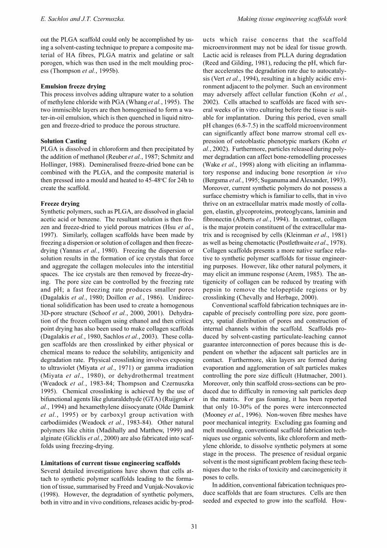
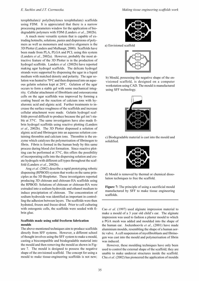
Scaffolds made using solid freeform fabricationmouldsThe above-mentioned techniques aim to produce scaffoldsdirectly from SFF systems. However, a different schoolof thought involves using the SFF system to make a mould,casting a biocompatible and biodegradable material intothe mould and then removing the mould as shown in Fig-ure 7. The mould is designed to possess the negativeshape of the envisioned scaffold. The concept for using amould to make tissue-engineering scaffolds is not new;
Cao et al. (1997) used alginate impression material tomake a mould of a 3 year old child’s ear. The alginateimpression was used to fashion a plaster mould to whicha PGA mesh was added and moulded into the shape ofthe human ear. Jockenhoevle et al., (2001) have madealuminium moulds, resembling the shape of a human aor-tic valve. A cell suspension of myofibroblasts and fibrino-gen was cast into the mould and polymerisation of fibrinwas induced.
However, these moulding techniques have only beenused to control the external shape of the scaffold; they areunable to make undercut structures inside the scaffold.Chu et al. (2002) has pioneered the application of moulds
a) Envisioned scaffold
b) Mould, possessing the negative shape of the en-visioned scaffold, is designed on a computerworkstation using CAD. The mould is manufacturedusing SFF technology.
c) Biodegradable material is cast into the mould andsolidified.
d) Mould is removed by thermal or chemical disso-lution techniques to free the scaffold.
Figure 7: The principle of using a sacrificial mouldmanufactured by SFF to make tissue engineeringscaffolds.

36
E. Sachlos and J.T. Czernuszka. Making tissue engineering scaffolds work
made by SFF to control the internal architecture of thescaffold. Moulds were fabricated with a SLA system us-ing a commercial epoxy resin. A 40 vol% HA suspensionwas cast into the mould and the acrylic-based binder ther-mally cured. The epoxy mould and acrylic binders wereremoved by pyrolysis and the remaining HA sintered.Several mould designs were produced, all of which pos-sessed a series of interconnecting shafts of controlled di-mensions which dictate the channel structure to be formedin the HA scaffold. The size of channels in the HA scaf-fold was reported to be 469±20µm in height and334±10µm in width. The reported average compressivestrength of 30±8 MPa of the HA scaffolds is comparableto that of coralline HA (Shors and Holmes, 1993). Im-plantation of cylindrical shaped scaffolds, with orthogonaland radial channel designs, in the mandible of Yucatanminipigs for 9 weeks revealed a bone penetration depthof 0.7-1.4mm in the end sections of both designs, and1.08+/-4.9mm and 1.28+/-0.6mm in the middle sectionsof the orthogonal and radial designs, respectively. Simi-larly, Bose et al. (2002) fabricated alumina scaffolds withmoulds made by FDM using commercially available ICW-06 thermoplastic wax. The alumina scaffolds were coatedwith HA to make the scaffolds bioactive. These bioceramicscaffolds are limited to bone tissue engineering and thehigh binder burn out temperatures (550oC) eliminates thepossibility of making biodegradable polymer scaffolds withthis process.
Taboas et al. (2003) have cast a solution of PLLA andchloroform into wax and ceramic moulds to create chan-nelled structures. Salt particles were added to the mould,and removed by leaching, to create interconnected poreswithin the polymer matrix. PLLA/PGA composites werealso made by a two-step melt casting procedure using aceramic mould. HA/PLLA composites were producedusing a similar process. The ceramic moulds were re-moved by chemical dissolution using RDO (a commer-cial decalcifier). This process uses organic solvents forcasting and mould removal, thus raising the issue ofwhether toxic residues are present on the scaffold.
A process developed at the University of Oxford andUMIST has resulted in the production of collagen scaf-folds with controlled and predefined internal morphol-ogy (Sachlos et al., 2003). The process begins with amould, which is manufactured using a phase-change ink-jet printer, the Model-Maker II. The mould possesses aseries of interconnected and branched shafts runningacross the walls of the mould. A dispersion of collagentype I is cast into the mould and frozen at –20oC. Themould containing the frozen collagen is then immersedin ethanol, which dissolves the mould and ice crystals toleave a porous collagen structure containing channels thatare predefined by the mould. The channel width andheight size can be adjusted to a minimum of 200±20µm.Ethanol is probably a more appropriate organic solvent totissue engineering scaffold processing because any residuesshould not be significantly deleterious to cells. The etha-nol is then removed by critical point drying with liquidcarbon dioxide. The result is a dry collagen scaffold,shown in Figure 8, which is ready for subsequentcrosslinking, rehydration and cell culturing. The process
is attractive in that collagen is used as the scaffold mate-rial. The processing temperature never exceeds 36oC,therefore preventing the denaturing of the collagen mol-ecules and offers the possibility of incorporating otherbiological molecules, e.g. growth factors. These collagenscaffolds are intended for the engineering of soft tissues.Composite scaffolds have also been formed by adding 70%weight HA particles into the collagen dispersion and thenprocessing as shown in Figure 9. These composite scaf-folds provide a chemical environment characteristic ofbone and are intended for engineering of this tissue. Themoulds are made from the commercial thermoplasticmaterials, ProtoBuildTM. Research efforts are aimed atmaking for scaffolds of type I collagen for use in hard andsoft tissue augmentation using non-toxic solvents andmaterials.
Figure 8: SEM image showing cross-section througha collagen scaffold made with SFF mould. Note thewell-defined square channels and the size of thesechannels which range from 170-450µm.
Figure 9: SEM image showing a composite scaffoldwith hydroxyapatite particles embedded throughoutthe collagen matrix. Note the open-cell porosity ofthe collagen matrix.

37
E. Sachlos and J.T. Czernuszka. Making tissue engineering scaffolds work
Conclusion
In summary, tissue-engineering scaffolds made from con-ventional fabrication techniques are inadequate for thegrowth of thick cross-sections of tissue due to the diffu-sion constraints posed by foam structures. Solid freeformfabrication systems provide a solution to this problem bycreating scaffolds with controlled internalmicroarchitecture, which should increase the mass trans-port of oxygen and nutrients deep into the scaffold. Sev-eral systems have been successfully adapted to producesynthetic and natural biodegradable polymer, bioceramicand hydrogel scaffolds. However, shortcomings arise fromdifficulty in support powder removal, use of toxic organicbinders, high temperature processing preventing the useof biological molecules and poor mechanical strength. Theuse of sacrificial moulds made by SFF systems avoids someof these processing issues whilst retaining the computercontrol and high resolution offered by SFF. Optimisingthe scaffold is essential if tissue engineering is going tobe successful in replacing or repairing damaged humantissues. SFF has the potential to optimise these scaffoldsand make them work.
References
Agrawal CM, Ray RB (2001) Biodegradable polymericscaffolds for musculoskeletal tissue engineering. J BiomedMater Res 55: 141-150.
Alberts B, Bray D, Lewis J, Raff M, Roberts K, WatsonJD (1994). Molecular Biology of the Cell, 3rd edition.Garland Publishing Ltd, New York. pp 971-1000.
Ang TH, Sultana FSA, Hutmacher DW, Wong YS,Fuh JYH, Mo XM, Loh HT, Burdet E, Teoh SH (2002)Fabrication of 3D chitosan-hydroxyapatite scaffolds us-ing a robotic dispensing system. Mater Sci Eng C20: 35-42.
Arem A (1985) Collagen modifications. Clin PlastSurg 12: 209-220.
Bose S, Darsell J, Hosick H, Yany L, Sarrkar DK,Bandyopahhyay A (2002): Processing and characteriza-tion of porous alumina scaffolds. J Mater Sci: Mat M 13:23-28.
Bredt JF, Sach E, Brancazio D, Cima M, Curodeau A,Fan T (1998) Three dimensional printing system. US Pat-ent 5807437.
Bergsma JE, de Bruijn WC, Rozema FR, Bos RR,Boering G (1995) Late degradation tissue response topoly(L-lactide) bone plates and screws. Biomaterials 16:25-31.
Burg KJL, Porter S, Kellam JF (2000) Biomaterialdevelopments for bone tissue engineering. Biomaterials21: 2347-2359.
Cao Y, Vacanti JP, Paige KT, Upton J, Vacanti CA(1997) Transplantation of chondrocytes utilizing a poly-mer-cell construct to produce tissue-engineered cartilagein the shape of a human ear. Plast Reconstr Surg 100:297-302.
Chevally B, Herbage D (2000) Collagen-basedbiomaterials as 3D scaffold for cell cultures: applications
for tissue engineering and gene therapy. Med Biol EngComput 38: 211-218.
Chu TMG, Orton DG, Hollister SJ, Feinberg SE,Halloran JW (2002) Mechanical and in vivo performanceof hydroxyapatite implants with controlled architectures.Biomaterials 23: 1283-1293.
Cima LG, Vacanti JP, Vacanti C, Inger D, Mooney D,Langer R (1991). Tissue engineering by cell transplanta-tion using degradable polymer substrates. J Biomech Eng-T ASME 113: 143-151.
Dagalakis N, Flink J, Stasikelis P, Burke JF, YannasIV (1980) Design of an artificial skin. Part III. Control ofpore structure. Biomaterials 14: 511-528.
Doillon CJ, Whyne CF, Brandwein S, Silver FH.(1986) Collagen-based wound dressings: Control of thepore structure and morphology. J Biomed Mater Res 20:1219-1228.
Eaglstein WH, Falanga V (1997): Tissue engineeringand the development of Apligraf®, a human skin equiva-lent, Clin Ther 19: 894-905.
Federal Register (1997). Q3C Impurities: Residual Sol-vents. US Department of Health and Human Services:Food and Drug Administration. Federal Register 62, pp.67377-67388.
Freed LE, Vunjak-Novakovic G (1998) Culture of or-ganized cell communities. Adv Drug Deliver Rev 33: 15-30.
Giordano RA, Wu BM, Borland SW, Cima LG, SachsEM, Cima MJ (1996) Mechanical properties of densepolylactic acid structures fabricated by three dimensionalprinting. J Biomat Sci-Polym E 8: 63-75.
Glicklis R, Shapiro L, Agbaria R, Merchuk JC, CohenS (2000) Hepatocyte behavior within three-dimensionalporous alginate scaffolds. Biotechnol Bioeng 67: 344-353.
Griffith ML, Halloran JW (1996) Freeform fabrica-tion of ceramics via stereolithography. J Am Ceram Soc79: 2601-2608.
Griffith LG (2000). Polymeric biomaterials. ActaMater 48: 263-277.
Griffith LG, Wu BM, Cima MJ, Powers MJ,Chaignaud B, Vacanti JP (1997) In vitro organogenesisof liver tissue. Ann N Y Acad Sci 831: 382-397.
Guyton AC, Hall JE (1996) Textbook of Medical Physi-ology, 6th edition. WB Saunders, Philadelphia. pp. 183-197.
Hayashi T (1994) Biodegradable polymers for biomedi-cal uses. Prog Polym Sci 19: 663-702.
Hsu YY, Gresser JD, Trantolo DJ, Lyons CM,Gangadharam PRJ, Wise DL (1997) Effect of polymerfoam morphology and density on kinetics of in vitro con-trolled release of isoniazid from compressed foam matri-ces. J Biomed Mater Sci 35: 107-116.
Hull C (1990) Method for production of three-dimen-sional objects by stereolithography. US Patent 4929402
Hutmacher DW (2001) Scaffold design and fabrica-tion technologies for engineering tissues-state of the artand future perspectives. J Biomat Sci-Polym E 12: 107-124.
Hutmacher DW, Schantz T, Zein I, Ng KW, Teoh SH,Tan KC (2001) Mechanical properties and cell cultural

38
E. Sachlos and J.T. Czernuszka. Making tissue engineering scaffolds work
response of polycaprolactone scaffolds designed and fab-ricated via fused deposition modeling. J Biomed MaterRes 55: 203-216.
Ishaug-Riley SL, Crane GM, Gurlek A, Miller MJ,Yasko AW, Yaszemski MJ, Mikos AG (1997): Ectopicbone formation by marrow stromal osteoblast transplan-tation using poly(DL-lactic-co-glycolic acid) foams im-planted into the rat mesentery. J Biomed Mater Res 36: 1-8.
Jockenhoevle S, Zund G, Hoerstrup SP, Chalabi K,Sachweh JS, Demircan L, Messmer BJ, Turina M (2001)Fibrin gel-advantages of a new scaffold in cardiovasculartissue engineering. Eur J Cardio-Thorac 13: 424-430.
Kim SS, Utsunomiya H, Koski JA, Wu BM, Cima MJ,Sohn J, Mukai M, Griffith LG, Vacanti JP (1998) Sur-vival and function of hepatocytes on a novel three-dimen-sional synthetic biodegradable polymer scaffold with anintrinsic network of channels. Ann Surg 228: 8-13
Kleinman HK, Klebe RJ, Martin GR (1981). Role ofcollagenous matrices in the adhesion and growth of cells.J Cell Biol 88: 473-485.
Koegler WS, Patrick C, Cima MJ, Griffith LG (2002)Carbon dioxide extraction of residual chloroform frombiodegradable polymers. J Biomed Mater Res 63: 567-576.
Kohn DG, Sarmadi M, Helman JI, Krebsbach PH(2002) Effects of pH on human bone marrow stromal cellsin vitro: Implications for tissue engineering of bone. JBiomed Mater Res 60: 292-299.
Lam EXF, Mo XM, Teoh SH, Hutmacher DW (2002)Scaffold development using 3D printing with a starch-based polymer. Mat Sci Eng C20: 49-56.
Landers R, Mulhaupt R (2000) Desktop manufactur-ing of complex objects, prototypes and biomedical scaf-folds by means of computer-assisted design combined withcomputer-guided 3D plotting of polymers and reactiveoligomers. Macromol Mater Eng 282: 17-21.
Landers R, Hubner U, Schmelzeisen R, Mulhaupt R(2002a). 3D Plotting. Proceedings of Rapid Prototypingfor Biomedical Applications: from Implants to OrganPrinting Workshop, Freiburg, Germany, 31 May 2002.Workshop organised by Landers R, Pfister A, MulhauptR, Freiburger Materialforschungszentrum and Institut furMakromolekulare Chemie of the Albert Ludwigs Univer-sity Freiburg, Germany.
Landers R, Pfister A, Hubner U, John H, SchmelzeisenR, Mulhaupt R (2002b) Fabrication of soft tissue engi-neering scaffolds by means of rapid prototyping tech-niques. J Mater Sci 37: 3107-3116.
Langer R, Vacanti JP (1993) Tissue engineering. Sci-ence 260: 920-926.
LeGeros RZ (2002) Properties of osteoconductivebiomaterials: calcium phosphates. Clin Orthop Relat Res395: 81-98.
Levy RA, Chu TGM, Halloran JW, Feinberg SE,Hollister S (1997) CT-generated porous hydroxyapatiteorbital floor prosthesis as a prototype bioimplant. Am JNeuroradiol 18: 1522-1525.
Lo H, Ponticiello MS, Leong KW (1995): Fabricationof controlled release biodegradable foams by phase sepa-ration. Tissue Eng 1: 15-28.
Madihally SV, Matthew HWT (1999) Porous chitosanscaffolds for tissue engineering. Biomaterials 20: 1133-1142.
Martin I, Padera RF, Vunjak-Novakovic G, Freed LE(1998) In vitro differentiation of chick embryo bone mar-row stromal cells into cartilaginous and bone-like tissues.J Orthopaed Res 16: 181-189.
Matsuda T, Mizutani M (2002) Liquid acrylate-endcapped biodegradable poly(e-caprolactone-co-trimeth-ylene carbonate). II. Computer-aided stereolithographicmicroarchitectural surface photoconstructs. J BiomedMater Res 62: 395-403.
Mikos AG, Sarakinos G, Leite SM, Vacanti JP, LangerR (1993a): Laminated three-dimensional biodegradablefoams for use in tissue engineering. Biomaterials 14: 323-330.
Mikos AG, Bao Y, Cima LG, Ingber DE, Vacanti JP,Langer R (1993b): Preparation of poly (glycolic acid)bonded fibres structures for cell attachment and trans-plantation. J Biomed Mater Res, 27: 183-189.
Mikos AG, Thorsen AJ, Czerwonka LA, Bao Y,Langer R (1994) Preparation and characterisation ofpoly(L-lactic acid) foams. Polymer 35: 1068-1077.
Mikos AG, Sarakinos G, Vacanti JP, Langer R, CimaLG (1996) Biocompatible polymer membranes and meth-ods of preparation of three dimensional membrane struc-tures. US Patent Number 5514378
Miyata T, Sohde T, Rubin AL, Stenzel KH (1971) Ef-fects of ultraviolet irradiation on native and telopeptide-poor collagen. Biochim Biophy Acta 229: 672-680.
Miyata T, Rubin AL, Dunn MW, Stenzel KH (1980)Collagen soft contact lens. US Patent 4 223 984
Mooney DJ, Baldwin DF, Suh NP, Vacanti JP, LangerR (1996) Novel approach to fabricate porous sponges ofpoly(D,L-lactic co-glycolic acid) without the use of or-ganic solvents. Biomaterials 17: 1417-1422.
Olde Damink LHH, Dijkstra PJ, Van Luyn MJA, VanWachem PB, Nieuwenhuis P, Feijen J (1995) Crosslinkingof dermal sheep collagen using hexamethylenediisocyanate. J Mat Sci: Mater Med 6: 429-434
Park A, Wu B, Griffith LG (1998) Integration of sur-face modification and 3D fabrication techniques to pre-pare patterned poly(L-lactide) substrates allowingregionally selective cell adhesion. J Biomater Sci-PolymE 9: 89-110.
Patrick CW, Mikos AG, McIntire LV (1998) Prospectsof tissue engineering. In: Frontiers in Tissue Engineer-ing. Patrick CW, Mikos AG, McIntire LV, eds. ElsevierScience Ltd, Oxford. pp. 3-11.
Pham DT, Dimov SS (2000) Rapid prototyping proc-esses. In: Rapid Manufacturing: the Technologies and Ap-plications of Rapid Prototyping and Rapid Tooling. PhamDT, Dimov SS, eds. Springer, London. pp.19-42.
Philbrook KF, Sanders JR, Royden C, Forsyth JL(1996) 3-D model maker. US Patent 5506607.
Porter NL, Pilliar RM, Grynpas MD (2001) Fabrica-tion of porous calcium polyphosphate implants by solidfreeform fabrication: A study of processing and in vitrodegradation characteristics. J Biomed Mat Res 56: 504-515.
Postlethwaite AE, Seyer JM, Kang AH (1978).

39
E. Sachlos and J.T. Czernuszka. Making tissue engineering scaffolds work
Chemotactic attraction of human fibroblasts by type I, IIand III collagens and collagen-derived peptides. Proc NatlAcad Sci USA 75: 871-875.
Reed AM, Gilding DK (1981) Biodegradable poly-mer for use in surgery – poly(glycolic)/poly(lactic acid)homo and copolymers: 2. In vitro degradation. Polymer,22: 342-346.
Reuber M, Yu LS, Kolff WJ (1987) Effect of process-ing temperature on the properties of polyurethane andcomparison of vacuum forming and solution casting tomake artificial hearts. Artif Organs 11: 323-323.
Ruijgrok JM, De Wijhn JR, Boon ME (1994)Optimizing glutaraldehyde crosslinking of collagen: ef-fect of time, temperature and concentration as measuredby shrinkage temperature. J Mater Sci: Mater Med 5: 80-87.
Sachlos E, Reis N, Ainsley C, Derby B, CzernuszkaJT (2003) Novel collagen scaffolds with predefined inter-nal morphology made by solid freeform fabrication.Biomaterials 24: 1487-1497.
Schmitz JP, Hollinger JO (1988) A preliminary studyof the osteogenic potential of a biodegradable alloplastic-osteoinductive alloimplant. Clin Orthop 237, 245-255
Schoof H, Apel J, Heschel I, Rau G (2001) Control ofpore structure and size in freeze-dried collagen sponges.J Biomed Mater Res-A 58: 352-357.
Schoof H, Burns L, Fisher A, Heschel I, Rau G (2000)Dendritic ice morphology in unidirectionally solidifiedcollagen suspensions. J Cryst Growth 209: 122-129.
Scott CS (1991) Apparatus and method for creatingthree-dimensional objects. US Patent 5121329.
Shalak R, Fox CF (1988). Preface. In: Tissue Engi-neering. Shalak R, Fox CF, eds. Alan R.Liss, New York.pp. 26-29.
Shors E, Holmes R (1993) Porous hydroxyapatite. In:An Introduction to Bioceramics. Hench L, Wilson J, eds.World Scientific, Singapore. pp 181-198.
Suganuma J, Alexander H (1993) Biological responseof intramedullary bone to poly-L-lactic acid. J ApplBiomater 4: 13-27.
Taboas JM, Maddox RD, Krebsbach PH, Hollister SJ(2003) Indirect solid free form fabrication of local andglobal porous, biomimetic and composite 3D polymer-ceramic scaffolds. Biomaterials 24: 181-194.
Thompson JI, Czernuszka JT (1995) The effect of twotypes of crosslinking on some mechanical properties ofcollagen. Biomed Mater Eng 5: 37-48.
Thompson RC, Yaszemski MJ, Powers JM, Mikos AG(1995a) Fabrication of biodegradable polymer scaffoldsto engineering trabecular bone. J Biomater Sci-Polym E7: 23-38.
Thompson RC, Yaszembksi MJ, Powers JM, HarriganTP, Mikos AG (1995b) Poly(a-hydroxy ester)/short fiberhydroxyapatite composite foams for orthopedic applica-tions. In: Polymers in Medicine and Pharmacy, Vol 394.Mikos AG, Leong KW, Yaszemski MJ, eds. MaterialsResearch Society Symposium Proceedings, Pittsburgh. pp.25-30.
Vander AJ, Sherman JH, Luciano DS (1985). HumanPhysiology. McGraw-Hill, New York. pp. 341, 366.
Vert M., Mauduit J, Li S (1994) Biodegradation ofPLA/GA polymers: increasing complexity. Biomaterials15: 1209-1213.
Wake MC, Gerecht PD, Lu LC, Mikos AG (1998) Ef-fects of biodegradable polymer particles on rat marrow-derived stromal osteoblasts in vitro. Biomaterials 19, 1255-1268.
Weadock K, Olson RM, Silver FH (1983-84): Evalua-tion of collagen crosslinking techniques. Biomater ArtifCell 11: 293-318.
Whang K, Thomas CK, Nuber G, Healy KE (1995) Anovel method to fabricate bioabsorbable scaffolds. Poly-mer 36: 837.
Woodfield T, Malda J, de Wijn J , Riesle J, vanBlitterwijk CA (2002) Proceedings of Rapid Prototypingfor Biomedical Applications: from Implants to OrganPrinting Workshop, Freiburg, Germany, 31 May 2002.Workshop organised by Landers R, Pfister A, MulhauptR, Freiburger Materialforschungszentrum and Institut furMakromolekulare Chemie of the Albert Ludwigs Univer-sity Freiburg, Germany.
Yannas IV, Burke JF, Gordon PL, Huang C, RubensteinRH (1980) Design of an artificial skin. Part II. Control ofchemical composition. Biomaterials 14: 107-131.
Zein I, Hutmacher DW, Tan KC, Teoh SH (2002) Fuseddeposition modelling of novel scaffold architectures fortissue engineering applications. Biomaterials 23: 1169-1185.
Discussion with Reviewers
S. Gogolewski: The authors state “Conventional scaffoldfabrication techniques are incapable of precisely control-ling pore size, pore geometry, spatial distribution of poresand construction of internal channels within the scaffold.Scaffolds produced by solvent-casting particulate-leach-ing cannot guarantee interconnection of pores because thisis dependent on whether the adjacent salt particles are incontact. Furthermore, skin layers are formed duringevaporation and agglomeration of salt particles makescontrolling the pore size difficult (Hutmacher, 2001).Moreover, only thin scaffold cross-sections can be pro-duced due to difficulty in removing salt particles deep inthe matrix “. This statement is too general and not justi-fied. Look for example in Gugala and Gogolewski (2000).The polylactide scaffold has interconnected open pores,well-controlled pore size, no skin layers and no salt parti-cles deep in the matrix.Authors: The use of SFF allows us to control channelsand their interconnectivity with a diameter of approxi-mately 200µm. The use of a porogen will give rise to adistribution of pore sizes and their interconectivity (whichmy well be present) cannot be controlled in the same wayas by SFF. It is this level of control of the internal (andexternal) architecture which makes SFF so potentiallyexciting. Of course, the matrix between the channels canbe porous with interconectivity if so desired. We stand byour assertion that current tissue engineering scaffoldswithout controlled internal channels cannot allow cellsto maintain viability deep within the scaffold.

40
E. Sachlos and J.T. Czernuszka. Making tissue engineering scaffolds work
M. Dalby: For the collagen / HA composite intended forbone tissue engineering, how mechanically similar to boneis the material and what is the percentage of HA incorpo-rated - will there be enough to truly effect osteoblast re-sponse? In order for such a material to be used in load-bearing applications, the material should be able to sup-port the rigors of walking etc. Perhaps it is intended thatonce osseointegration has taken place the formation ofbone within the scaffold will be able to take the load?A. Curtis: Would the authors like to comment on (1) Thedegree of accuracy and reproducibility in fabrication thatis desired, and (2) The desirable mechanical properties ofthe polymer?Authors: These two questions are answered together. Themechanical properties of the scaffold can be tailored tosuit the particular requirements. Thus, a material with alow elastic modulus would be more suitable as a scaffoldfor an arterial graft, whereas higher values of the YoungModulus would be required for bone augmentation. The
modulus is controlled via several routes - firstly by thecomponents comprising the scaffold itself, secondly thescale and size of the porosity and thirdly, the dimensionsand shape of any internal channels. It is this degree ofcontrol which makes solid free-form fabrication such auseful tool in making scaffolds. The scaffold of bone tis-sue augmentation has a Young’s modulus of approximately2 GPa.. This is similar to many artificial polymers, butwe use the natural components - collagen and hydroxya-patite. The response of osteoprogenitor cells to this classof biomaterial is superior to that found with artificial poly-mers.
Additional Reference
Gugala Z, Gogolewski S (2000) In vitro growth andactivity of primary chondrocytes on a resorbablepolylactide three-dimensional scaffold. J Biomed MaterRes 49: 183-191.