magnetic resonance imaging findings of grafted cancellous allografts after removal of bone tumors...
TRANSCRIPT

Magnetic Resonance Imaging Findings of Grafted Cancellous Allografts after Removal of Bone Tumors
Seungcheol Kang, Ilkyu Han,
Sunghwan Hong, Hwan Seong Cho, Han-Soo Kim
Musculoskeletal Tumor Center, Seoul National University Cancer Hospital, Seoul, Korea
- CTOS 2014 -

Cancellous Allograft
• Allograft
: Donor site morbidity↓ / Quantity ↑
→ Commonly used in bone tumor surgery
• Cancellous allograft
: Fill defect after curettage of bone tumor

MRI in Bone Tumors
• Follow-up modality for surveillance of recurrence
• Curettage + Cancellous allograft
→ MRI DDx. cancellous allograft vs. recurred tumor
• No report on MRI findings of cancellous allograft

Purpose
To investigate the MRI features of
• Cancellous allograft & their change over time
• Cancellous allograft vs. Recurred tumors

Reviewed (n=53)
Curettage and cancellous allograft between 2002~2011
MRI for follow-up
Metallic device, BMP, or calcium component (n=17)
Lost to follow-up within 1 year after imaging (n=2)
Analyzed (n=34)
Excluded (n=19)
Patient Selection

• Age: 27 years (11-64)
• Sex: M (18) F (14)
• Tumor typeChondroblastoma (11), LG chonsa (10), ABC (3), GCT (2), FD (2), SBC (2), IO lipoma (1), PVNS (1)
• Site Femur (12), tibia (8), pelvis (5), humerus (4), foot (4)
• Tumor size: 6.7 ± 6.3 cm
Patient Characteristics

• 1.5- or 3.0-Tesla MRI scanners
• Sequences• T1WI: spin-echo T1-weighted • T2WI: fast spin-echo T2-weighted • Enhanced T1: gadolinium-enhanced spin-echo T1WI
• Review by 2 independent authors
Acquisition of MRIs
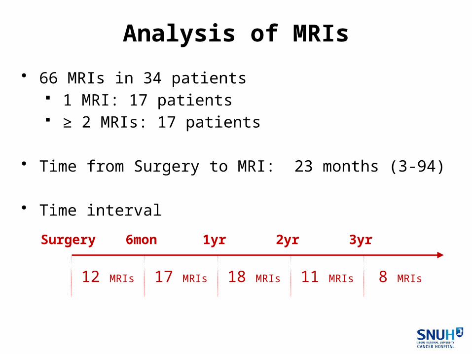
• 66 MRIs in 34 patients 1 MRI: 17 patients ≥ 2 MRIs: 17 patients
• Time from Surgery to MRI: 23 months (3-94)
• Time interval
Analysis of MRIs
Surgery 6mon 1yr 2yr 3yr
12 MRIs 17 MRIs 18 MRIs 11 MRIs 8 MRIs
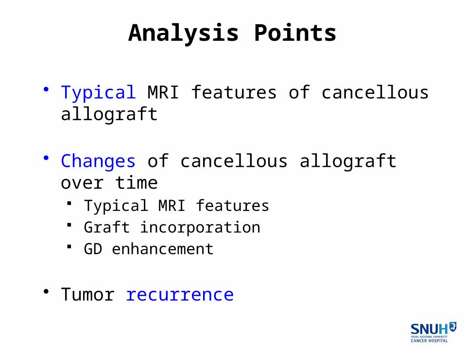
• Typical MRI features of cancellous allograft
• Changes of cancellous allograft over time Typical MRI features Graft incorporation GD enhancement
• Tumor recurrence
Analysis Points

Results

Typical MRI Features• T1
Homogeneous intermediate SI ±Speckled hyperintensities

Typical MRI Features• T1
Homogeneous intermediate SI ± Speckled hyperintensities
• T2 High SI ± Speckled hypointensities

Typical MRI Features• T1
Homogeneous intermediate SI ± Speckled hyperintensities
• T2 High SI ± Speckled hypointensities
• Enhanced T1 Peripheral rim enhance ± Central heterogeneous enhance

Typical MRI Features• T1
Homogeneous intermediate SIs ± Speckled hyperintensities
• T2 High SIs ± Speckled hypointensities
• Enhanced T1 Peripheral rim enhance ± Central heterogeneous enhance
Present in
all cases (n=12)

Changes of Typical MRI Features
T1 100% 82% 67% 60% 0%
T2 100% 59% 33% 17% 0%
En-T1 100% 100% 87% 50% 0%
Surgery 6mon 1yr 2yr 3yr
Changes of Over Time

Graft Incorporation
Peripheral 0% 24% 50% 54% -
Complete 0% 0% 17% 46% 100%
Total 0% 24% 67% 100% 100%
Surgery 6mon 1yr 2yr 3yr
Changes of Over Time

Graft IncorporationPO 10mon PO 18mon
Changes of Over Time

GD Enhancement
100% 59% 33% 17% 0%
Surgery 6mon 1yr 2yr 3yr
Changes of Over Time

PO 5mon PO 34mon
GD EnhancementChanges of Over Time

Suspicious Recurrence on MRI (n=8)
No recurrence (n=3)
Recurrence on MRI
Recurrence (n=5)
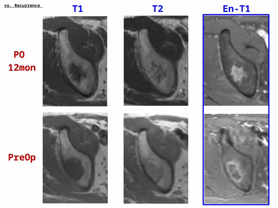
vs. Recurrence

Suspicious Recurrence on MRI (n=8)
No recurrence (n=3)
Recurrence on MRI
Recurrence (n=5)
Same SI with primary tumor
vs. Recurrence

Recur Initial
Recurrence PO 9 mon
vs. Recurrence

Suspicious Recurrence on MRI (n=8)
MRIs < 1 year after surgery Chondrosarcomas
No recurrence (n=3)
Recurrence on MRI
Recurrence (n=5)
vs. Recurrence

No Recurrence PO 23 mon
Pre-op FU
vs. Recurrence

T1 T2 En-T1
PO
12mon
vs. Recurrence

T1 T2 En-T1
PO
12mon
PreOp
vs. Recurrence

T1 T2 En-T1
PO
12mon
PreOp
vs. Recurrence

T1 T2 En-T1
PO
12mon
PreOp
vs. Recurrence
T1 T2 En-T1
PO
12mon
PreOp
vs. Recurrence

T1 T2 En-T1
PO
12mon
PreOp
vs. Recurrence

Summary & Conclusion
• This study assessed the MRI features of cancellous allo-grafts & their change over time, and the differentiating fea-tures between cancellous allografts and recurred tumors.

Summary & Conclusion
• This study assessed the MRI features of cancellous allo-grafts & their change over time, and the differentiating fea-tures between cancellous allografts and recurred tumors.
• Cancellous allografts typically showed homogeneous inter-mediate or low SIs on T1, high SIs with speckled hypointen-sities on T2, and peripheral rim enhancements on enhanced T1.

Summary & Conclusion
• This study assessed the MRI features of cancellous allo-grafts & their change over time, and the differentiating fea-tures between cancellous allografts and recurred tumors.
• Cancellous allografts typically showed homogeneous inter-mediate or low Sis on T1, high SIs with speckled hypointen-sities on T2, and peripheral rim enhancements on enhanced T1.
• These typical features were maintained until 6 months and then gradually disappeared by 3 years.

Summary & Conclusion
• Graft incorporation occurred from periphery to center and were completed within 3 years.

Summary & Conclusion
• Graft incorporation occurred from periphery to center and were completed within 3 years.
• The single most differentiating feature of a recurred tumor was showing the similar pattern and SI of the primary tumor.

Summary & Conclusion
• Graft incorporation occurred from periphery to center and were completed within 3 years.
• The single most differentiating feature of a recurred tumor was showing the similar pattern and SI of the primary tumor.
• Careful interpretation is warranted when dealing with chon-droid tumors, especially for MRIs performed within 1 year of surgery.
