
Magnetic Resonance Imaging Findings of Grafted Cancellous Allografts after Removal of Bone Tumors
Seungcheol Kang, Ilkyu Han,
Sunghwan Hong, Hwan Seong Cho, Han-Soo Kim
Musculoskeletal Tumor Center, Seoul National University Cancer Hospital, Seoul, Korea
- CTOS 2014 -

Cancellous Allograft
• Allograft
: Donor site morbidity↓ / Quantity ↑
→ Commonly used in bone tumor surgery
• Cancellous allograft
: Fill defect after curettage of bone tumor

MRI in Bone Tumors
• Follow-up modality for surveillance of recurrence
• Curettage + Cancellous allograft
→ MRI DDx. cancellous allograft vs. recurred tumor
• No report on MRI findings of cancellous allograft

Purpose
To investigate the MRI features of
• Cancellous allograft & their change over time
• Cancellous allograft vs. Recurred tumors

Reviewed (n=53)
Curettage and cancellous allograft between 2002~2011
MRI for follow-up
Metallic device, BMP, or calcium component (n=17)
Lost to follow-up within 1 year after imaging (n=2)
Analyzed (n=34)
Excluded (n=19)
Patient Selection

• Age: 27 years (11-64)
• Sex: M (18) F (14)
• Tumor typeChondroblastoma (11), LG chonsa (10), ABC (3), GCT (2), FD (2), SBC (2), IO lipoma (1), PVNS (1)
• Site Femur (12), tibia (8), pelvis (5), humerus (4), foot (4)
• Tumor size: 6.7 ± 6.3 cm
Patient Characteristics

• 1.5- or 3.0-Tesla MRI scanners
• Sequences• T1WI: spin-echo T1-weighted • T2WI: fast spin-echo T2-weighted • Enhanced T1: gadolinium-enhanced spin-echo T1WI
• Review by 2 independent authors
Acquisition of MRIs
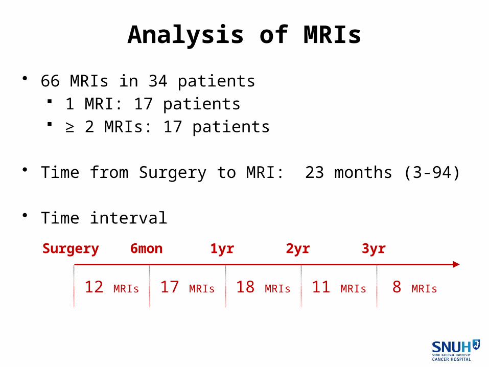
• 66 MRIs in 34 patients 1 MRI: 17 patients ≥ 2 MRIs: 17 patients
• Time from Surgery to MRI: 23 months (3-94)
• Time interval
Analysis of MRIs
Surgery 6mon 1yr 2yr 3yr
12 MRIs 17 MRIs 18 MRIs 11 MRIs 8 MRIs
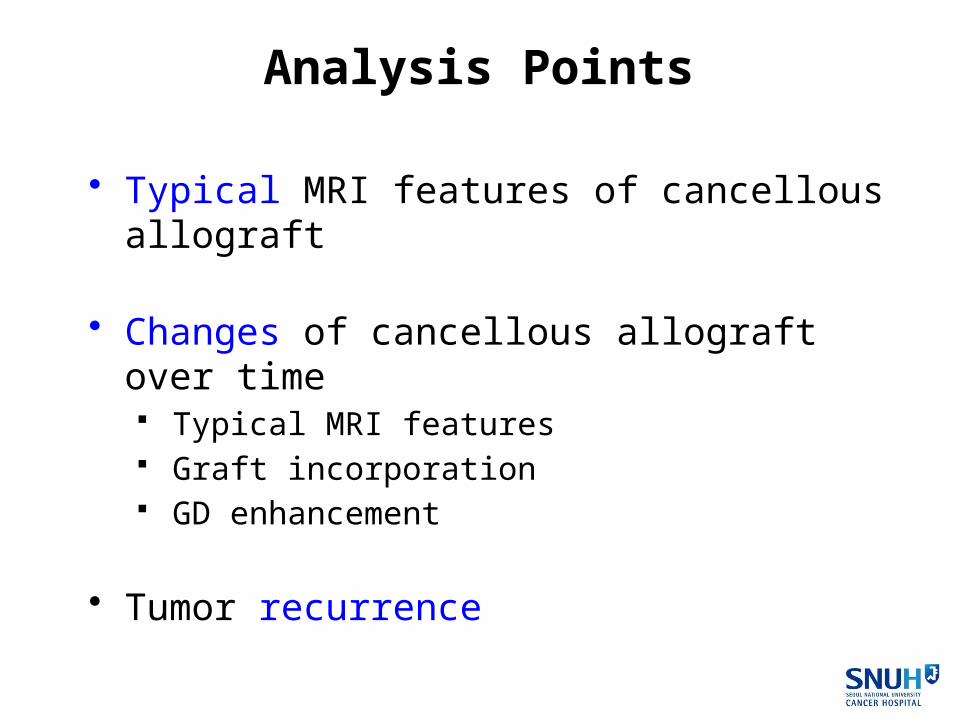
• Typical MRI features of cancellous allograft
• Changes of cancellous allograft over time Typical MRI features Graft incorporation GD enhancement
• Tumor recurrence
Analysis Points

Results

Typical MRI Features• T1
Homogeneous intermediate SI ±Speckled hyperintensities

Typical MRI Features• T1
Homogeneous intermediate SI ± Speckled hyperintensities
• T2 High SI ± Speckled hypointensities

Typical MRI Features• T1
Homogeneous intermediate SI ± Speckled hyperintensities
• T2 High SI ± Speckled hypointensities
• Enhanced T1 Peripheral rim enhance ± Central heterogeneous enhance

Typical MRI Features• T1
Homogeneous intermediate SIs ± Speckled hyperintensities
• T2 High SIs ± Speckled hypointensities
• Enhanced T1 Peripheral rim enhance ± Central heterogeneous enhance
Present in
all cases (n=12)

Changes of Typical MRI Features
T1 100% 82% 67% 60% 0%
T2 100% 59% 33% 17% 0%
En-T1 100% 100% 87% 50% 0%
Surgery 6mon 1yr 2yr 3yr
Changes of Over Time

Graft Incorporation
Peripheral 0% 24% 50% 54% -
Complete 0% 0% 17% 46% 100%
Total 0% 24% 67% 100% 100%
Surgery 6mon 1yr 2yr 3yr
Changes of Over Time

Graft IncorporationPO 10mon PO 18mon
Changes of Over Time

GD Enhancement
100% 59% 33% 17% 0%
Surgery 6mon 1yr 2yr 3yr
Changes of Over Time

PO 5mon PO 34mon
GD EnhancementChanges of Over Time

Suspicious Recurrence on MRI (n=8)
No recurrence (n=3)
Recurrence on MRI
Recurrence (n=5)
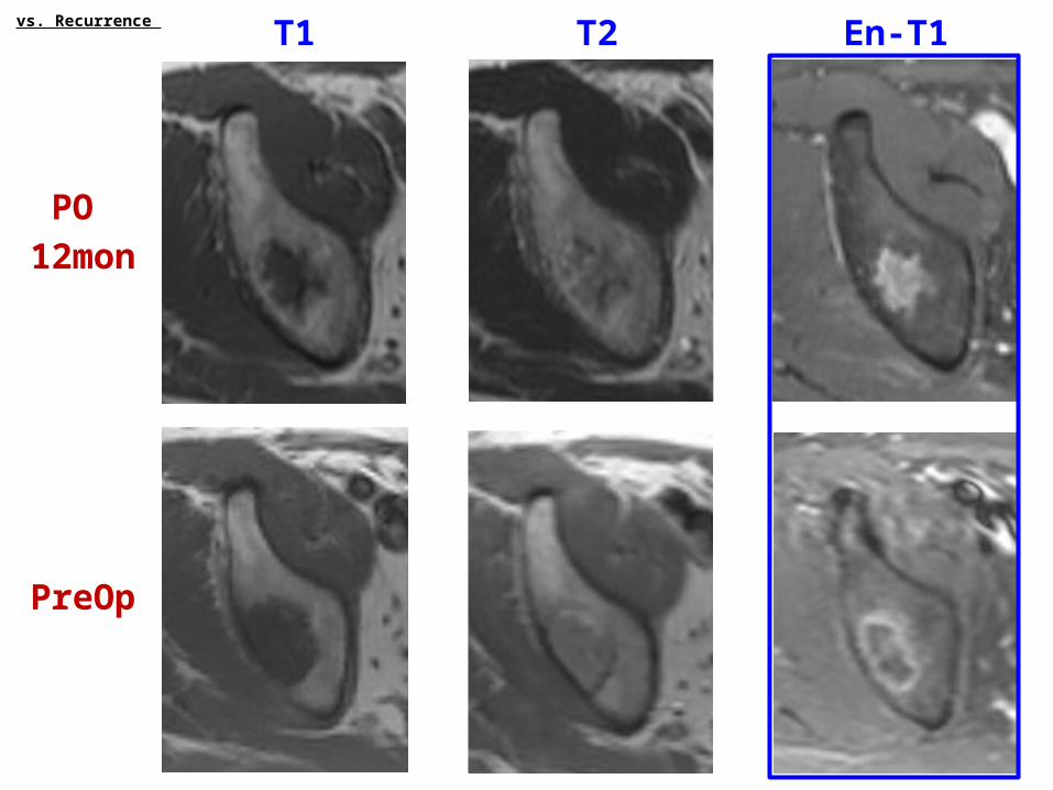
vs. Recurrence

Suspicious Recurrence on MRI (n=8)
No recurrence (n=3)
Recurrence on MRI
Recurrence (n=5)
Same SI with primary tumor
vs. Recurrence

Recur Initial
Recurrence PO 9 mon
vs. Recurrence

Suspicious Recurrence on MRI (n=8)
MRIs < 1 year after surgery Chondrosarcomas
No recurrence (n=3)
Recurrence on MRI
Recurrence (n=5)
vs. Recurrence

No Recurrence PO 23 mon
Pre-op FU
vs. Recurrence

T1 T2 En-T1
PO
12mon
vs. Recurrence

T1 T2 En-T1
PO
12mon
PreOp
vs. Recurrence

T1 T2 En-T1
PO
12mon
PreOp
vs. Recurrence

T1 T2 En-T1
PO
12mon
PreOp
vs. Recurrence
T1 T2 En-T1
PO
12mon
PreOp
vs. Recurrence

T1 T2 En-T1
PO
12mon
PreOp
vs. Recurrence

Summary & Conclusion
• This study assessed the MRI features of cancellous allo-grafts & their change over time, and the differentiating fea-tures between cancellous allografts and recurred tumors.

Summary & Conclusion
• This study assessed the MRI features of cancellous allo-grafts & their change over time, and the differentiating fea-tures between cancellous allografts and recurred tumors.
• Cancellous allografts typically showed homogeneous inter-mediate or low SIs on T1, high SIs with speckled hypointen-sities on T2, and peripheral rim enhancements on enhanced T1.

Summary & Conclusion
• This study assessed the MRI features of cancellous allo-grafts & their change over time, and the differentiating fea-tures between cancellous allografts and recurred tumors.
• Cancellous allografts typically showed homogeneous inter-mediate or low Sis on T1, high SIs with speckled hypointen-sities on T2, and peripheral rim enhancements on enhanced T1.
• These typical features were maintained until 6 months and then gradually disappeared by 3 years.

Summary & Conclusion
• Graft incorporation occurred from periphery to center and were completed within 3 years.

Summary & Conclusion
• Graft incorporation occurred from periphery to center and were completed within 3 years.
• The single most differentiating feature of a recurred tumor was showing the similar pattern and SI of the primary tumor.

Summary & Conclusion
• Graft incorporation occurred from periphery to center and were completed within 3 years.
• The single most differentiating feature of a recurred tumor was showing the similar pattern and SI of the primary tumor.
• Careful interpretation is warranted when dealing with chon-droid tumors, especially for MRIs performed within 1 year of surgery.


