lytic cycle of toxoplasma gondii - microbiology and...
TRANSCRIPT
MICROBIOLOGY AND MOLECULAR BIOLOGY REVIEWS,1092-2172/00/$04.0010
Sept. 2000, p. 607–623 Vol. 64, No. 3
Copyright © 2000, American Society for Microbiology. All Rights Reserved.
Lytic Cycle of Toxoplasma gondiiMICHAEL W. BLACK AND JOHN C. BOOTHROYD*
Department of Microbiology and Immunology, Stanford University School of Medicine,Stanford, California 94305-5124
INTRODUCTION .......................................................................................................................................................607Taxonomy .................................................................................................................................................................607Life Cycle .................................................................................................................................................................608Pathogenesis ............................................................................................................................................................608Genetics ....................................................................................................................................................................608
Toxoplasma is amenable to genetic analysis....................................................................................................608Molecular tools for manipulation of the Toxoplasma genome......................................................................608Gene discovery using expressed sequence tags...............................................................................................609
Ultrastructure..........................................................................................................................................................609Multiple, regulated secretory organelles .........................................................................................................609Cytoskeletal network ..........................................................................................................................................609Trimembrane pellicle .........................................................................................................................................610
HOST CELL ATTACHMENT...................................................................................................................................610Surface of Toxoplasma ............................................................................................................................................610Host Ligand .............................................................................................................................................................611
PARASITE INVASION ..............................................................................................................................................611Toxoplasma Demonstrates Substrate-Dependent Gliding Motility ..................................................................611Toxoplasma Invasion Requires Parasite Motility................................................................................................611Gliding Motility Is Dependent on an Actomyosin Motor .................................................................................612Candidate Bridge Molecules between the Exterior and the Actomyosin Motors ..........................................612Current Models for Toxoplasma Motility.............................................................................................................613Secretory Events during Invasion.........................................................................................................................614
VACUOLE FORMATION .........................................................................................................................................614The Parasitophorous Vacuole Is Distinct from Phagosomes ...........................................................................614Postinvasion Secretory Events Further Modify the Parasitophorous Vacuole ..............................................615
PARASITE REPLICATION ......................................................................................................................................616Morphological Examination of Endodyogeny .....................................................................................................616Toxoplasma Cell Cycle ............................................................................................................................................617
PARASITE EGRESS ..................................................................................................................................................617Egress Is a Rapid, Cytolytic Event.......................................................................................................................617Calcium Acts as a Signal for Egress....................................................................................................................618Dithiothreitol Acts as a Signal for Egress ..........................................................................................................619
INFLUENCE OF CA21 ON OTHER ASPECTS OF TOXOPLASMA GROWTH ..............................................619Rearrangement of the Toxoplasma Cytoskeleton ................................................................................................619Parasite Motility and Invasion .............................................................................................................................619Intracellular Survival and Replication ................................................................................................................619
CONCLUSION............................................................................................................................................................620ACKNOWLEDGMENTS ...........................................................................................................................................620REFERENCES ............................................................................................................................................................620
INTRODUCTION
Before discussing the Toxoplasma lytic cycle itself, we willprovide brief overviews of its taxonomy, life cycle, pathogene-sis, genetics, and ultrastructure. A basic understanding of theseaspects will help appreciate how the lytic cycle fits into theoverall biology of the parasite and the technical aspects ofstudying it. Following this introduction, we will describe ourcurrent understanding of the five components of the lytic cycle:attachment, invasion, vacuole formation, replication, and
egress. Because of the special role of calcium signaling in theseprocesses, a sixth section devoted to this aspect will also beincluded.
Taxonomy
Toxoplasma gondii is an obligate intracellular protozoanpathogen that was first described in 1908 by Nicolle andManceaux working in North Africa and by Splendore workingin Brazil. The species designation originated from the name ofthe North African rodent (Ctenodactylus gondi) from whichthis parasite was isolated. The genus name is derived from theGreek work toxon, meaning “bow” and referring to the cres-cent shape of the organism.
Toxoplasma belongs to the phylum Apicomplexa, which con-sists of intracellular parasites that have a characteristically po-
* Corresponding author. Mailing address: Department of Microbi-ology and Immunology, Fairchild Building, Room D305, Stanford Uni-versity School of Medicine, 299 Campus Drive, Stanford, CA 94305-5124. Phone: (650) 723-7984. Fax: (650) 723-6853. E-mail: [email protected].
607
on July 3, 2018 by guesthttp://m
mbr.asm
.org/D
ownloaded from
larized cell structure and a complex cytoskeletal and organellararrangement at their apical end (42). Other members of thisphylum include the human pathogens Plasmodium (the causeof malaria) and Cryptosporidium as well as the animal patho-gens Eimeria (the cause of chicken coccidiosis) and Sarcocystis.
Life Cycle
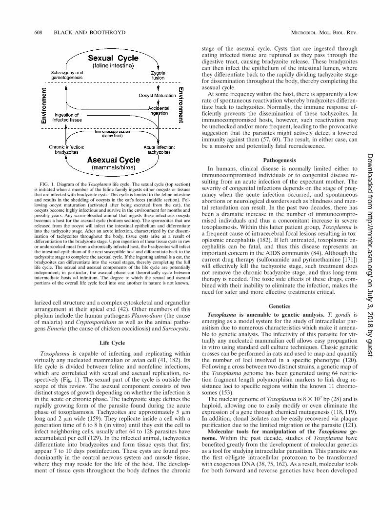
Toxoplasma is capable of infecting and replicating withinvirtually any nucleated mammalian or avian cell (41, 182). Itslife cycle is divided between feline and nonfeline infections,which are correlated with sexual and asexual replication, re-spectively (Fig. 1). The sexual part of the cycle is outside thescope of this review. The asexual component consists of twodistinct stages of growth depending on whether the infection isin the acute or chronic phase. The tachyzoite stage defines therapidly growing form of the parasite found during the acutephase of toxoplasmosis. Tachyzoites are approximately 5 mmlong and 2 mm wide (159). They replicate inside a cell with ageneration time of 6 to 8 h (in vitro) until they exit the cell toinfect neighboring cells, usually after 64 to 128 parasites haveaccumulated per cell (129). In the infected animal, tachyzoitesdifferentiate into bradyzoites and form tissue cysts that firstappear 7 to 10 days postinfection. These cysts are found pre-dominantly in the central nervous system and muscle tissue,where they may reside for the life of the host. The develop-ment of tissue cysts throughout the body defines the chronic
stage of the asexual cycle. Cysts that are ingested througheating infected tissue are ruptured as they pass through thedigestive tract, causing bradyzoite release. These bradyzoitescan then infect the epithelium of the intestinal lumen, wherethey differentiate back to the rapidly dividing tachyzoite stagefor dissemination throughout the body, thereby completing theasexual cycle.
At some frequency within the host, there is apparently a lowrate of spontaneous reactivation whereby bradyzoites differen-tiate back to tachyzoites. Normally, the immune response ef-ficiently prevents the dissemination of these tachyzoites. Inimmunocompromised hosts, however, such reactivation maybe unchecked and/or more frequent, leading to the provocativesuggestion that the parasites might actively detect a loweredimmunity against them (57, 60). The result, in either case, canbe a massive and potentially fatal recrudescence.
PathogenesisIn humans, clinical disease is normally limited either to
immunocompromised individuals or to congenital disease re-sulting from an acute infection of the expectant mother. Theseverity of congenital infections depends on the stage of preg-nancy when the acute infection occurred, and spontaneousabortions or neurological disorders such as blindness and men-tal retardation can result. In the past two decades, there hasbeen a dramatic increase in the number of immunocompro-mised individuals and thus a concomitant increase in severetoxoplasmosis. Within this latter patient group, Toxoplasma isa frequent cause of intracerebral focal lesions resulting in tox-oplasmic encephalitis (182). If left untreated, toxoplasmic en-cephalitis can be fatal, and thus this disease represents animportant concern in the AIDS community (84). Although thecurrent drug therapy (sulfonamide and pyrimethamine [171])will effectively kill the tachyzoite stage, such treatment doesnot remove the chronic bradyzoite stage, and thus long-termtherapy is needed. The toxic side effects of these drugs, com-bined with their inability to eliminate the infection, makes theneed for safer and more effective treatments critical.
GeneticsToxoplasma is amenable to genetic analysis. T. gondii is
emerging as a model system for the study of intracellular par-asitism due to numerous characteristics which make it amena-ble to genetic analysis. The infectivity of this parasite for vir-tually any nucleated mammalian cell allows easy propagationin vitro using standard cell culture techniques. Classic geneticcrosses can be performed in cats and used to map and quantifythe number of loci involved in a specific phenotype (120).Following a cross between two distinct strains, a genetic map ofthe Toxoplasma genome has been generated using 64 restric-tion fragment length polymorphism markers to link drug re-sistance loci to specific regions within the known 11 chromo-somes (153).
The nuclear genome of Toxoplasma is 8 3 107 bp (28) and ishaploid, allowing one to easily modify or even eliminate theexpression of a gene through chemical mutagenesis (118, 119).In addition, clonal isolates can be easily recovered via plaquepurification due to the limited migration of the parasite (121).
Molecular tools for manipulation of the Toxoplasma ge-nome. Within the past decade, studies of Toxoplasma havebenefited greatly from the development of molecular geneticsas a tool for studying intracellular parasitism. This parasite wasthe first obligate intracellular protozoan to be transformedwith exogenous DNA (38, 75, 162). As a result, molecular toolsfor both forward and reverse genetics have been developed
FIG. 1. Diagram of the Toxoplasma life cycle. The sexual cycle (top section)is initiated when a member of the feline family ingests either oocysts or tissuesthat are infected with bradyzoite cysts. This cycle is limited to the feline intestineand results in the shedding of oocysts in the cat’s feces (middle section). Fol-lowing oocyst maturation (activated after being excreted from the cat), theoocysts become highly infectious and survive in the environment for months andpossibly years. Any warm-blooded animal that ingests these infectious oocystsbecomes a host for the asexual cycle (bottom section). The sporozoites that arereleased from the oocyst will infect the intestinal epithelium and differentiateinto the tachyzoite stage. After an acute infection, characterized by the dissem-ination of tachyzoites throughout the body, tissue cysts arise as a result ofdifferentiation to the bradyzoite stage. Upon ingestion of these tissue cysts in rawor undercooked meat from a chronically infected host, the bradyzoites will infectthe intestinal epithelium of the next susceptible host and differentiate back to thetachyzoite stage to complete the asexual cycle. If the ingesting animal is a cat, thebradyzoites can differentiate into the sexual stages, thereby completing the fulllife cycle. The sexual and asexual components of the life cycle are potentiallyindependent; in particular, the asexual phase can theoretically cycle betweenintermediate hosts ad infinitum. The degree to which the sexual and asexualportions of the overall life cycle feed into one another in nature is not known.
608 BLACK AND BOOTHROYD MICROBIOL. MOL. BIOL. REV.
on July 3, 2018 by guesthttp://m
mbr.asm
.org/D
ownloaded from
to manipulate the genome of T. gondii (reviewed in refer-ences 16, 133, and 160). A variety of markers have been de-veloped to specifically select for transformed parasites withoutkilling the host cells. These markers include chloramphenicolacetyltransferase (75), dihydrofolate reductase-thymidylate syn-thase (38), hypoxanthine-xanthine-guanine phosphoribosyltrans-ferase (HXGPRT) (39), tryptophan synthase (154), and phleo-mycin resistance (91, 161). These markers have been used ingene replacement strategies via homologous recombination(40, 76) as well as in random insertional mutagenesis (37, 78).
The HXGPRT gene is of particular value due to its versa-tility for use in both positive and negative selections by treat-ment with mycophenolic acid or 6-thioxanthine, respectively(39). This positive and negative selection has permitted hit-and-run strategies to knock out the expression of genes (36).However, unlike the other selectable markers, HXGPRT isnormally expressed by this parasite and thus its expressionmust be repressed to enable selection for the construct-derivedcopy. Since it is not an essential gene, the endogenous copy wascompletely removed in the RH strain, providing a clean back-ground for selection (117, 133). Chloramphenicol acetyltrans-ferase is unique among these selectable markers because inaddition to its parasiticidal activity, it may be readily used as areporter (162). Other reporters include the Escherichia coliderived b-galactosidase (146) and modified versions of thegreen fluorescence protein (167).
Gene discovery using expressed sequence tags. In additionto the development of molecular tools, a new database ofexpressed sequence tags (EST) has been generated for T. gon-dii (5, 64, 87, 178). Using cDNA libraries of both tachyzoitesand bradyzoites, a single sequencing reaction from each of;10,000 clones was entered into the database to sample thetranscripts from the two stages (5). One of the applications ofthis database is to identify genes that are stage specific in theirtranscript levels (87). This database has also been used touncover previously unidentified proteins that show homologyto known attachment and motility proteins in Plasmodium (24,178). By searching the EST sequences for proteins with ho-mology to SAG1, the major surface antigen in Toxoplasma, afamily of related proteins has been identified that coat the
surface of the parasite and may be involved in attachmentand/or regulation of the host immune response (17, 87).
Ultrastructure
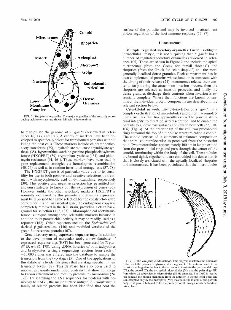
Multiple, regulated secretory organelles. Given its obligateintracellular lifestyle, it is not surprising that T. gondii has anumber of regulated secretory organelles (reviewed in refer-ence 105). These are shown in Figure 2 and include the apicalmicronemes (from the Greek for “small threads”) andrhoptries (from the Greek for “club-shaped”) and the moregenerally localized dense granules. Each compartment has itsown complement of proteins whose function is consistent withthe timing of their release (24): micronemes release their con-tents early during the attachment-invasion process, then therhoptries are released as invasion proceeds, and finally thedense granules discharge their contents when invasion is es-sentially complete. Where their functions are known or sur-mised, the individual protein components are described in therelevant section below.
Cytoskeletal network. The cytoskeleton of T. gondii is acomplex orchestration of microtubules and other macromolec-ular structures that has apparently evolved to provide struc-tural integrity, to direct polarized secretion, and to enable theparasite to glide across surfaces and invade host cells (53, 104,106) (Fig. 3). At the anterior tip of the cell, two preconoidalrings surround the top of a tube-like structure called a conoid.The conoid consists of 14 elements of unknown compositionthat spiral counterclockwise as perceived from the posteriorpole. Two microtubules approximately 400 nm in length extendfrom the preconoidal rings and pass through the center of theconoid, terminating within the body of the cell. These tubulesare bound tightly together and are embedded in a dense matrixthat is closely associated with the apically localized rhoptriesand micronemes. It has been postulated that the microtubules
FIG. 2. Toxoplasma organelles. The major organelles of the asexually repro-ducing tachyzoite stage are shown. Mitoch., mitochondrion.
FIG. 3. The Toxoplasma cytoskeleton. This diagram illustrates the dominantfeatures of the parasite’s cytoskeletal arrangement. The anterior end of theparasite is enlarged in the box to the left so as to illustrate the preconoidal rings(CR), the conoid (C), the two apical microtubules (M), and the polar ring (PR)from which 22 subpellicular microtubules (SPM) emanate. The IMC is locatedjust beneath the plasma membrane from the anterior to the posterior poles andis interrupted only by the micropore (MP) located in the middle of the parasitebody. This pore is believed to be the primary portal through which endocytosistakes place.
VOL. 64, 2000 LYTIC CYCLE OF T. GONDII 609
on July 3, 2018 by guesthttp://m
mbr.asm
.org/D
ownloaded from

function as a scaffold directing these organelles to pass throughthe conoid and secrete their contents from the apical tip (106).
Just posterior to the conoid, a polar ring functions as amicrotubule-organizing center from which 22 microtubulesemanate and spiral down two-thirds of the body (;4 to 5 mm)aligned in the same counterclockwise orientation as the conoidsubunits (134). Although microfilaments have not been ob-served in this organism (27), actin has been detected in theconoid, preconoidal rings, and subpellicular microtubules byimmunoelectron microscopy (183). Immunofluorescence of in-tracellular tachyzoites demonstrates that actin resides predom-inantly in the anterior portion of 75% of extracellular parasites(the remaining 25% demonstrated staining throughout thecell) (50).
Actin is encoded by a single gene in Toxoplasma and isgenerally found in a monomeric, soluble form within the cyto-plasm (34). Toxoplasma also has a novel actin-binding protein(dubbed “toxofilin”), which sequesters G-actin and may play akey role in the creation and function of actin filaments (124).Myosin, a mechanoprotein that interacts with actin, colocalizeswith actin in the anterior portion of the parasite as well asalong the inner membrane complex (33, 143). Three uncon-ventional myosins have been cloned from Toxoplasma by aPCR screen using generic myosin head primers (65). Twoclones (TgM-B and TgM-C) appear to be products of differ-ential RNA splicing with estimated protein sizes of ;114 and125 kDa, respectively. Both TgM-B and TgM-C contain asingle IQ motif (consensus, IQXXXRGXXXRK) in the neckregion, which is normally important in light-chain binding andregulation of activity (89, 99). The third clone (TgM-A) definesa novel class of myosins since it is the only myosin identified todate that lacks a definable neck domain or IQ motif. Thisprotein is one of the smallest myosins known, with an expectedsize of 93 kDa. The tail regions of all three proteins have nohomology to other known myosins apart from a highly basiccharge (65). Their size resembles that of the small myosins ofAcanthamoeba and Dictyostelium (79), which were determinedto be membrane associated and which have been implicated incell translocation (96). There are currently no data on thefunction or regulation of the Toxoplasma myosins.
Trimembrane pellicle. T. gondii is a member of the Alveo-lates because it possesses an inner membrane complex (IMC)consisting of flattened membrane vesicles that lie just beneaththe plasma membrane (110). In the Apicomplexa, this trimem-branous structure (two membranes from the IMC and onefrom the plasmalemma) is called the pellicle; it runs from theanterior preconoidal rings to the posterior end of the cell (47).The only apparent interruption in this complex is at the mi-cropore, which is positioned in the middle of the parasite body(Fig. 3). This pore is believed to be the active site of endocy-tosis, and vesicles have been observed in this region that ap-pear to have clathrin-like coats (108).
The vesicles that make up the IMC measure ;20 to 100 nmin diameter (128) and are “sutured” together in a spiral withthe same pitch as that observed for the subpellicular microtu-bules (104). The function of the pellicle has not been deter-mined, although its close association with the microtubulessuggest that it may be involved in structural integrity and mo-tility of the cell (see below). Fourier analysis of electron mi-crographs demonstrates that microtubule-associated proteins(MAPs) and intramembranous particles (IMPs) found withinthe IMC both exhibit a 32-nm periodicity longitudinally (104).These structures appear to overlap and are further ordered bya second dimension of IMPs that are found in rows betweenthe microtubules. The interdigitating rows of IMPs are ori-ented at an angle of ;75° relative to the vertical MAP-associ-
ated rows and are constrained to a uniform lateral spacing of30 nm and the 32-nm longitudinal repeat observed in theMAPs. This ordered arrangement of IMPs in the IMC is nei-ther actin based nor dependent on the microtubules, since itextends beyond the localization of these proteins and is not loston exposure to drugs which disrupt their polymerization (104).Since there are large open spaces of membrane between theIMPs, the distribution is thought to be maintained by some-thing other than nearest-neighbor contacts and may require asecond set of filaments. In related apicomplexans, a filamen-tous lattice of unknown composition extends throughout thelength of the cell both parallel and perpendicular to the mi-crotubules (32). This framework has not been observed inToxoplasma but is likely to be responsible for the orderedorientation of these IMPs.
In addition to the interaction with microtubules, the pellicleis connected to the plasma membrane by bridges of unknowncomposition (127). The function of these filamentous networkslinking the plasma membrane, IMC, and subpellicular micro-tubules has not been determined but is most probably re-quired, at least in part, for directing parasite motility.
HOST CELL ATTACHMENT
Surface of Toxoplasma
For an intracellular pathogen to gain entry into a cell, it firstmust make intimate contact with the surface of the cell. Sincethe lipid membranes of the host and pathogen normally pos-sess a negative net charge, receptor-ligand interactions arerequired to overcome this repulsive force and to attain the firmattachment required for both motility and invasion (describedbelow). T. gondii is unusual among the known intracellularpathogens in being quite promiscuous in its ability to invade awide variety of host cells. When cultured in vitro, this parasiteis capable of invading almost any mammalian cell type andeven insect and fish cell lines (20, 179). This broad range ofpossible host cells would require Toxoplasma to express eithermultiple receptors for a variety of ligands or a few receptorsthat bind to ligands common to numerous cell types.
To discriminate between these two scenarios, the surface ofToxoplasma has been extensively characterized to identify thecomponents involved in this process (17). The plasma mem-brane of this parasite appears to consist predominantly of avariety of proteins that are linked to the membrane by a gly-cosylphosphatidylinositol (GPI) moiety. Upon identifying thegenes for these proteins, it has been determined that most ofthe surface consists of a family of proteins related to the sur-face antigen SAG1 (87). SAG1 (21) is the most abundant ofthese surface proteins (72, 73) and has been implicated, at leastin part, in the initial events of attachment to the host mem-brane (59, 92, 93, 131). This protein is clearly not the onlyparasite molecule involved in attachment since Sag12 mutantsare still infectious although with altered properties: they attachless well (92) but take less time on average to enter a host cell:1 h after adding syringe-released tachyzoites to a fresh host cellmonolayer, about twice as many Sag12 mutants had invadedcompared to wild-type parasites (M. Grigg and J. C. Boothroyd,unpublished results).
The interaction between SAG1 and the host cell can bepartially blocked using the neoglycoprotein bovine serum al-bumin glucosamide as a competitive inhibitor (74, 94, 131).There is also evidence demonstrating the ability of the para-sites to bind the extracellular matrix protein laminin, and thismay be used as a bridge for the ubiquitous laminin receptorsfound on the host cell (54, 55). While antibodies against lami-
610 BLACK AND BOOTHROYD MICROBIOL. MOL. BIOL. REV.
on July 3, 2018 by guesthttp://m
mbr.asm
.org/D
ownloaded from
nin blocked attachment at a similar level to that seen withanti-SAG1 antibodies (54, 92), the laminin receptor on Toxo-plasma has not been identified.
Host Ligand
The contribution of the host cell surface to attachment hasbeen less well characterized. The expression of the as yet un-identified attachment ligand(s) on the surface of the host cellappears to be cell cycle dependent in at least some of the linestested in vitro (48, 58). In synchronized populations of Chinesehamster ovary (CHO) and bovine kidney (MDBK) cells, par-asite attachment increased threefold as the cells proceededfrom the G1 phase to the mid-S phase and decreased back tobaseline as the cells entered G2/M (58). This association be-tween attachment and cell cycle may explain the observationthat within a population of fibroblasts, there is significant,nonrandom variation in the number of vacuoles within indi-vidual host cells (although this cannot be the entire explana-tion because most of these host cells grown to near confluencywould probably be in G0). Antibodies raised against synchro-nized MDBK cells that were harvested at different points dur-ing the cell cycle showed that polyclonal serum raised againstcells taken at mid-S phase was almost threefold more effectiveat blocking attachment than was serum raised against cells inthe G1 phase (58). This block was not specific to the MDBKcell line, since a similar effect was noted using CHO cells as thehost. These data would suggest that a similar antigenic ligandin both cell lines is utilized in attachment by Toxoplasma,although it does not exclude the possibility of a shared, cellcycle-dependent epitope that blocks the interaction throughsteric hindrance.
Some clues to the nature of the interaction between host andparasite have come from studies that use polysaccharides asinhibitors and probes (111). These experiments show thatthere is a sugar-lectin type of interaction involved in the at-tachment of the parasite because certain polysaccharides (hep-arin, fucoidan, and dextran sulfate) can facilitate or block par-asite attachment to host cells depending on the concentrationof the polysaccharide used. Cell lines deficient in proteoglycansynthesis also show a decreased ability to bind the parasites,again implicating a parasite lectin-like activity in the attach-ment phenomenon. The identity of these lectins is not yetknown.
PARASITE INVASION
The entry of Toxoplasma into nonphagocytic host cells is anactive process that apparently involves actin/myosin motorsinside the parasite and certain transmembrane proteins thatlink these motors to the ligands outside. This conclusion isbased on data summarized in the following sections.
Toxoplasma Demonstrates Substrate-DependentGliding Motility
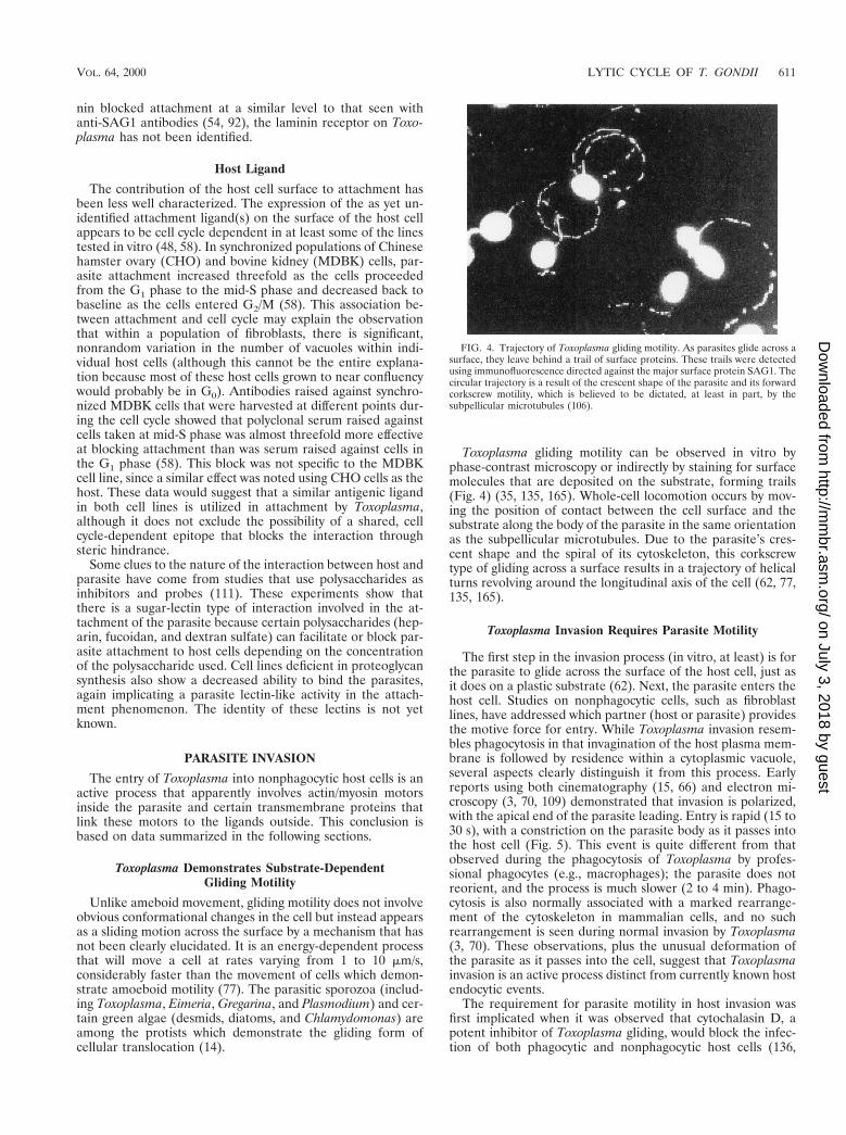
Unlike ameboid movement, gliding motility does not involveobvious conformational changes in the cell but instead appearsas a sliding motion across the surface by a mechanism that hasnot been clearly elucidated. It is an energy-dependent processthat will move a cell at rates varying from 1 to 10 mm/s,considerably faster than the movement of cells which demon-strate amoeboid motility (77). The parasitic sporozoa (includ-ing Toxoplasma, Eimeria, Gregarina, and Plasmodium) and cer-tain green algae (desmids, diatoms, and Chlamydomonas) areamong the protists which demonstrate the gliding form ofcellular translocation (14).
Toxoplasma gliding motility can be observed in vitro byphase-contrast microscopy or indirectly by staining for surfacemolecules that are deposited on the substrate, forming trails(Fig. 4) (35, 135, 165). Whole-cell locomotion occurs by mov-ing the position of contact between the cell surface and thesubstrate along the body of the parasite in the same orientationas the subpellicular microtubules. Due to the parasite’s cres-cent shape and the spiral of its cytoskeleton, this corkscrewtype of gliding across a surface results in a trajectory of helicalturns revolving around the longitudinal axis of the cell (62, 77,135, 165).
Toxoplasma Invasion Requires Parasite Motility
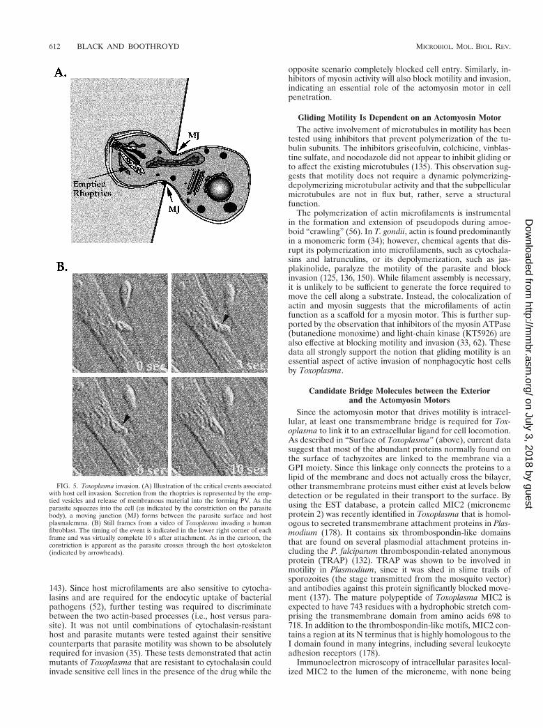
The first step in the invasion process (in vitro, at least) is forthe parasite to glide across the surface of the host cell, just asit does on a plastic substrate (62). Next, the parasite enters thehost cell. Studies on nonphagocytic cells, such as fibroblastlines, have addressed which partner (host or parasite) providesthe motive force for entry. While Toxoplasma invasion resem-bles phagocytosis in that invagination of the host plasma mem-brane is followed by residence within a cytoplasmic vacuole,several aspects clearly distinguish it from this process. Earlyreports using both cinematography (15, 66) and electron mi-croscopy (3, 70, 109) demonstrated that invasion is polarized,with the apical end of the parasite leading. Entry is rapid (15 to30 s), with a constriction on the parasite body as it passes intothe host cell (Fig. 5). This event is quite different from thatobserved during the phagocytosis of Toxoplasma by profes-sional phagocytes (e.g., macrophages); the parasite does notreorient, and the process is much slower (2 to 4 min). Phago-cytosis is also normally associated with a marked rearrange-ment of the cytoskeleton in mammalian cells, and no suchrearrangement is seen during normal invasion by Toxoplasma(3, 70). These observations, plus the unusual deformation ofthe parasite as it passes into the cell, suggest that Toxoplasmainvasion is an active process distinct from currently known hostendocytic events.
The requirement for parasite motility in host invasion wasfirst implicated when it was observed that cytochalasin D, apotent inhibitor of Toxoplasma gliding, would block the infec-tion of both phagocytic and nonphagocytic host cells (136,
FIG. 4. Trajectory of Toxoplasma gliding motility. As parasites glide across asurface, they leave behind a trail of surface proteins. These trails were detectedusing immunofluorescence directed against the major surface protein SAG1. Thecircular trajectory is a result of the crescent shape of the parasite and its forwardcorkscrew motility, which is believed to be dictated, at least in part, by thesubpellicular microtubules (106).
VOL. 64, 2000 LYTIC CYCLE OF T. GONDII 611
on July 3, 2018 by guesthttp://m
mbr.asm
.org/D
ownloaded from
143). Since host microfilaments are also sensitive to cytocha-lasins and are required for the endocytic uptake of bacterialpathogens (52), further testing was required to discriminatebetween the two actin-based processes (i.e., host versus para-site). It was not until combinations of cytochalasin-resistanthost and parasite mutants were tested against their sensitivecounterparts that parasite motility was shown to be absolutelyrequired for invasion (35). These tests demonstrated that actinmutants of Toxoplasma that are resistant to cytochalasin couldinvade sensitive cell lines in the presence of the drug while the
opposite scenario completely blocked cell entry. Similarly, in-hibitors of myosin activity will also block motility and invasion,indicating an essential role of the actomyosin motor in cellpenetration.
Gliding Motility Is Dependent on an Actomyosin MotorThe active involvement of microtubules in motility has been
tested using inhibitors that prevent polymerization of the tu-bulin subunits. The inhibitors griseofulvin, colchicine, vinblas-tine sulfate, and nocodazole did not appear to inhibit gliding orto affect the existing microtubules (135). This observation sug-gests that motility does not require a dynamic polymerizing-depolymerizing microtubular activity and that the subpellicularmicrotubules are not in flux but, rather, serve a structuralfunction.
The polymerization of actin microfilaments is instrumentalin the formation and extension of pseudopods during amoe-boid “crawling” (56). In T. gondii, actin is found predominantlyin a monomeric form (34); however, chemical agents that dis-rupt its polymerization into microfilaments, such as cytochala-sins and latrunculins, or its depolymerization, such as jas-plakinolide, paralyze the motility of the parasite and blockinvasion (125, 136, 150). While filament assembly is necessary,it is unlikely to be sufficient to generate the force required tomove the cell along a substrate. Instead, the colocalization ofactin and myosin suggests that the microfilaments of actinfunction as a scaffold for a myosin motor. This is further sup-ported by the observation that inhibitors of the myosin ATPase(butanedione monoxime) and light-chain kinase (KT5926) arealso effective at blocking motility and invasion (33, 62). Thesedata all strongly support the notion that gliding motility is anessential aspect of active invasion of nonphagocytic host cellsby Toxoplasma.
Candidate Bridge Molecules between the Exteriorand the Actomyosin Motors
Since the actomyosin motor that drives motility is intracel-lular, at least one transmembrane bridge is required for Tox-oplasma to link it to an extracellular ligand for cell locomotion.As described in “Surface of Toxoplasma” (above), current datasuggest that most of the abundant proteins normally found onthe surface of tachyzoites are linked to the membrane via aGPI moiety. Since this linkage only connects the proteins to alipid of the membrane and does not actually cross the bilayer,other transmembrane proteins must either exist at levels belowdetection or be regulated in their transport to the surface. Byusing the EST database, a protein called MIC2 (micronemeprotein 2) was recently identified in Toxoplasma that is homol-ogous to secreted transmembrane attachment proteins in Plas-modium (178). It contains six thrombospondin-like domainsthat are found on several plasmodial attachment proteins in-cluding the P. falciparum thrombospondin-related anonymousprotein (TRAP) (132). TRAP was shown to be involved inmotility in Plasmodium, since it was shed in slime trails ofsporozoites (the stage transmitted from the mosquito vector)and antibodies against this protein significantly blocked move-ment (137). The mature polypeptide of Toxoplasma MIC2 isexpected to have 743 residues with a hydrophobic stretch com-prising the transmembrane domain from amino acids 698 to718. In addition to the thrombospondin-like motifs, MIC2 con-tains a region at its N terminus that is highly homologous to theI domain found in many integrins, including several leukocyteadhesion receptors (178).
Immunoelectron microscopy of intracellular parasites local-ized MIC2 to the lumen of the microneme, with none being
FIG. 5. Toxoplasma invasion. (A) Illustration of the critical events associatedwith host cell invasion. Secretion from the rhoptries is represented by the emp-tied vesicles and release of membranous material into the forming PV. As theparasite squeezes into the cell (as indicated by the constriction on the parasitebody), a moving junction (MJ) forms between the parasite surface and hostplasmalemma. (B) Still frames from a video of Toxoplasma invading a humanfibroblast. The timing of the event is indicated in the lower right corner of eachframe and was virtually complete 10 s after attachment. As in the cartoon, theconstriction is apparent as the parasite crosses through the host cytoskeleton(indicated by arrowheads).
612 BLACK AND BOOTHROYD MICROBIOL. MOL. BIOL. REV.
on July 3, 2018 by guesthttp://m
mbr.asm
.org/D
ownloaded from

detectable on the surface of the parasite or within the parasi-tophorous vacuole (PV) (2, 178). In extracellular parasites,however, MIC2 is secreted onto the apical surface of Toxo-plasma but only when there is contact with the host cell (24).This protein can also be seen in gliding trails (61), and itappears to be capped to the posterior end during invasion (24).A smaller, soluble form of MIC2 can be recovered from me-dium containing extracellular parasites and is believed to beproteolytically cleaved from the surface, leaving the transmem-brane portion in the membrane (24, 178). Micronemal proteinssharing this TRAP-like conservation have also been identifiedin other Apicomplexans (e.g., Eimeria [173] and Cryptospo-ridium [163]), suggesting a family of proteins that may have acommon function in attachment and motility.
The intimate association between TRAP-dependent motilityand host cell entry was observed by using knockout mutantsthat were engineered in the erythrocytic stage of Plasmodiumberghei (a stage that does not express this gene). After differ-entiation to the sporozoite stage, this mutant not only failed toglide but also was unable to invade either mosquito salivaryglands or human hepatocytes (168). Similar studies have yet tobe conducted in Toxoplasma.
Another Plasmodium protein that may serve as a bridgebetween extracellular ligands and the intracellular milieu is theapical membrane antigen AMA1, found in the rhoptries (115).
Toxoplasma has a similarly apical homologue of this protein(albeit in the Toxoplasma micronemes instead of the rhoptries),complete with putative extracellular, transmembrane and in-tracellular domains (A. Hehl, C. Lekutis, E. Ortega, J.-F.Dubremetz, P. Bradley, and J. C. Boothroyd, unpublished re-sults). However, the molecules inside and outside the parasitethat are interacting with Toxoplasma AMA1 and MIC2 haveyet to be identified.
Current Models for Toxoplasma Motility
Two models of how Toxoplasma is able to glide across asubstrate have been suggested. The first scenario (Fig. 6A) iscalled linear motion (77) and requires that short actin fila-ments associate with the outer face of the IMC. Myosin wouldthen interact directly, or via linking proteins, with the cytoplas-mic tail of a transmembrane attachment protein (e.g., MIC2 orAMA1) and the microfilament scaffold. The path of these actinfilaments would be guided by the subpellicular microtubulesthrough IMPs that cross both membranes of the pellicle tomimic the helical trajectory set by the microtubular network.As the myosin contracts down the spiraling actin filaments, itpulls along the attachment protein toward the posterior end,where cleavage would occur to release the external portion ofthe protein. Since the subpellicular microtubules are found
FIG. 6. Current models for the gliding motility exhibited by T. gondii. (A) Graphic illustration of the linear motion model (77). In this model, a transmembraneattachment protein(s) binds to an extracellular ligand and associates with myosin via its cytoplasmic tail. Myosin binds to the actin microfilaments located just beneaththe plasmalemma and ratchets the transmembrane protein down the length of the parasite. The microfilaments are aligned to the orientation of the subpellicularmicrotubules via trans-IMC linkages, which results in the characteristic forward corkscrew locomotion. (B) The second model suggests that the IMC plays a more activerole in motility (104). This model localizes both actin and myosin beneath the IMC in direct contact with the subpellicular microtubules. Instead of myosin, the IMPsassociated with the IMC bind to the cytoplasmic tail of the attachment protein(s). These IMPs are connected to a secondary network of filaments that encapsulatesthe individual flattened vesicles. Myosin binds to either the same IMPs or distinct particles that are on the inner face of the IMC and move along microfilamentsassociated with the subpellicular microtubules. As these myosin-associated IMPs move along the actin tracks, they pull the secondary network like a conveyor belt. Sincethe outer surface IMPs are also connected to this filamentous network, they transduce this force to the attachment protein and move it down the length of the vesicle.
VOL. 64, 2000 LYTIC CYCLE OF T. GONDII 613
on July 3, 2018 by guesthttp://m
mbr.asm
.org/D
ownloaded from
only in the anterior two-thirds of the parasite, they may serveonly to guide the second network of unknown filaments thatrun the full length of the parasite and are apparently used toorder the interdigitating IMPs (see above). This model of Tox-oplasma motility closely resembles that of capping ligands (of-ten seen in the removal of antigen-antibody complexes), whichalso use an actin-myosin motor (51).
The second model (Fig. 6B) suggests that the inner mem-brane complex may not act as a stationary support matrix butinstead moves like a conveyor belt driven by a microtubule-associated motor (104). The interaction between short actinmicrofilaments and microtubules has been documented in nu-merous events including axonal transport and mitotic spindleformation (123). In T. gondii motility, myosin may transientlybind to the tethered IMPs found associated with the pellicleand move these particles along microtubule-associated actinfilaments. As the IMPs are transported along the actin, theentire surface of the vesicle would move as a result of thelattice-like connections between these particles. The cytoplas-mic tails of transmembrane attachment proteins at the surfacemay bind to the IMPs on the outer surface of the pellicle andbe pulled along by this moving belt with the same spiral asdirected by the microtubules. At the end of each vesicle, theattachment protein may either be passed on to the next con-veyor belt or cleaved to maintain uninterrupted movementalong the length of the cell.
Two major assumptions are required by this second modelthat present formidable obstacles and may weaken its plausi-bility. The first is the supposition that the filamentous networkused to order the IMPs is set up individually for each of thevesicular subunits of the pellicle. Since the pellicle is compart-mentalized with the components sutured together, it would bedifficult to envision the pellicle with a continuous belt of IMPsmoving across these junctions and traversing the length of theparasite. The second assumption is the requirement for a di-rect association between the subpellicular microtubules andthis secondary IMP network. In this model, vesicles that areposterior to the microtubules must be influenced by those inthe anterior two-thirds of the cell. If, as mentioned above, thevesicles function as independent units with each demonstratingthe same direction of movement, the force would be in oppo-site directions at the points of contact between the anterior andposterior vesicles. Although there are insufficient data to for-mally confirm or reject either of the two scenarios, the linear-motion model lacks the mechanistic problems presented by thepellicular-belt proposal.
While other forms of movement, such as the anterior-pos-terior flexing and forward protrusion of the conoid, have alsobeen observed in Toxoplasma, they do not appear to be coor-dinated with gliding motility (26, 106, 145). The localization ofactin in the space between the anterior end of the inner mem-brane complex and the conoid suggests that extrusion of theconoid may be powered by a similar actomyosin motor (183).The involvement of actin microfilaments in conoid motility isfurther supported by the observation that cytochalasin Dblocks conoid protrusion (97). The regulation of this activity,as well as that demonstrated in gliding motility, is unknown.
Secretory Events during Invasion
It has long been suggested that the organelles within theapical complex, namely, the rhoptries and micronemes, areinvolved in invasion. The micronemes are thin, possibly vesic-ular organelles that are adjacent to the rhoptries at the apicalend of the parasite. These organelles appear to secrete theircontents just after making contact with the host cell (24). The
possible role of micronemal proteins in the invasion process isdiscussed above.
There are generally 8 to 16 rhoptries per parasite that areidentified by electron microscopy as club-shaped, electrondense organelles (149). These highly acidic organelles (148)are thought to be derived from Golgi secretory vesicles and arefaithfully reproduced in each daughter cell during endodyog-eny (69, 95). During invasion, rhoptries secrete their contentsinto the nascent PV through the narrow duct formed at theconoid by fusion of the rhoptry membrane with the apical tip(107, 127). Rhoptries in T. gondii contain at least eight knownproteins, ROP1 to ROP8 (12, 82, 113), and while some becomeassociated with the PV membrane (11, 139), their role in cellpenetration and vacuole formation remains largely unknown.The basic pI of several of the major rhoptry proteins (ROP2 toROP5) has led to the proposal of a role for polycationicpolypeptides in host penetration (180). This is based on theobservation that a histidine-rich protein isolated from Plasmo-dium was shown to induce the invagination of the erythrocyticplasma membrane (10). The mode of activity of polycationicpolypeptides is suspected to be through energy-dependent al-terations of membrane proteins or altered membrane micro-viscosity through intercalation with the lipid bilayer (180).
A penetration enhancement factor (PEF) (86) ascribed tothese organelles will cause morphological degeneration of hostcell membranes at high concentrations (85). In Toxoplasma,PEF activity is dependent on temperature, Ca21 and Mg21
concentrations, and pH (139, 172). The effect of PEF on hostmembranes and its dependence on calcium suggested that aparasite-derived phospholipase may be responsible for this ac-tivity. Phospholipase A2 (PLA2) activity is commonly usedamong pathogens for the penetration of host cells (e.g., rick-ettsiae [181]) and cytolysis (e.g., Entamoeba histolytica [130]).When host cells are treated with exogenous PLA2 from snakevenom (Naja naja), a morphological disruption of their mem-branes is observed concurrent with an augmented invasionfrequency by Toxoplasma (138). Similarly, treating parasiteswith the irreversible PLA2 inhibitor p-bromophenacyl bromideor the cPLA2-specific inhibitor LY311727 or AACOCF3 re-duces penetration without metabolically disabling the parasites(46, 116). This inhibition is specific to the parasite-derivedenzyme(s), since pretreatment of host cells does not alter par-asite invasion. A soluble phospholipase has been identified incrude lysates of Toxoplasma (140), and it is proposed that thisenzyme may play a role in the initial events of invasion eitherthrough augmenting rhoptry exocytosis or by increasing mem-brane fluidity in the host plasmalemma during vacuole forma-tion (140).
VACUOLE FORMATION
The Parasitophorous Vacuole Is Distinct from Phagosomes
After intracellular pathogens enter a cell, they must confrontor avoid the endocytic apparatus which has evolved to degradeingested material (including a phagocytosed microbe). When avacuole is formed by receptor-mediated endocytosis, the lu-men of the compartment is rapidly acidified by ATP-drivenproton pumps derived from the plasma membrane (88). Thisacidic environment plays a crucial role in the preparation ofthe vacuole for fusion with lysosomes bearing hydrolytic en-zymes. Since the host cell repertoire of Toxoplasma includesprofessional phagocytes, the parasite must circumvent a varietyof additional antimicrobial effectors including reactive oxygenand nitrogen intermediates (31).
The vacuole generated during Toxoplasma invasion (83) is
614 BLACK AND BOOTHROYD MICROBIOL. MOL. BIOL. REV.
on July 3, 2018 by guesthttp://m
mbr.asm
.org/D
ownloaded from
unusual in that it does not acidify (156) or fuse with anycytoplasmic vesicle, even those which are taken up at the timeof parasite entry (70, 152). This difference distinguishes thetwo types of vacuoles that can be observed within professionalphagocytes: the phagosome and the PV. When opsonized par-asites are phagocytosed, a vacuole is formed that is spaciousand noticeably distinct from the more confined PV. Thisphagosome will rapidly acidify and follow the fate of the en-docytic pathway, resulting in the digestion of the internalizedparasite (101, 103). To prevent acidification and endocyticprocessing, the PV formed by parasite invasion must eitherexclude or rapidly eliminate the proton pumps and surfaceproteins that are used for vesicular fusion. This proposed ac-tivity is supported by the observation that a newly formed PVis smooth and almost completely free of intramembranousparticles (44).
The site where protein exclusion is most likely to occur is atthe moving junction (Fig. 5), which is observed only duringinvasion, not in host-mediated phagocytosis. This structure wasbest defined in malaria during the erythrocytic invasion (4) andrepresents a bridge between the parasite and host membranes.In freeze-fracture electron microscopy, the junction appearsvery tight with a crystalline array of lipids in the host plasma-lemma that may be responsible for preventing the diffusion ofproteins into the PV membrane (43). The exclusive nature ofthis structure has been demonstrated in two ways. First, anti-gen-antibody complexes, but not uncoupled antigen, are shedfrom the parasite surface as it passes through the movingjunction (45). Second, host plasmalemma proteins are selec-tively excluded depending on the nature of their membraneassociation: GPI-anchored proteins pass through the movingjunction and associate with the nascent vacuole, whereas trans-membrane-anchored proteins are excluded from it (100). Theactual structure and composition of the moving junction arepoorly understood due to its transient appearance in bothPlasmodium and Toxoplasma (for which invasion is completewithin 15 to 30 s).
An alternative explanation for the absence of host proteinsin the PV is that the parasite provides much of the membra-nous material for this vacuole after punching a hole in the hostplasmalemma. Electron micrographs of early invasion eventsshow rhoptries releasing membranous whirls which may beresponsible for extending this membrane into the cytoplasm(107). Additional evidence for this model comes from obser-vations of aborted invasion events by parasites which havebeen paralyzed with cytochalasin D (24). Although these par-asites were prevented from penetrating the host, small vacu-oles were still formed at the point of contact between the apicalend of the parasite and its host. Immunoelectron microscopydemonstrated that these empty vacuoles contain proteins se-creted from the rhoptries.
To determine the relative contribution of these two potentialsources of the newly formed PV, invagination of host plasma-lemma versus parasite-derived, time-resolved capacitancemeasurements were used to measure the host cell surface areaduring parasite invasion (169). Since the nascent PV appears tobe continuous with the host membrane during the invasionevent (43), extension of this membrane by adding parasite-released lipids would be expected to increase the host surfacearea. Conversely, if the vacuole was derived from host plasma-lemma, the surface area of the host cell should be measurablyreduced once invasion is complete and the vacuole has buddedoff. The result was that no significant change in capacitancewas seen during invasion but a marked decrease occurred afterthe vacuole had budded off. These results indicate that whileup to 20% of the vacuole may be provided by the parasite
during penetration, the majority of the initial membrane isderived from the host cell. These electrophysiology experi-ments also indicated that one of the earliest events in theinvasion process is a brief spike of conductance between theinside and outside of the host cell. This may represent a tran-sient permeabilization of the host plasma membrane, perhapsinvolving the PLA2 activity discussed above, although no datato suggest a role for this enzyme have yet been obtained.
Postinvasion Secretory Events Further Modifythe Parasitophorous Vacuole
After the vacuole has formed, a third set of organelles,called dense granules, release their contents from both theanterior and posterior poles of the parasite (24). Multilamellarvesicles, similar to those secreted from the rhoptries duringinvasion, are secreted from a specialized invagination at theposterior end 10 to 20 min postinvasion (155). These longtubules appear to be responsible for the construction of thespaghetti-like, tubulovesicular network that extends from thePV membrane into the lumen of the vacuole (151). The func-tion of this network is unclear but appears to dramaticallyincrease the surface area of the PV and is associated with atleast four dense-granule proteins (GRA1, GRA2, GRA4, andGRA6) (1, 80). Three other dense-granule proteins, GRA3,GRA5, and GRA8, localize with the vacuolar membrane andremain segregated from those within the tubular network (22,112). Although the two membranous structures (vacuole andnetwork) appear to be contiguous in some regions, this appar-ent partition suggests that they are maintained as separatedomains.
The mechanism by which dense-granule proteins go frombeing soluble within the secretory organelles to having a trans-membrane location in the PV membrane or the tubulovesicu-lar network is poorly understood but presumably involves aprofound change in their topology. Studies of selected mem-bers of this group of proteins show a dependence on certainelements within their sequence, especially within the eventualcytoplasmic domain of GRA4 (71), the transmembrane do-main of GRA5 (81), and the amphipathic helices of GRA2(90).
The PV membrane is a porous structure with a size exclusionlimit of ;1,300 Da (142). It has been proposed that GRA3, asoluble protein that oligomerizes and inserts into the PV mem-brane, may function as the pore-forming complex (112). SinceToxoplasma is auxotrophic for purine biosynthesis (114, 144),these pores may be required for the acquisition of purines inthe form of host cytosolic ATP. Using mutant host cells thatare incapable of purine synthesis, it was determined that par-asites do not require or directly utilize the host ATP as anenergy source but, instead, hydrolyze the molecule to adeno-sine for purine salvage (144). An NTP hydrolase (NTPase) hasbeen identified in the lumen of the PV and may be partlyresponsible for this salvage process (8).
In addition to purine salvage, the PV membrane is respon-sible for recruiting host mitochondria and endoplasmic retic-ulum (ER) immediately following invasion (127, 158). Theseorganelles will form an intimate interaction with the PV mem-brane, covering almost 75% of the vacuolar membrane by 4 hpostinfection (158). The PV-associated mitochondria may bephysiologically distinct since they label more intensely whenstained with rhodamine 123 than do those which are not soassociated (170). A similar recruitment of host mitochondriaoccurs in the vacuole formed during Legionella pneumophilainvasion of monocytes (67). While this interaction is essential
VOL. 64, 2000 LYTIC CYCLE OF T. GONDII 615
on July 3, 2018 by guesthttp://m
mbr.asm
.org/D
ownloaded from
for the intracellular growth of this bacterial pathogen (13), itsrole in intracellular survival is not known. For Toxoplasma, ithas been proposed that recruitment of host ER and mitochon-dria may serve to provide lipids for the PV (174). Similarregions of membrane apposition between the mitochondriaand ER have been observed in a variety of cell types and arethought to be sites of lipid trafficking (174, 175). Since prelim-inary data suggest that Toxoplasma may be incapable of denovo fatty acid synthesis (158), this interaction may provide themeans for lipid transport to intravacuolar parasites through thetubulovesicular network.
PARASITE REPLICATION
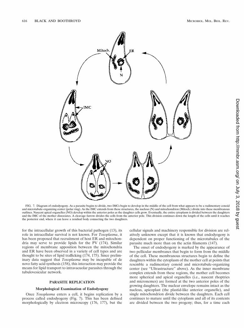
Morphological Examination of EndodyogenyOnce Toxoplasma enters a cell, it begins replication by a
process called endodyogeny (Fig. 7). This has been definedmorphologically by electron microscopy (176, 177), but the
cellular signals and machinery responsible for division are rel-atively unknown except that it is known that endodyogeny isdependent on proper functioning of the microtubules of theparasite much more than on the actin filaments (147).
The onset of endodyogeny is marked by the appearance oftwo pellicular membranes that begin to form from the middleof the cell. These membranous structures begin to define thedaughters within the cytoplasm of the mother cell at points thatresemble a rudimentary conoid and microtubule-organizingcenter (see “Ultrastructure” above). As the inner membranecomplex extends from these regions, the mother cell becomesmore spherical and apical organelles (i.e., nascent rhoptriesand micronemes) are formed at the two anterior poles of thegrowing daughters. The nuclear envelope remains intact as thenucleus, apicoplast (the plastid-like anterior organelle), andsingle mitochondrion divide between the daughters. Each cellcontinues to mature until the cytoplasm and all of its contentsare divided between the two progeny; thus, for a time each
FIG. 7. Diagram of endodyogeny. As a parasite begins to divide, two IMCs begin to develop in the middle of the cell from what appears to be a rudimentary conoidand microtubule-organizing center (polar ring). As the IMC extends from these structures, the nucleus (N) and mitochondrion (Mitoch.) divide into these membranousoutlines. Nascent apical organelles (NO) develop within the anterior poles as the daughter cells grow. Eventually, the entire cytoplasm is divided between the daughtersand the IMC of the mother dissociates. A cleavage furrow divides the cells from the anterior pole. This division continues down the length of the cells until it reachesthe posterior end, where it can leave a residual body connecting the two daughters.
616 BLACK AND BOOTHROYD MICROBIOL. MOL. BIOL. REV.
on July 3, 2018 by guesthttp://m
mbr.asm
.org/D
ownloaded from
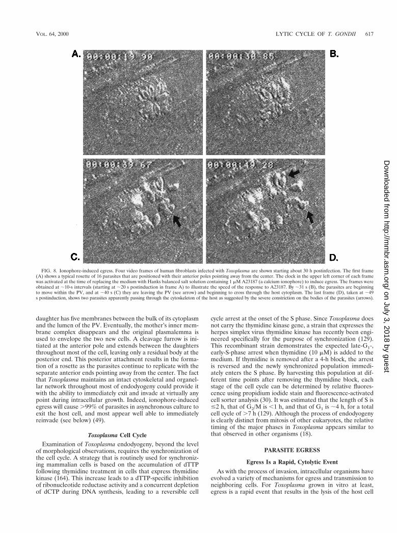
daughter has five membranes between the bulk of its cytoplasmand the lumen of the PV. Eventually, the mother’s inner mem-brane complex disappears and the original plasmalemma isused to envelope the two new cells. A cleavage furrow is ini-tiated at the anterior pole and extends between the daughtersthroughout most of the cell, leaving only a residual body at theposterior end. This posterior attachment results in the forma-tion of a rosette as the parasites continue to replicate with theseparate anterior ends pointing away from the center. The factthat Toxoplasma maintains an intact cytoskeletal and organel-lar network throughout most of endodyogeny could provide itwith the ability to immediately exit and invade at virtually anypoint during intracellular growth. Indeed, ionophore-inducedegress will cause .99% of parasites in asynchronous culture toexit the host cell, and most appear well able to immediatelyreinvade (see below) (49).
Toxoplasma Cell CycleExamination of Toxoplasma endodyogeny, beyond the level
of morphological observations, requires the synchronization ofthe cell cycle. A strategy that is routinely used for synchroniz-ing mammalian cells is based on the accumulation of dTTPfollowing thymidine treatment in cells that express thymidinekinase (164). This increase leads to a dTTP-specific inhibitionof ribonucleotide reductase activity and a concurrent depletionof dCTP during DNA synthesis, leading to a reversible cell
cycle arrest at the onset of the S phase. Since Toxoplasma doesnot carry the thymidine kinase gene, a strain that expresses theherpes simplex virus thymidine kinase has recently been engi-neered specifically for the purpose of synchronization (129).This recombinant strain demonstrates the expected late-G1-,early-S-phase arrest when thymidine (10 mM) is added to themedium. If thymidine is removed after a 4-h block, the arrestis reversed and the newly synchronized population immedi-ately enters the S phase. By harvesting this population at dif-ferent time points after removing the thymidine block, eachstage of the cell cycle can be determined by relative fluores-cence using propidium iodide stain and fluorescence-activatedcell sorter analysis (30). It was estimated that the length of S is#2 h, that of G2/M is ,1 h, and that of G1 is ;4 h, for a totalcell cycle of .7 h (129). Although the process of endodyogenyis clearly distinct from mitosis of other eukaryotes, the relativetiming of the major phases in Toxoplasma appears similar tothat observed in other organisms (18).
PARASITE EGRESS
Egress Is a Rapid, Cytolytic Event
As with the process of invasion, intracellular organisms haveevolved a variety of mechanisms for egress and transmission toneighboring cells. For Toxoplasma grown in vitro at least,egress is a rapid event that results in the lysis of the host cell
FIG. 8. Ionophore-induced egress. Four video frames of human fibroblasts infected with Toxoplasma are shown starting about 30 h postinfection. The first frame(A) shows a typical rosette of 16 parasites that are positioned with their anterior poles pointing away from the center. The clock in the upper left corner of each framewas activated at the time of replacing the medium with Hanks balanced salt solution containing 1 mM A23187 (a calcium ionophore) to induce egress. The frames wereobtained at ;10-s intervals (starting at ;20 s postinduction in frame A) to illustrate the speed of the response to A23187. By ;31 s (B), the parasites are beginningto move within the PV, and at ;40 s (C) they are leaving the PV (see arrow) and beginning to cross through the host cytoplasm. The last frame (D), taken at ;49s postinduction, shows two parasites apparently passing through the cytoskeleton of the host as suggested by the severe constriction on the bodies of the parasites (arrows).
VOL. 64, 2000 LYTIC CYCLE OF T. GONDII 617
on July 3, 2018 by guesthttp://m
mbr.asm
.org/D
ownloaded from
and release of very motile parasites. By video microscopy, asynchronized mass exodus of all the intracellular parasiteswithin a single host can be seen passing through the PV mem-brane, the host cytoplasm, and finally the plasmalemma. In Fig.8, video frames of ionophore-induced egress are shown (de-scribed further below). As the parasites pass through each ofthe membranes, an obvious constriction runs down the body ofthe parasite, resembling the junction ring observed during in-vasion. Although there have been no reports of secretoryevents associated with Toxoplasma egress, the merozoite stageof Plasmodium appears to secrete from their rhoptries justprior to erythrocyte exit (29, 115). If micronemal proteins areindeed required for Toxoplasma motility (see “Candidatebridge molecules between the exterior and the actomyosinmotors” above), it would be expected that these organelles(and possibly rhoptries) are induced to exocytose duringegress. Once outside the confines of the lysed host cell, theparasites will quickly invade neighboring cells and halt afterthey are enveloped by their new vacuole, thus completing thelytic cycle (Fig. 9).
Calcium Acts as a Signal for Egress
While the actual stimulus that signals the timing of naturalegress is unknown, Ca21 has been shown to play a major rolein the activation of this process (49, 142). The relationship
between calcium and egress was first demonstrated using thecalcium ionophore A23187 on infected murine macrophages(49). This drug will increase the permeability of membranes forthe passive diffusion of divalent cations (preference for Ca21)along a concentration gradient (126). It was observed thatintracellular parasites which are exposed to this drug wouldrapidly leave their host cells in a manner resembling that ofnatural egress. This ionophore-induced egress appears to be adestructive event for the host cell, resulting in the formation ofmembranous blebs and lysis (observed even in cells infected bysingle parasites [49]). This cytolysis is dependent on viableintracellular parasites, since uninfected macrophages andthose that had phagocytosed dead parasites were not affectedby the ionophore.
Upon closer examination, it appears that during egress, theparasite pushes the host plasmalemma out from the surface,much like “invading” out of the cell (49). Eventually this mem-brane is ruptured as the parasite escapes, resulting in host celllysis. Nonspecific effects of A23187 (e.g., pH shifts resultingfrom the exchange of protons for Ca21) do not appear tobe responsible for ionophore-induced egress, because it isspecifically blocked by chelation of intracellular Ca21 withBAPTA-AM (a cell-permeable Ca21 chelator [M. W. Black andJ. C. Boothroyd, unpublished results]) and can also be inducedby the direct microinjection of Ca21 into infected cells (142).
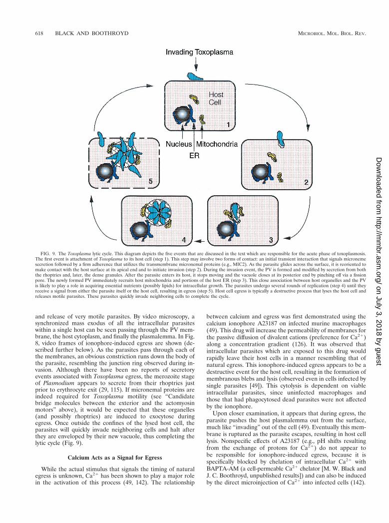
FIG. 9. The Toxoplasma lytic cycle. This diagram depicts the five events that are discussed in the text which are responsible for the acute phase of toxoplasmosis.The first event is attachment of Toxoplasma to its host cell (step 1). This step may involve two forms of contact: an initial transient interaction that signals micronemesecretion followed by a firm adherence that utilizes the transmembrane micronemal proteins (e.g., MIC2). As the parasite glides across the surface, it is reoriented tomake contact with the host surface at its apical end and to initiate invasion (step 2). During the invasion event, the PV is formed and modified by secretion from boththe rhoptries and, later, the dense granules. After the parasite enters its host, it stops moving and the vacuole closes at its posterior end by pinching off via a fissionpore. The newly formed PV immediately recruits host mitochondria and portions of the host ER (step 3). This close association between host organelles and the PVis likely to play a role in acquiring essential nutrients (possibly lipids) for intracellular growth. The parasites undergo several rounds of replication (step 4) until theyreceive a signal from either the parasite itself or the host cell, resulting in egress (step 5). Host cell egress is typically a destructive process that lyses the host cell andreleases motile parasites. These parasites quickly invade neighboring cells to complete the cycle.
618 BLACK AND BOOTHROYD MICROBIOL. MOL. BIOL. REV.
on July 3, 2018 by guesthttp://m
mbr.asm
.org/D
ownloaded from
Investigation of the influence of elevated extracellular [Ca21]on Toxoplasma has uncovered a putative polyvalent cationreceptor similar to those observed in kidney cells (9). In mam-malian cells, receptors such as these function via coupling toheterotrimeric GTP-binding proteins (G-proteins) which influ-ence the regulation of Ca21 channels by the generation ofinositol 1,4,5-trisphosphate (19). Although there is evidencefor such G-proteins in Toxoplasma (63), there is no informa-tion about their function or association with such a receptor.
Dithiothreitol Acts as a Signal for Egress
In addition to directly elevating host cytosolic [Ca21], Tox-oplasma egress has been induced by treating infected mono-layers with dithiothreitol (DTT) (157, 166). Infected cellstreated with this reducing agent demonstrated a 15 to 50%increase in the level of cytoplasmic Ca21 immediately beforethe activation of parasite motility and egress (166). This pecu-liar response to DTT appears to be dependent on both the hostand parasite, since DTT will not increase the cytoplasmic[Ca21] of uninfected cells or the motility of extracellular par-asites. Although the calcium flux and parasite egress could beinduced using other dithiol reducing agents, including dithio-erythritol and to a lesser extent dimercaptopropanol, there wasno response to monothiols such as 2-mercaptoethanol (166).
This preference for dithiols directly correlates with data onthe in vitro activation of parasite vacuolar NTPases (7, 157). Inthe virulent RH strain, there are two isoforms of this enzymecalled NTPase 1 (equally active against NTP and NDP) andNTPase 3 (specific for NTP) (7). They function as an apyrase,degrading ATP to ADP and AMP, and are expected to beinvolved in purine salvage of host cytosolic ATP that diffusesinto the vacuole (see above). An unusual feature of Toxo-plasma NTPases is that they are each equipped with 15 cysteineresidues that require dithiol reducing agents for in vitro acti-vation. This DTT-dependent form of NTPase is unique toToxoplasma among the Apicomplexans, and its role in intra-cellular parasitism is not understood (6). It is estimated thatless than 5% of the enzyme expressed within the PV is func-tional during intracellular growth. A plausible compensationfor this inactivity is the gross excess of NTPase that is secretedinto the vacuolar space; a sufficient amount is secreted from asingle parasite to deplete the entire pool of host ATP in amatter of minutes if activated (157). Infected host cells treatedwith DTT demonstrated a dramatic increase in ATP degrada-tion that correlated with the induction of parasite egress (157).As seen in the DTT-induced calcium flux, degradation of ATPwas observed only in parasite-infected cells.
While the connection between NTPase activity and cytoplas-mic [Ca21] is unclear, a candidate may be the Ca21-ATPasechannels found within host ER and mitochondria. When ATPis depleted, these cation pumps would not be expected toprevent the release of Ca21 into the cytosol. Interestingly,activation of Toxoplasma NTPases also appears to be triggeredby protein-protein interactions, as determined by their spon-taneous activation during immunoprecipitation (157). It hasbeen postulated that NTPases may use this trait as a noveltiming mechanism for the induction of a burst of activity uponreaching a critical threshold. As observed with DTT, this en-zymatic activation could result in (i) the rapid degradation ofhost ATP, (ii) a flux in cytosolic [Ca21], and (iii) parasiteegress (157, 166).
INFLUENCE OF CA21 ON OTHER ASPECTS OFTOXOPLASMA GROWTH
Rearrangement of the Toxoplasma Cytoskeleton
Although Ca21 homeostasis in T. gondii has not been wellcharacterized, it is clear that its regulation is key for severalcellular activities. Three distinct intracellular Ca21 stores havebeen identified in this parasite including the ER, mitochondria,and an unusual acidic compartment called the acidocalcisome(102). Using the calcium ionophores A23187 and ionomycin, itwas determined that Ca21 release from one or more of theseorganelles results in the forward protrusion of the conoid (97).Although the presence or absence of extracellular Ca21 in themedium had little influence on protrusion, when parasites werepretreated with BAPTA-AM to chelate intracellular Ca21,ionophore induction of conoid extrusion was almost com-pletely abolished. This BAPTA-AM paralysis was reversed byadding excess (1 mM) CaCl2 to the medium containing theionophore, indicating that elevation of cytosolic [Ca21] is di-rectly responsible for conoid extrusion. As predicted by theseresults, Ca21 fluxes also stimulate the discharge of mi-cronemes, an event closely tied to conoid protrusion (23).
Parasite Motility and Invasion
The gliding motility of Toxoplasma appears to be activatedby elevated cytosolic [Ca21] levels. When extracellular para-sites are exposed to calcium ionophores (97) or treated withlow concentrations of trypsin in the presence of calcium andATP (98), they begin to exhibit the characteristic gliding mo-tility. Trypsinization of these parasites appears to briefly per-meabilize their plasma membrane, resulting in the diffusion ofCa21 and ATP into (and presumably out of) the cytosol. Thisis supported by the fact that parasites trypsinized in Ca21- orATP-free medium did not become motile. The requirementfor intracellular Ca21 was demonstrated by the BAPTA-AMinhibition of gliding in both the trypsinization induction andionophore induction procedures. As one might expect, pre-treatment of extracellular parasites with BAPTA-AM also re-sulted in a .90% reduction in host cell invasion compared tountreated controls (97). Interestingly, the same study revealedthat invasion was inhibited almost fourfold when parasiteswere briefly treated with ionomycin compared to untreatedcontrols. Although it is possible that this is a consequence ofexhausting the resources required for motility and invasion,little is known about the overall effect of calcium ionophoreson Toxoplasma. The unconventional myosins found in this par-asite (see “Ultrastructure” above) represent possible targetsfor the Ca21-dependent control of conoid extrusion and glid-ing, since both these processes involve actomyosin motors.
Intracellular Survival and Replication
While the influence of [Ca21] on intracellular growth hasnot been directly examined in Toxoplasma, a synthesized drugthat blocks calcium import across the host plasmalemma (L-651,582) has been found to effectively inhibit intracellulargrowth (68). This dependence on host [Ca21] or Ca21-depen-dent pathways is consistent with data obtained concerning theantiparasitic effects of Ca21 and calmodulin antagonists in P.falciparum (141). An investigation of host cytosolic [Ca21] inToxoplasma-infected cells demonstrates that the PV importsCa21 from the host cytoplasm (122). After 48 h of intracellularreplication, a significant decrease in the level of host cytosolicCa21 was observed compared to that in uninfected cells. Sincethe porous PV would allow free diffusion of Ca21 across the
VOL. 64, 2000 LYTIC CYCLE OF T. GONDII 619
on July 3, 2018 by guesthttp://m
mbr.asm
.org/D
ownloaded from
membrane, these data suggest that products secreted or im-ported into the vacuole bind Ca21. A candidate for this activityis the 23-kDa dense-granule protein GRA1 (25). This proteinhas a high affinity for Ca21 and is closely associated with thetubulovesicular network of the PV. The stability of this net-work appears to be calcium dependent and can be recoveredfrom intracellular parasites only in the presence of 1 mM Ca21
(151). Other than this apparent stabilization, no functionalroles have been ascribed to vacuolar sequestration of hostCa21.
CONCLUSION
In this review, we have described the current knowledgeabout the lytic cycle of the asexual, tachyzoite form of T. gondii.While many of the basic phenomena are beginning to be welldescribed, relatively few of the molecules involved have beenidentified. Many of the most important questions remain un-answered. For example, which specific receptor-ligand interac-tions mediate the attachment and invasion processes? What isthe mechanism by which gliding motility is used to activelypenetrate the host cell? What functions do the many novelproteins found within the various secretory compartments ofthe parasite (micronemes, rhoptries and dense granules) servein this cycle? What is the role of the spaghetti-like networkelaborated inside the parasitophorous vacuole? Which criticalmolecules enter (or exit?) via the micropore? And, last but notleast, which signals lead to egress? This is no simple agenda,but the intricacies are likely to be as interesting as they areimportant.
ACKNOWLEDGMENTS
We thank our many colleagues at and outside Stanford for theircomments and helpful suggestions and for permission to cite unpub-lished data. We are especially grateful to Gustavo Arrizabalaga, PeterBradley, and Chris Lekutis for their comments on the manuscript.
Work from this laboratory was supported by grants from the NIH(AI21423 and AI45057), the University of California AIDS ResearchProgram, and a Predoctoral Fellowship from the Howard HughesMedical Institute (to M.B.).
REFERENCES
1. Achbarou, A., P. Mercereau, A. Sadak, B. Gortier, M. A. Leriche, D. Ca-mus, and J. F. Dubremetz. 1991. Differential targeting of dense granuleproteins in the parasitophorous vacuole of Toxoplasma gondii. Parasitology3:321–329.
2. Achbarou, A., O. Mercereau-Puijalon, J. M. Autheman, B. Fortier, D.Camus, and J. F. Dubremetz. 1991. Characterization of microneme pro-teins of Toxoplasma gondii. Mol. Biochem. Parasitol. 47:223–233.
3. Aikawa, M., Y. Komata, T. Asai, and O. Midorikawa. 1977. Transmissionand scanning electron microscopy of host cell entry by Toxoplasma gondii.Am. J. Pathol. 87:285–295.
4. Aikawa, M., L. H. Miller, J. Johnson, and J. Rabbege. 1978. Erythrocyteentry by malarial parasites: a moving junction between erythrocyte andparasite. J. Cell Biol. 77:72–82.
5. Ajioka, J. W., J. C. Boothroyd, B. P. Brunk, A. Hehl, L. Hillier, I. D.Manger, M. Marra, G. C. Overton, D. S. Roos, K. L. Wan, R. Waterston,and L. D. Sibley. 1998. Gene discovery by EST sequencing in Toxoplasmagondii reveals sequences restricted to the Apicomplexa. Genome Res. 8:18–28.
6. Asai, T., T. Kanazawa, S. Kubayashi, T. Takeuchi, and T. Kim. 1986. Doprotozoa conceal a high potency of nucleoside triphosphate hydrolysispresent in Toxoplasma gondii? Comp. Biochem. Physiol. 85:365–367.
7. Asai, T., S. Miura, L. D. Sibley, H. Okabayashi, and T. Takeuchi. 1995.Biochemical and molecular characterization of nucleoside triphosphatehydrolase isozymes from the parasitic protozoan Toxoplasma gondii. J. Bio-chem. 270:11391–11397.
8. Asai, T., W. J. O’Sullivan, and M. Tatibana. 1983. A potent nucleosidetriphosphate hydrolase from the parasitic protozoan Toxoplasma gondii.Purification, some properties, and activation by thiol compounds. J. Biol.Chem. 258:6816–6822.
9. Bai, M., S. Quinn, S. Trivedi, O. Kifor, S. H. S. Pearce, M. R. Pollak, K.Krapcho, S. C. Hebert, and E. M. Brown. 1996. Expression and character-
ization of inactivating and activating mutations in the human Ca21-sensingreceptor. J. Biol. Chem. 271:19537–19545.
10. Bannister, L. H., G. A. Butcher, and G. H. Mitchell. 1977. Recent advancesin understanding the invasion of erythrocytes by merozoites of Plasmodiumknowlesi. Bull. W. H. O. 55:163–169.
11. Beckers, C. J. M., J. F. Dubremetz, O. Mercereau-Puijalon, and K. A.Joiner. 1994. The Toxoplasma gondii rhoptry protein ROP2 is inserted intothe parasitophorous vacuole membrane, surrounding the intracellular par-asite, and is exposed to the host cell cytoplasm. J. Cell Biol. 127:947–961.
12. Beckers, C. J. M., T. Wakefield, and K. A. Joiner. 1997. The expression ofToxoplasma proteins in Neospora caninum and the identification of a geneencoding a novel rhoptry protein. Mol. Biochem. Parasitol. 89:209–223.
13. Berger, K. H., and R. R. Isberg. 1993. Two distinct defects in intracellulargrowth complemented by a single genetic locus in Legionella pneumophila.Mol. Microbiol. 7:7–19.
14. Bloodgood, R. A. 1989. Gliding motility: can regulated protein movementsin the plasma membrane drive whole cell locomotion? Cell Motil. Cytoskel-eton 14:340–344.
15. Bommer, W. 1969. The life cycle of virulent Toxoplasma in cell cultures.Aust. J. Exp. Med. Sci. 47:505–512.
16. Boothroyd, J. C., M. W. Black, S. Bonnefoy, A. Hehl, L. J. Knoll, I. D.Manger, E. Ortega-Barria, and S. Tomavo. 1997. Genetic and biochemicalanalysis of development in Toxoplasma gondii. Philos. Trans. R. Soc. Lon-don Ser. B 352:1347–1354.
17. Boothroyd, J. C., A. Hehl, L. J. Knoll, and I. D. Manger. 1998. The surfaceof Toxoplasma: more and less. Inter. J. Parasitol. 28:3–9.
18. Bootsma, D., L. Budke, and O. Vos. 1964. Studies on synchronous divisionof tissue culture cells initiated by excess thymidine. Exp. Cell Res. 33:301–309.
19. Brown, E., P. Enyedi, M. LeBoff, J. Rotberg, J. Preston, and C. Chen. 1987.High extracellular Ca21 and Mg21 stimulate accumulation of inositol phos-phates in bovine parathyroid cells. FEBS Lett. 218:113–118.
20. Buckly, S. M. 1973. Survival of Toxoplasma gondii in mosquito cell lines andestablishment of continuous infection in Vero cell cultures. Exp. Parasitol.33:23–26.
21. Burg, J. L., D. Perelman, L. H. Kasper, P. L. Ware, and J. C. Boothroyd.1988. Molecular analysis of the gene encoding the major surface antigen ofToxoplasma gondii. J. Immunol. 141:3584–3591.
22. Carey, K. L., C. G. Donahue, and G. E. Ward. 2000. Identification andmolecular characterization of GRA8, a novel, proline-rich, dense granuleprotein of Toxoplasma gondii. Mol. Biochem. Parasitol 105:25–37.
23. Carruthers, V. B., S. N. Moreno, and L. D. Sibley. 1999. Ethanol andacetaldehyde elevate intracellular [Ca21] and stimulate microneme dis-charge in Toxoplasma gondii. Biochem. J. 342:379–386.
24. Carruthers, V. B., and L. D. Sibley. 1997. Sequential protein secretion fromthree distinct organelles of Toxoplasma gondii accompanies invasion ofhuman fibroblasts. Eur. J. Cell Biol. 73:114–123.
25. Cesbron-Delauw, M. F., B. Guy, R. J. Pierce, G. Lenzen, J. Y. Cesbron, H.Charif, P. Lepage, F. Darcy, J. P. Lecocq, and A. Capron. 1989. Molecularcharacterization of a 23 kDa major antigen secreted by Toxoplasma gondii.Proc. Natl. Acad. Sci. USA 86:7537–7541.
26. Chiappino, M. L., B. A. Nichols, and G. R. O’Connor. 1984. Scanningelectron microscopy of Toxoplasma gondii: parasite torsion and host-cellresponses during invasion. J. Protozool. 31:288–292.
27. Cintra, W. M., and W. DeSouza. 1985. Immunocytochemical localization ofcytoskeletal proteins and electron microscopy of detergent extractedtachyzoites of Toxoplasma gondii. J. Submicrosc. Cytol. 17:503–508.
28. Cornelissen, A. W., J. P. Overdulve, and M. van der Ploeg. 1984. Determi-nation of nuclear DNA of five eucoccidian parasites, Isospora (Toxoplasma)gondii, Sarcocystis cruzi, Eimeria tenella, E. acervulina and Plasmodiumberghei, with special reference to gamontogenesis and meiosis in I. (T.)gondii. Parasitology 88:531–553.
29. Crewther, P. E., J. G. Culvenor, A. Silva, J. A. Cooper, and R. F. Anders.1990. Plasmodium falciparum: two antigens of similar size are located indifferent compartments of the rhoptry. Exp. Parasitol. 70:193–206.
30. Darzynkiewicz, Z., F. Traganos, and M. Kimmel. 1987. Assay of cell cyclekinetics by multivariate flow cytometry using the principle of stathmokine-sis, p. 291–336. In J. W. Gray and Z. Daryzynkiewicz (ed.), Techniques incell cycle analysis. Humana Press, San Diego, Calif.
31. Densen, P., and G. L. Mandell. 1995. Granulocytic phagocytes, p. 78–101.In G. L. Mandell, R. G. Douglas, and J. E. Bennett (ed.), Principles andpractice of infectious diseases. Churchill Livingstone, Inc., New York, N.Y.
32. D’Haese, J., H. Melhorn, and W. Peters. 1977. Comparative electron mi-croscope study of pellicular structures in coccidia (Sarcocystis, Besnoitia,Eimeria). Int. J. Parasitol. 7:505–518.
33. Dobrowolski, J. M., V. B. Carruthers, and L. D. Sibley. 1997. Participationof myosin in gliding motility and host cell invasion by Toxoplasma gondii.Mol. Microbiol. 26:163–173.
34. Dobrowolski, J. M., I. R. Niesman, and L. D. Sibley. 1997. Actin in Toxo-plasma gondii is encoded by a single-copy gene, ACT1, and exists primarilyin a globular form. Cell Motil. Cytoskeleton 37:253–262.
35. Dobrowolski, J. M., and L. D. Sibley. 1996. Toxoplasma invasion of mam-
620 BLACK AND BOOTHROYD MICROBIOL. MOL. BIOL. REV.
on July 3, 2018 by guesthttp://m
mbr.asm
.org/D
ownloaded from
malian cells is powered by the actin cytoskeleton of the parasite. Cell84:933–939.
36. Donald, R. G., and D. S. Roos. 1998. Gene knock-outs and allelic replace-ments in Toxoplasma gondii: HXGPRT as a selectable marker for hit-and-run mutagenesis. Mol. Biochem. Parasitol. 91:295–305.
37. Donald, R. G., and D. S. Roos. 1995. Insertional mutagenesis and markerrescue in a protozoan parasite: cloning of the uracil phosphoribosyltrans-ferase locus from Toxoplasma gondii. Proc. Natl. Acad. Sci. USA 92:5749–5753.
38. Donald, R. G., and D. S. Roos. 1993. Stable molecular transformation ofToxoplasma gondii: a selectable dihydrofolate reductase-thymidylate syn-thase marker based on drug-resistance mutations in malaria. Proc. Natl.Acad. Sci. USA 90:11703–11707.
39. Donald, R. G. K., D. Carter, B. Ullman, and D. S. Roos. 1996. Insertionaltagging, cloning, and expression of the Toxoplasma gondii hypoxanthine-xanthine-guanine-phosphoribosyltransferase gene. J. Biol. Chem. 271:14010–14019.
40. Donald, R. G. K., and D. S. Roos. 1994. Homologous recombination andgene replacement at the dihydrofolate reductase-thymidylate synthase locusin Toxoplasma gondii. Mol. Biochem. Parasitol. 63:243–253.
41. Dubey, J. P. 1998. Advances in the life cycle of Toxoplasma. Int. J. Parasitol.28:1019–1024.
42. Dubey, J. P., D. S. Lindsay, and C. A. Speer. 1998. Structures of Toxoplasmagondii tachyzoites, bradyzoites, and sporozoites and biology and develop-ment of tissue cysts. Clin. Microbiol. Rev. 11:267–299.
43. Dubremetz, J. F. 1998. Host cell invasion by Toxoplasma gondii. TrendsMicrobiol. 6:27–30.
44. Dubremetz, J. F., A. Achbarou, D. Bermudes, and K. A. Joiner. 1993.Kinetics and pattern of organelle exocytosis during Toxoplasma gondii-host-cell interaction. Parasitol. Res. 79:402–408.
45. Dubremetz, J. F., C. Rodriguez, and E. Ferreira. 1985. Toxoplasma gondii:redistribution of monoclonal antibodies on tachyzoites during host cellinvasion. Exp. Parasitol. 59:24–31.
46. Dubremetz, J. F., and J. D. Schwarzman. 1993. Subcellular organelles ofToxoplasma gondii and host cell invasion. Res. Immunol. 144:31–33.
47. Dubremetz, J. F., and G. Torpier. 1978. Freeze fracture study of the pellicleof an Eimerian sporozoite (Protozoa, Coccidia). J. Ultrastruct. Res. 62:94–109.
48. Dvorak, J. A., and M. S. J. Crane. 1981. Vertebrate cell cycle modulatesinfection by protozoan parasites. Science 214:1034–1036.
49. Endo, T., K. K. Sethi, and G. Piekarski. 1982. Toxoplasma gondii: calciumionophore A23187-mediated exit of trophozoites from infected murinemacrophages. Exp. Parasitol. 53:179–188.
50. Endo, T., K. Yagita, T. Yasuda, and T. Nakamura. 1988. Detection andlocalization of actin in Toxoplasma gondii. Parasitol. Res. 75:102–106.
51. Espinosa-Cantellano, M., and A. Martinez-Palomo. 1994. Entamoeba his-tolytica: mechanism of surface receptor capping. Exp. Parasitol. 79:424–435.
52. Falkow, S., R. R. Isberg, and D. A. Portnoy. 1992. The interaction ofbacteria with mammalian cells. Annu. Rev. Cell Biol. 8:333–363.
53. Frixione, E., R. Mondragon, and I. Meza. 1996. Kinematic analysis ofToxoplasma gondii motility. Cell Motil. Cytoskeleton 34:152–163.
54. Furtado, G. C., Y. Cao, and K. A. Joiner. 1992. Laminin on Toxoplasmagondii mediates parasite binding to the b1 integrin receptor a6b1 on humanforeskin fibroblasts and Chinese hamster ovary cells. Infect. Immun. 60:4925–4931.
55. Furtado, G. C., M. Slowik, H. K. Kleinman, and K. A. Joiner. 1992. Lamininenhances binding of Toxoplasma gondii tachyzoites to J774 murine macro-phage cells. Infect. Immun. 60:2337–2342.
56. Gawlitta, W., H. Hinssen, and W. Stockem. 1980. The influence of anactin-modulating protein (AM-protein) from Physarum polycephalum onthe cell motility of Amoeba proteus. Eur. J. Cell Biol. 23:43–52.
57. Gazzinelli, R. T., I. Eltoum, T. A. Wynn, and A. Sher. 1993. Acute cerebraltoxoplasmosis is induced by in vivo neutralization of TNF-alpha and cor-relates with the down-regulated expression of inducible nitric oxide syn-thase and other markers of macrophage activation. J. Immunol. 151:3672–3681.
58. Grimwood, J., J. R. Mineo, and L. H. Kasper. 1996. Attachment of Toxo-plasma gondii to host cells is host cell cycle dependent. Infect. Immun.64:4099–4104.
59. Grimwood, J., and J. E. Smith. 1992. Toxoplasma gondii: the role of a30-kDa surface protein in host cell invasion. Exp. Parasitol. 74:106–111.
60. Gross, U., and F. Pohl. 1996. Influence of antimicrobial agents on replica-tion and stage conversion of Toxoplasma gondii. Curr. Top. Microbiol.Immunol. 219:235–245.
61. Hakansson, S., V. Carruthers, J. Heuser, and D. Sibley. 1997. A putativerole for MIC2 in gliding motility of Toxoplasma gondii, p. 421. In MolecularParasitology Meeting VIII.
62. Hakansson, S., H. Morisaki, J. Heuser, and L. D. Sibley. 1999. Time-lapsevideo microscopy of gliding motility in Toxoplasma gondii reveals a novel,biphasic mechanism of cell locomotion. Mol. Biol. Cell 10:3539–3547.
63. Halonen, S. K., E. Weidner, and J. F. Siebenaller. 1996. Evidence for
heterotrimeric GTP-binding proteins in Toxoplasma gondii. J. Eukaryot.Microbiol. 43:187–193.
64. Hehl, A., I. D. Manger, and J. C. Boothroyd. 1997. Genetic analysis inToxoplasma: gene discovery with expressed sequence tags and rapid map-ping of natural polymorphisms. Methods 13:89–102.
65. Heintzelman, M. B., and J. D. Schwartzman. 1997. A novel class of uncon-ventional myosins from Toxoplasma gondii. J. Mol. Biol. 271:139–146.
66. Hirai, K., K. Hirato, and R. Yanagawa. 1966. A cinematographic study ofthe penetration of cultured cells by Toxoplasma gondii. Jpn. J. Vet. Res.14:81–99.
67. Horwitz, M. A. 1983. Formation of a novel phagosome by the Legionnairs’disease bacterium (Legionella pneumophila). J. Exp. Med. 158:1319–1331.
68. Hupe, D. J., E. R. Pfefferkorn, N. D. Behrens, and K. Peters. 1991.L-651,582 inhibition of intracellular parasitic protozoal growth correlateswith host-cell directed effects. J. Pharmacol. Exp. Ther. 156:462–467.
69. Joiner, K. A., and J. F. Dubremetz. 1993. Toxoplasma gondii: a protozoanfor the nineties. Infect. Immun. 61:1169–1172.
70. Jones, T. C., S. Yeh, and J. G. Hirsch. 1972. The interaction betweenToxoplasma gondii and mammalian cells. I. Mechanism of entry and intra-cellular fate of the parasite. J. Exp. Med. 136:1157–1172.
71. Karsten, V., H. Qi, C. J. Beckers, A. Reddy, J. F. Dubremetz, P. Webster,and K. A. Joiner. 1998. The protozoan parasite Toxoplasma gondii targetsproteins to dense granules and the vacuolar space using both conserved andunusual mechanisms. J. Cell Biol. 141:1323–1333.
72. Kasper, L. H., J. H. Crabb, and E. R. Pfefferkorn. 1983. Purification of amajor membrane protein of Toxoplasma gondii by immunoabsorption witha monoclonal antibody. J. Immunol. 130:2407–2412.
73. Kasper, L. H., and J. C. Boothroyd. 1993. Toxoplasma gondii: immunologyand molecular biology, p. 269–301. In K. S. Warren (ed.), Immunology andmolecular biology of parasitic infections. Blackwell, Cambridge, Mass.
74. Kasper, L. H., and J. R. Mineo. 1994. Attachment and invasion of host cellsby Toxoplasma gondii. Parasitol. Today 10:184–188.
75. Kim, K., and J. C. Boothroyd. 1993. Stable transformation of the opportu-nistic pathogen Toxoplasma using chloramphenicol selection. Clin. Res.41:209A.
76. Kim, K., D. Soldati, and J. C. Boothroyd. 1993. Gene replacement inToxoplasma gondii with chloramphenicol acetyltransferase as selectablemarker. Science 262:911–914.
77. King, C. A. 1988. Cell motility of sporozoan protozoa. Parasitol. Today4:315–319.
78. Knoll, L. J., and J. C. Boothroyd. 1998. Isolation of developmentally reg-ulated genes from Toxoplasma gondii by a gene trap with the positive andnegative selectable marker hypoxanthine-xanthine-guanine phosphoribosyl-transferase. Mol. Cell. Biol. 18:1–8.
79. Korn, E. D., and J. A. Hammer. 1988. Myosins of nonmuscle cells. Annu.Rev. Biophys. Biophys. Chem. 17:23–45.
80. Labruyere, E., M. Lingnau, C. Mercier, and L. D. Sibley. 1999. Differentialmembrane targeting of the secretory proteins GRA4 and GRA6 within theparasitophorous vacuole formed by Toxoplasma gondii. Mol. Biochem.Parasitol. 102:311–324.
81. Lecordier, L., C. Mercier, L. D. Sibley, and M. F. Cesbron-Delauw. 1999.Transmembrane insertion of the Toxoplasma gondii GRA5 protein occursafter soluble secretion into the host cell. Mol. Biol. Cell 10:1277–1287.
82. Leriche, M. A., and J. F. Dubremetz. 1991. Characterization of the proteincontents of rhoptries and dense granules of Toxoplasma gondii tachyzoitesby subcellular fractionation and monoclonal antibodies. Mol. Biochem.Parasitol. 45:249–260.
83. Lingelbach, K., and K. A. Joiner. 1998. The parasitophorous vacuole mem-brane surrounding Plasmodium and Toxoplasma: an unusual compartmentin infected cells. J. Cell Sci. 111:1467–1475.
84. Luft, B., and J. S. Remington. 1992. AIDS commentary: toxoplasmic en-cephalitis in AIDS. Clin. Infect. Dis. 15:211–222.
85. Lycke, E., K. Carlberg, and R. Norrby. 1975. Interactions between Toxo-plasma gondii and its host cells: function of the penetration-enhancementfactor of Toxoplasma. Infect. Immun. 11:853–861.
86. Lycke, E., and R. Norrby. 1966. Demonstration of a factor of Toxoplasmagondii enhancing the penetration of toxoplasma parasites into cultured hostcells. Br. J. Exp. Pathol. 47:248–256.
87. Manger, I. D., A. B. Hehl, and J. C. Boothroyd. 1998. The surface ofToxoplasma tachyzoites is dominated by a family of glycosylphosphatidyl-inositol-anchored antigens related to SAG1. Infect. Immun. 66:2237–2244.
88. Mellman, I., R. Fuchs, and A. Helenius. 1986. Acidification of the endocyticand exocytic pathways. Annu. Rev. Biochem. 55:663–700.
89. Mercer, J. A., P. K. Seperack, M. C. Strobel, N. G. Copeland, and N. A.Jenkins. 1991. Novel myosin heavy chain encoded by murine dilute coatcolor locus. Nature 349:709–713.
90. Mercier, C., M. F. Cesbron-Delauw, and L. D. Sibley. 1998. The amphi-pathic alpha helices of the toxoplasma protein GRA2 mediate post-secre-tory membrane association. J. Cell Sci. 111:2171–2180.
91. Messina, M., I. Niesman, C. Mercier, and L. D. Sibley. 1995. Stable DNAtransformation of Toxoplasma gondii using phleomycin selection. Gene165:213–217.
VOL. 64, 2000 LYTIC CYCLE OF T. GONDII 621
on July 3, 2018 by guesthttp://m
mbr.asm
.org/D
ownloaded from
92. Mineo, J. R., and L. H. Kasper. 1994. Attachment of Toxoplasma gondii tohost cells involves major surface protein, SAG-1 (P30). Exp. Parasitol.79:11–20.
93. Mineo, J. R., R. McLeod, D. Mack, J. Smith, I. A. Khan, K. H. Ely, andL. H. Kasper. 1993. Antibodies to Toxoplasma gondii major surface protein(SAG-1, P30) inhibit infection of host cells and are produced in murineintestine after peroral infection. J. Immunol. 150:3951–3964.
94. Mineo, J. R., R. McLeod, D. Mack, J. Smith, I. A. Khan, K. H. Ely, andL. H. Kasper. 1993. Antibodies to Toxoplasma gondii major surface protein(SAG-1, P30) inhibit infection of host cells and are produced in murineintestine after peroral infection. J. Immunol. 150:3951–3964.
95. Mitchell, G. H., and L. H. Bannister. 1988. Malarial parasite invasion:interactions with the red cell membrane. Crit. Rev. Oncol. Hematol. 8:255–310.
96. Miyata, H., B. Bowers, and E. D. Korn. 1989. Plasma membrane associationof Acanthamoeba myosin I. J. Cell Biol. 109:1519–1528.
97. Mondragon, R., and E. Frixione. 1996. Ca21 dependence of conoid extru-sion in Toxoplasma gondii tachyzoites. J. Eukaryot. Microbiol. 43:120–127.
98. Mondragon, R., I. Meza, and E. Frixione. 1994. Divalent cation and ATPdependent motility of Toxoplasma gondii tachyzoites after mild treatmentwith trypsin. J. Eukaryot. Microbiol. 41:330–337.
99. Mooseker, M. S., and R. E. Cheney. 1996. Unconventional myosins. Annu.Rev. Cell Dev. Biol. 11:633–675.
100. Mordue, D. G., N. Desai, M. Dustin, and L. D. Sibley. 1999. Invasion byToxoplasma gondii establishes a moving junction that selectively excludeshost cell plasma membrane proteins on the basis of their membrane an-choring. J. Exp. Med. 190:1783–1792.
101. Mordue, D. G., and L. D. Sibley. 1997. Intracellular fate of vacuoles con-taining Toxoplasma gondii is determined at the time of formation anddepends on the mechanism of entry. J. Immunol. 159:4452–4459.
102. Moreno, S. N. J., and L. Zhong. 1996. Acidocalcisomes in Toxoplasmagondii tachyzoites. Biochem. J. 313:655–659.
103. Morisaki, J. H., J. E. Heuser, and L. D. Sibley. 1995. Invasion of Toxo-plasma gondii occurs by active penetration of the host cell. J. Cell Sci.108:2457–2464.
104. Morrissette, N. S., J. M. Murray, and D. S. Roos. 1997. Subpellicularmicrotubules associate with an intramembranous particle lattice in theprotozoan parasite Toxoplasma gondii. J. Cell Sci. 110:35–42.
105. Ngo, H. M., H. C. Hoppe, and K. A. Joiner. 2000. Differential sorting andpost-secretory targeting of proteins in parasitic invasion. Trends Cell Biol.10:67–72.
106. Nichols, B. A., and M. L. Chiappino. 1987. Cytoskeleton of Toxoplasmagondii. J. Protozool. 34:217–226.
107. Nichols, B. A., M. L. Chiappino, and G. R. O’Connor. 1983. Secretion fromthe rhoptries of Toxoplasma gondii during host-cell invasion. J. Ultrastruct.Res. 83:85–98.
108. Nichols, B. A., M. L. Chiappino, and C. E. N. Pravesio. 1994. Endocytosisat the micropore of Toxoplasma gondii. Parasitol. Res. 80:91–98.
109. Nichols, B. A., and R. G. O’Connor. 1981. Penetration of mouse peritonealmacrophages by the protozoan Toxoplasma gondii. Lab. Investig. 44:324–355.
110. Ogino, N., and C. Yondeda. 1966. The fine structure and mode of divisionof Toxoplasma gondii. Arch. Opthalmol. 75:218–227.
111. Ortega-Barria, E., and J. C. Boothroyd. 1999. A Toxoplasma lectin-likeactivity specific for sulfated polysaccharides is involved in host cell infec-tion. J. Biol. Chem. 274:1267–1276.
112. Ossorio, P. N., J. F. Dubremetz, and K. A. Joiner. 1994. A soluble secretoryprotein of the intracellular parasite Toxoplasma gondii associates with theparasitophorous vacuole membrane through hydrophobic interactions.J. Biol. Chem. 269:15350–15357.
113. Ossorio, P. N., J. D. Schwartzman, and J. C. Boothroyd. 1992. A Toxo-plasma gondii rhoptry protein associated with host cell penetration hasunusual charge asymmetry. Mol. Biochem. Parasitol. 50:1–15.
114. Perrotto, J., D. B. Keister, and A. H. Gelderman. 1971. Incorporation ofprecursors into Toxoplasma DNA. J. Protozool. 18:470–473.
115. Peterson, M. G., V. M. Marshall, J. A. Smythe, P. E. Crewther, A. Lew, A.Silva, R. F. Anders, and D. J. Kemp. 1989. Integral membrane proteinlocated in the apical complex of Plasmodium falciparum. Mol. Cell. Biol.9:3151–3154.
116. Pezzella, N., A. Bouchot, A. Bonhomme, L. Pingret, C. Klein, H. Burlet, G.Balossier, P. Bonhomme, and J. M. Pinon. 1997. Involvement of calciumand calmodulin in Toxoplasma gondii tachyzoite invasion. Eur. J. Cell Biol.74:92–101.
117. Pfefferkorn, E. R., and S. E. Borotz. 1994. Toxoplasma gondii: character-ization of a mutant resistant to 6-thioxanthine. Exp. Parasitol. 79:374–382.
118. Pfefferkorn, E. R., and L. H. Kasper. 1983. Toxoplasma gondii: geneticcrosses reveal phenotypic suppression of hydroxyurea resistance by fluoro-deoxyuridine resistance. Exp. Parasitol. 55:207–218.
119. Pfefferkorn, E. R., and L. C. Pfefferkorn. 1979. Quantitative studies on themutagenesis of Toxoplasma gondii. J. Parasitol. 65:364–370.
120. Pfefferkorn, E. R., and L. C. Pfefferkorn. 1980. Toxoplasma gondii: genetic
recombination between drug resistant mutants. Exp. Parasitol. 50:305–316.121. Pfefferkorn, E. R., and L. C. Pfefferkorn. 1976. Toxoplasma gondii: isolation
and preliminary characterization of temperature-sensitive mutants. Exp.Parasitol. 39:365–376.
122. Pingret, L., J. M. Millot, S. Sharonov, A. Bonhomme, M. Manfait, and J. M.Pinon. 1996. Relationship between intracellular free calcium concentra-tions and the intracellular development of Toxoplasma gondii. J. Histo-chem. Cytochem. 44:1123–1129.
123. Pollard, T. D., S. C. Selden, and P. Maupin. 1984. Interaction of actinfilaments with microtubules. J. Cell Biol. 99:33S–37S.
124. Poupel, O., H. Boleti, S. Axisa, E. Couture-Tosi, and I. Tardieux. 2000.Toxofilin, a novel actin-binding protein from toxoplasma gondii, sequestersactin monomers and caps actin filaments. Mol. Biol. Cell 11:355–368.
125. Pouple, O., and I. Tardieux. 1999. Toxoplasma gondii motility and host cellinvasiveness are drastically impaired by jasplakinolide, a cyclic peptidestabilizing F-actin. Microbes Infect. 1:653–662.
126. Pressmann, B. C. 1976. Biological applications of ionophores. Annu. Rev.Biochem. 45:501–530.
127. Prochet-Hennere, E., and G. Nicolas. 1983. Are rhoptries of Coccidia reallyextrusomes? J. Ultrastruct. Res. 84:194–203.
128. Rabjeau, A., F. Foussard, G. Mauras, and J. F. Dubremetz. 1997. Enrich-ment and biochemical characterization of Toxoplasma gondii tachyzoiteplasmalemma. Parasitology 114:421–426.
129. Radke, J. R., and M. W. White. 1998. A cell cycle model for the tachyzoiteof Toxoplasma gondii using the Herpes simplex virus thymidine kinase. Mol.Biochem. Parasitol. 94:237–247.
130. Ravdin, J. I., C. F. Murphy, R. L. Guerrant, and S. A. Long-Krug. 1985.Effect of antagonists of calcium and phospholipase A on the cytopathoge-nicity of Entamoeba histolytica. J. Infect. Dis. 152:542–549.
131. Robert, R., P. L. DeLaJarrige, C. Mahaza, J. Cottin, A. Marot-Leblond, andJ. M. Senet. 1991. Specific binding of neoglycoproteins to Toxoplasmagondii tachyzoites. Infect. Immun. 59:4670–4673.
132. Robson, K. J. H., J. R. S. Hall, M. W. Jennings, T. J. R. Harris, K. Marsh,C. I. Newbold, V. E. Tate, and D. J. Weatherall. 1988. A highly conservedamino-acid sequence in thrombospondin, properdin and in proteins fromsporozoites and blood stages of a human malaria parasite. Nature 335:79–82.
133. Roos, D. S., R. G. K. Donald, N. S. Morrissette, and A. L. C. Moulton. 1995.Molecular tools for genetic dissection of the protozoan parasite Toxoplasmagondii. Methods Cell Biol. 45:28–63.
134. Russell, D. G., and R. G. Burns. 1984. The polar ring of coccidian sporo-zoites: a unique microtubule-organizing centre. J. Cell Sci. 65:193–207.
135. Russell, D. G., and R. E. Sinden. 1981. The role of the cytoskeleton in themotility of Coccidian sporozoites. J. Cell Sci. 50:345–359.
136. Ryning, F. W., and J. S. Remington. 1978. Effect of cytochalasin D onToxoplasma gondii cell entry. Infect. Immun. 20:739–743.
137. Saccapelo, R., S. Naitza, K. J. H. Robson, and A. Crisanti. 1997. Throm-bospondin-related adhesive protein (TRAP) of Plasmodium berghei andparasite motility. Lancet 350:335.
138. Saffer, L. D., S. A. Long-Krug, and J. D. Schwartzman. 1989. The role ofphospholipase in host cell penetration by Toxoplasma gondii. Am. J. Trop.Med. Hyg. 40:145–149.
139. Saffer, L. D., O. Mercereau-Puijalon, J. F. Dubremetz, and J. D. Schwartz-man. 1992. Localization of a Toxoplasma gondii rhoptry protein by immu-noelectron microscopy during and after host cell penetration. J. Protozool.39:526–530.
140. Saffer, L. D., and J. D. Schwartzman. 1991. A soluble phospholipase ofToxoplasma gondii associated with host cell penetration. J. Protozool. 38:454–460.
141. Scheibel, L. W., P. M. Colomani, A. D. Hess, M. Aikawa, C. T. Atkinson,and W. K. Milhous. 1987. Calcium and calmodulin antagonists inhibithuman malaria parasites (Plasmodium falciparum): implications for drugdesign. Proc. Natl. Acad. Sci. USA 84:7310–7314.
142. Schwab, J. C., C. J. M. Beckers, and K. A. Joiner. 1994. The parasitopho-rous vacuole membrane surrounding intracellular Toxoplasma gondii func-tions as a molecular sieve. Proc. Natl. Acad. Sci. USA 91:509–513.
143. Schwartzman, J. D., and E. R. Pfefferkorn. 1983. Immunofluorescent lo-calization of myosin at the anterior pole of the Coccidian, Toxoplasmagondii. J. Protozool. 30:657–661.
144. Schwartzman, J. D., and E. R. Pfefferkorn. 1982. Toxoplasma gondii: purinesynthesis and salvage in mutant host cells and parasites. Exp. Parasitol.53:77–86.
145. Schwartzman, J. D., and L. D. Saffer. 1992. How Toxoplasma gondii getsinto and out of host cells. Subcell. Biochem. 18:867–874.
146. Seeber, F., and J. C. Boothroyd. 1995. E. coli b-galactosidase as an in vitroand in vivo reporter enzyme and stable transfection marker in the intracel-lular protozoan parasite Toxoplasma gondii. Gene 22:39–45.
147. Shaw, M. K., H. L. Compton, D. S. Roos, and L. G. Tilney. 2000. Micro-tubules, but not actin filaments, drive daughter cell budding and cell divi-sion in Toxoplasma gondii. J. Cell Sci. 113:1241–1254.
148. Shaw, M. K., D. S. Roos, and L. G. Tilney. 1998. Acidic compartments andrhoptry formation in Toxoplasma gondii. Parasitology 117:435–443.
622 BLACK AND BOOTHROYD MICROBIOL. MOL. BIOL. REV.
on July 3, 2018 by guesthttp://m
mbr.asm
.org/D
ownloaded from
149. Sheffield, H. G., and M. L. Melton. 1968. The fine structure and reproduc-tion of Toxoplasma gondii. J. Parasitol. 54:209–226.
150. Sibley, L. D., S. Håkansson, and V. B. Carruthers. 1998. Gliding motility:an efficient mechanism for cell penetration. Curr. Biol. 8:R12–R14.
151. Sibley, L. D., and J. L. Krahenbuhl. 1986. Toxoplasma modifies macro-phage phagosomes by secretion of a vesicular network rich in surfaceproteins. J. Cell Biol. 103:867–874.
152. Sibley, L. D., J. L. Krahenbuhl, and E. Weidner. 1985. Lymphokine acti-vation of J774G8 cells and mouse peritoneal macrophages challenged withToxoplasma gondii. Infect. Immun. 49:760–764.
153. Sibley, L. D., A. J. LeBlanc, E. R. Pfefferkorn, and J. C. Boothroyd. 1992.Generation of a restriction fragment length polymorphism linkage map forToxoplasma gondii. Genetics 132:1003–1015.
154. Sibley, L. D., M. Messina, and I. R. Niesman. 1994. Stable DNA transfor-mation in the obligate intracellular parasite Toxoplasma gondii by comple-mentation of tryptophan auxotrophy. Proc. Natl. Acad. Sci. USA 91:5508–5512.
155. Sibley, L. D., I. R. Niesman, S. F. Parmley, and M. F. Cesbron-Delauw.1995. Regulated secretion of multi-lamellar vesicles leads to formation of atubulovesicular network in host-cell vacuoles occupied by Toxoplasma gon-dii. J. Cell Sci. 108:1669–1677.
156. Sibley, L. D., E. Weidner, and J. L. Krahenbuhl. 1985. Phagosome acidifi-cation is blocked by intracellular Toxoplasma gondii. Nature 315:416–419.
157. Silverman, J. A., H. Qi, A. Riehl, C. Beckers, V. Nakaar, and K. A. Joiner.1998. Induced activation of the Toxoplasma gondii nucleoside triphosphatehydrolase leads to depletion of host cell ATP levels and rapid exit ofintracellular parasites from infected cells. J. Biol. Chem. 273:12352–12359.
158. Sinai, A. P., P. Webster, and K. A. Joiner. 1997. Association of host cellmitochondria and endoplasmic reticulum with the Toxoplasma gondii para-sitophorous vacuole. J. Cell Sci. 110:2117–2128.
159. Smith, J. E. 1995. A ubiquitous intracellular parasite: the cellular biology ofToxoplasma gondii. Int. J. Parasitol. 25:1301–1309.
160. Soete, M., C. Hettman, and D. Soldati. 1999. The importance of reversegenetics in determining gene function in apicomplexan parasites. Parasitol-ogy 118:S53–S61.
161. Soldati, D., and J. C. Boothroyd. 1995. A selector of transcription initiationin the protozoan parasite Toxoplasma gondii. Mol. Cell. Biol. 15:87–93.
162. Soldati, D., and J. C. Boothroyd. 1993. Transient transfection and expres-sion in the obligate intracellular parasite Toxoplasma gondii. Science 260:349–352.
163. Spano, F., L. Putignani, S. Naitza, C. Puri, S. Wright, and A. Crisanti. 1998.Molecular cloning and expression analysis of a Cryptosporidium parvumgene encoding a new member of the thrombospondin family. Mol. Bio-chem. Parasitol. 92:147–162.
164. Stein, G. S., J. L. Stein, J. B. Lian, T. J. Last, T. Owen, and L. McCabe.1994. Synchronization of normal diploid and transformed mammalian cells,p. 282–287. In J. E. Celis (ed.), Cell biology: a laboratory handbook. Aca-demic Press, Inc., San Diego, Calif.
165. Stewart, M. J., and J. P. Vanderberg. 1988. Malaria sporozoites leavebehind gliding trails of circumsporozoite protein during gliding motility. J.Protozool. 35:389–393.
166. Stommel, E. W., H. E. Kenneth, J. D. Schwartzman, and L. H. Kasper.1997. Toxoplasma gondii: dithiol induced Ca21 flux causes egress of para-sites from the parasitophorous vacuole. Exp. Parasitol. 87:88–97.
167. Striepen, B., C. Y. He, M. Matrajt, D. Soldati, and D. S. Roos. 1998.Expression, selection, and organellar targeting of the green fluorescentprotein in Toxoplasma gondii. Mol. Biochem. Parasitol. 92:325–338.
168. Sultan, A. A., V. Thathy, U. Frevert, K. J. H. Robson, A. Crisanti, V.Nussenzweig, R. Nussenzweig, and R. Menard. 1997. TRAP is necessary forgliding, motility and infectivity of Plasmodium berghei. Cell 90:511–522.
169. Suss-Toby, E., J. Zimmerberg, and G. E. Ward. 1996. Toxoplasma invasion:the parasitophorous vacuole is formed from host cell plasma membraneand pinches off via a fission pore. Proc. Natl. Acad. Sci. USA 93:8413–8418.
170. Tanabe, K., and K. Murakami. 1984. Reduction in the mitochondrial mem-brane potential of Toxoplasma gondii after invasion of host cells. J. Cell Sci.70:73–81.
171. Tenant-Flowers, M., M. J. Boyle, D. Carey, D. J. Marriot, J. L. Harkness,R. Penny, and D. A. Cooper. 1991. Suphadiazine desensitization in patientswith AIDS and cerebral toxoplasmosis. AIDS 5:311–315.
172. Thardin, J. F., C. M’Rini, M. Beraud, J. Vandaele, M. F. Frisach, M. H.Bessieres, J. P. Seguela, and B. Pipy. 1993. Eicosanoid production by mouseperitoneal macrophages during Toxoplasma gondii penetration: role of par-asite and host cell phospholipases. Infect. Immun. 61:1432–1441.
173. Tomley, F. M., L. E. Clarke, U. Kawazoe, R. Dijdema, and J. J. Kok. 1991.Sequence of the gene encoding an immunodominant microneme protein ofEimeria tenella. Mol. Biochem. Parasitol. 49:277–288.
174. Trotter, P. J., and D. R. Voelker. 1994. Lipid transport processes in eukary-otic cells. Biochim. Biophys. Acta 1213:241–262.
175. Vance, J. E., and Y. J. Shiao. 1996. Intracellular trafficking of phospholip-ids: import of phosphatidylserine into mitochondria. Anticancer Res. 16:1333–1340.
176. van der Zypen, E., and G. Piekarski. 1967. Endodyogeny in Toxoplasmagondii. A morphological analysis. Z. Parasitenkd. 29:15–35. (In German.)
177. van der Zypen, E., and G. Piekarski. 1968. Ultrastructure on endodyogenyin Toxoplasma gondii. Bol. Chil. Parasitol. 23:90–94. (In Spanish.)
178. Wan, K. L., V. B. Carruthers, L. D. Sibley, and J. W. Ajioka. 1997. Molec-ular characterization of an expressed sequence tag locus of Toxoplasmagondii encoding the micronemal protein MIC2. Mol. Biochem. Parasitol.84:203–214.
179. Werk, R. 1985. How does Toxoplasma gondii enter host cells? Rev. Infect.Dis. 7:449–457.
180. Werk, R., R. Dunker, and S. Fischer. 1984. Polycationic polypeptides: apossible model for the penetration-enhancing factor in the invasion of hostcells by Toxoplasma gondii. J. Gen. Microbiol. 130:927–933.
181. Winkler, H. H., and E. T. Miller. 1980. Phospholipase A activity in thehemolysis of sheep and human erythrocytes by Rickettsia prowazeki. Infect.Immun. 29:316–321.
182. Wong, S. Y., and J. S. Remington. 1993. Biology of Toxoplasma gondii.AIDS 7:299–316.
183. Yasuda, T., K. Yagita, T. Nakamura, and T. Endo. 1988. Immunocytochem-ical localization of actin in Toxoplasma gondii. Parasitol. Res 75:107–113.
VOL. 64, 2000 LYTIC CYCLE OF T. GONDII 623
on July 3, 2018 by guesthttp://m
mbr.asm
.org/D
ownloaded from