murein lytic enzyme tgaa of bifidobacterium bifidum mimbb75
TRANSCRIPT

Murein Lytic Enzyme TgaA of Bifidobacterium bifidum MIMBb75Modulates Dendritic Cell Maturation through Its Cysteine- andHistidine-Dependent Amidohydrolase/Peptidase (CHAP) AmidaseDomain
Simone Guglielmetti,a Ivan Zanoni,b Silvia Balzaretti,a Matteo Miriani,a Valentina Taverniti,a Ivano De Noni,a Ilaria Presti,a
Milda Stuknyte,a Alessio Scarafoni,a Stefania Arioli,a Stefania Iametti,a Francesco Bonomi,a Diego Mora,a Matti Karp,c
Francesca Granuccib
Department of Food Environmental and Nutritional Sciences, Università degli Studi di Milano, Milan, Italya; Department of Biotechnology and Biosciences, University ofMilano-Bicocca, Milan, Italyb; Department of Chemistry and Bioengineering, Tampere University of Technology, Tampere, Finlandc
Bifidobacteria are Gram-positive inhabitants of the human gastrointestinal tract that have evolved close interaction with theirhost and especially with the host’s immune system. The molecular mechanisms underlying such interactions, however, arelargely unidentified. In this study, we investigated the immunomodulatory potential of Bifidobacterium bifidum MIMBb75, abacterium of human intestinal origin commercially used as a probiotic. Particularly, we focused our attention on TgaA, a proteinexpressed on the outer surface of MIMBb75’s cells and homologous to other known bacterial immunoactive proteins. TgaA is apeptidoglycan lytic enzyme containing two active domains: lytic murein transglycosylase (LT) and cysteine- and histidine-de-pendent amidohydrolase/peptidase (CHAP). We ran immunological experiments stimulating dendritic cells (DCs) with the B.bifidum MIMBb75 and TgaA, with the result that both the bacterium and the protein activated DCs and triggered interleukin-2(IL-2) production. In addition, we observed that the heterologous expression of TgaA in Bifidobacterium longum transferred tothe bacterium the ability to induce IL-2. Subsequently, immunological experiments performed using two purified recombinantproteins corresponding to the single domains LT and CHAP demonstrated that the CHAP domain is the immune-reactive regionof TgaA. Finally, we also showed that TgaA-dependent activation of DCs requires the protein CD14, marginally involves TRIF,and is independent of Toll-like receptor 4 (TLR4) and MyD88. In conclusion, our study suggests that the bacterial CHAP domainis a novel microbe-associated molecular pattern actively participating in the cross talk mechanisms between bifidobacteria andthe host’s immune system.
The human intestinal microbiota comprises more than 1,000microbial taxa, which through evolution have adopted differ-
ent strategies to interact with the host, from commensalism orsymbiosis to parasitism (1). A well-established and rapidly grow-ing body of literature demonstrates how deeply the intestinal mi-crobiota is involved in several host physiologic dysfunctions suchas obesity, diabetes, autoimmune diseases, and cancer (1, 2). Fur-thermore, there is scientific evidence that certain members of themicrobiota more than others play a crucial role in maintaining aphysiological homeostasis in the host (3, 4). For instance, bifido-bacteria are Gram-positive inhabitants of the human gastrointes-tinal tract that have evolved a deep interaction with the host (5, 6,7, 8). In particular, bifidobacteria, which colonize the human in-testine immediately after birth, are the predominant taxon of themicrobiota of breast-fed infants and affect the maturation ofthe host’s immune system during the neonatal period, especiallythe TH1/TH2 balance (9, 10). Accordingly, a relation has beenestablished between allergic diseases and bifidobacterial coloniza-tion (11, 12, 13, 14). In fact, allergic patients have exhibited lowerBifidobacterium counts than healthy control subjects. In addition,the species Bifidobacterium adolescentis and Bifidobacteriumlongum subsp. longum have been isolated from allergic infants asthe predominant bifidobacteria, whereas the predominant onesisolated from age-matched healthy infants have been Bifidobacte-rium breve, Bifidobacterium longum subsp. infantis, and Bifidobac-terium bifidum (13). These findings testify to a plausible link be-
tween bifidobacteria and atopy/tolerance balance (15, 16), withcertain species such as B. bifidum being potentially crucial in lim-iting the development of a long-term TH2-skewed immunologicalmemory in infants and, therefore, in preventing allergies. Theability of B. bifidum strains to interact with the host immune sys-tem has been reported in several studies (17, 18), but very littleinformation is available concerning the molecular componentsthat support the host-B. bifidum cross talk (8, 19).
Whereas adaptive immunity recognizes a microorganism by itsspecific microbial molecular components known as antigens, in-nate immunity relies on recognizing microbe-associated molecu-lar patters (MAMPs), that is, conserved structures present withina class of microorganisms but not in the host tissue (20). SeveralMAMPs have been identified, such as lipopolysaccharide (LPS,also referred to as endotoxin), peptidoglycan, lipoteichoic acid,
Received 5 March 2014 Accepted 1 May 2014
Published ahead of print 9 May 2014
Editor: M. W. Griffiths
Address correspondence to Simone Guglielmetti, [email protected].
Supplemental material for this article may be found at http://dx.doi.org/10.1128/AEM.00761-14.
Copyright © 2014, American Society for Microbiology. All Rights Reserved.
doi:10.1128/AEM.00761-14
5170 aem.asm.org Applied and Environmental Microbiology p. 5170 –5177 September 2014 Volume 80 Number 17
on April 4, 2018 by guest
http://aem.asm
.org/D
ownloaded from

and bacterial CpG DNA motifs. However, many other yet unchar-acterized MAMPs govern the intricate mechanisms of host-mi-crobiota interactions and either help maintain or compromiseimmunological homeostasis. Consequently, it is critically impor-tant to study the immunological role of microbial molecular cellcomponents to decipher the operating principles of the immunesystem. In this regard, our use of a reductionist molecular ap-proach allowed us to define the immunological role of an intra-molecular region belonging to an outer surface enzyme, TgaA,identified in an accompanying paper (21) by comparativegenomic analysis in strain B. bifidum MIMBb75.
MATERIALS AND METHODSBifidobacterial culture conditions. Bifidobacteria were grown under an-aerobic conditions (Anaerocult A System; Merck, Darmstadt, Germany)at 37°C in prereduced DeMan-Rogosa-Sharpe (MRS) broth (Difco Lab-oratories, Inc., Detroit, MI) supplemented with 0.05% L-cysteine hydro-chloride (cMRS).
Overproduction and purification of TgaA-derived recombinantproteins. All enzymes and reagents for molecular biology reactions werefrom Fermentas (Vilnius, Lithuania) or New England BioLabs (Euro-Clone S.p.A., Pero, Italy). Commercial kits for the extraction and purifi-cation of nucleic acids were from MoBio Laboratories (Cabru s.a.s., Ar-core, Italy) or Qiagen s.r.l. (Milan, Italy). Vectors pET-Tga, pET-�CHAP(where CHAP is cysteine- and histidine-dependent amidohydrolase/pep-tidase) and pET-�LT (where LT is lytic murein transglycosylase) (pre-pared as described in the accompanying paper [21]) were used for theisopropyl-�-D-thiogalactopyranoside (IPTG)-dependent overexpressionin Escherichia coli of the recombinant proteins �SP-TgaA-His (containingboth domains of TgaA protein; also named here recombinant TgaA, orrTgaA, and SPPelB-TgaA in the accompanying paper [21]), protein �SP-TgaA-�CHAP-His (containing only the LT domain; rLT), and �SP-TgaA-�LT-His (containing only the CHAP domain; rCHAP), respec-tively. In brief, mutant E. coli strains were grown in 2� YT (yeast extract,tryptone) broth at 37°C for 2 h before 1 mM IPTG was added, and incu-bation was prolonged for 4 h. Afterwards, proteins were extracted underdenaturing conditions with PerfectPro Ni-nitrilotriacetic acid (NTA) aga-rose (5 Prime; Eppendorf Italia, s.r.l., Milan, Italy) according to the man-ufacturer’s instructions. The molecular mass of recombinant �SP-TgaA-His, �SP-TgaA-�CHAP-His, and �SP-TgaA-�LT-His proteins wasconfirmed by reverse-phase high-pressure liquid chromatography (RP-HPLC)/electrospray ionization mass spectrometry (ESI-MS) analysis un-der conditions reported by Taverniti et al. (22). A further purification andremoval of residue LPS from recombinant proteins was carried out byanalytical or preparative RP-HPLC. After acidification to pH 2 by addi-tion of 0.1% of trifluoroacetic acid (TFA), aliquots of 0.1 ml of Ni-NTA-purified protein were injected into a Waters 600 HPLC system fitted witha VYDAC C4 (4.6 by 250 mm; 300-Å pore size; 5-�m particle size) column(Grace, Deerfield, IL). The eluents used for the separation were solvent A,consisting of 0.1% trifluoroacetic acid (TFA) in MilliQ-treated water, andsolvent B, consisting of 0.1% TFA in acetonitrile. The protein separationwas performed at room temperature by using a linear elution gradient(60% to 30% of solvent A in 25 min). Proteins were eluted at a flow rate of0.8 ml min�1 and monitored at 220 and 280 nm. Protein-containingfractions were collected, and the degree of purification was checked bySDS-PAGE. Finally, eluted fractions were lyophilized and stored at�20°C.
Generation of �SP-TgaA-�LT-His-specific antibodies and immu-nogold labeling. The preparation of an antibody against the recombinantprotein �SP-TgaA-�LT-His (rCHAP) was raised in rabbits by Primms.r.l. (Milan, Italy). Immunogold labeling was performed as described inthe companion paper (21).
Expression of TgaA in Bifidobacterium longum NCC2705. All clon-ing steps for the preparation of the vectors used to transform B. longum
NCC2705 were carried out in E. coli XL1-Blue (see Fig. S1 in the supple-mental material). In detail, the promoter region from phage T5 (PT5) wasobtained from vector pGBL8b (23) through digestion with restrictionenzymes BamHI and HindIII. The resulting DNA fragment was intro-duced in vector pUC19 in the same restriction sites, yielding vector pUC-T5. At the same time, the DNA region containing the gene coding for thechloramphenicol acetyltransferase of pC194 (a natural plasmid of Staph-ylococcus aureus) with its original promoter was obtained through PCRwith primers catBC-f (5=-CATGGATCCATCGATCTGCA-3=; restrictionsites BamHI and ClaI are underlined) and catS-r 5=-TACCCGGAGCTCCTCTAGA-3=; the restriction site SalI is underlined) from vectorpGBL8b. The resulting amplicon was digested with BamHI and SalI andcloned in pUC-T5 digested with the same restriction enzymes, yieldingvector pUT5cat. Subsequently, the gene tgaA was amplified from thechromosomal DNA of B. bifidum MIMBb75 with primers tgaDie-f (5=-AACTTCGTGAGATCTGCCGCGTCCCGCGCCAT-3=; restriction siteBglII is underlined) and tgaC-r (5=-GCTCATCGATTTTACTTTCCTT-3=; restriction site ClaI is underlined); the amplicon was digested withenzymes BglII and ClaI and cloned in sites BamHI and ClaI of vectorpUT5cat, yielding vector pUTgaCat. Plasmid pUTgaCat was then di-gested with HindIII and XbaI; then, the restriction fragment containingthe gene tgaA was extracted from the agarose gel, purified, and cloned inthe same restriction sites of vector pGOSBif33 (24), yielding the finalvector pTgaBif5, which was employed for the expression of tgaA in B.longum. The control vector pT5CatBif21 (empty vector) was preparedaccording to the same protocol, with the exception of the cloning step forthe introduction of gene tgaA, which was not carried out. Finally, vectorspTgaBif5 and pT5CatBif21 were introduced in B. longum NCC2705(courteously provided by Nestlé Research Center, Lausanne, Switzerland)as previously described (23, 24).
RT-PCR. The expression of the gene tgaA from plasmid pTgaBif5 wasverified in B. longum NCC2705 by reverse transcription-PCR (RT-PCR).For the extraction of RNA, 20 ml of exponentially growing cultures of B.bifidum MIMBb75 were centrifuged for 10 min at 4,000 � g at 4°C in thepresence of RNA-Later (Ambion). The pellet was then immediately frozenin liquid nitrogen and subjected to RNA extraction using a previouslydescribed method, which includes a DNase treatment (25). The qualityand integrity of the RNA were checked by Experion (Bio-Rad) analysis.cDNA was synthesized using an iScript cDNA synthesis kit (Bio-Rad),according to the supplier’s instructions. PCR was performed with primersB-7x (5=-CACAGTTCATACCGTCCACA-3=) and Bext-f-II (5=-GTAGTTGGTGGTCTCCGTGA-3=) according to the following PCR protocol: 1cycle of 95°C for 3 min, 39 cycles consisting of 95°C for 30 s, 58°C for 40 sand 72°C for 30 s, and a final elongation step of 72°C for 7 min. Theobtainment of an amplicon with the expected size of 260 bp demonstratedthe presence of tgaA mRNA in the recombinant NCC2705 strain.
Preparation and analysis of immunofluorescent B. bifidumMIMBb75. B. bifidum MIMBb75 cells were cultivated until early station-ary growth phase, harvested by centrifugation, washed once with deion-ized sterile water, and incubated at room temperature for 10 min with�SP-TgaA-�LT-His antiserum (1:100 diluted in deionized water). Sub-sequently, bacterial cells were centrifuged, resuspended in a solution ofCy5-conjugated goat anti-rabbit secondary antibody (Molecular Probes,Eugene, OR) (1:10 diluted in deionized water), and incubated for 10 minat room temperature in the dark. Afterwards, stained cells were visualizedwith a fluorescence optical digital microscope (Leica DM1000; Leica Mi-crosystems, Wetzlar, Germany) at a magnification of �1,000.
Study of BMDC activation. Dendritic cells (DCs) were obtained invitro from bone marrow hematopoietic precursors isolated from C57BL/6mouse femurs as described previously (26). In brief, hematopoietic pre-cursors were recovered from mouse femoral bone marrow and resus-pended in conditioned medium (complete medium supplemented with10% of the growth supernatant of granulocyte-macrophage colony-stim-ulating factor [GM-CSF]-transduced B16 cells). About 7 � 106 cells wereplated in 100-mm suspension plates. The proportion of CD11c� cells
CHAP Domain from B. bifidum Modulates DC Maturation
September 2014 Volume 80 Number 17 aem.asm.org 5171
on April 4, 2018 by guest
http://aem.asm
.org/D
ownloaded from
(corresponding to dendritic cells) was monitored periodically by flowcytometry until it reached 90% (ca. 8 days). The bone marrow-deriveddendritic cells (BMDCs) were then used for bacterial activation assays. Onthe day of bacterial infection, BMDCs were plated at a concentration of 0.5million per ml in 96-well plates (105 cells/well). After 1 h, BMDCs wereincubated with four different concentrations of bacterial cells for 2 h,washed with saline, resuspended in a culture medium containing penicil-lin G, streptomycin, tetracycline, and gentamicin, and incubated over-night. Finally, interleukin-2 (IL-2) and tumor necrosis factor alpha(TNF-�) in the supernatant were quantified by enzyme-linked immu-nosorbent assay (ELISA) using DuoSet kits (R&D Systems, Minneapolis,MN). The same procedure was followed to prepare BMDCs from fourmutant mouse lines. Wild-type animals were supplied by Harlan Italy.Ticam1Lps2 (Trif�/�) mice were purchased from The Jackson Labora-tory. Myd88�/� and Tlr4�/� mice were provided by S. Akira (IFReC,Japan). Cd14�/� mice were from CNRS d’Orléans (Orléans, France). D1cell line was cultured in Iscove’s modified Dulbecco’s medium (IMDM;Sigma, St. Louis, MO) containing 10% heat-inactivated fetal bovine se-rum (Gibco-BRL, Gaithersburg, MD), 100 IU of penicillin, 100 �g ml�1
of streptomycin, 2 mM L-glutamine (all from Sigma), and 50 �M �-mer-captoethanol (in complete IMDM) with 30% supernatant from R1 me-dium (supernatant from NIH 3T3 fibroblasts transfected with GM-CSF).
Unpaired Student’s t tests were run to determine statistically significantdifferences.
RESULTS AND DISCUSSION
Recently, we generated a draft genome sequence of Bifidobacte-rium bifidum MIMBb75, a probiotic strain with demonstratedability to interact with the host (19, 27–29). Comparative genom-ics revealed in MIMBb75’s genome the presence of a gene encod-ing TgaA, a peptidoglycan-lytic enzyme which contains two con-served domains: lytic murein transglycosylase (LT; cd00254.3)and cysteine- and histidine-dependent amidohydrolase/peptidase(CHAP; pfam05257.4) (21).
The biomolecular composition of a bacterium’s cell wallmainly determines the cross talk processes with the host. In fact,most known bacterial molecules governing bacterial adhesion toepithelia or interaction with immune cells are cell wall constitu-ents (lipopolysaccharides, teichoic and lipoteichoic acids, murein,and proteinaceous adhesins) or cell wall appendages (flagella, pili,and fimbriae) (6, 8, 19, 22). Interestingly, TgaA protein was foundto be abundantly expressed and located on the outer surface of
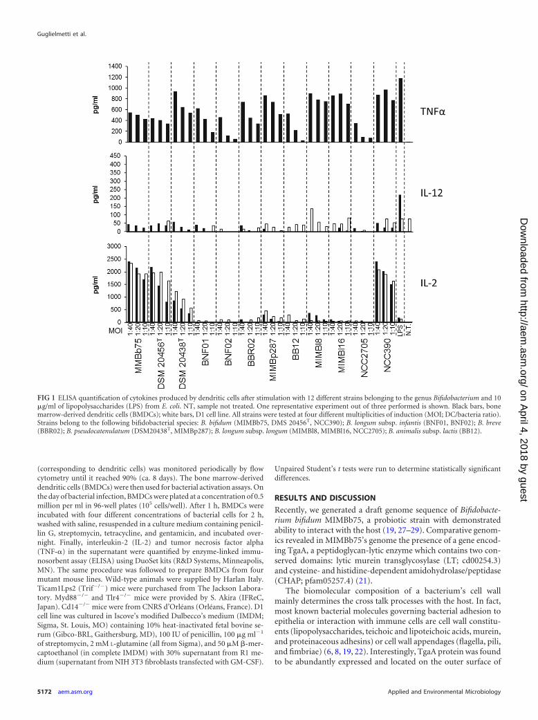
FIG 1 ELISA quantification of cytokines produced by dendritic cells after stimulation with 12 different strains belonging to the genus Bifidobacterium and 10�g/ml of lipopolysaccharides (LPS) from E. coli. NT, sample not treated. One representative experiment out of three performed is shown. Black bars, bonemarrow-derived dendritic cells (BMDCs); white bars, D1 cell line. All strains were tested at four different multiplicities of induction (MOI; DC/bacteria ratio).Strains belong to the following bifidobacterial species: B. bifidum (MIMBb75, DMS 20456T, NCC390); B. longum subsp. infantis (BNF01, BNF02); B. breve(BBR02); B. pseudocatenulatum (DSM20438T, MIMBp287); B. longum subsp. longum (MIMBl8, MIMBl16, NCC2705); B. animalis subsp. lactis (BB12).
Guglielmetti et al.
5172 aem.asm.org Applied and Environmental Microbiology
on April 4, 2018 by guest
http://aem.asm
.org/D
ownloaded from

MIMBb75 bacterial cells (21). Furthermore, notably, a search ofthe Conserved Domain Database (30) for the TgaA protein pro-duced a significant match (E value of 8.30e-23) between the CHAPmodule and the conserved domain COG3942, which has beenannotated as a surface antigen. The description of COG3942 orig-inated from two studies that identified the CHAP domain-con-taining protein as an immune-reactive molecule of Listeria mono-cytogenes (31) and Streptococcus pyogenes (32). Stimulated by theabove data, we evaluated the potential contribution of the TgaAprotein to the immunomodulatory activity of the strainMIMBb75.
We assessed the immunological properties of B. bifidumMIMBb75 in parallel and independently of the TgaA proteinstudy and discovered through our immunological model a pecu-liar immunomodulatory capacity of this strain. In particular, wefocused on dendritic cells (DCs), the sentinels of the immunesystem present at various mucosal sites and especially in the intes-tinal mucosa. At this site, they may sense the intestinal lumencontent (including bacteria) via transepithelial processes by re-sponding to the ligation of the DCs’ specific microbe recognitionreceptors (MRRs) to MAMPs (such as LPS, peptidoglycan, andbacterial and viral nucleic acids) to trigger immune responses (33,34). Particularly, upon activation and migration to secondarylymphoid organs, DCs induce immunocompetence in lympho-cytes through antigen presentation and cytokine production.Hence, DCs form a link between innate and adaptive immunityand are considered arbiters of immunological tolerance (34).
In DCs, a major outcome of the ligation of pattern recognitionreceptors (PRRs) may be the production of interleukin-2 (IL-2),which has several effects. In fact, DC-derived IL-2 increasesgamma interferon (IFN-) production by the natural killer (NK)cells (35), enhances T cell responses in both mice (26) and humans(36), regulates thymic development (37), and promotes regula-tory T cell (Treg) expansion and function (38). Furthermore, ithas been linked with TH1-skewing stimuli (35). Inducing IL-2
production in DCs can thus steer the adaptive immune systemtoward preventing TH2 cell-dominated immune responses, suchas those associated with allergy (39, 40).
Our experiments showed that out of 12 bifidobacterial strainsbelonging to six different species and subspecies, the strain B. bi-fidum MIMBb75 was among the strongest inducers of IL-2 pro-duction by murine bone marrow-derived DCs (BMDCs) (Fig. 1).
Subsequently, we studied the potential immunomodulatoryactivity of TgaA with the same immunological model we used forbifidobacterial cells. Initially, we cloned a complete tgaA gene (in-cluding the original signal sequence for exporting the protein at acell wall level) in Bifidobacterium longum NCC2705 using a shuttlevector previously developed and used for successful expression ofheterologous genes in B. longum (23, 24). We followed this strat-egy because all of our previous efforts to knock out tgaA had beenunsuccessful since this species has been proved to be recalcitrantto genetic manipulation (41, 42). We selected B. longum NCC2705as the host for tgaA expression after analysis of its whole genomerevealed that the tgaA gene as absent in this human intestinalbacterium. Furthermore, our immunological data (Fig. 1) showedthat the strain NCC2705, unlike MIMBb75, is incapable of induc-ing IL-2 expression by BMDCs. We named the recombinant strainwe obtained NCC:tga�. After verifying by reverse transcription-PCR (RT-PCR) and immunogold labeling (see Fig. S2A and B inthe supplemental material) that NCC:tga� expressed the recom-binant tgaA gene, we incubated BMDCs with supernatant-freewild-type live cells and recombinant NCC2705 strains and thenmeasured by ELISA the levels of the secreted tumor necrosis factoralpha (TNF-�) and IL-2. Secretion by BMDCs of the TNF-� cy-tokine, the principal marker of DC activation, was induced by allbacterial preparations (Fig. 2). In contrast, our experimentsshowed a significant secretion of IL-2 only when DCs were stim-ulated with the recombinant strain NCC:tga� (i.e., NCC2705transformed with the vector containing the gene tgaA) and notwith control preparations (wild-type NCC2705 and NCC2705
FIG 2 ELISA quantification of cytokine production by BMDCs after stimulation with E. coli lipopolysaccharides (LPS), Bifidobacterium longum NCC2705, andits recombinant strains NCC:tga� (expressing TgaA protein) and NCC:tga� (harboring the empty vector). All samples were tested at four different multiplicitiesof infection (MOI; BMDC/bacteria ratio). One representative experiment out of two performed is shown.
CHAP Domain from B. bifidum Modulates DC Maturation
September 2014 Volume 80 Number 17 aem.asm.org 5173
on April 4, 2018 by guest
http://aem.asm
.org/D
ownloaded from

transformed with the empty vector) (Fig. 2). Therefore, our datasuggest that the TgaA protein may be directly involved in inducingthe cytokine IL-2 in dendritic cells.
To further test our hypothesis, we thoroughly purified from E.coli cells the recombinant proteins rTgaA, rLT, and rCHAP byNi-NTA affinity chromatography and subsequent RP-HPLC toremove any residual LPS and extraneous proteins (Fig. 3). Purifiedproteins were then used to stimulate two dendritic cell models:BMDCs and the D1 cell line, which is a long-term growth factor-dependent immature myeloid (CD11c� CD8��) DC line ofsplenic origin. Our results show clearly that the rTgaA protein canin vitro activate dendritic cells and induce dose-dependent secre-tion of IL-2 (Fig. 4). A similar stimulatory effect was found with
the recombinant protein rCHAP, whereas rLT activated both den-dritic cell models only marginally but could not markedly triggerIL-2 expression (Fig. 4). Therefore, the TgaA protein activatesDCs and triggers IL-2 secretion by DCs by means of the CHAPdomain.
Finally, we studied the pathway induced by the protein TgaA inDCs. Since TgaA can induce TNF-� production by DCs and sincethe cluster of differentiation 14 (CD14) protein is essential in in-ducing such cytokines in LPS-stimulated DCs (43), we first testedif CD14 might have a role also in recognizing TgaA. As shown inFig. 5, BMDC activation was totally abolished in the absence ofCD14. Since CD14 is the best characterized as a coreceptor forToll-like receptor 4 (TLR4), we then tested if BMDCs isolated
FIG 3 Preparation of TgaA-derived recombinant proteins. (A) Functional map of the recombinant proteins originated from a tgaA gene sequence and producedby overexpression in E. coli BL21(DE3) pLysS using the pET26b(�) expression vector. (B) SDS-PAGE of the purified six-His-tagged TgaA-derived recombinantproteins. Lane 1, �SP-TgaA-His (containing both TgaA domains; rTgaA); lane 2, �SP-TgaA-�LT-His (containing the CHAP domain; rCHAP), lane 3,�SP-TgaA-�CHAP-His (containing the LT domain; rLT). The relative molecular masses (in kDa) of standard proteins are indicated on the left. aa, amino acids.
FIG 4 ELISA quantification of cytokine production by BMDCs and the D1 cell line stimulated with LPS or TgaA-derived recombinant proteins: rTgaAcorresponds to �SP-TgaA-His, which contains both TgaA domains; rLT corresponds to �SP-TgaA-�CHAP-His, which contains only the LT domain; rCHAPcorresponds to �SP-TgaA-�LT-His, which contains only the CHAP domain. Statistically significant differences were determined by an unpaired Student’s t test(*, P 0.05; ***, P 0.001). mBMDC, murine BMDCs.
Guglielmetti et al.
5174 aem.asm.org Applied and Environmental Microbiology
on April 4, 2018 by guest
http://aem.asm
.org/D
ownloaded from

from knockout mice lacking the expression of TLR4 can be acti-vated by TgaA. According to our data, the receptor TLR4 was notnecessary to induce TNF-� in BMDCs by TgaA (Fig. 5). There-fore, whereas LPS required TLR4 to induce TNF-� (43), the pro-tein TgaA directly activated the downstream pathways throughCD14 or involved CD14 for its presentation to a different un-known receptor, independently of TLR4.
To test if other TLRs might be important to recognizing TgaAby DCs, we used BMDCs derived from mice lacking the trans-ducer of TLR signaling. In particular, TLR downstream signalingcan occur via two pathways, which depend on two proteins: (i) themyeloid differentiation primary response gene 88 (MyD88) and(ii) the Toll IL-1 receptor (TIR) domain-containing adaptor-in-ducing IFN-� (TRIF) (44). Surprisingly, we found that without afunctional TRIF or MyD88, production of TNF-� by TgaA-stim-ulated BMDCs was largely preserved (Fig. 5), suggesting that, indendritic cells, the downstream signaling induced by TgaA couldfollow a CD14-dependent, TLR-independent signaling pathway.
Collectively, the above data show that TgaA is a cell wall pro-tein of B. bifidum MIMBb75 and contains a C-terminal domainthat initiates a CD14-dependent and TLR4-independent signalingpathway, which only partially involves the TRIF adapter protein.
A putative CHAP coding sequence is present in many bacterialgenomes and is included in a number of different deduced proteinarchitectures (45). Interestingly, several proteins from Gram-pos-itive bacteria that include the CHAP domain actively interact withthe host’s immune system. For instance, the Streptococcus pyogenesSibA protein binds all immunoglobulin G (IgG) subclasses, the Fcand Fab fragments, and also IgA and IgM (32). PcsB, anotherCHAP-containing protein of Streptococcus spp. and a known an-tigen of Streptococcus pneumoniae, is capable of activating T cellsin humans by inducing strong IL-17A responses (46). Further-more, P40, a CHAP-containing protein produced by Lactobacillusrhamnosus GG (perhaps the most studied probiotic strain so far),can inhibit cytokine-induced epithelial cell apoptosis, reduceTNF-induced colon epithelial damage, and promote cell growth
in human and mouse colon epithelial cells and cultured mousecolon explants (47, 48). In addition, our study showed that TgaAfrom B. bifidum MIMBb75 is a cell surface-exposed molecule ca-pable alone through its C-terminal CHAP domain of inducing DCactivation and IL-2 production. Based on observed induction ofIL-2 secretion, the TgaA stimulatory capacity here described maystimulate dendritic cells to acquire TH1 stimulatory capacity (35).Furthermore, the ability of B. bifidum MIMBb75 and its proteinTgaA to induce IL-2 by DCs, together with the previous observa-tion that B. bifidum strains induce an immune response affectingTreg/TH17 plasticity (49), supports the hypothesis that these com-mensal bacteria have a key role in mucosal tolerance. The presenceof specific microbial stimuli, such as the CHAP-containing TgaAprotein, may then have served as a way for commensals to co-evolve with the host and produce mechanisms for maintaininghomeostasis and controlled reactions in the adaptive immune sys-tem (i.e., prevent an imbalance in T-helper cell subsets).
Interestingly, a BLASTP search revealed the presence of a geneclosely similar to the CHAP domain of TgaA (with more than 80%identity in the amino acid sequence) not only in B. bifidum but
FIG 5 ELISA quantification of TNF-� produced by rTgaA-stimulatedBMDCs isolated from wild-type (WT) and knockout mice. tlr4�/�, Toll-likereceptor 4 (TLR4) KO mutant mouse; cd14�/�, CD14 protein KO mutantmouse; myd88�/�, myeloid differentiation primary response gene 88(MYD88) KO mutant mouse; trif�/�, Toll IL-1 receptor (TIR) domain-con-taining adaptor-inducing IFN-� (TRIF) KO mutant mouse. Statistically sig-nificant differences were determined by an unpaired Student’s t test (���, P 0.001). nt, not treated.
FIG 6 DC-mediated host immune response to TgaA protein from Bifidobac-terium bifidum MIMBb75: possible consequences of IL-2 production. Thecontact between B. bifidum MIMBb75 and dendritic cells (DCs) determinesDC activation and IL-2 secretion. Reportedly, stimuli able to induce IL-2 pro-duction by DCs are associated with a Th1-skewing of the immune responses,Treg lymphocyte proliferation, and natural killer (NK) cell activation (for areview, see reference 50).
CHAP Domain from B. bifidum Modulates DC Maturation
September 2014 Volume 80 Number 17 aem.asm.org 5175
on April 4, 2018 by guest
http://aem.asm
.org/D
ownloaded from

also in the genome of B. breve (e.g., GenBank accession numberWP_016462813), which is another Bifidobacterium species typ-ically associated with healthy breast-fed infants (13). Therefore,the presence in B. bifidum and other bifidobacteria of moleculessimilar to the TgaA protein and capable of stimulating the im-mune system could help explain the ability of these bacteria toprotect the host and nurture the immune system in early life (15,16).
Conclusions. The results of this study can be summarized asfollows: (i) both the cells of B. bifidum MIMBb75 and its surfaceprotein TgaA can activate dendritic cells and trigger IL-2 produc-tion; (ii) TgaA activates dendritic cells through its CHAP amidasedomain; and (iii) the TgaA-dependent activation of dendritic cellsrequires the protein CD14, only marginally involves TRIF, and isindependent of TLR4 and MyD88.
The bacterium B. bifidum MIMBb75, which is of human ori-gin, has probiotic properties (28), and its ability to interact withthe host may partly depend on the presence of the CHAP domainof its TgaA surface protein. In conclusion, the bacterial CHAPdomain may well be considered a novel, potentially widespreadMAMP that may participate in the cross talk mechanisms amongGram-positive bacteria and their mammalian host. The possiblephysiological consequences of the ability of B. bifidum MIMBb75and its TgaA protein to activate DCs are schematically summa-rized in Fig. 6.
ACKNOWLEDGMENT
This work was financially supported by Fondazione Cariplo (grant 2010-0678).
REFERENCES1. Tremaroli V, Backhed F. 2012. Functional interactions between the gut
microbiota and host metabolism. Nature 489:242–249. http://dx.doi.org/10.1038/nature11552.
2. Tlaskalova-Hogenova H, Stepankova R, Kozakova H, Hudcovic T,Vannucci L, Tuckova L, Rossmann P, Hrncir T, Kverka M, ZakostelskaZ, Klimesova K, Pribylova J, Bartova J, Sanchez D, Fundova P, Boro-vska D, Srutkova D, Zidek Z, Schwarzer M, Drastich P, Funda DP.2011. The role of gut microbiota (commensal bacteria) and the mucosalbarrier in the pathogenesis of inflammatory and autoimmune diseases andcancer: contribution of germ-free and gnotobiotic animal models of hu-man diseases. Cell. Mol. Immunol. 8:110 –120. http://dx.doi.org/10.1038/cmi.2010.67.
3. Sokol H, Pigneur B, Watterlot L, Lakhdari O, Bermudez-Humaran LG,Gratadoux JJ, Blugeon S, Bridonneau C, Furet JP, Corthier G, Grang-ette C, Vasquez N, Pochart P, Trugnan G, Thomas G, Blottiere HM,Dore J, Marteau P, Seksik P, Langella P. 2008. Faecalibacterium praus-nitzii is an anti-inflammatory commensal bacterium identified by gut mi-crobiota analysis of Crohn disease patients. Proc. Natl. Acad. Sci. U. S. A.105:16731–16736. http://dx.doi.org/10.1073/pnas.0804812105.
4. Belzer C, de Vos WM. 2012. Microbes inside-from diversity to function:the case of Akkermansia. ISME J. 6:1449 –1458. http://dx.doi.org/10.1038/ismej.2012.6.
5. Sela DA, Chapman J, Adeuya A, Kim JH, Chen F, Whitehead TR,Lapidus A, Rokhsar DS, Lebrilla CB, German JB, Price NP, RichardsonPM, Mills DA. 2008. The genome sequence of Bifidobacterium longumsubsp. infantis reveals adaptations for milk utilization within the infantmicrobiome. Proc. Natl. Acad. Sci. U. S. A. 105:18964 –18969. http://dx.doi.org/10.1073/pnas.0809584105.
6. Fanning S, Hall LJ, Cronin M, Zomer A, MacSharry J, Goulding D,Motherway MO, Shanahan F, Nally K, Dougan G, van Sinderen D.2012. Bifidobacterial surface-exopolysaccharide facilitates commensal-host interaction through immune modulation and pathogen protection.Proc. Natl. Acad. Sci. U. S. A. 109:2108 –2113. http://dx.doi.org/10.1073/pnas.1115621109.
7. Ventura M, Turroni F, Motherway MO, MacSharry J, van Sinderen D.
2012. Host-microbe interactions that facilitate gut colonization by com-mensal bifidobacteria. Trends Microbiol. 20:467– 476. http://dx.doi.org/10.1016/j.tim.2012.07.002.
8. Turroni F, Serafini F, Foroni E, Duranti S, Motherway MO, TavernitiV, Mangifesta M, Milani C, Viappiani A, Roversi T, Sanchez B, SantoniA, Gioiosa L, Ferrarini A, Delledonne M, Margolles A, Piazza L, PalanzaP, Bolchi A, Guglielmetti S, van Sinderen D, Ventura M. 2013. Role ofsortase-dependent pili of Bifidobacterium bifidum PRL2010 in modulatingbacterium-host interactions. Proc. Natl. Acad. Sci. U. S. A. 110:11151–11156. http://dx.doi.org/10.1073/pnas.1303897110.
9. Gaboriau-Routhiau V, Raibaud P, Dubuquoy C, Moreau MC. 2003.Colonization of gnotobiotic mice with human gut microflora at birthprotects against Escherichia coli heat-labile enterotoxin-mediated abroga-tion of oral tolerance. Pediatr. Res. 54:739 –746. http://dx.doi.org/10.1203/01.PDR.0000086902.52137.C9.
10. Dong P, Yang Y, Wang WP. 2010. The role of intestinal bifidobacteria onimmune system development in young rats. Early Hum. Dev. 86:51–58.http://dx.doi.org/10.1016/j.earlhumdev.2010.01.002.
11. Bjorksten B, Naaber P, Sepp E, Mikelsaar M. 1999. The intestinalmicroflora in allergic Estonian and Swedish 2-year-old children. Clin.Exp. Allergy 29:342–346. http://dx.doi.org/10.1046/j.1365-2222.1999.00560.x.
12. He F, Ouwehand AC, Isolauri E, Hashimoto H, Benno Y, Salminen S.2001. Comparison of mucosal adhesion and species identification of bifi-dobacteria isolated from healthy and allergic infants. FEMS Immunol.Med. Microbiol. 30:43– 47. http://dx.doi.org/10.1111/j.1574-695X.2001.tb01548.x.
13. Ouwehand AC, Isolauri E, He F, Hashimoto H, Benno Y, Salminen S.2001. Differences in Bifidobacterium flora composition in allergic andhealthy infants. J. Allergy Clin. Immunol. 108:144 –145. http://dx.doi.org/10.1067/mai.2001.115754.
14. Watanabe S, Narisawa Y, Arase S, Okamatsu H, Ikenaga T, Tajiri Y,Kumemura M. 2003. Differences in fecal microflora between patientswith atopic dermatitis and healthy control subjects. J. Allergy Clin. Immu-nol. 111:587–591. http://dx.doi.org/10.1067/mai.2003.105.
15. Young SL, Simon MA, Baird MA, Tannock GW, Bibiloni R, Spencely K,Lane JM, Fitzharris P, Crane J, Town I, Addo-Yobo E, Murray CS,Woodcock A. 2004. Bifidobacterial species differentially affect expressionof cell surface markers and cytokines of dendritic cells harvested. Clin.Diagn. Lab. Immunol. 11:686 – 690. http://dx.doi.org/10.1128/CDLI.11.4.686-690.2004.
16. Menard O, Butel MJ, Gaboriau-Routhiau V, Waligora-Dupriet AJ.2008. Gnotobiotic mouse immune response induced by Bifidobacteriumsp. strains isolated from infants. Appl. Environ. Microbiol. 74:660 – 666.http://dx.doi.org/10.1128/AEM.01261-07.
17. Weiss G, Rasmussen S, Nielsen Fink L, Jarmer H, Nøhr Nielsen B,Frøkiaer H. 2010. Bifidobacterium bifidum actively changes the gene ex-pression profile induced by Lactobacillus acidophilus in murine dendriticcells. PLoS One 5:e11065. http://dx.doi.org/10.1371/journal.pone.0011065.
18. Turroni F, Taverniti V, Ruas-Madiedo P, Duranti S, Guglielmetti S,Lugli GA, Gioiosa L, Palanza P, Margolles A, van Sinderen D, VenturaM. 2014. Bifidobacterium bifidum PRL2010 modulates the host innateimmune response. Appl. Environ. Microbiol. 80:730 –740. http://dx.doi.org/10.1128/AEM.03313-13.
19. Guglielmetti S, Tamagnini I, Mora D, Minuzzo M, Scarafoni A, ArioliS, Hellman J, Karp M, Parini C. 2008. Implication of an outer surfacelipoprotein in adhesion of Bifidobacterium bifidum to Caco-2 cells. Appl.Environ. Microbiol. 74:4695– 4702. http://dx.doi.org/10.1128/AEM.00124-08.
20. Janeway CA, Jr. 1989. Approaching the asymptote? Evolution and revo-lution in immunology. Cold Spring Harbor Symp. Quant. Biol. 54:1–13.http://dx.doi.org/10.1101/SQB.1989.054.01.003.
21. Guglielmetti S, Balzaretti S, Taverniti V, Miriani M, Milani C, ScarafoniA, Corona S, Arioli S, Santala V, Iametti S, Bonomi F, Ventura M, MoraD, Karp M. 2014. TgaA, a VirB1-like component belonging to a putativetype IV secretion system of Bifidobacterium bifidum MIMBb75. Appl. En-viron. Microbiol. 80:5161–5169. http://dx.doi.org/10.1128/AEM.01413-14.
22. Taverniti V, Stuknyte M, Minuzzo M, Arioli S, De Noni I, ScabiosiC, Cordova ZM, Junttila I, Hämäläinen S, Turpeinen H, Mora D,Karp M, Pesu M, Guglielmetti S. 2013. S-layer protein mediates thestimulatory effect of Lactobacillus helveticus MIMLh5 on innate immu-
Guglielmetti et al.
5176 aem.asm.org Applied and Environmental Microbiology
on April 4, 2018 by guest
http://aem.asm
.org/D
ownloaded from

nity. Appl. Environ. Microbiol. 79:1221–1231. http://dx.doi.org/10.1128/AEM.03056-12.
23. Guglielmetti S, Ciranna A, Mora D, Parini C, Karp M. 2008. Construc-tion, characterization and exemplificative application of bioluminescentBifidobacterium longum biovar longum. Int. J. Food Microbiol. 124:285–290. http://dx.doi.org/10.1016/j.ijfoodmicro.2008.03.033.
24. Guglielmetti S, Karp M, Mora D, Tamagnini I, Parini C. 2007. Molec-ular characterization of Bifidobacterium longum biovar longum NAL8 plas-mids and construction of a novel replicon screening system. Appl. Micro-biol. Biotechnol. 74:1053–1061. http://dx.doi.org/10.1007/s00253-006-0755-1.
25. Ventura M, Zhang Z, Cronin M, Canchaya C, Kenny JG, Fitzgerald GF,van Sinderen D. 2005. The ClgR protein regulates transcription of theclpP operon in Bifidobacterium breve UCC 2003. J. Bacteriol. 187:8411–8426. http://dx.doi.org/10.1128/JB.187.24.8411-8426.2005.
26. Granucci F, Vizzardelli C, Pavelka N, Feau S, Persico M, Virzi E,Rescigno M, Moro G, Ricciardi-Castagnoli P. 2001. Inducible IL-2 pro-duction by dendritic cells revealed by global gene expression analysis. Nat.Immunol. 2:882– 888. http://dx.doi.org/10.1038/ni0901-882.
27. Guglielmetti S, Tamagnini I, Minuzzo M, Arioli S, Parini C, Comelli E,Mora D. 2009. Study of the adhesion of Bifidobacterium bifidumMIMBb75 to human intestinal cell lines. Curr. Microbiol. 59:167–172.http://dx.doi.org/10.1007/s00284-009-9415-x.
28. Guglielmetti S, Mora D, Gschwender M, Popp K. 2011. Randomisedclinical trial: Bifidobacterium bifidum MIMBb75 significantly alleviates ir-ritable bowel syndrome and improves quality of life—a double-blind, pla-cebo-controlled study. Aliment. Pharmacol. Therap. 33:1123–1132. http://dx.doi.org/10.1111/j.1365-2036.2011.04633.x.
29. Singh N, Arioli S, Wang A, Villa CR, Jahani R, Song YS, Mora D,Guglielmetti S, Comelli EM. 2013. Impact of Bifidobacterium bifidumMIMBb75 on mouse intestinal microorganisms. FEMS Microbiol. Ecol.85:369 –375. http://dx.doi.org/10.1111/1574-6941.12124.
30. Marchler-Bauer A, Lu SN, Anderson JB, Chitsaz F, Derbyshire MK,DeWeese-Scott C, Fong JH, Geer LY, Geer RC, Gonzales NR, Gwadz M,Hurwitz DI, Jackson JD, Ke ZX, Lanczycki CJ, Lu F, Marchler GH,Mullokandov M, Omelchenko MV, Robertson CL, Song JS, Thanki N,Yamashita RA, Zhang DC, Zhang NG, Zheng CJ, Bryant SH. 2011.CDD: a Conserved Domain Database for the functional annotation ofproteins. Nucleic Acids Res. 39:D225–D229. http://dx.doi.org/10.1093/nar/gkq1189.
31. Schubert K, Bichlmaier AM, Mager E, Wolff K, Ruhland G, Fiedler F.2000. P45, an extracellular 45 kDa protein of Listeria monocytogenes withsimilarity to protein p60 and exhibiting peptidoglycan lytic activity. Arch.Microbiol. 173:21–28. http://dx.doi.org/10.1007/s002030050003.
32. Fagan PK, Reinscheid D, Gottschalk B, Chhatwal GS. 2001. Identifica-tion and characterization of a novel secreted immunoglobulin bindingprotein from group A Streptococcus. Infect. Immun. 69:4851– 4857. http://dx.doi.org/10.1128/IAI.69.8.4851-4857.2001.
33. Granucci F, Feau S, Zanoni I, Pavelka N, Vizzardelli C, Raimondi G,Ricciardi-Castagnoli P. 2003. The immune response is initiated by den-dritic cells via interaction with microorganisms and interleukin-2 produc-tion. J. Infect. Dis. 187:S346 –S350. http://dx.doi.org/10.1086/374748.
34. Lewis KL, Reizis B. 2012. Dendritic cells: arbiters of immunity and im-munological tolerance. Cold Spring Harb. Perspect. Biol. 4:a007401. http://dx.doi.org/10.1101/cshperspect.a007401.
35. Zanoni I, Foti M, Ricciardi-Castagnoli P, Granucci F. 2005. TLR-dependent activation stimuli associated with Th1 responses confer NK cellstimulatory capacity to mouse dendritic cells. J. Immunol. 175:286 –292.http://dx.doi.org/10.4049/jimmunol.175.1.286.
36. Wuest SC, Edwan JH, Martin JF, Han S, Perry JSA, Cartagena CM,Matsuura E, Maric D, Waldmann TA, Bielekova B. 2011. A role forinterleukin-2 trans-presentation in dendritic cell-mediated T cell activa-tion in humans, as revealed by daclizumab therapy. Nat. Med. 17:604 –U125. http://dx.doi.org/10.1038/nm.2365.
37. Cheng GY, Yu AX, Malek TR. 2011. T-cell tolerance and the multi-functional role of IL-2R signaling in T-regulatory cells. Immunol. Rev.241:63–76. http://dx.doi.org/10.1111/j.1600-065X.2011.01004.x.
38. Guiducci C, Valzasina B, Dislich H, Colombo MP. 2005. CD40/CD40Linteraction regulates CD4� CD25� Treg homeostasis through dendriticcell-produced IL-2. Eur. J. Immunol. 35:557–567. http://dx.doi.org/10.1002/eji.200425810.
39. Lampinen M, Hakansson L, Venge P. 2001. Interleukin-2 inhibits eosin-ophil migration but is counteracted by IL-5 priming. Clin. Exp. Allergy31:249 –258. http://dx.doi.org/10.1046/j.1365-2222.2001.00968.x.
40. Palm NW, Rosenstein RK, Medzhitov R. 2012. Allergic host defences.Nature 484:465– 472. http://dx.doi.org/10.1038/nature11047.
41. Serafini F, Turroni F, Guglielmetti S, Gioiosa L, Foroni E, Sanghez V,Bartolomucci A, Motherway MO, Palanza P, van Sinderen D, VenturaM. 2012. An efficient and reproducible method for transformation ofgenetically recalcitrant bifidobacteria. FEMS Microbiol. Lett. 333:146 –152. http://dx.doi.org/10.1111/j.1574-6968.2012.02605.x.
42. Guglielmetti S, Mayo B, Alvarez-Martin P. 2013. Mobilome and geneticmodification of bifidobacteria. Benef. Microbes 4:143–166. http://dx.doi.org/10.3920/BM2012.0031.
43. Zanoni I, Ostuni R, Capuano G, Collini M, Caccia M, Ronchi AE,Rocchetti M, Mingozzi F, Foti M, Chirico G, Costa B, Zaza A, Ricciardi-Castagnoli P, Granucci F. 2009. CD14 regulates the dendritic cell lifecycle after LPS exposure through NFAT activation. Nature 460:264 –U130. http://dx.doi.org/10.1038/nature08118.
44. Takeda K, Akira S. 2005. Toll-like receptors in innate immunity. Int.Immunol. 17:1–14. http://dx.doi.org/10.1093/intimm/dxh186.
45. Bateman A, Rawlings ND. 2003. The CHAP domain: a large family ofamidases including GSP amidase and peptidoglycan hydrolases.Trends Biochem. Sci. 28:234 –237. http://dx.doi.org/10.1016/S0968-0004(03)00061-6.
46. Lundgren A, Bhuiyan TR, Novak D, Kaim J, Reske A, Lu YJ, Qadri F,Malley R. 2012. Characterization of Th17 responses to Streptococcus pneu-moniae in humans: Comparisons between adults and children in a devel-oped and a developing country. Vaccine 30:3897–3907. http://dx.doi.org/10.1016/j.vaccine.2012.03.082.
47. Yan F, Cao HW, Cover TL, Whitehead R, Washington MK, Polk DB.2007. Soluble proteins produced by probiotic bacteria regulate intestinalepithelial cell survival and growth. Gastroenterology 132:562–575. http://dx.doi.org/10.1053/j.gastro.2006.11.022.
48. Yan F, Cao HW, Cover TL, Washington MK, Shi Y, Liu LS, ChaturvediR, Peek RM, Wilson KT, Polk DB. 2011. Colon-specific delivery of aprobiotic-derived soluble protein ameliorates intestinal inflammation inmice through an EGFR-dependent mechanism. J. Clin. Invest. 121:2242–2253. http://dx.doi.org/10.1172/JCI44031.
49. Lopez P, Gonzalez-Rodriguez I, Gueimonde M, Margolles A, Suarez A.2011. Immune response to Bifidobacterium bifidum strains support Treg/Th17 plasticity. PLoS One 6:e24776. http://dx.doi.org/10.1371/journal.pone.0024776.
50. Zanoni I, Granucci F. 2012. Regulation and dysregulation of innateimmunity by NFAT signaling downstream of pattern recognition recep-tors (PRRs). Eur. J. Immunol. 42:1924 –1931. http://dx.doi.org/10.1002/eji.201242580.
CHAP Domain from B. bifidum Modulates DC Maturation
September 2014 Volume 80 Number 17 aem.asm.org 5177
on April 4, 2018 by guest
http://aem.asm
.org/D
ownloaded from