lymhocyte chromosomal aberration assay in … · lymhocyte chromosomal aberration assay in...
TRANSCRIPT
Dr. Birutė Gricienė 1,2
1Radiation Protection Centre 2Vilnius University
LYMHOCYTE CHROMOSOMAL ABERRATION ASSAY IN RADIATION BIODOSIMETRY
Introduction
Ionising radiation is a well-known mutagenic and carcinogenic agent.
The intensive use of ionizing radiation sources and development of radiation technology is related with increased exposure risk.
In the case of nuclear or radiological event it is important to identify exposed individuals in order to determine whether the medical intervention is needed
In many emergency cases the dose assessments can only be conducted using biological assays.
Chromosomal aberrations assay in peripheral blood lymphocyte is a most reliable, sensitive and mostly used biological dosimetry method for assessing radiation dose.
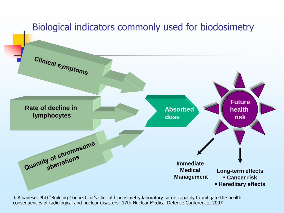
Rate of decline in
lymphocytes
Absorbed
dose
Biological indicators commonly used for biodosimetry
Future
health
risk
Immediate
Medical
Management Long-term effects
Cancer risk
Hereditary effects
J. Albanese, PhD “Building Connecticut’s clinical biodosimetry laboratory surge capacity to mitigate the health consequences of radiological and nuclear disasters” 17th Nuclear Medical Defence Conference, 2007
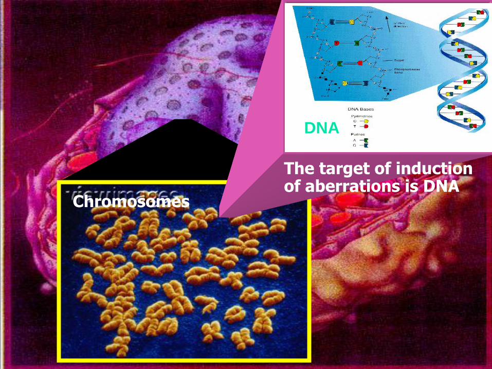
DNA
The target of induction of aberrations is DNA
Chromosomes
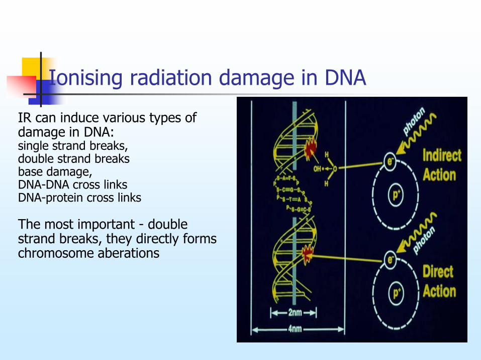
Ionising radiation damage in DNA
IR can induce various types of damage in DNA: single strand breaks, double strand breaks base damage, DNA-DNA cross links DNA-protein cross links The most important - double strand breaks, they directly forms chromosome aberations
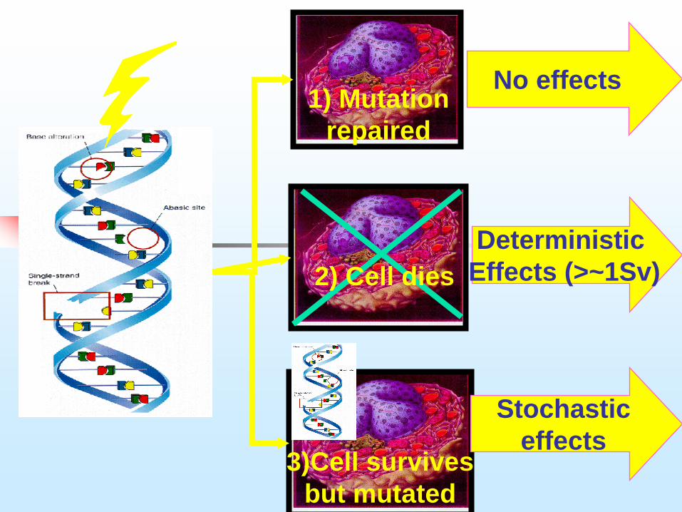
3)Cell survives
but mutated
Stochastic
effects
1) Mutation
repaired
Deterministic
Effects (>~1Sv)
No effects
2) Cell dies
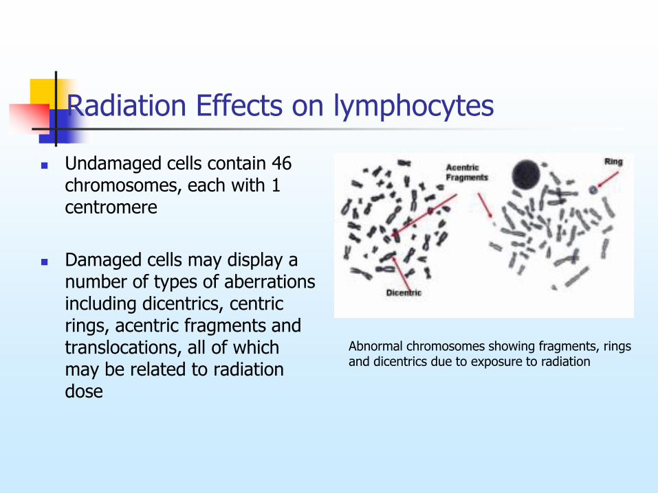
Radiation Effects on lymphocytes
Undamaged cells contain 46 chromosomes, each with 1 centromere
Damaged cells may display a number of types of aberrations including dicentrics, centric rings, acentric fragments and translocations, all of which may be related to radiation dose
Abnormal chromosomes showing fragments, rings and dicentrics due to exposure to radiation
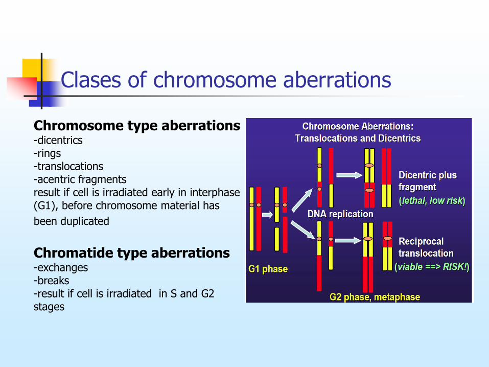
Clases of chromosome aberrations
Chromosome type aberrations -dicentrics -rings -translocations -acentric fragments result if cell is irradiated early in interphase (G1), before chromosome material has
been duplicated Chromatide type aberrations -exchanges -breaks -result if cell is irradiated in S and G2 stages
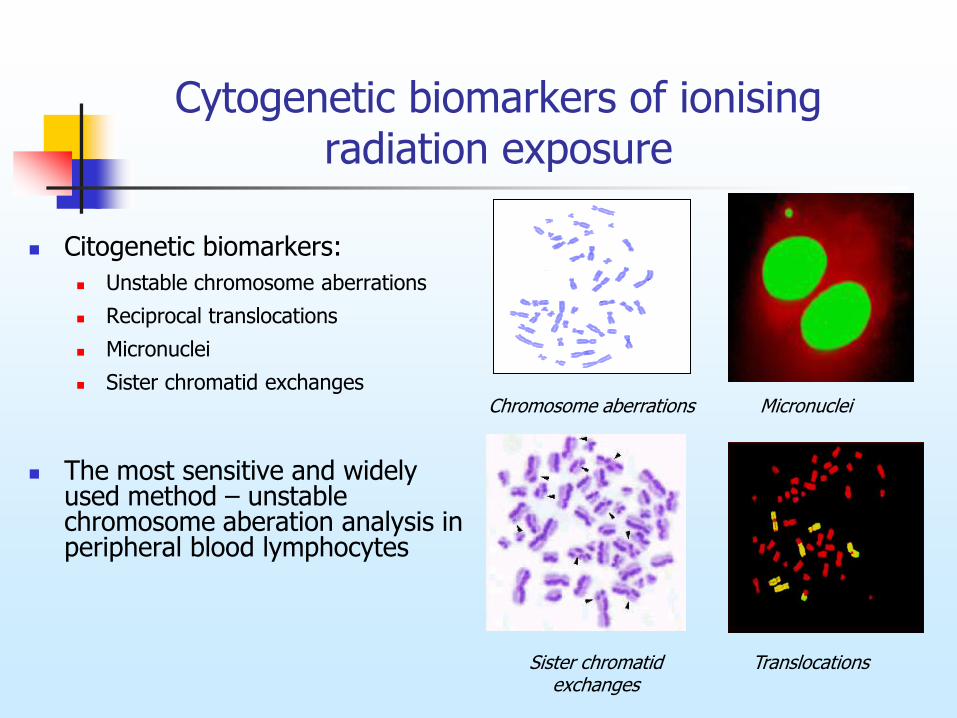
Cytogenetic biomarkers of ionising radiation exposure
Citogenetic biomarkers:
Unstable chromosome aberrations
Reciprocal translocations
Micronuclei
Sister chromatid exchanges
The most sensitive and widely used method – unstable chromosome aberation analysis in peripheral blood lymphocytes
Micronuclei
Translocations
Chromosome aberrations
Sister chromatid exchanges
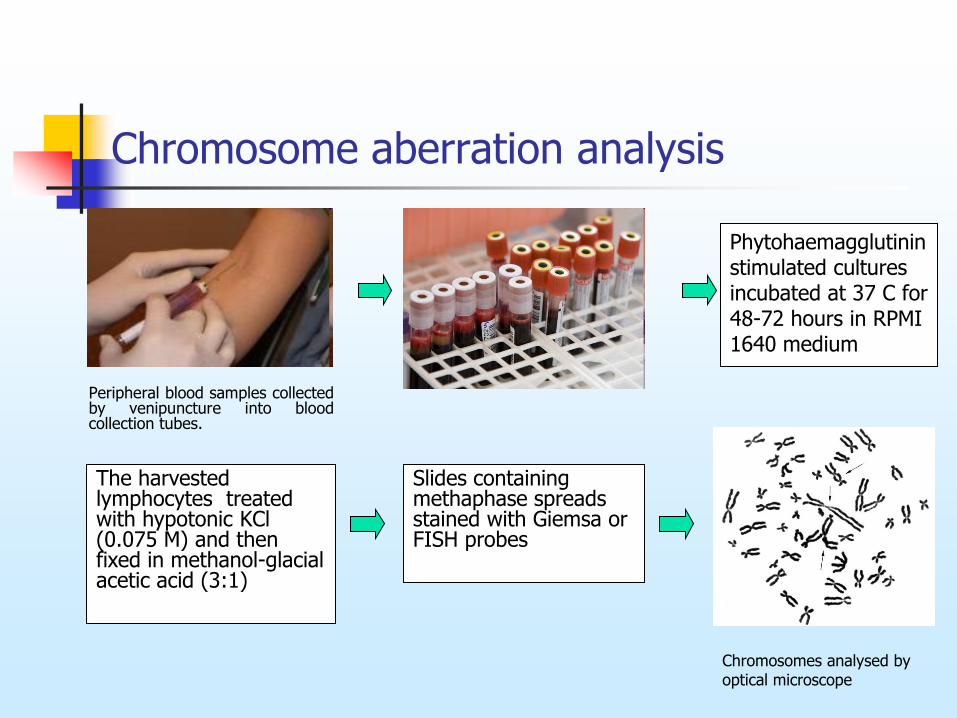
Peripheral blood samples collected by venipuncture into blood collection tubes.
Slides containing methaphase spreads stained with Giemsa or FISH probes
.
Phytohaemagglutinin stimulated cultures incubated at 37 C for 48-72 hours in RPMI 1640 medium
Chromosomes analysed by optical microscope
Chromosome aberration analysis
The harvested lymphocytes treated with hypotonic KCl (0.075 M) and then fixed in methanol-glacial acetic acid (3:1)
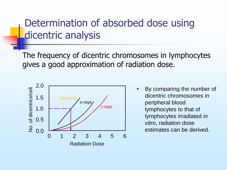
Determination of absorbed dose using dicentric analysis
0 1 2 3 4 5 6 0.0
2.0
0.5
1.0
1.5
No. of
dic
entr
ics/c
ell
Radiation Dose
x-rays -rays
The frequency of dicentric chromosomes in lymphocytes gives a good approximation of radiation dose.
Neutrons
By comparing the number of
dicentric chromosomes in
peripheral blood
lymphocytes to that of
lymphocytes irradiated in
vitro, radiation dose
estimates can be derived.
Production of gamma radiation (Co-60) dose-response curves
In 2009 was produced gamma radiation dose-response curve at Vilnius University, faculty of Nature sciences.
Experiment was designed as follows: blood samples from three donors were irradiated with Co-60 (0,256 Gy/min) to various doses (0.5-4 Gy).
Samples were cultured according to the standard procedures.
200-1000 first cycle metaphases were analysed for each radiation dose per donor after the preparation and staining of the slides.
For fitting a linear-quadratic dose-response relationship the CABAS software was used.
Gricienė B, Slapšytė G, Mierauskienė J. Gama spinduliuote sukeltų chromosomų aberacijų dozės ir atsako kreivės sudarymas bei panaudojimas biologinei dozimetrijai. Visuomenės sveikata. 2009;1:19-23.
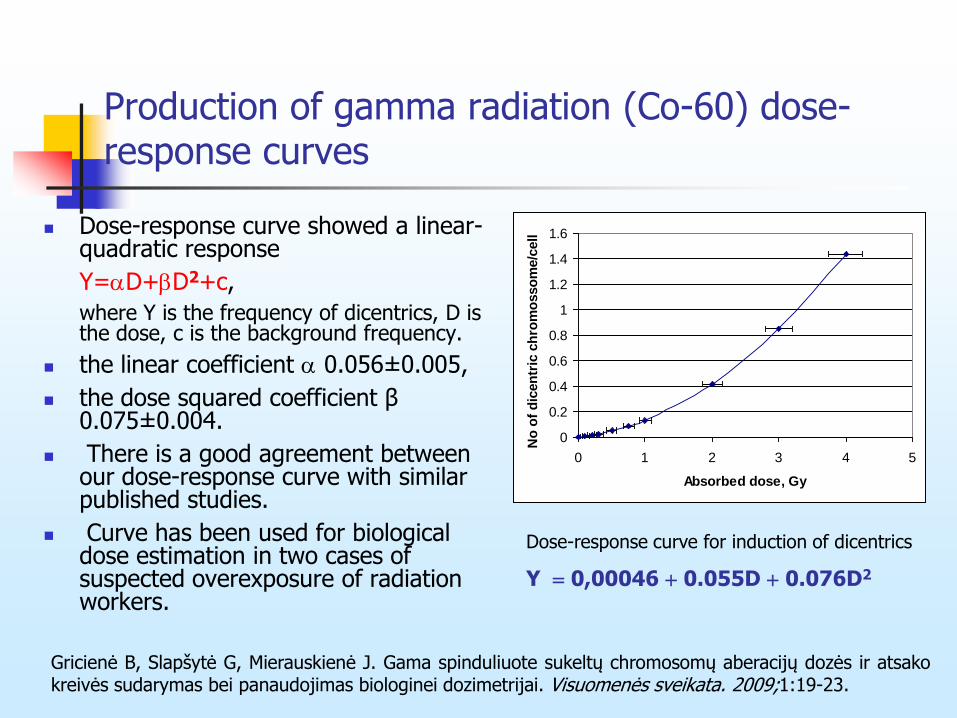
Production of gamma radiation (Co-60) dose-response curves
Dose-response curve showed a linear-quadratic response
Y=D+D2+c,
where Y is the frequency of dicentrics, D is the dose, c is the background frequency.
the linear coefficient 0.056±0.005,
the dose squared coefficient β 0.075±0.004.
There is a good agreement between our dose-response curve with similar published studies.
Curve has been used for biological dose estimation in two cases of suspected overexposure of radiation workers.
Dose-response curve for induction of dicentrics
Y 0,00046 0.055D 0.076D2
0
0.2
0.4
0.6
0.8
1
1.2
1.4
1.6
0 1 2 3 4 5
Absorbed dose, Gy
No
of
dic
en
tric
ch
rom
osso
me/c
ell
Gricienė B, Slapšytė G, Mierauskienė J. Gama spinduliuote sukeltų chromosomų aberacijų dozės ir atsako kreivės sudarymas bei panaudojimas biologinei dozimetrijai. Visuomenės sveikata. 2009;1:19-23.
Application of lymhocyte chromosomal aberration assay in radiation biodosimetry
Dose-response curves are used:
For accidental exposure dose assessment (Chernobyl NPP, 1986; Brazil, 1987; Estonia, 1994; Japan, 1999 ir kt.)
For epidemiological studies to investigate absorbed doses long time after exposure (Japan A bomb survivors, exposed persons after Chernobyl NPP accident)
For determination of individual radiosensitivity
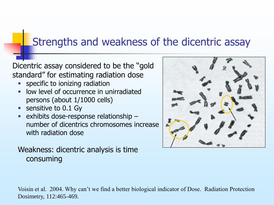
Strengths and weakness of the dicentric assay
Dicentric assay considered to be the “gold standard” for estimating radiation dose specific to ionizing radiation low level of occurrence in unirradiated
persons (about 1/1000 cells) sensitive to 0.1 Gy exhibits dose-response relationship –
number of dicentrics chromosomes increase with radiation dose
Weakness: dicentric analysis is time consuming
Voisin et al. 2004. Why can’t we find a better biological indicator of Dose. Radiation Protection
Dosimetry, 112:465-469.
Limitations of scoring dicentrics
Major limitation of using dicentrics for dosimetry is loss of lymphocytes from the blood
Yield of measured dicentrics after an irradiation would decrease with time (1/2 life is about 3 yrs), although this is uncertain
When replaced by stem cell division, new lymphocytes will tend not to contain dicentrics due to elimination at anaphase
unstable aberration since it is lethal to the cell
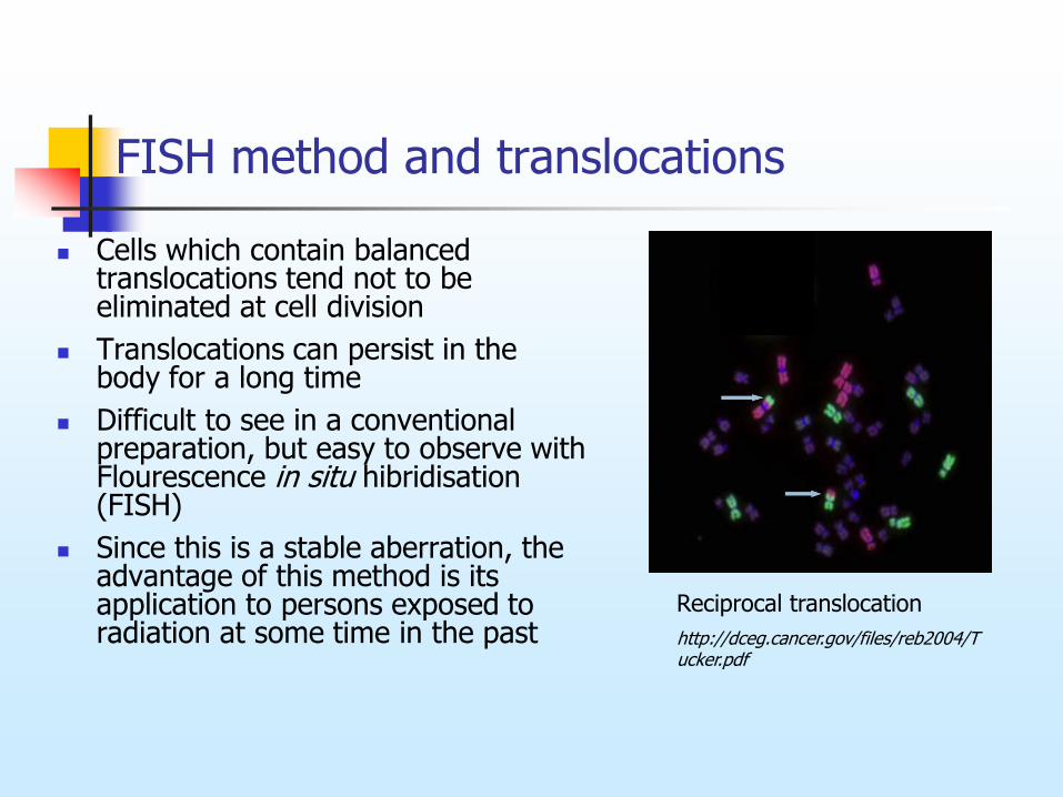
FISH method and translocations
Cells which contain balanced translocations tend not to be eliminated at cell division
Translocations can persist in the body for a long time
Difficult to see in a conventional preparation, but easy to observe with Flourescence in situ hibridisation (FISH)
Since this is a stable aberration, the advantage of this method is its application to persons exposed to radiation at some time in the past
Reciprocal translocation
http://dceg.cancer.gov/files/reb2004/Tucker.pdf
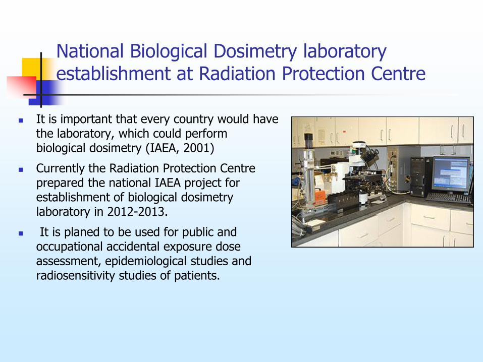
It is important that every country would have the laboratory, which could perform biological dosimetry (IAEA, 2001)
Currently the Radiation Protection Centre prepared the national IAEA project for establishment of biological dosimetry laboratory in 2012-2013.
It is planed to be used for public and occupational accidental exposure dose assessment, epidemiological studies and radiosensitivity studies of patients.
National Biological Dosimetry laboratory establishment at Radiation Protection Centre
Summary
Ionising radiation induces chromosome aberrations at all stages of the cell cycle.
Since in vitro and in vivo irradiation of lymphocytes induces similar yields of chromosome damage per unit dose, the absorbed dose to individual can be estimated by comparing the aberration yield to calibration curve generated in vitro.
Biological dosimetry based on chromosome aberration analysis can be used for accidental exposure dose assessment, radiosensitivity studies of radiotherapy patients.
Methods are available to perform biodosimetry using dicentric frequencies following the ionising radiation exposure.
Dicentrics are unstable aberrations and eliminated with time.
FISH technique using chromosome specific libraries has proven to be very efficient to detect translocations, which are stable and persist in the body for many years and allows estimating absorbed dose from past accidents.