long-term stability of maxillary and mandibular arch ... · man scientist named korkhaus visited...
TRANSCRIPT

Long-term stability of maxillary and mandibular arch dimensions when using rapid palatal expansion and edgewise mechanotherapy in growing patients
Objective: The purpose of this study was to assess the long-term stability of rapid palatal expansion (RPE) followed by full fixed edgewise appliances. Methods: This study included 67 patients treated using Haas-type RPE and non-extraction edgewise appliance therapy at a single orthodontic practice. Serial dental casts were obtained at three different time points: pretreatment (T1), after expansion and fixed appliance therapy (T2), and at long-term recall (T3). The mean duration of the T1–T2 and T2–T3 periods was 4.8 ± 3.5 years and 11.0 ± 5.4 years, respectively. The dental casts were digitized, and the computed measurements were compared with untreated reference data. Results: The majority of treatment-related increases in the maxillary and mandibular arch measurements were statistically significant (p < 0.05) and greater than expected for the untreated controls. Although many measurements decreased postretention (T2–T3), the net gains persisted for all of the measurements evaluated. Conclusions: The use of RPE therapy followed by full fixed edgewise appliances is an effective method for increasing maxillary and mandibular arch width dimensions in growing patients.[Korean J Orthod 2019;49(2):89-96]
Key words: Expansion, Stability, Digital models
Ki Beom Kima
Renee E. Doyleb Eustáquio A. Araújoa Rolf G. Behrentsa Donald R. Olivera Guilherme Thiesena,c
aDepartment of Orthodontics, Saint Louis University, St. Louis, MO, USAbPrivate Practice, Columbia, IL, USAcPrivate Practice, Florianopolis, SC, Brazil
Received August 8, 2018; Revised September 24, 2018; Accepted October 5, 2018.
Corresponding author: Ki Beom Kim.Program Director, Department of Orthodontics, Saint Louis University, 3320 Rutger Street, Saint Louis, MO 63104, USA.Tel +1-314-977-8367 e-mail [email protected]
How to cite this article: Kim KB, Doyle RE, Araújo EA, Behrents RG, Oliver DR, Thiesen G. Long-term stability of maxillary and mandibular arch dimensions when using rapid palatal expansion and edgewise mechanotherapy in growing patients. Korean J Orthod 2019;49:89-96.
89
© 2019 The Korean Association of Orthodontists.
This is an Open Access article distributed under the terms of the Creative Commons Attribution Non-Commercial License (http://creativecommons.org/licenses/by-nc/4.0) which permits unrestricted non-commercial use, distribution, and reproduction in any medium, provided the original work is properly cited.
THE KOREAN JOURNAL of ORTHODONTICSOriginal Article
pISSN 2234-7518 • eISSN 2005-372Xhttps://doi.org/10.4041/kjod.2019.49.2.89
Kim et al • Long-term stability of rapid palatal expansion
www.e-kjo.org90 https://doi.org/10.4041/kjod.2019.49.2.89
INTRODUCTION
The dawn of rapid palatal expansion (RPE) dates back to 1860, with Angell cited as the founding father. His claim, however, was perceived with much resistance as many disputed the validity and capability of maxillary separation. It was not until the advent of radiology that RPE re-emerged in the United States.1 In 1956, a Ger-man scientist named Korkhaus visited the department of orthodontics at the University of Illinois. His cephalo-metric records of cases treated using RPE provoked the curiosity of colleagues, such as Brodie and Haas, and ul-timately led to the reintroduction of RPE in the country.2
Since then, much has been learned about the biol-ogy of the midpalatal suture, the concomitant skeletal and dental effects, and the various indications of palatal expansion in correcting a malocclusion. However, as its popularity in correcting transverse maxillary deficiency began to increase, so did interest in the long-term sta-bility of the procedure.
Previous studies have evaluated the long-term stabil-ity of RPE by using various measurement techniques ranging from manual measurement of dental casts3 to plain film radiographic techniques4,5 to digital imaging.6 However, more sophisticated techniques for evaluating morphological changes in the dentofacial complex have been developed. The advent of the surface laser scanner and companion software has made it possible to gener-ate three-dimensional (3D) images from plaster casts and, through various viewing functions, offer greater accuracy and precision in measurement. The purpose of this study was to assess the long-term maxillary and mandibular arch width stability of RPE followed by fixed edgewise appliances. One experienced practitioner treated the entire sample at his private practice, and the arch dimensions were analyzed over the longest known period of time reported in the literature.
MATERIALS AND METHODS
SubjectsThis study was approved by the Saint Louis University
Institutional Review Board (protocol #22020). All pa-tients and parents provided written informed consents.
The sample consisted of 197 paired dental casts ob-tained from 67 patients (53 female and 14 male) treated by a single practitioner. To be included in the study, the pretreatment and posttreatment dental casts had to be available, the pretreatment dental casts had to be obtained under 18 years of age, and all the cases had to be treated using Haas-type RPE and subsequent non-extraction edgewise appliance therapy. All patients exhibited posterior crossbites and/or narrow maxillary arches (i.e., ≤ 31 mm maxillary intermolar width, as sug-
gested by McNamara et al.6).Sample size calculation for 80% power and a 5%
significance level was performed with the Epi Info® 7 software (Centers for Disease Control and Prevention, Atlanta, GA, USA) by using the parameters from a previ-ous study.6 This calculation showed that the available sample was sufficient.
The patients underwent a standardized protocol of Haas-type RPE with two turns a day (0.25 mm per turn) until the expansion screw reached 11 to 14 mm. The de-sired expansion was achieved when the mandibular arch was completely contained by the maxillary arch. The Haas expander was retained on the teeth as a passive retainer for an average of 3 months. After expansion, all patients received full maxillary and mandibular fixed standard edgewise appliances. The retention protocol af-ter orthodontic treatment consisted of an upper remov-able appliance and a lower fixed lingual retainer from canine to canine, worn for approximately 6.5 years.
Dental casts were obtained at three observation times: pretreatment (T1), after expansion and fixed appliance therapy (T2), and at long-term recall (T3). The mean age was 12 years 3 months ± 2 years 5 months at T1, 17 years 0 months ± 3 years 11 months at T2, and 27 years 11 months ± 6 years 2 months at T3. The mean duration of the T1–T2 and T2–T3 periods was 4 years 10 months ± 3 years 6 months and 11 years 0 months ± 5 years 5 months, respectively.
Data collectionThe dental casts were digitized using the R700TM in-
office model scanner (3Shape, Copenhagen, Denmark). Three scans were performed using the R700TM for each set of dental models: the full maxillary model, the full mandibular model, and the models together in occlu-sion. The software used a best-fit algorithm to automat-ically fit the individual full scans with the occlusion scan to produce on-screen digital models with an accurate occlusion.7
Landmark acquisitionOrtho AnalyzerTM (3Shape) was used to compute the
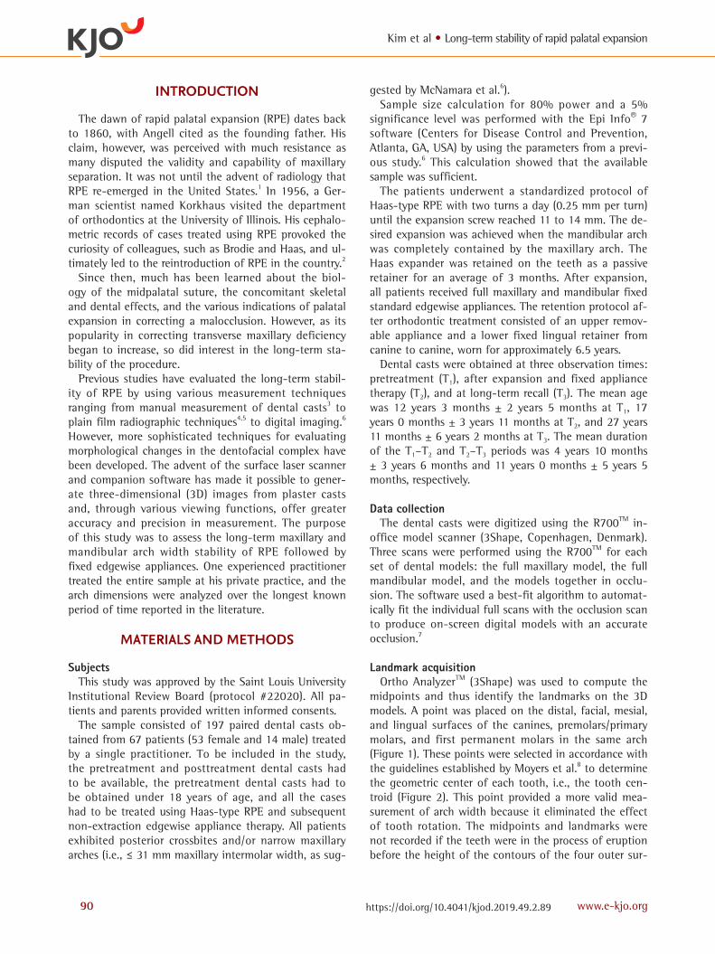
midpoints and thus identify the landmarks on the 3D models. A point was placed on the distal, facial, mesial, and lingual surfaces of the canines, premolars/primary molars, and first permanent molars in the same arch (Figure 1). These points were selected in accordance with the guidelines established by Moyers et al.8 to determine the geometric center of each tooth, i.e., the tooth cen-troid (Figure 2). This point provided a more valid mea-surement of arch width because it eliminated the effect of tooth rotation. The midpoints and landmarks were not recorded if the teeth were in the process of eruption before the height of the contours of the four outer sur-
Kim et al • Long-term stability of rapid palatal expansion
www.e-kjo.org 91https://doi.org/10.4041/kjod.2019.49.2.89
faces (mesial, distal, facial, and lingual) were visible.
MeasurementsThe Ortho AnalyzerTM software was also used to mea-
sure arch width at the following teeth: primary canines/permanent canines, first primary molars/first premolars, second primary molars/second premolars, and first per-
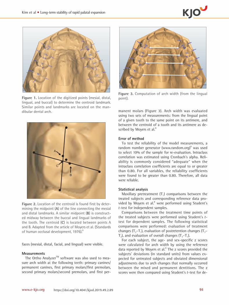
manent molars (Figure 3). Arch width was evaluated using two sets of measurements: from the lingual point of a given tooth to the same point on its antimere, and between the centroid of a tooth and its antimere as de-scribed by Moyers et al.8
Error of methodTo test the reliability of the model measurements, a
random number generator (www.random.org)9 was used to select 10% of the sample for re-evaluation. Intraclass correlation was estimated using Cronbach’s alpha. Reli-ability is commonly considered “adequate” when the intraclass correlation coefficients are equal to or greater than 0.80. For all variables, the reliability coefficients were found to be greater than 0.80. Therefore, all data were reliable.
Statistical analysisMaxillary pretreatment (T1) comparisons between the
treated subjects and corresponding reference data pro-vided by Moyers et al.8 were performed using Student’s t-test for independent samples.
Comparisons between the treatment time points of the treated subjects were performed using Student’s t-test for dependent samples. The following statistical comparisons were performed: evaluation of treatment changes (T2–T1), evaluation of postretention changes (T3–T2), and evaluation of overall changes (T3–T1).
For each subject, the age- and sex-specific z scores were calculated for arch width by using the reference data reported by Moyers et al.8 The z scores provided the subjects’ deviations (in standard units) from values ex-pected for untreated subjects and obviated dimensional adjustments due to arch changes that normally occurred between the mixed and permanent dentitions. The z scores were then compared using Student’s t-test for de-
Figure 1. Location of the digitized points (mesial, distal, lingual, and buccal) to determine the centroid landmark. Similar points and landmarks are located on the man-dibular dental arch.
Figure 2. Location of the centroid is found first by deter-mining the midpoint (A) of the line connecting the mesial and distal landmarks. A similar midpoint (B) is construct-ed midway between the buccal and lingual landmarks of the tooth. The centroid (C) is located between points A and B. Adapted from the article of Moyers et al. (Standards of human occlusal development, 1976).8
Figure 3. Computation of arch width (from the lingual point).

Kim et al • Long-term stability of rapid palatal expansion
www.e-kjo.org92 https://doi.org/10.4041/kjod.2019.49.2.89
pendent samples. The following statistical comparisons were performed: z score evaluation of treatment changes (T2–T1), z score evaluation of postretention changes (T3–T2), and z score evaluation of overall changes (T3–T1).
RESULTS
Maxillary archAt T1, the maxillary arch widths of the treated pa-
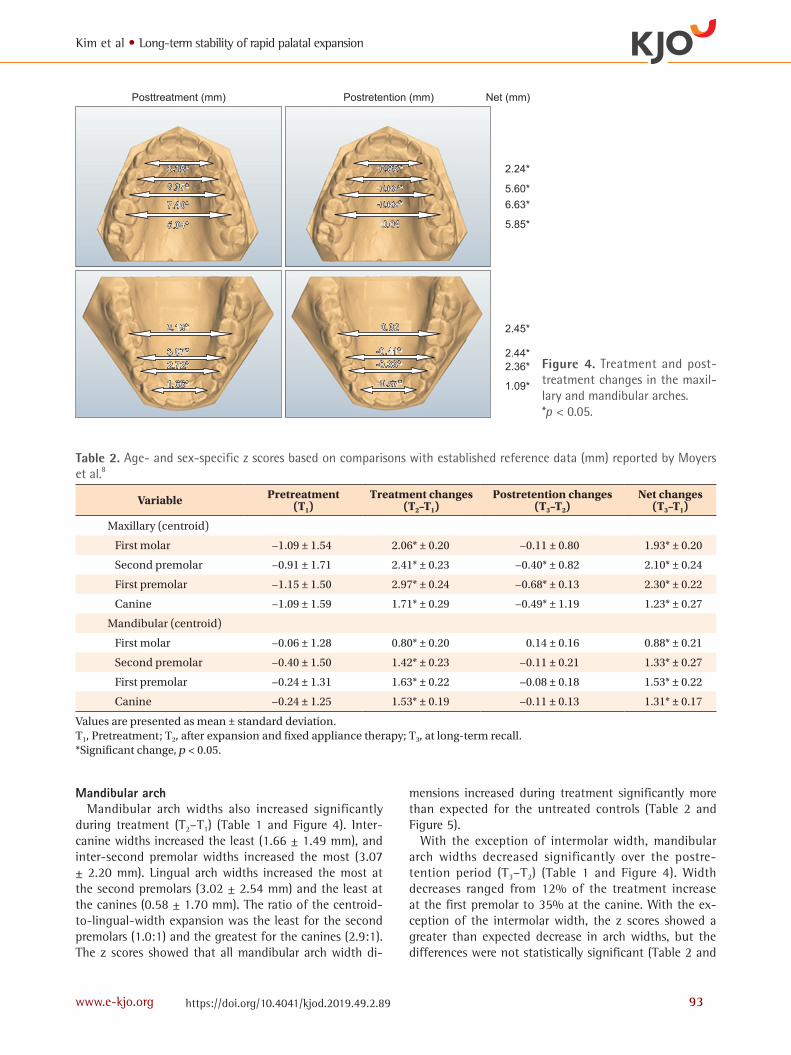
tients were significantly narrower than the correspond-ing dental arch widths of the established reference data. Maxillary arch widths increased significantly (p < 0.05) during treatment (T2–T1) (Table 1 and Figure 4). At the centroid level, intercanine widths increased the least (3.18 ± 2.47 mm) and inter-second premolar widths in-creased the most (7.40 ± 2.56 mm). Lingual arch widths followed a similar pattern, increasing the most at the second premolars (7.10 ± 2.43 mm) and the least at the canines (1.56 ± 2.60 mm). The ratio of the centroid width increase to the corresponding lingual width in-crease, which provides a rough measure of tipping, was the greatest for the canines (2.0:1) and the least for the
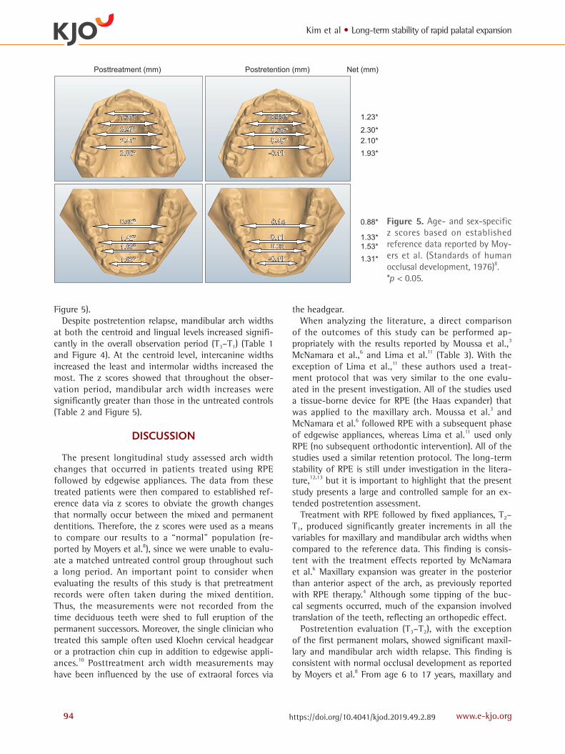
second premolars (1.0:1). The z scores, adjusted for age and sex, showed significant treatment-related increases in arch width at the centroid (Table 2 and Figure 5).
With the exception of intermolar width, maxillary arch widths decreased significantly postretention (T3–T2) (Table 1 and Figure 4). Width decreases ranged from 9% of the treatment-related increase at the second pre-molars to 27% of the treatment-related increase at the canines. Compared to untreated reference data (Table 2 and Figure 5), with the exception of the first molar, arch width significantly decreased more than was expected posttreatment. Arch width at the first molar decreased more than expected; however, the difference was not statistically significant.
When considering the net change of treatment (T3–T1), maxillary arch widths at both the centroid and lingual levels increased significantly (Table 1 and Figure 4). At the centroid level, intercanine widths increased the least and inter-second premolar widths increased the most. The z scores were greater than expected and significant for the overall observation period (Table 2 and Figure 5).
Table 1. Maxillary and mandibular arch width (lingual and centroid) changes (mm)
Variable Pretreatment(T1)
Treatment changes(T2–T1)
Postretention changes (T3–T2)
Net changes(T3–T1)
Maxillary (centroid)
First molar 41.37 ± 3.78 6.04* ± 2.90 0.04 ± 1.20 5.85* ± 2.89
Second premolar 36.33 ± 3.55 7.40* ± 2.56 −0.66* ± 1.19 6.63* ± 2.75
First premolar 32.52 ± 3.02 6.37* ± 2.25 −0.68* ± 1.00 5.60* ± 2.38
Canine 29.16 ± 2.74 3.18* ± 2.47 −0.85* ± 1.50 2.24* ± 2.38
Maxillary (lingual)
First molar 31.58 ± 3.74 4.86* ± 2.83 0.22 ± 1.35 4.89* ± 0.35
Second premolar 27.38 ± 3.56 7.10* ± 2.43 −0.47* ± 1.07 6.41* ± 0.32
First premolar 23.59 ± 2.66 5.72* ± 1.62 −0.42* ± 0.95 5.23* ± 0.22
Canine 24.10 ± 2.90 1.56* ± 2.60 −0.61* ± 1.67 1.23* ± 0.42
Mandibular (centroid)
First molar 41.24 ± 2.89 2.19* ± 2.19 0.32 ± 1.34 2.45* ± 2.11
Second premolar 35.68 ± 2.95 3.07* ± 2.20 −0.41* ± 0.96 2.44* ± 2.05
First premolar 30.75 ± 2.38 2.72* ± 1.60 −0.33* ± 0.72 2.36* ± 1.65
Canine 24.26 ± 1.72 1.66* ± 1.49 −0.57* ± 0.65 1.09* ± 1.48
Mandibular (lingual)
First molar 32.42 ± 2.96 1.43* ± 2.61 0.61 ± 2.32 1.99* ± 0.36
Second premolar 28.36 ± 3.20 3.02* ± 2.54 −0.08* ± 2.08 2.63* ± 0.34
First premolar 24.65 ± 2.52 2.44* ± 1.86 −0.04* ± 1.02 2.38* ± 0.23
Canine 19.33 ± 1.92 0.58* ± 1.70 −0.42 ± 0.75 0.26 ± 0.23
Values are presented as mean ± standard deviation. T1, Pretreatment; T2, after expansion and fixed appliance therapy; T3, at long-term recall.*Significant change, p < 0.05.
Kim et al • Long-term stability of rapid palatal expansion
www.e-kjo.org 93https://doi.org/10.4041/kjod.2019.49.2.89
Mandibular archMandibular arch widths also increased significantly
during treatment (T2–T1) (Table 1 and Figure 4). Inter-canine widths increased the least (1.66 ± 1.49 mm), and inter-second premolar widths increased the most (3.07 ± 2.20 mm). Lingual arch widths increased the most at the second premolars (3.02 ± 2.54 mm) and the least at the canines (0.58 ± 1.70 mm). The ratio of the centroid-to-lingual-width expansion was the least for the second premolars (1.0:1) and the greatest for the canines (2.9:1). The z scores showed that all mandibular arch width di-
mensions increased during treatment significantly more than expected for the untreated controls (Table 2 and Figure 5).
With the exception of intermolar width, mandibular arch widths decreased significantly over the postre-tention period (T3–T2) (Table 1 and Figure 4). Width decreases ranged from 12% of the treatment increase at the first premolar to 35% at the canine. With the ex-ception of the intermolar width, the z scores showed a greater than expected decrease in arch widths, but the differences were not statistically significant (Table 2 and
Figure 4. Treatment and post-treatment changes in the maxil-lary and mandibular arches. *p < 0.05.
Table 2. Age- and sex-specific z scores based on comparisons with established reference data (mm) reported by Moyers et al.8
Variable Pretreatment(T1)
Treatment changes(T2–T1)
Postretention changes(T3–T2)
Net changes(T3–T1)
Maxillary (centroid)
First molar −1.09 ± 1.54 2.06* ± 0.20 −0.11 ± 0.80 1.93* ± 0.20
Second premolar −0.91 ± 1.71 2.41* ± 0.23 −0.40* ± 0.82 2.10* ± 0.24
First premolar −1.15 ± 1.50 2.97* ± 0.24 −0.68* ± 0.13 2.30* ± 0.22
Canine −1.09 ± 1.59 1.71* ± 0.29 −0.49* ± 1.19 1.23* ± 0.27
Mandibular (centroid)
First molar −0.06 ± 1.28 0.80* ± 0.20 0.14 ± 0.16 0.88* ± 0.21
Second premolar −0.40 ± 1.50 1.42* ± 0.23 −0.11 ± 0.21 1.33* ± 0.27
First premolar −0.24 ± 1.31 1.63* ± 0.22 −0.08 ± 0.18 1.53* ± 0.22
Canine −0.24 ± 1.25 1.53* ± 0.19 −0.11 ± 0.13 1.31* ± 0.17
Values are presented as mean ± standard deviation. T1, Pretreatment; T2, after expansion and fixed appliance therapy; T3, at long-term recall.*Significant change, p < 0.05.
Kim et al • Long-term stability of rapid palatal expansion
www.e-kjo.org94 https://doi.org/10.4041/kjod.2019.49.2.89
Figure 5).Despite postretention relapse, mandibular arch widths
at both the centroid and lingual levels increased signifi-cantly in the overall observation period (T3–T1) (Table 1 and Figure 4). At the centroid level, intercanine widths increased the least and intermolar widths increased the most. The z scores showed that throughout the obser-vation period, mandibular arch width increases were significantly greater than those in the untreated controls (Table 2 and Figure 5).
DISCUSSION
The present longitudinal study assessed arch width changes that occurred in patients treated using RPE followed by edgewise appliances. The data from these treated patients were then compared to established ref-erence data via z scores to obviate the growth changes that normally occur between the mixed and permanent dentitions. Therefore, the z scores were used as a means to compare our results to a “normal” population (re-ported by Moyers et al.8), since we were unable to evalu-ate a matched untreated control group throughout such a long period. An important point to consider when evaluating the results of this study is that pretreatment records were often taken during the mixed dentition. Thus, the measurements were not recorded from the time deciduous teeth were shed to full eruption of the permanent successors. Moreover, the single clinician who treated this sample often used Kloehn cervical headgear or a protraction chin cup in addition to edgewise appli-ances.10 Posttreatment arch width measurements may have been influenced by the use of extraoral forces via
the headgear. When analyzing the literature, a direct comparison
of the outcomes of this study can be performed ap-propriately with the results reported by Moussa et al.,3 McNamara et al.,6 and Lima et al.11 (Table 3). With the exception of Lima et al.,11 these authors used a treat-ment protocol that was very similar to the one evalu-ated in the present investigation. All of the studies used a tissue-borne device for RPE (the Haas expander) that was applied to the maxillary arch. Moussa et al.3 and McNamara et al.6 followed RPE with a subsequent phase of edgewise appliances, whereas Lima et al.11 used only RPE (no subsequent orthodontic intervention). All of the studies used a similar retention protocol. The long-term stability of RPE is still under investigation in the litera-ture,12,13 but it is important to highlight that the present study presents a large and controlled sample for an ex-tended postretention assessment.
Treatment with RPE followed by fixed appliances, T2–T1, produced significantly greater increments in all the variables for maxillary and mandibular arch widths when compared to the reference data. This finding is consis-tent with the treatment effects reported by McNamara et al.6 Maxillary expansion was greater in the posterior than anterior aspect of the arch, as previously reported with RPE therapy.4 Although some tipping of the buc-cal segments occurred, much of the expansion involved translation of the teeth, reflecting an orthopedic effect.
Postretention evaluation (T3–T2), with the exception of the first permanent molars, showed significant maxil-lary and mandibular arch width relapse. This finding is consistent with normal occlusal development as reported by Moyers et al.8 From age 6 to 17 years, maxillary and
Figure 5. Age- and sex-specific z scores based on established reference data reported by Moy-ers et al. (Standards of human occlusal development, 1976)8. *p < 0.05.
Kim et al • Long-term stability of rapid palatal expansion
www.e-kjo.org 95https://doi.org/10.4041/kjod.2019.49.2.89
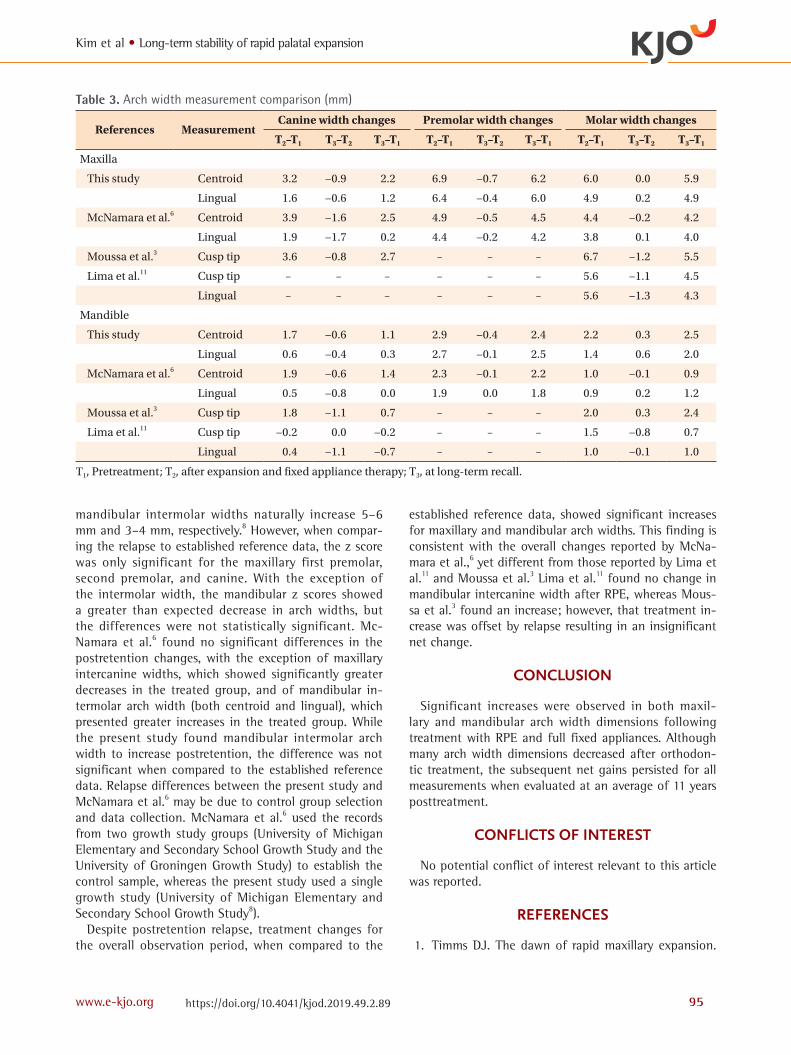
mandibular intermolar widths naturally increase 5–6 mm and 3–4 mm, respectively.8 However, when compar-ing the relapse to established reference data, the z score was only significant for the maxillary first premolar, second premolar, and canine. With the exception of the intermolar width, the mandibular z scores showed a greater than expected decrease in arch widths, but the differences were not statistically significant. Mc-Namara et al.6 found no significant differences in the postretention changes, with the exception of maxillary intercanine widths, which showed significantly greater decreases in the treated group, and of mandibular in-termolar arch width (both centroid and lingual), which presented greater increases in the treated group. While the present study found mandibular intermolar arch width to increase postretention, the difference was not significant when compared to the established reference data. Relapse differences between the present study and McNamara et al.6 may be due to control group selection and data collection. McNamara et al.6 used the records from two growth study groups (University of Michigan Elementary and Secondary School Growth Study and the University of Groningen Growth Study) to establish the control sample, whereas the present study used a single growth study (University of Michigan Elementary and Secondary School Growth Study8).
Despite postretention relapse, treatment changes for the overall observation period, when compared to the
established reference data, showed significant increases for maxillary and mandibular arch widths. This finding is consistent with the overall changes reported by McNa-mara et al.,6 yet different from those reported by Lima et al.11 and Moussa et al.3 Lima et al.11 found no change in mandibular intercanine width after RPE, whereas Mous-sa et al.3 found an increase; however, that treatment in-crease was offset by relapse resulting in an insignificant net change.
CONCLUSION
Significant increases were observed in both maxil-lary and mandibular arch width dimensions following treatment with RPE and full fixed appliances. Although many arch width dimensions decreased after orthodon-tic treatment, the subsequent net gains persisted for all measurements when evaluated at an average of 11 years posttreatment.
CONFLICTS OF INTEREST
No potential conflict of interest relevant to this article was reported.
REFERENCES
1. Timms DJ. The dawn of rapid maxillary expansion.
Table 3. Arch width measurement comparison (mm)
References MeasurementCanine width changes Premolar width changes Molar width changes
T2–T1 T3–T2 T3–T1 T2–T1 T3–T2 T3–T1 T2–T1 T3–T2 T3–T1
Maxilla
This study Centroid 3.2 −0.9 2.2 6.9 −0.7 6.2 6.0 0.0 5.9
Lingual 1.6 −0.6 1.2 6.4 −0.4 6.0 4.9 0.2 4.9
McNamara et al.6 Centroid 3.9 −1.6 2.5 4.9 −0.5 4.5 4.4 −0.2 4.2
Lingual 1.9 −1.7 0.2 4.4 −0.2 4.2 3.8 0.1 4.0
Moussa et al.3 Cusp tip 3.6 −0.8 2.7 – – – 6.7 −1.2 5.5
Lima et al.11 Cusp tip – – – – – – 5.6 −1.1 4.5
Lingual – – – – – – 5.6 −1.3 4.3
Mandible
This study Centroid 1.7 −0.6 1.1 2.9 −0.4 2.4 2.2 0.3 2.5
Lingual 0.6 −0.4 0.3 2.7 −0.1 2.5 1.4 0.6 2.0
McNamara et al.6 Centroid 1.9 −0.6 1.4 2.3 −0.1 2.2 1.0 −0.1 0.9
Lingual 0.5 −0.8 0.0 1.9 0.0 1.8 0.9 0.2 1.2
Moussa et al.3 Cusp tip 1.8 −1.1 0.7 – – – 2.0 0.3 2.4
Lima et al.11 Cusp tip −0.2 0.0 −0.2 – – – 1.5 −0.8 0.7
Lingual 0.4 −1.1 −0.7 – – – 1.0 −0.1 1.0
T1, Pretreatment; T2, after expansion and fixed appliance therapy; T3, at long-term recall.

Kim et al • Long-term stability of rapid palatal expansion
www.e-kjo.org96 https://doi.org/10.4041/kjod.2019.49.2.89
Angle Orthod 1999;69:247-50.2. Haas AJ. The treatment of maxillary deficiency
by opening the midpalatal suture. Angle Orthod 1965;35:200-17.
3. Moussa R, O'Reilly MT, Close JM. Long-term stabili-ty of rapid palatal expander treatment and edgewise mechanotherapy. Am J Orthod Dentofacial Orthop 1995;108:478-88.
4. Wertz RA. Skeletal and dental changes accompany-ing rapid midpalatal suture opening. Am J Orthod 1970;58:41-66.
5. Asanza S, Cisneros GJ, Nieberg LG. Comparison of Hyrax and bonded expansion appliances. Angle Or-thod 1997;67:15-22.
6. McNamara JA Jr, Baccetti T, Franchi L, Herberger TA. Rapid maxillary expansion followed by fixed ap-pliances: a long-term evaluation of changes in arch dimensions. Angle Orthod 2003;73:344-53.
7. Barry M. In-office digital study models. J Clin Or-thod 2011;45:385-9.
8. Moyers RE, vander Linden FP, Riolo ML, McNamara JA. Standards of human occlusal development.
Monograph 5. Craniofacial growth series. Ann Ar-bor: Center for Human Growth and Development, University of Michigan; 1976. 371 p.
9. Haahr M. True random number service [Internet]. Dublin: RANDOM.ORG; 1998 [cited 2014 May 4]. Available from: www.random.org.2010.
10. Haas AJ. Palatal expansion: just the beginning of dentofacial orthopedics. Am J Orthod 1970;57:219-55.
11. Lima AC, Lima AL, Filho RM, Oyen OJ. Spontaneous mandibular arch response after rapid palatal expan-sion: a long-term study on Class I malocclusion. Am J Orthod Dentofacial Orthop 2004;126:576-82.
12. Pinheiro FH, Garib DG, Janson G, Bombonatti R, de Freitas MR. Longitudinal stability of rapid and slow maxillary expansion. Dental Press J Orthod 2014;19:70-7.
13. Mohan CN, Araujo EA, Oliver DR, Kim KB. Long-term stability of rapid palatal expansion in the mixed dentition vs the permanent dentition. Am J Orthod Dentofacial Orthop 2016;149:856-62.