logan’s evidence-based diagnostic imaging pathways
TRANSCRIPT
Logan’s Evidence-Based Diagnostic Imaging Pathways
January 2014
1

These pathways are adapted with permission from the Educational Manual for Evidence-Based Chiropractic: Chapter 2 Diagnostic Imaging, Oregon Board of Chiropractic Examiners, 2005. We thank Norman Kettner, DC, DACBR, and Martha Kaeser, MA, DC, for updating the literature included in the document. The following Acknowledgements apply to the original document.
Acknowledgements We wish to acknowledge the hard work and expertise of the volunteers who comprised the steering committee, the seed panels that produced the seed statements, the nominal and Delphi panels who refined these statements, and the facilitators who conducted the consensus process. In addition we wish to thank the efforts of Meridel Gatterman who has served as process consultant, process manager, and compiler of the manuscript, and Kelly Bird and Dave McTeague who have edited the final copy. Diagnostic Imaging Seed Panel: Drs. Ann Goldeen DC, Gary Smith DC DACBR, Lisa Hoffman DC DACBR, Scott Conklin DC, Michael Underhill DC. Sectional seed panel: Peggy Seron DC DACBR, John Hyland DC DACBR DABCO MPH, Brian Enebo MS DC. Videofluoroscopy Seed Panel: Drs. Ann Goldeen DC, Don Ferrante DC, Alexe Bellingham DC, Beverly Harger DC DACBR, K.C. Snellgrove DC, Tyrone Wei DC DACBR. Facilitators: Drs. Cathy Cummins DC DACBR, John Colwell DC & Meridel Gatterman MA DC M.Ed. Nominal Panel Members: Drs. Jim Bartley, Paula Conklin, Thomas Freedland, Meridel Gatterman, Kevin Holzapfel, Sunny Kierstyn, Ron LeFebvre, John Noren, Christene Olshove, Bruce Pace, Don Peterson, David Saboe, LaVerne Saboe Jr., Steve Sebers. Steering Committee: Members (as of 6-3-05) Drs. David Day-Chair, Thomas Dobson, Kathleen Galligan, John Colwell and Meridel Gatterman.
2
DIAGNOSTIC IMAGING
INTRODUCTION The fundamental purpose of diagnostic imaging is to provide information to assist in the development of a diagnosis or otherwise impact the treatment plan. It is the responsibility of the chiropractic physician to keep abreast of advancements in diagnostic imaging (ie. http://www.radiologyeducation.com/). The chiropractic physician should strive to provide patient-centered and cost-effective imaging decisions.1 This pathway takes an evidence-informed approach to the utilization of diagnostic imaging in the assessment of chiropractic patients.
UTILIZATION OF RADIOGRAPHIC STUDIES Decisions to utilize any diagnostic imaging procedure should be based on a demonstrated need (i.e. clinical necessity determined by indications) following an adequate case history, physical examination and the provision of a differential diagnosis. Since there is no safe level of radiation exposure the use of radiography requires careful consideration of its risk-benefit. Plain radiography is the most frequently utilized diagnostic imaging test for the evaluation of the musculoskeletal system. It has reasonable sensitivity for a range of pathologies and functional disorders, ie. fractures, osteoarthritis, joint instability. However, negative radiographic exams do not exclude underlying pathology. Listed below are the indications and contraindications for radiography and are designed to assist in the decision-making process. All relevant clinical and historical information needs to be considered. In keeping with the tenets of evidence-based practice, decisions on the use of radiography depend on a combination of the best available evidence, patient values and preferences, and the practitioner's clinical judgment.2
PATHWAYS FOR CHIROPRACTIC UTILIZATION OF RADIOGRAPHY Indications3-7
• History of malignancy (with unexplained new symptoms)
• Significant trauma, recent trauma, repetitive trauma with significant clinical findings • Suspected fractures • Clinically significant neurologic signs and symptoms • Unexplained weight loss or gain • Unrelenting night pain • Suspicion or history of inflammatory arthritis with change in symptoms • Known or suspected bone density loss • Palpable mass • Substance abuse • Prolonged corticosteroid use • Fever of unknown origin (>100° F) • Suspected infection
3

• Abnormal laboratory finding (Erythrocyte Sedimentation Rate [ESR], or C-reactive protein [CRP], White Blood Cell Count [WBC], etc.)
• Recent surgery or invasive procedure related to chief complaint • Failure to improve without prior radiography • Patients over 50 years of age are at greater risk of having significant pathologies • Surgical history at area of chief complaint • Failed surgery • Postural abnormalities (scoliosis, lordosis, kyphosis) • Hyper/hypomobility • Segmental instability (traumatic, degenerative, post-operative)
Additional Indications
• Suspected physical abuse3 • Environmental exposure to toxic or infectious agents4 • Recent immigration or foreign travel4 • Medicolegal implications when combined with clinical indicators
Contraindications5
• Pregnancy3,6 • Financial gain • Patient education • Routine (habitual) radiographic screening and follow-up • Research without sanctioned review-board approval • Unnecessary duplication of services • Routine pre-employment screening • Routine discharge radiography
IMAGING MODALITIES
There are a number of imaging modalities available to the chiropractic physician to utilize in the diagnostic work-up and treatment of patients. The following will be a discussion emphasizing the advantages and disadvantages of modalities including plain film radiography, computed tomography (CT), magnetic resonance (MR) imaging, radionuclide imaging, PET, DEXA, and ultrasound. Plain Film Radiography7 The use of plain film radiography in the chiropractic profession began in 1910. It was initially used as a research tool and later as the imaging modality of choice for diagnosis of pathology as well as evaluation of postural and biomechanical disorders of the spinal column and pelvis. Use has expanded to include the appendicular skeleton. Plain radiography of the vertebral column may offer insight into pathology, indications and contraindications for chiropractic management, as well as postural and biomechanical disorders. The risk of exposure to ionizing radiation mandates that a thorough history, physical examination and differential diagnosis be performed prior to the decision to utilize radiography.
4
Chest and Abdomen Radiography Radiography of the chest and abdomen,8,9 are also utilized by the chiropractic physician. Radiography of the chest, thorax and abdomen may be necessary to evaluate for differential diagnoses that are contributory to the patient’s complaints. Minimal Standard Radiographic Views Standard projections are employed in plain radiography with the minimum of orthogonal projections (two views 90 degrees to each other). Additional views to the minimum diagnostic series may include oblique views, angulated spot views, and dynamic stress studies. Oblique projections evaluate the apophyseal joints of the cervical and lumbar spine as well as the intervertebral foramina (IVF) in the cervical spine. In the appendicular skeleton, oblique projections more fully demonstrate complex anatomy. The sacroiliac joints are more clearly demonstrated on the angulated projection than on any other projection. Dynamic stress views include flexion/extension and lateral bending of the cervical and lumbar spine. These studies reveal information related to the end range of motion and are indicated when segmental instability is suspected. Stress radiography may also be utilized to evaluate injured joints of the appendicular skeleton.
5
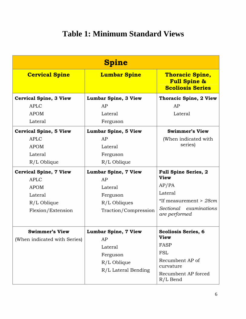
Table 1: Minimum Standard Views
Spine Cervical Spine Lumbar Spine Thoracic Spine,
Full Spine & Scoliosis Series
Cervical Spine, 3 View APLC APOM Lateral
Lumbar Spine, 3 View AP Lateral Ferguson
Thoracic Spine, 2 View AP Lateral
Cervical Spine, 5 View APLC APOM Lateral R/L Oblique
Lumbar Spine, 5 View AP Lateral Ferguson R/L Oblique
Swimmer’s View (When indicated with
series)
Cervical Spine, 7 View APLC APOM Lateral R/L Oblique Flexion/Extension
Lumbar Spine, 7 View AP Lateral Ferguson R/L Obliques Traction/Compression
Full Spine Series, 2 View AP/PA Lateral *If measurement > 28cm Sectional examinations are performed
Swimmer’s View (When indicated with Series)
Lumbar Spine, 7 View AP Lateral Ferguson R/L Oblique R/L Lateral Bending
Scoliosis Series, 6 View FASP FSL Recumbent AP of curvature Recumbent AP forced R/L Bend
6
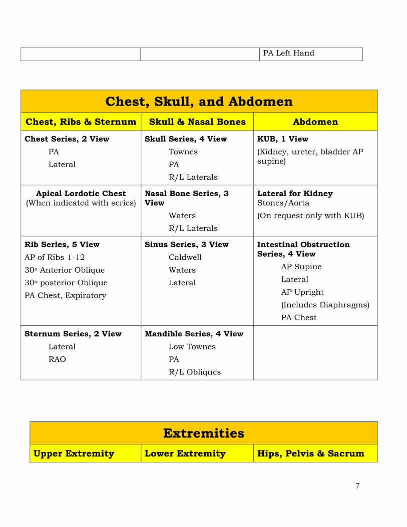
PA Left Hand
Chest, Skull, and Abdomen Chest, Ribs & Sternum Skull & Nasal Bones Abdomen
Chest Series, 2 View PA Lateral
Skull Series, 4 View Townes PA R/L Laterals
KUB, 1 View (Kidney, ureter, bladder AP supine)
Apical Lordotic Chest (When indicated with series)
Nasal Bone Series, 3 View
Waters R/L Laterals
Lateral for Kidney Stones/Aorta (On request only with KUB)
Rib Series, 5 View AP of Ribs 1-12 30o Anterior Oblique 30o posterior Oblique PA Chest, Expiratory
Sinus Series, 3 View Caldwell Waters Lateral
Intestinal Obstruction Series, 4 View
AP Supine Lateral AP Upright (Includes Diaphragms) PA Chest
Sternum Series, 2 View Lateral RAO
Mandible Series, 4 View Low Townes PA R/L Obliques
Extremities Upper Extremity Lower Extremity Hips, Pelvis & Sacrum
7
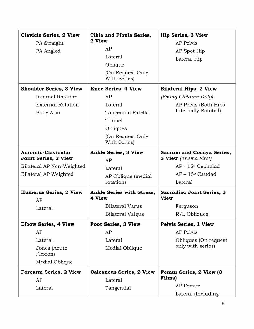
Clavicle Series, 2 View PA Straight PA Angled
Tibia and Fibula Series, 2 View
AP Lateral Oblique (On Request Only With Series)
Hip Series, 3 View AP Pelvis AP Spot Hip Lateral Hip
Shoulder Series, 3 View Internal Rotation External Rotation Baby Arm
Knee Series, 4 View AP Lateral Tangential Patella Tunnel Obliques (On Request Only With Series)
Bilateral Hips, 2 View (Young Children Only)
AP Pelvis (Both Hips Internally Rotated)
Acromio-Clavicular Joint Series, 2 View Bilateral AP Non-Weighted Bilateral AP Weighted
Ankle Series, 3 View AP Lateral AP Oblique (medial rotation)
Sacrum and Coccyx Series, 3 View (Enema First)
AP - 15o Cephalad AP – 15o Caudad Lateral
Humerus Series, 2 View AP Lateral
Ankle Series with Stress, 4 View
Bilateral Varus Bilateral Valgus
Sacroiliac Joint Series, 3 View
Ferguson R/L Obliques
Elbow Series, 4 View AP Lateral Jones (Acute Flexion) Medial Oblique
Foot Series, 3 View AP Lateral Medial Oblique
Pelvis Series, 1 View AP Pelvis Obliques (On request only with series)
Forearm Series, 2 View AP Lateral
Calcaneus Series, 2 View Lateral Tangential
Femur Series, 2 View (3 Films)
AP Femur Lateral (Including
8

Knee) Lateral Hip
Wrist Series, 4 View PA Hand Lateral Oblique (Pronated) PA with Ulnar Deviation
Toe Series, 3 View AP Foot Lateral Toe Oblique Toe (Marked)
Hand Series, 3 View
PA Hand Lateral (With Fingers Spread) Oblique
Leg Length (Scan-O-Gram), 6 View Right: AP Hip/Knee/Ankle Left: AP Hip/Knee/Ankle
Finger/Thumb Series, 3 View
PA Hand Lateral Affected Finger Oblique Finger
Finger Series (digits 2 through 5), 5 view AP Pronated oblique Lateral Supinated oblique PA
IMAGING OF BIOMECHANICAL ABNORMALITIES Radiography that includes appropriate views, when combined with clinical findings, is intended to provide a better understanding of the patient’s condition and to establish a diagnosis. Biomechanical analysis is used to determine misalignment, postural and motion abnormalities,
9

and to guide decisions including the use of manipulation or mobilization. Many radiographic lines, angles, and measurements have been demonstrated to be reliable indicators of postural and biomechanical abnormalities. Spinal Radiographic Analysis Most chiropractic methods of radiographic analysis have stressed the importance of assessing the patient in the upright, weight-bearing position. This allows for both full spine and regional postural evaluation, such as the knee joint in osteoarthritis. Specific consideration is given to the identification of abnormal spinal curvatures that may compromise efficient biomechanical function. Studies that evaluate the reliability, validity and clinical relevance of radiographic line drawing have produced conflicting evidence.10 Reliability Reliability is the repeatability of a measurement and indicates consistency and precision when a procedure is done by different examiners and at multiple times.11 Factors that influence the reliability of spinal radiographic analysis include: anatomic variants, positioning of patient, the person performing the study, and x-ray equipment. In addition to these and other potential sources of systematic error, random measurement error adversely affects the reliability of measurement methods. While inter-examiner reliability of the actual marking of x-rays has been demonstrated,12,13 the reliability of the entire procedure has not been established.11 Reliability does not establish the clinical relevance or validity of measurement procedures. Validity and Clinical Efficacy Validity refers to how accurately an assessment procedure measures, identifies or predicts the true state of the patient. While construct validity (a measure of the theoretical concept of x-ray line marking) has been evaluated,13 the predictive validity (the clinical relevance of x-ray line marking, i.e. can it identify current spine problems, predict future occurrences, or measure resolution) has not been established through well-designed clinical trials.14 Predictive validity is crucial; it is far more relevant than construct validity or reliability tests in establishing the clinical efficacy of assessment procedures. Functional Radiographic Analysis Functional radiographs are practical tools for the evaluation of spinal segmental motion. Functional radiography may be used to evaluate the segmental range of motion by comparing the neutral position to the end range of movement in either the sagittal or coronal planes. However, clinical information may be lost when the information from the neutral position is not included in the assessment. The key to accurately evaluating motion on functional spinal radiographs is precise standards of patient positioning. Meticulous attention to the details of positioning cannot be overemphasized if the information obtained from the resultant radiography is to be considered a reliable assessment of that particular patient’s function. Functional radiographic studies have
10

traditionally been performed with active movement by the patient. The reliability and clinical validation of cervical flexion/extension studies have been demonstrated.15 Full Spine Radiography16 Depending on history and clinical findings, the need for full spine radiography is based on the clinical judgment of the doctor. The choice of sectional or full spine views is dependent on clinical necessity and the ability to produce diagnostic quality radiographs. AP/PA and lateral exams with a central ray measurement over 28 cm should be performed a sectional exam because larger patients will result in non-diagnostic images17. AP/PA full spine radiographs are used for evaluation of pathology and biomechanical analysis. Single exposure, lateral full spine radiographs are not recommended.7
The use of full spine radiography is of value when clinical findings indicate the involvement of multiple spinal levels. In the following circumstances PA full spine radiography may be preferred over sectional radiography:
• cases in which clinical examination disclosed the need for radiography of several spinal sections;
• cases in which severe postural distortions are evident, (scoliosis) after clinical assessment;
• cases in which a mechanical problem in one spinal area adversely affects other regions; • to specifically evaluate complex biomechanical or postural disorders of the spine and
pelvis under weight-bearing conditions. Full spine radiography can be considered to be of diagnostic quality with less radiation exposure to the patient compared to sectionals of the multiple levels involved. This requires appropriate technology and optimal radiographic technique with careful attention to exposure factors and shielding. The evaluation of suspected pathology requires sectional series and/or spot views to attain better detail. Analysis of full spine radiography has been used to identify biomechanical disorders,, chiropractic subluxations and joint dysfunction. There are a variety of line marking systems used to evaluate radiographs. The validity and reliability of the full spine analytical systems has been studied with mixed results.
PATIENT SAFETY18
Patient safety in diagnostic imaging encompasses a range of activities performed before, during and after the actual imaging exam. The primary goal of these efforts is to provide the most clinically significant information with the lowest possible risk and cost to the patient. The following key areas should be addressed: patient education and informed consent, patient comfort, selection criteria, radiation safety, image quality control, facilities maintenance and record keeping.
11
Patient Education and Informed Consent The clinician should explain the diagnostic imaging procedures and follow up, the time and cost involved, risks and contraindications, and patient preparatory procedures. This should be done regardless of whether the clinician will perform the imaging or order it from another facility. Patient Comfort A clean, safe, comfortable environment should be provided for waiting, changing garments, securing personal items, and performing the imaging procedure. The privacy of the patient should be guarded during preparation for and execution of the exam, as well as with the storage of radiographic images and reports. Radiation Safety The most important aspect of patient safety is to minimize the radiation dose to the patient18. There is no known safe dose of ionizing radiation. Even the smallest dose may produce genetic damage. However, diagnostic imaging doses do not typically produce clinical manifestations. The benefit to the patient must outweigh the risk19-21 As Low As Reasonably Achievable (ALARA): Efforts should be made in all areas of the imaging procedure to provide the lowest possible dose to the patient without compromising image quality.19,22,23 Patient Selection Criteria The planned diagnostic imaging procedures must supply significant clinical information that cannot be otherwise determined. If the diagnosis, treatment or prognosis will not likely change based on imaging findings, the imaging is not appropriate. Every exposure, including post-treatment exposures and scanograms, must have clinical justification with adequate documentation consistent with the patient’s case history. Clinicians are responsible for ordering necessary and appropriate imaging studies. More than one study may be indicated to fully evaluate a patient. Previous imaging studies should be accessed if possible for interval change. Studies may be repeated if timely access to previous exams is not feasible, they are of poor quality or are not clinically relevant. Consultation with a radiologist may be helpful in determining which studies are most appropriate for a case.
ADVANCED IMAGING PROCEDURES The choice of an appropriate imaging modality depends on the patient’s differential diagnosis. A given patient may have specific needs or limitations that affect the choices of imaging modality. These factors and the continuing development of complex advanced imaging protocols make consultation with a radiologist invaluable prior to the selection of the advanced imaging procedure. The information provided here is intended as a general guide.5,24-31
12

Magnetic Resonance Imaging Magnetic resonance imaging (MRI) is a valuable diagnostic tool in neuromusculoskeletal imaging. Sectional images can be obtained through all body areas in axial (transverse), sagittal and coronal planes, or at oblique angles for smaller anatomical areas. No ionizing radiation is produced with MRI and risks to appropriately chosen patients have not been identified. The use of contrast exams should be carefully weighed in consultation with a radiologist as patients with renal insufficiency may develop nephrogenic systemic sclerosis from gadolinium exposure32. Patients with some pacemakers, some aneurysm clips, metallic foreign bodies, and other ferromagnetic artifacts are not appropriate candidates for MRI. In general, MRI images tissues based on their hydrogen atom content, reflecting total quantity and molecular bonds. Therefore, both free and intracellular water, and fat produce the majority of the MRI "signal" which creates the image. MRI is an excellent procedure for imaging the brain, spinal cord peripheral nervous system, intervertebral discs, articular cartilage, muscles, tendons, ligaments, menisci, and most organs. MRI is rarely used as the initial imaging procedure. In many cases, MRI will provide additional information after evaluation by plain radiography. MRI may be used as the initial study in cases of significant or rapidly progressing neurologic changes, especially those that indicate central nervous system (CNS) pathology. MRI is also useful as a follow-up imaging procedure after surgical treatment for intervertebral disc (IVD) herniation or neoplasm.
Computed Tomography Computed tomography (CT) combines the imaging physics of plain radiography with the advantages of sectional and tomographic imaging. Like plain film radiography, CT produces images through the interaction of x-ray photons with the tissues of the body, and is quite valuable in imaging osseous structures. CT also carries the same consideration of the potential harmful effects of ionizing radiation. The radiation dose should be kept as low as possible without losing diagnostic information and the risk-benefit ratio carefully weighed and discussed with a radiologist. Pathologies containing calcium densities may also be evaluated with CT. Some soft tissues, particularly of the chest and abdomen are best imaged with CT or ultrasound. Multidetector computed tomorgraphy (MDCT) acquires multiple slices per gantry rotation and has replaced single detector systems. Over the last 10-15 years, there has been a progressive increase in the number of detectors and slices, resulting in marked reduction of the scanning acquisition times and improved image quality. Most scanners operate with 16 or 64 detectors. Both the 16 slice and 64 slice CT scanners acquire data as isotropic voxels so images can be viewed in 3D with all imaging planes (axial, coronal, sagittal) displaying similar spatial resolution33. Computed tomography is used extensively, with and without intravenous or ingested contrast agents, for chest and abdomen examinations. It is superior to MRI in most scenarios for the chest and abdomen since the motion artifacts produced by heart contractions and bowel peristalsis may interfere with the acquisition of MRI images. Plain radiography, as scout films, will often be used for preliminary examination of the chest and abdomen before CT imaging.
13

CT provides detailed evaluation of fractures. This is particularly useful in unusually shaped bones or areas difficult to image with plain radiography such as the pelvis, craniovertebral junction, posterior elements of the spine, and ankle. CT may be combined with arthrography (contrast) when the differential list includes cartilaginous and bony abnormalities or when MRI is inconclusive, as in some cases of glenoid labrum tear. CT evaluation in the musculoskeletal system typically follows radiographic examination. CT is also used extensively, though less than MRI, in evaluation of the spine, spinal canal, and intervertebral discs. CT is superior to MRI in detailing significant osseous changes, but MRI is usually more valuable in evaluating the impact on neurologic structures. Myelography can improve the ability of CT (CTM) to evaluate neurologic structures, especially the thecal sac or nerve roots. In some cases, both procedures will be used to reach an accurate diagnosis and provide information for surgical planning. In cases where MRI is not available or not appropriate, CT, with or without myelography, is typically the imaging procedure of choice.
CT is also used to evaluate head and facial trauma where fracture and acute intracranial bleed are in the differential diagnosis. Radionuclide Imaging Radionuclide imaging of bone (also known as bone scan or skeletal scintigraphy) involves the intravenous administration of a radionuclide tagged to a phosphate analog, which is incorporated in the hydroxyapatite crystal of bone. Gamma rays emitted by the radionuclide are then detected quantitatively to produce an image. The image produced reflects blood flow and areas of increased bone production. Bone scan is much more sensitive than plain radiography for detecting osseous abnormalities but is distinctly nonspecific and would not be used as the only imaging procedure. It requires a complementary imaging exam, such as plain radiography or CT. A bone scan is typically used when the differential diagnosis raises the suspicion of skeletal disease. Since almost all pathologies of bone lead to some reactive bone growth, bone scan may be applicable in a wide variety of suspected pathologies. It is most commonly used in the detection of radiographically occult stress fractures, neoplasms, and infection. It is used extensively in the evaluation of skeletal metastasis since the entire skeleton can be imaged at once.
Single photon emission computerized tomography (SPECT) is a very useful method for displaying multiple planes of radionuclide activity. SPECT is especially useful to identify small areas of osseous pathology, particularly in the spine. Positron emission tomography (PET) utilizes a positron emitting radionuclide labeled with a biological agent, such as glucose, to localize the metabolism of tumor activity. It has become the workhorse of oncological imaging capable of staging a wide range of benign and malignant disorders and monitoring treatment responses34 Dual energy x-ray absorpiometry (DEXA) DEXA is the imaging modality of choice to evaluate bone mineral density and predict the probability of fractures in the lumbar spine and hip. DEXA is a low dose x-ray exam that does not require lead shielding for the patient. An examination of total body composition, i.e. android, gynoid fat percentage is also available35.
14
Diagnostic Ultrasound Diagnostic ultrasound (US) is a multiplanar (tomographic) imaging procedure that relies on the reflection or transmission of sound waves by body tissues for producing images. There is no ionizing radiation. The added capabilities of Doppler ultrasound allows for the quantification of flow rates in given structures, such as arteries. Among the most significant advantages of US are availability, low cost, noninvasiveness, and lack of known harmful effects. This procedure is used frequently in abdominal imaging where it is capable of determining organ size, organ masses, and in distinguishing between cystic, solid, and complex masses. It is typically the first imaging procedure chosen for thyroid abnormalities and can provide useful information in breast imaging. Diagnostic ultrasound is also increasing in use for musculoskeletal imaging and it is capable of detecting tears or hypertrophy in some of the commonly injured and more superficial soft tissue structures. Examples include rotator cuff tendon tears, peripheral neuropathy, deQuervain’s syndrome, joint instability (dynamic exams) and plantar fasciitis. It is very sensitive for the diagnosis of stress or conventional fractures. Superficial masses may also be initially evaluated by ultrasound. The large quantity of cartilage relative to bone in the pediatric skeleton, especially the very young, lends itself to evaluation by ultrasound. Research has evolved on using color Doppler ultrasonography to visualize lumbar artery blood flow36 and B-mode ultrasonography to assess segmental spinal motion in the lumbar spine37 and the relationship to low back pain.
15

PATHOLOGY
PLAIN FILM COMPUTED TOMOGRAPHY
MRI RADIONUCLIDE STUDY
ULTRASOUND CLINICAL CONSIDERATIONS
Table 2: Comparison of Imaging Procedures PATHOLOGY
PLAIN FILM
COMPUTED TOMOGRAPHY
MRI
RADIONUCLIDE STUDY
ULTRASOUND
CLINICAL CONSIDERATIONS
Muscle or tendon injury of extremities
Minimal use: May identify secondary effects, such as subluxation, gross disruption of Achilles’ and quadriceps tendons.
No routine use; may add info regarding associated osseous structures
Ideal imaging in most cases
No routine use Best imaging choice in some cases, particularly where structure is superficial (rotator cuff, Achilles’ tendon, quadriceps tendon, many muscles)
Imaging often not required; most useful in evaluating for suspected instability and the need for surgery
Ligamentous injury of extremities
May identify secondary effects such as subluxation stress studies may be diagnostic
No routine use; may add info regarding associated osseous structures
Ideal imaging in most cases
No routine use Best imaging choice where structure is superficial
Imaging often not required; most useful in evaluating for instability and need for surgery
Fibrocartilage injury
Offers little or no diagnostic information
Offers little or no diagnostic information
Imaging of choice in most cases
No routine use Limited specific applications
Arthroscopy may be the most appropriate procedure
Muscle, tendon or ligament injury of spine
May identify secondary effects such as subluxation, especially on stress studies.
No routine use; May add info regarding associated osseous structures
No routine use; gross soft tissue disruptions may be appreciated
No routine use Limited specific applications
PATHOLOGY
PLAIN FILM COMPUTED TOMOGRAPHY
MRI RADIONUCLIDE STUDY
ULTRASOUND CLINICAL CONSIDERATIONS
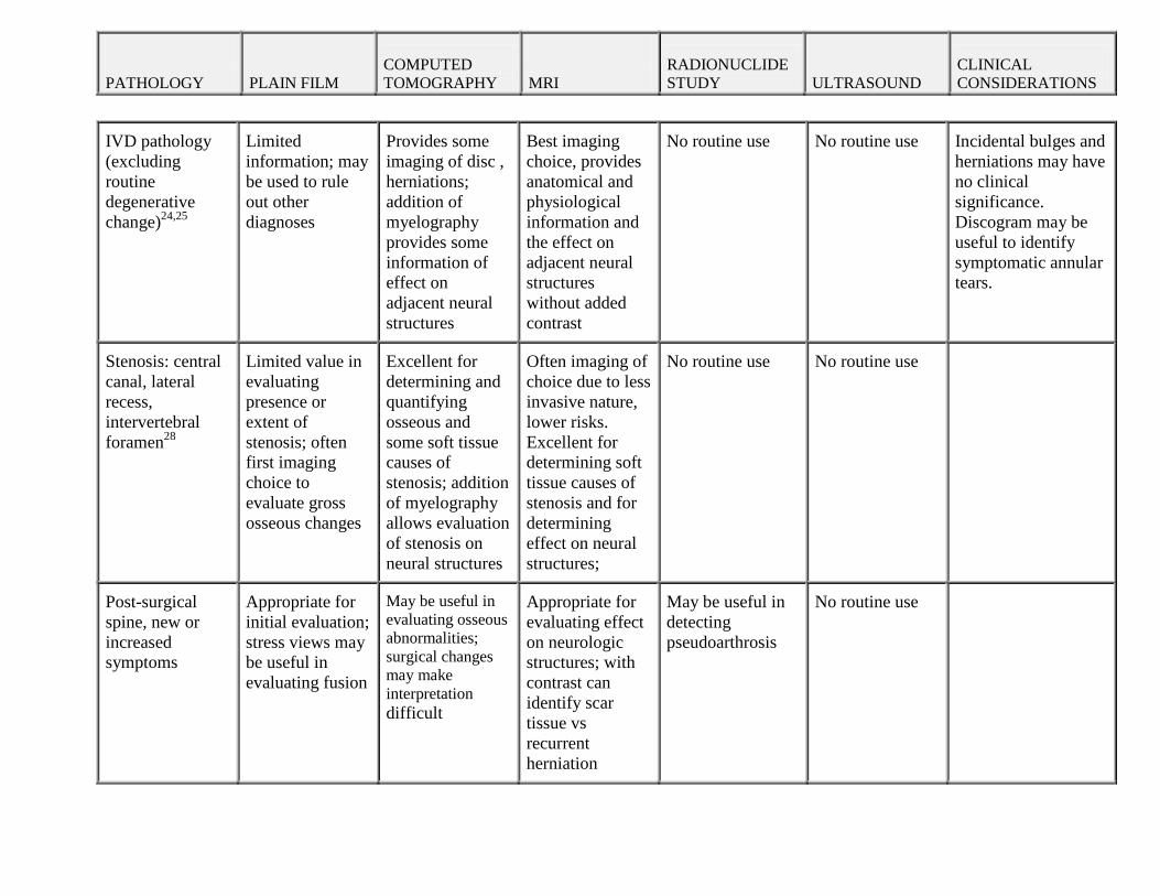
IVD pathology (excluding routine degenerative change)24,25
Limited information; may be used to rule out other diagnoses
Provides some imaging of disc , herniations; addition of myelography provides some information of effect on adjacent neural structures
Best imaging choice, provides anatomical and physiological information and the effect on adjacent neural structures without added contrast
No routine use No routine use Incidental bulges and herniations may have no clinical significance. Discogram may be useful to identify symptomatic annular tears.
Stenosis: central canal, lateral recess, intervertebral foramen28
Limited value in evaluating presence or extent of stenosis; often first imaging choice to evaluate gross osseous changes
Excellent for determining and quantifying osseous and some soft tissue causes of stenosis; addition of myelography allows evaluation of stenosis on neural structures
Often imaging of choice due to less invasive nature, lower risks. Excellent for determining soft tissue causes of stenosis and for determining effect on neural structures;
No routine use No routine use
Post-surgical spine, new or increased symptoms
Appropriate for initial evaluation; stress views may be useful in evaluating fusion
May be useful in evaluating osseous abnormalities; surgical changes may make interpretation difficult
Appropriate for evaluating effect on neurologic structures; with contrast can identify scar tissue vs recurrent herniation
May be useful in detecting pseudoarthrosis
No routine use
PATHOLOGY
PLAIN FILM COMPUTED TOMOGRAPHY
MRI RADIONUCLIDE STUDY
ULTRASOUND CLINICAL CONSIDERATIONS
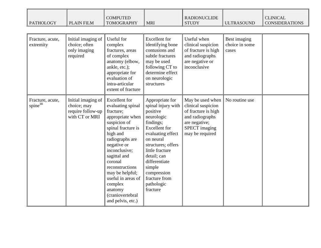
Fracture, acute, extremity
Initial imaging of choice; often only imaging required
Useful for complex fractures, areas of complex anatomy (elbow, ankle, etc.); appropriate for evaluation of intra-articular extent of fracture
Excellent for identifying bone contusions and subtle fractures may be used following CT to determine effect on neurologic structures
Useful when clinical suspicion of fracture is high and radiographs are negative or inconclusive
Best imaging choice in some cases
Fracture, acute, spine38
Initial imaging of choice; may require follow-up with CT or MRI
Excellent for evaluating spinal fracture; appropriate when suspicion of spinal fracture is high and radiographs are negative or inconclusive; sagittal and coronal reconstructions may be helpful; useful in areas of complex anatomy (craniovertebral and pelvis, etc.)
Appropriate for spinal injury with positive neurologic findings; Excellent for evaluating effect on neural structures; offers little fracture detail; can differentiate simple compression fracture from pathologic fracture
May be used when clinical suspicion of fracture is high and radiographs are negative; SPECT imaging may be required
No routine use

PATHOLOGY
PLAIN FILM COMPUTED TOMOGRAPHY
MRI RADIONUCLIDE STUDY
ULTRASOUND CLINICAL CONSIDERATIONS
Fracture, stress26 Initial imaging of choice; many will be radiographically occult, especially in early stages
May be used to determine extent; not usually required; may be useful for pars interarticularis
Sensitive to early changes; may be difficult to differentiate stress fracture from other pathologies
Appropriate for detection of radiographically occult, clinically suspected stress fracture; may require SPECT imaging, especially in the spine and other areas of complex osseous anatomy
Best imaging choice within extremities
Dislocation Most appropriate initial imaging
Useful if radiographic findings questionable; may be used for additional detail, especially to detect associated fracture
May be useful in detailing associated soft tissue injuries and/or effect on adjacent neurovascular structures
No routine use No routine use unless assessing neurovascular compromise post dislocation
Articular cartilage pathology
Depicts general cartilage loss; may show calcinosis secondary to crystal deposition; not effective for focal defects
No routine use Diagnostic in most cases; intra-articular contrast (MRI-arthrogram) may improve sensitivity
No routine use May identify chondral injury focal or diffuse depending on the region
PATHOLOGY
PLAIN FILM COMPUTED TOMOGRAPHY
MRI RADIONUCLIDE STUDY
ULTRASOUND CLINICAL CONSIDERATIONS
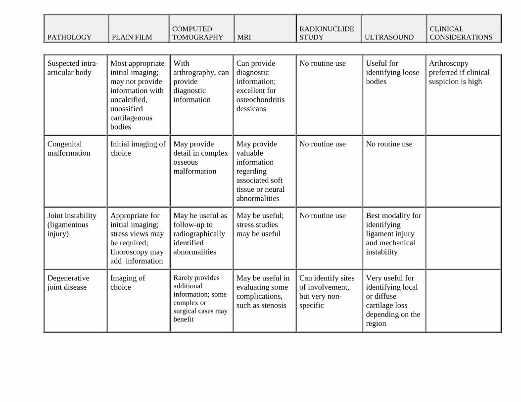
Suspected intra-articular body
Most appropriate initial imaging; may not provide information with uncalcified, unossified cartilagenous bodies
With arthrography, can provide diagnostic information
Can provide diagnostic information; excellent for osteochondritis dessicans
No routine use Useful for identifying loose bodies
Arthroscopy preferred if clinical suspicion is high
Congenital malformation
Initial imaging of choice
May provide detail in complex osseous malformation
May provide valuable information regarding associated soft tissue or neural abnormalities
No routine use No routine use
Joint instability (ligamentous injury)
Appropriate for initial imaging; stress views may be required; fluoroscopy may add information
May be useful as follow-up to radiographically identified abnormalities
May be useful; stress studies may be useful
No routine use Best modality for identifying ligament injury and mechanical instability
Degenerative joint disease
Imaging of choice
Rarely provides additional information; some complex or surgical cases may benefit
May be useful in evaluating some complications, such as stenosis
Can identify sites of involvement, but very non-specific
Very useful for identifying local or diffuse cartilage loss depending on the region

PATHOLOGY
PLAIN FILM COMPUTED TOMOGRAPHY
MRI RADIONUCLIDE STUDY
ULTRASOUND CLINICAL CONSIDERATIONS
Inflammatory arthritis27,29,39
Imaging of choice
Rarely provides additional information
Can detect some changes earlier than plain film
No routine use Best modality for extremity joint disease
Crystal deposition disease
Imaging of choice
More sensitive to calcium deposition, but rarely provides additional information
Can detect articular cartilage involvement
No routine use High sensitivity with low specificity
Infection38
Initial imaging of choice; radiographic latent period from several days to several weeks
May be useful as follow-up to radiographically identified abnormalities
Very sensitive; no significant latent period; useful in radiographically occult cases and to determine extent of involvement
Much more sensitive than plain film; non-specific; useful in cases of high clinical suspicion and negative radiographs
Sensitive but non-specific
Neoplasm, osseous38
Initial imaging of choice
May be useful as follow-up to radiographically identified abnormalities or in areas of complex anatomy
Very sensitive; may provide useful histologic information; useful in radiographically occult cases and to determine extent of involvement. Procedure of choice for multiple myeloma
Much more sensitive than plain film; non-specific; useful in cases of high clinical suspicion and negative radiographs, and to determine the extent of skeletal metastasis
No routine use Metastasis evaluation requires very specific protocols based on a number of patient variables
PATHOLOGY
PLAIN FILM COMPUTED TOMOGRAPHY
MRI RADIONUCLIDE STUDY
ULTRASOUND CLINICAL CONSIDERATIONS
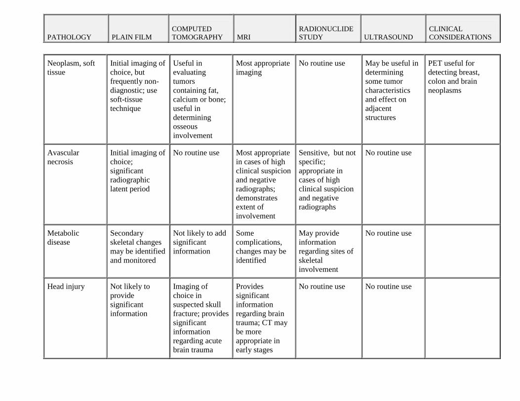
Neoplasm, soft tissue
Initial imaging of choice, but frequently non-diagnostic; use soft-tissue technique
Useful in evaluating tumors containing fat, calcium or bone; useful in determining osseous involvement
Most appropriate imaging
No routine use May be useful in determining some tumor characteristics and effect on adjacent structures
PET useful for detecting breast, colon and brain neoplasms
Avascular necrosis
Initial imaging of choice; significant radiographic latent period
No routine use Most appropriate in cases of high clinical suspicion and negative radiographs; demonstrates extent of involvement
Sensitive, but not specific; appropriate in cases of high clinical suspicion and negative radiographs
No routine use
Metabolic disease
Secondary skeletal changes may be identified and monitored
Not likely to add significant information
Some complications, changes may be identified
May provide information regarding sites of skeletal involvement
No routine use
Head injury
Not likely to provide significant information
Imaging of choice in suspected skull fracture; provides significant information regarding acute brain trauma
Provides significant information regarding brain trauma; CT may be more appropriate in early stages
No routine use No routine use

PATHOLOGY
PLAIN FILM COMPUTED TOMOGRAPHY
MRI RADIONUCLIDE STUDY
ULTRASOUND CLINICAL CONSIDERATIONS
Chronic sinus disease
Appropriate for initial evaluation; not as sensitive or specific as CT
Most appropriate imaging; initial imaging in most cases
May be used as follow-up to CT findings in unusual cases
No routine use No routine use
GI disease
Abdomen plain film does not provide adequate information in most scenarios; used as initial evaluation for suspected acute obstruction or perforation; barium studies may be diagnostic
Provides best imaging of many organs; frequently used with addition of barium
Useful for evaluation of some organs; presence of gas and intestinal motility often provides for poor imaging
Scans for specific organs can be useful
Frequently used in evaluation of abdominal disease; especially useful for solid organs and cystic abnormalities
GU disease
Frequently used as initial study, but usually requires additional imaging; addition of contrast often required
Often provides best imaging; usually includes contrast agent
Frequently useful; may not provide adequate imaging of some areas
No routine use Frequently used for evaluation of kidney and bladder disease
Peripheral Neuropathy
No routine use No routine use Useful when NCV is negative
No routine use Best imaging modality
REFERENCES
1. Haldeman S C-SD, Peterson DM,. Guidelines for chiropractic quality assurance and practice parameters. Aspen Publisher: Gaithersburg, MD; 1992.
2. Sackett DL, Rosenberg WM, Gray JA, Haynes RB, Richardson WS. Evidence based medicine: what it is and what it isn't. Bmj. Jan 13 1996;312(7023):71-72.
3. De Santis M, Di Gianantonio E, Straface G, et al. Ionizing radiations in pregnancy and teratogenesis: a review of literature. Reprod Toxicol. Sep-Oct 2005;20(3):323-329.
4. Downie A, Williams CM, Henschke N, et al. Red flags to screen for malignancy and fracture in patients with low back pain: systematic review. BMJ. 2013;347:f7095.
5. Chou R, Deyo RA, Jarvik JG. Appropriate use of lumbar imaging for evaluation of low back pain. Radiol Clin North Am. Jul 2012;50(4):569-585.
6. Ursprung WM, Howe JW, Yochum TR, Kettner NW. Plain film radiography, pregnancy, and therapeutic abortion revisited. J Manipulative Physiol Ther. Jan 2006;29(1):83-87.
7. Keating JC. James F. McGinnis, D.C., N.D., C.P. (1873-1947): spinographer, educator, marketer and bloodless surgeon. Chiropr Hist. Dec 1998;18(2):63-79.
8. ACR–SPR Practice Guideline for the Performance of Chest Radiography. http://www.acr.org/~/media/ACR/Documents/PGTS/
s/Chest_Radiography.pdf. 2011. 9. ACR–SPR Practice Guideline for the Performance of Abdominal Radiography.
http://www.acr.org/~/media/ACR/Documents/PGTS/guidelines/Abdominal_Radiography.pdf. 2011.
10. Malfair D, Flemming AK, Dvorak MF, et al. Radiographic evaluation of scoliosis: review. AJR. American journal of roentgenology. Mar 2010;194(3 Suppl):S8-22.
11. Haas M, Taylor JA, Gillette RG. The routine use of radiographic spinal displacement analysis: a dissent. J Manipulative Physiol Ther. May 1999;22(4):254-259.
12. Harrison DE, Harrison DD, Cailliet R, Troyanovich SJ, Janik TJ, Holland B. Cobb method or Harrison posterior tangent method: which to choose for lateral cervical radiographic analysis. Spine (Phila Pa 1976). Aug 15 2000;25(16):2072-2078.
13. Harrison DE, Holland B, Harrison DD, Janik TJ. Further reliability analysis of the Harrison radiographic line-drawing methods: crossed ICCs for lateral posterior tangents and modified Risser-Ferguson method on AP views. J Manipulative Physiol Ther. Feb 2002;25(2):93-98.
14. Gore DR. Roentgenographic findings in the cervical spine in asymptomatic persons: a ten-year follow-up. Spine (Phila Pa 1976). Nov 15 2001;26(22):2463-2466.
15. Dvorak J, Panjabi MM, Grob D, Novotny JE, Antinnes JA. Clinical validation of functional flexion/extension radiographs of the cervical spine. Spine (Phila Pa 1976). Jan 1993;18(1):120-127.
16. Taylor JA. Full-spine radiography: a review. J Manipulative Physiol Ther. Sep 1993;16(7):460-474.
17. Peterson C.K. HW, Grace K., Lumsden R., Tanner L. Comparison of full-spine film quality with patient measurement, gender and age. Journal of the American Chiropractic Association. 2006;43(9):9-12.
18. Fazel R, Krumholz HM, Wang Y, et al. Exposure to low-dose ionizing radiation from medical imaging procedures. N Engl J Med. Aug 27 2009;361(9):849-857.
19. Johnston DA, Brennan PC. Reference dose levels for patients undergoing common diagnostic X-ray examinations in Irish hospitals. Br J Radiol. Apr 2000;73(868):396-402.
20. Rommens C, Ringeard C, Hubert P. Exposure of red bone marrow to ionising radiation from natural and medical sources in France. J Radiol Prot. Sep 2001;21(3):209-219.
21. Williams JR, Catling MK. An investigation of X-ray equipment factors influencing patient dose in radiography. Br J Radiol. Nov 1998;71(851):1192-1198.
22. Mettler FA, Jr., Huda W, Yoshizumi TT, Mahesh M. Effective doses in radiology and diagnostic nuclear medicine: a catalog. Radiology. Jul 2008;248(1):254-263.
23. Mettler FA, Jr., Bhargavan M, Faulkner K, et al. Radiologic and nuclear medicine studies in the United States and worldwide: frequency, radiation dose, and comparison with other radiation sources--1950-2007. Radiology. Nov 2009;253(2):520-531.
24. Albeck MJ, Hilden J, Kjaer L, et al. A controlled comparison of myelography, computed tomography, and magnetic resonance imaging in clinically suspected lumbar disc herniation. Spine (Phila Pa 1976). Feb 15 1995;20(4):443-448.
25. Herzog RJ. The radiologic assessment for a lumbar disc herniation. Spine (Phila Pa 1976). Dec 15 1996;21(24 Suppl):19S-38S.
26. Kaiser JA, Holland, B.A. Using imaging studies in the diagnosis of low back pain. . Journal of Musculoskeletal Medicine 1995(July):20-35.
27. Keat A. ABC of rheumatology. Spondyloarthropathies. BMJ. May 20 1995;310(6990):1321-1324.
28. Postacchini F. Surgical management of lumbar spinal stenosis. Spine (Phila Pa 1976). May 15 1999;24(10):1043-1047.
29. Rubin DA. The radiology of early arthritis. Semin Roentgenol. Jul 1996;31(3):185-197. 30. Wilmink JT. MR Myelography in Patients with Lumbosacral Radicular Pain: Diagnostic
Value and Technique. Neuroradiol J. Aug 31 2011;24(4):570-576. 31. Perez FA, Jarvik JG. Evidence-based imaging and effective utilization: lessons in
neuroradiology. Neuroimaging Clin N Am. Aug 2012;22(3):467-476. 32. Marckmann P, Skov L, Rossen K, et al. Nephrogenic systemic fibrosis: suspected
causative role of gadodiamide used for contrast-enhanced magnetic resonance imaging. J Am Soc Nephrol. Sep 2006;17(9):2359-2362.
33. Prokop M. General principles of MDCT. Eur J Radiol. Mar 2003;45 Suppl 1:S4-10. 34. Basu S, Alavi A. Unparalleled contribution of 18F-FDG PET to medicine over 3 decades.
J Nucl Med. Oct 2008;49(10):17N-21N, 37N. 35. Lorente Ramos RM, Azpeitia Arman J, Arevalo Galeano N, Munoz Hernandez A, Garcia
Gomez JM, Gredilla Molinero J. Dual energy X-ray absorptimetry: fundamentals, methodology, and clinical applications. Radiologia. Sep-Oct 2012;54(5):410-423.
36. Espahbodi S, Dore CJ, Humphries KN, Hughes SP. Color Doppler ultrasonography of lumbar artery blood flow in patients with low back pain. Spine (Phila Pa 1976). Feb 15 2013;38(4):E230-236.
37. Chleboun GS, Amway MJ, Hill JG, Root KJ, Murray HC, Sergeev AV. Measurement of segmental lumbar spine flexion and extension using ultrasound imaging. J Orthop Sports Phys Ther. Oct 2012;42(10):880-885.
38. Staiger TO, Paauw DS, Deyo RA, Jarvik JG. Imaging studies for acute low back pain. When and when not to order them. Postgrad Med. Apr 1999;105(4):161-162, 165-166, 171-162.
39. McQueen FM. Imaging in early rheumatoid arthritis. Best Pract Res Clin Rheumatol. Aug 2013;27(4):499-522.