localized olfactory representation in mushroom bodies of … · 2010-05-26 · liria m....
TRANSCRIPT

Localized olfactory representation in mushroombodies of Drosophila larvaeLiria M. Masuda-Nakagawaa,1, Nanae Gendreb, Cahir J. O’Kanec, and Reinhard F. Stockerb
aInstitute of Molecular and Cellular Biosciences, University of Tokyo, Yayoi 1-1-1, Bunkyo-ku, Tokyo 113-0032, Japan; bDepartment of Biology, University ofFribourg, Chemin du Musee 10, CH-1700 Fribourg, Switzerland; and cDepartment of Genetics, University of Cambridge, Downing Street, Cambridge CB23EH, United Kingdom
Edited by Obaid Siddiqi, Tata Institute for Fundamental Research, Bangalore, India, and approved May 4, 2009 (received for review January 7, 2009)
Odor discrimination in higher brain centers is essential for behav-ioral responses to odors. One such center is the mushroom body(MB) of insects, which is required for odor discrimination learning.The calyx of the MB receives olfactory input from projectionneurons (PNs) that are targets of olfactory sensory neurons (OSNs)in the antennal lobe (AL). In the calyx, olfactory information istransformed from broadly-tuned representations in PNs to sparserepresentations in MB neurons (Kenyon cells). However, the extentof stereotypy in olfactory representations in the calyx is unknown.Using the anatomically-simple larval olfactory system of Drosoph-ila in which odor ligands for the entire set of 21 OSNs are known,we asked how odor identity is represented in the MB calyx. We firstmapped the projections of all larval OSNs in the glomeruli of theAL, and then followed the connections of individual PNs from theAL to different calyx glomeruli. We thus established a comprehen-sive olfactory map from OSNs to a higher olfactory associationcenter, at a single-cell level. Stimulation of single OSNs evokedstrong neuronal activity in 1 to 3 calyx glomeruli, showing thatbroadening of the strongest PN responses is limited to a few calyxglomeruli. Stereotypic representation of single OSN input in calyxglomeruli provides a mechanism for MB neurons to detect anddiscriminate olfactory cues.
calyx � genetically-encoded calcium indicator � olfactory sensory neurons �projection neurons
We now understand much about how odor information isdetected and conveyed to the brain by sensory neurons,
but less about how this information is represented in higher braincenters to influence behavioral outputs. The mushroom body(MB) of insects, which in Drosophila is essential for odordiscrimination learning, provides a model to understand olfac-tory coding in higher association centers (1, 2). In the periphery,odor identity is detected by sets of olfactory sensory neurons(OSNs) whose specificities are determined by the olfactoryreceptor (OR) that they express (3, 4). OSNs that express thesame OR converge on defined glomeruli in the first olfactorycenter of the brain, the antennal lobe (AL), analogously to theconvergence of OSNs on olfactory bulb glomeruli in mammals.Projection neurons (PNs) then carry olfactory information fromsingle AL glomeruli to the higher brain, the MB, and the lateralhorn. However, excitatory interneurons that innervate multipleAL glomeruli lead to broadening of PN specificity comparedwith OSNs (5, 6). PNs then connect to Kenyon cells (KCs) in thecalyx of the MB, where the representation of odor qualities isradically transformed; individual KCs respond much more se-lectively to odors than do either OSNs or PNs (7–9).
The extent of stereotypy in olfactory processing in the calyxhas been a matter of debate, in contrast to the clearly stereotypicconnections between OSNs and PNs in the AL. In Drosophilaadults, apparently stereotypic projections of PNs and KCs havebeen defined anatomically only at the level of broad zones(10–12), and odors can evoke localized PN activity in largeregions of the calyx (13). However, at least some subsets of KCsshow apparently nonstereotypic responses to odors (9). The level
of spatial stereotypy of odor representations in PNs in the calyxhas not yet been functionally defined, and the complexity of theadult calyx makes it difficult to visualize the representation ofodor qualities in single identified cells.
Drosophila larvae, which can perceive a wide variety of odors(14) and perform odor discrimination learning (15, 16), have anolfactory system with the same basic architecture as adults butnumerically much simpler. It contains only 21 unique OSNs (17),each expressing a different OR (3, 4) with known ligand spec-ificity (18). Each AL glomerulus appears to be connected to theMB calyx by a single PN (19). Whereas the adult calyx consistsof hundreds of microglomeruli that cannot be individuallyidentified, the larval calyx is organized in �34 glomeruli, in eachof which a single identifiable PN contacts hundreds of KCdendrites (20). Therefore, the larval olfactory system comprisesa small number of identifiable odor quality channels, eachcarried by a single neuron between successive layers of theolfactory pathway.
Here, we use the simple larval calyx to ask how individual odorqualities are represented in PN terminals and individual calyxglomeruli. We first generated a 3D map of OSN projections inlarval AL glomeruli. Using this map, we determined the con-nectivity of individual PNs between specific AL glomeruli andcalyx glomeruli. Although the 1-to-1 connectivity between OSNsand PNs in AL glomeruli predicts that activity in each OSNshould stimulate a single calyx glomerulus, it is not easy topredict how many calyx glomeruli are activated as a result ofbroadening of PN responses relative to OSNs. By carrying outthe imaging of odor-evoked activity in single PN terminals in theintact larval MB, we detect only limited dispersal of olfactorysignals during their transmission from single OSNs to the calyx.
Results3D Map of the Larval AL Based on OSN Projections. To identify themain sensory pathways from OSNs into PNs in the larval AL, wefirst generated a 3D map of AL glomeruli, defined by theinnervation patterns of larval OSNs. We analyzed confocalsections of the AL projections of 22 OrX-GAL4 lines that areeach expressed in a single OSN (3, 4). Terminals of single OSNseach ramified within a single AL glomerulus into branches andvaricosities of variable number, density, size, and shape (Fig.1A). The arrangement of OSN presynaptic terminals withinglomeruli was confirmed by visualizing terminals of all OSNsusing n-syb::GFP (Fig. 1B). In contrast, PN dendrites filled upmost of the volume of AL glomeruli in which they arborized(Fig. 1C).
Author contributions: L.M.M.-N., C.J.O., and R.F.S. designed research; L.M.M.-N., N.G., andR.F.S. performed research; C.J.O. contributed new reagents/analytic tools; L.M.M.-N., N.G.,C.J.O., and R.F.S. analyzed data; and L.M.M.-N., C.J.O., and R.F.S. wrote the paper.
The authors declare no conflict of interest.
This article is a PNAS Direct Submission.
1To whom correspondence should be addressed. E-mail: [email protected].
This article contains supporting information online at www.pnas.org/cgi/content/full/0900178106/DCSupplemental.
10314–10319 � PNAS � June 23, 2009 � vol. 106 � no. 25 www.pnas.org�cgi�doi�10.1073�pnas.0900178106

In general, glomerular position, shape and size were con-served between individuals (Fig. S1). The average center posi-tions of 21 glomeruli were mapped by calculating the relativepositions of OSN terminals within a virtual grid constructedfrom the average dimensions of the larval AL (Figs. S1–S3 andTable S1). Most of the glomeruli could be identified by anti-Dlgneuropile labeling that showed them as discrete structures atconserved locations, even though the borders were not alwaysclearly defined (Fig. 1D Fig. S4, and Movie S1). Based on thetypical terminal positions of each OSN, and visualization ofglomerular outlines using anti-Dlg, we prepared a 3D glomerularmodel that allows inspection of the larval AL from differentangles (Fig. 1E, Movies S2 and S3) and as a series of sectionsalong the antero-posterior axis (Fig. S4).
Arborization of PN Dendrites in Single AL Glomeruli. We next testedwhether PN dendritic arbors could be mapped onto specific AL
glomeruli. Labeled PN clones were generated by FLP-out (21)in the GH146-GAL4 line (Fig. 2A), reported to label �16–18larval PNs (22). GH146-expressing PNs arborized throughoutmost AL glomeruli except for 67b and 74a (Fig. 1C), suggestingthat the arborizations of PNs in 19 glomeruli could be studied bythis approach. Arborizations of 142 labeled PNs in the AL clearlymatched the glomerular organization of OSN terminals, thusallowing the identification of 19 classes of PN according to theiroverlap with the terminals of specific OSNs (examples in Fig. S5).PN arborizations were distributed more uniformly within glomer-uli, covered a larger volume than OSN terminals, and sometimessent collateral dendrites into neighboring glomeruli (Fig. S5).
Representation of OSN Input by PNs in the MB Calyx. To understandhow the sensory map of the larval AL is represented in calyxglomeruli, we used the GH146 PN clones described above. Weidentified calyx glomeruli by using published criteria of their
Fig. 1. 3D organization of the larval antennal lobe. All AL panels in this and subsequent figures show frontal confocal sections through the AL, with dorsalto the top, lateral to the left. (A) OSN terminals, labeled by expression of GFP under control of Or45b-GAL4, form varicosities within a single AL glomerulus (partlyoutlined by a broken line). (B) Expression of the synaptic vesicle marker n-syb::GFP in OSN presynaptic terminals using Or83b-GAL4, is found in varicosities withinglomeruli (outlined by broken lines and labeled using anti-Dlg). (C) Arborization of GH146 PN dendrites in a posterior section of a larval AL, in which mCD8::GFPis expressed using GH146-GAL4. Two glomeruli subsequently identified as 67b and 74a (broken lines) are not labeled by GH146. (D) Confocal sections fromsuccessive anteroposterior (AP) levels (from left to right, increments of 4 �m) of a representative larval AL, stained by anti-Dlg. Although glomeruli are not alwayscompletely separated from each other, prominent glomeruli are distinct (broken lines). For a complete AP sequence and for panels free of annotations, see Fig.S4. (E) 3D model of the larval AL, viewed from different angles. A, P, M, and L denote anterior, posterior, medial, and lateral faces of the AL, respectively. Fullrotations of the model are in Movie S2 and Movie S3. Colors of glomeruli denote predicted responses to broad classes of odorant (18): green, aromatic; red,broad-range aliphatic, principally esters and ketones; blue, esters; brown, alcohols; yellow, inhibitory responses; pale blue, no known responses.
Masuda-Nakagawa et al. PNAS � June 23, 2009 � vol. 106 � no. 25 � 10315
NEU
ROSC
IEN
CE
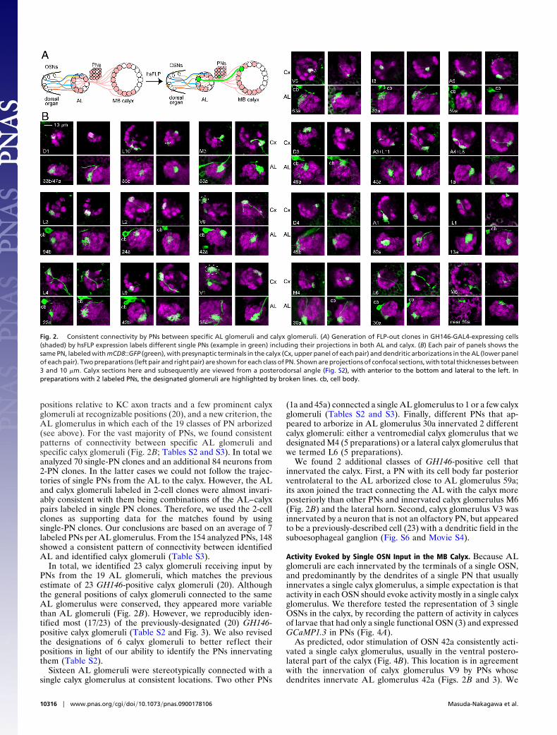
positions relative to KC axon tracts and a few prominent calyxglomeruli at recognizable positions (20), and a new criterion, theAL glomerulus in which each of the 19 classes of PN arborized(see above). For the vast majority of PNs, we found consistentpatterns of connectivity between specific AL glomeruli andspecific calyx glomeruli (Fig. 2B; Tables S2 and S3). In total weanalyzed 70 single-PN clones and an additional 84 neurons from2-PN clones. In the latter cases we could not follow the trajec-tories of single PNs from the AL to the calyx. However, the ALand calyx glomeruli labeled in 2-cell clones were almost invari-ably consistent with them being combinations of the AL–calyxpairs labeled in single PN clones. Therefore, we used the 2-cellclones as supporting data for the matches found by usingsingle-PN clones. Our conclusions are based on an average of 7labeled PNs per AL glomerulus. From the 154 analyzed PNs, 148showed a consistent pattern of connectivity between identifiedAL and identified calyx glomeruli (Table S3).
In total, we identified 23 calyx glomeruli receiving input byPNs from the 19 AL glomeruli, which matches the previousestimate of 23 GH146-positive calyx glomeruli (20). Althoughthe general positions of calyx glomeruli connected to the sameAL glomerulus were conserved, they appeared more variablethan AL glomeruli (Fig. 2B). However, we reproducibly iden-tified most (17/23) of the previously-designated (20) GH146-positive calyx glomeruli (Table S2 and Fig. 3). We also revisedthe designations of 6 calyx glomeruli to better reflect theirpositions in light of our ability to identify the PNs innervatingthem (Table S2).
Sixteen AL glomeruli were stereotypically connected with asingle calyx glomerulus at consistent locations. Two other PNs
(1a and 45a) connected a single AL glomerulus to 1 or a few calyxglomeruli (Tables S2 and S3). Finally, different PNs that ap-peared to arborize in AL glomerulus 30a innervated 2 differentcalyx glomeruli: either a ventromedial calyx glomerulus that wedesignated M4 (5 preparations) or a lateral calyx glomerulus thatwe termed L6 (5 preparations).
We found 2 additional classes of GH146-positive cell thatinnervated the calyx. First, a PN with its cell body far posteriorventrolateral to the AL arborized close to AL glomerulus 59a;its axon joined the tract connecting the AL with the calyx moreposteriorly than other PNs and innervated calyx glomerulus M6(Fig. 2B) and the lateral horn. Second, calyx glomerulus V3 wasinnervated by a neuron that is not an olfactory PN, but appearedto be a previously-described cell (23) with a dendritic field in thesuboesophageal ganglion (Fig. S6 and Movie S4).
Activity Evoked by Single OSN Input in the MB Calyx. Because ALglomeruli are each innervated by the terminals of a single OSN,and predominantly by the dendrites of a single PN that usuallyinnervates a single calyx glomerulus, a simple expectation is thatactivity in each OSN should evoke activity mostly in a single calyxglomerulus. We therefore tested the representation of 3 singleOSNs in the calyx, by recording the pattern of activity in calycesof larvae that had only a single functional OSN (3) and expressedGCaMP1.3 in PNs (Fig. 4A).
As predicted, odor stimulation of OSN 42a consistently acti-vated a single calyx glomerulus, usually in the ventral postero-lateral part of the calyx (Fig. 4B). This location is in agreementwith the innervation of calyx glomerulus V9 by PNs whosedendrites innervate AL glomerulus 42a (Figs. 2B and 3). We
Fig. 2. Consistent connectivity by PNs between specific AL glomeruli and calyx glomeruli. (A) Generation of FLP-out clones in GH146-GAL4-expressing cells(shaded) by hsFLP expression labels different single PNs (example in green) including their projections in both AL and calyx. (B) Each pair of panels shows thesame PN, labeled with mCD8::GFP (green), with presynaptic terminals in the calyx (Cx, upper panel of each pair) and dendritic arborizations in the AL (lower panelof each pair). Two preparations (left pair and right pair) are shown for each class of PN. Shown are projections of confocal sections, with total thicknesses between3 and 10 �m. Calyx sections here and subsequently are viewed from a posterodorsal angle (Fig. S2), with anterior to the bottom and lateral to the left. Inpreparations with 2 labeled PNs, the designated glomeruli are highlighted by broken lines. cb, cell body.
10316 � www.pnas.org�cgi�doi�10.1073�pnas.0900178106 Masuda-Nakagawa et al.

were unable to map the active glomerulus more accurately in livepreparations, because brain orientation was more variable thanin fixed preparations and Dlg background staining could not beapplied.
Unexpectedly, odor stimulation of either OSN 45b or 47ausually activated 2 calyx glomeruli strongly (Fig. 4 C and D).Approximately a quarter of preparations also had a third glo-merulus that showed weak activity (e.g., Fig. 4C), and occasionalbrains had only a single active glomerulus. For both OSNs, 1active glomerulus was usually in a location expected from the PNclonal analysis: in the case of OSN 45b, one was in the postero-medial region of the calyx, consistent with the expectation ofactivity in glomerulus D4, and in the case of OSN 47a, ananterodorsal glomerulus consistent with the expectation ofactivity in glomerulus D1 (Figs. 2B and 3) was found. The otheractive glomeruli tended to be found in an anterior or centrallocation in the case of OSN 45b stimulation and in a ventrome-dial location in the case of OSN 47a, but because of the absenceof landmarks in the live preparations we were not able to assigntheir identities.
DiscussionParallel Pathways from OSNs to the Calyx. We have generated acomprehensive olfactory circuit analysis that links identifiedsensory neurons to localized activity in a higher association
center and shows how activity in identified sensory neurons isrepresented in such a center.
We provide a 3D larval AL glomerular map of the input of thewhole set of 21 OSN classes. Comparison of the map with the
Fig. 3. OSN representation in calyx glomeruli. This is a 3D reconstruction ofa single calyx, viewed from different angles. GH146-expressing glomeruli arecolor-coded according to predicted input as in Fig. 1E; other glomeruli arewhite. Labeling indicates both the name of the best-fitting calyx glomerulusand the AL glomerulus connected to it. D, V, M, and L denote dorsal, ventral,medial, and lateral faces of the calyx, respectively. Fig. 4. PN activity in the calyx of larvae with a single functional OSN. (A)
Diagram of the larval olfactory circuitry, showing GCaMP1.3 expressed in mostPNs under control of GH146-GAL4 (shaded PNs that project to AL and calyx).A single functional OSN is expected to strongly stimulate a single PN thatnormally has a presynaptic terminal in a single calyx glomerulus (pathwayshown by dark thick shading). (B–D) Each row shows GCaMP fluorescence inGH146-expressing PN terminals of a single calyx at rest and during stimulation,with �F represented in false color, both alone and superimposed on a rawimage. Panels are single confocal sections, except where noted. Quantifica-tion and time courses of responses are shown in Fig. S7. (B) Activity in ventralsections of 2 calyces, evoked by ethyl acetate via OSN 42a. Note the increasedfluorescence in 1 calyx glomerulus, possibly V9. In the top row the activeglomerulus is surrounded by a broken line, and activity is also seen in an axoncoming from the inner antennocerebral tract (arrow). (C) Activity in 3 calyces,evoked by acetophenone via OSN 45b. The top and middle rows come fromdifferent brain hemispheres of the same larva (all calyces are presented withlateral left and anterior front, for consistency between panels). Most calycesshow a strongly-responding posterior (P) glomerulus, possibly D4, and acentral (C) glomerulus, and a few have a more weakly responding anterior (A)glomerulus. The middle row shows 2 superimposed sections from midway (Pglomerulus) and ventral (C and A glomeruli) levels along the dorsoventral axis.(D) Activity in 2 calyces, evoked by pentyl acetate via OSN 47a. The first 2 rowsshow dorsal (D) and ventral (V) sections from the same calyx. Most glomerulishow activity in an anterodorsal (AD) glomerulus, possibly D1, and a medio-ventral (MV) glomerulus.
Masuda-Nakagawa et al. PNAS � June 23, 2009 � vol. 106 � no. 25 � 10317
NEU
ROSC
IEN
CE
ligand specificities of larval ORs (18) shows a regional repre-sentation of some classes of odor qualities. For example, aro-matic odors are represented mainly in a cluster of lateralglomeruli, whereas alcohols appear to map to medial glomeruli(Fig. 1E; Movie S2, and Movie S3). The proximity of ALglomeruli dealing with similar odor qualities may facilitatecross-talk by local interneurons between glomeruli, broadeningolfactory representations during their passage through the AL,but preferentially among subsets of PNs that deal with similarodor qualities.
The criterion of AL glomerular arborization to identify PNssupports most features of the previous calyx map (20) and refinesit by providing a potential molecular criterion for identifyingcalyx glomeruli. We thus identified 23 calyx glomeruli thatreceive input from 19 AL glomeruli. In almost all cases, each ALglomerulus and OSN is represented uniquely by 1 or 2 calyxglomeruli. Unexpectedly, 1 apparent PN class (30a), innervatescalyx glomeruli at 2 alternative positions (L6 and M4), andspecific PN molecular markers will probably be required todetermine whether this is really a single class of PN.
In the calyx, the representation of odor quality classes appearsmore dispersed than in the AL. For example, ‘‘aromatic’’ ALglomeruli are connected to calyx glomeruli that are not obviouslyclustered. Also, the neighboring AL glomeruli 13a and 35a,which respond preferentially to alcohols, innervate nonadjacentcalyx glomeruli L1 and V1. Similar patterns are seen for the calyxrepresentations of other functionally-related groups of AL glo-meruli (Figs. 2B and 3, Movie S5, and Movie S6). Because mostKCs can integrate inputs from widely dispersed and apparentlyrandom subsets of calyx glomeruli (20), spatial proximity ofglomeruli may be less important for processing olfactory repre-sentations in the calyx than in the AL, where the spatialorganization of OSN input and PN output is very stereotypic.
GH146-positive PNs innervate only 22 of some 34 calyxglomeruli. From where do the remaining calyx glomeruli receivetheir input? Candidates are non-GH146-expressing PNs inner-vating AL glomeruli 67b and 74a or nonolfactory neurons suchas the one providing input to V3 from a broad region of thesuboesophageal ganglion, the main gustatory input region in thelarva (23). Neurons transmitting gustatory signals into the calyxcould allow larvae to use taste and smell as conditioned stimuliin associative learning.
Evoked Activity in Calyx Glomeruli. Our single-clone analysis suggeststhat each PN essentially connects a single AL glomerulus to a singlecalyx glomerulus. Consistent with this, stimulation of OSN 42aactivated a single calyx glomerulus, whose position was consistentwith it being V9, which is connected to AL glomerulus 42a.
In contrast, stimulation of either OSN 45b or OSN 47astrongly activated 2 calyx glomeruli in most preparations, withweak activation of an additional calyx glomerulus in a fewpreparations. For both OSNs, the position of 1 activated glo-merulus was generally consistent with the predictions from thePN clonal analysis, but the presence of additional active glomer-uli was unexpected. While our work was under review, Asahinaet al. (24) also reported that stimulation of single OSNs activatedPN terminals strongly in either 1 or 2 calyx glomeruli andsuggested that this finding was consistent with a 1-to-1 connec-tivity between OSNs and PNs in the AL.
Although our observations of strong activity evoked by singleOSNs in 1 to 3 calyx glomeruli are remarkably consistent withAsahina et al. (24), our analysis of the neuronal circuitry leadsus to a very different interpretation. OSNs 45b and 47a (thiswork) and 42b (24) all evoke activity in multiple calyx glomeruli,but the PNs that connect to these OSNs in the AL innervate onlya single calyx glomerulus; activity in multiple calyx glomerulitherefore cannot be explained by a 1-to-1 connectivity betweenOSNs and PNs. At least 2 explanations are possible. First, AL
glomeruli 45b and 47a might each contain arborizations from 2PNs, each of which innervates a different calyx glomerulus.However, this is unlikely, because arborizations of �1 GH146-positive PN in a single larval AL glomerulus were never ob-served (19). Second, interglomerular excitatory cholinergic con-nections in the AL, which apparently follow defined routes,broaden the response of PNs relative to that of OSNs (5, 6, 25).Olsen et al. (5) detected broadening of input from single adultOSNs to many more PNs than the one or two observed here, inthe form of depolarization of most PNs tested, from a subset ofthe entire PN population. However, they reported only occa-sional spikes. In contrast, our imaging experiments assay almostall PNs for presynaptic activity, and we detect probably highlevels of spiking in one or a few PN terminals, because GCaMPmay not be sensitive enough to detect lower levels of activity (26).Either the larval and adult calyces differ in the nature of lateralexcitatory spread, or the limited set of OSN and PN combinationssampled by Olsen et al. (5) may have missed high levels of lateralexcitation of a small number of PNs. In conclusion, the high levelsof activity in a small number of larval calyx glomeruli are consistentwith specific routes for broadening of some olfactory representa-tions in the AL, but suggest that the strongest signals from singleOSNs are not widely dispersed in the calyx.
ConclusionsThe simple organization of the Drosophila larval olfactorysystem has facilitated both a 3D sensory map of the larval ALof Drosophila and a map of the major OSN input to specificglomeruli in the calyx. Together with the apparently randomarborization of KC dendrites among calyx glomeruli, this worksuggests that KCs may receive many different combinations ofheterogeneous odor inputs, allowing them to discriminate verylarge numbers of odor bouquets. The future availability of GAL4lines that permit activation or silencing of spatially-localizedinputs into the calyx, or the ability to visualize activity of specificinput pathways, should help to reveal how representations ofodor qualities are transformed in the calyx and how they areencoded by KCs during learning.
We have produced a map of sensory input to a definedassociation center in the higher brain. The most analogous (andpossibly homologous) regions in the vertebrate brain are asso-ciation cortices, including olfactory cortex areas, where no overtspatial organization of sensory inputs has been detected. Thelarval calyx provides a very clear picture of how sensory inputsare organized and can potentially be a model for more complexassociation centers where the complexity obscures the underly-ing logic of the connectivity.
Materials and MethodsFly Strains and Clone Induction. Flies were raised on standard cornmealmedium at 25 °C. OSNs were labeled by crossing Or83b-Myc;UAS-GFP (3) toOrX-GAL4 lines (4). UAS-nsyb::GFP has been described by Ito et al. (27). PNclones were generated by FLP-out, in the progeny of, GH146-GAL4 (28, 29)crossed to hsFLP;CyO/Sp;UAS� y� CD2�CD8-GFP (21). FLP recombinase wasinduced by heat shock (35 °C) for 20–30 min at 12–18, 18–24, or 24–30 h afteregg laying. Neuronal activity in PN terminals of larvae with a single active OSNwas imaged in the female progeny of GC56;GH146-GAL4/CyO; or83b1 femalescrossed to GC56/Y;OrX-GAL4;or83b1 males. GC56 is an X chromosome with2 copies of UAS-GCaMP1.3 (30). OrX-GAL4 insertions (Fig. S8) were eitherhomozygous Or42a-GAL4 or Or47a-GAL4, or Or45b-GAL4 heterozygouswith CyO.
Specimen Orientation and Confocal Immunomicroscopy. The CNS of wanderingthird-instar larvae was dissected, fixed, and labeled as described (20). Orien-tation of the larval AL was defined relative to the body axis rather than theneuraxis, as described in SI Text and Fig. S2. To collect confocal images from thelarval MB calyx, the CNS was viewed from a posterodorsal angle as described(20) (Fig. S2). This allowed more standardized orientation of calyx landmarksthan the flattened preparation of Ramaekers et al. (19) and easier comparison
10318 � www.pnas.org�cgi�doi�10.1073�pnas.0900178106 Masuda-Nakagawa et al.

with imaging of calyx activity; we therefore adopted the nomenclature ofMasuda-Nakagawa et al. (20) for calyx glomeruli.
Imaging Neuronal Activity in Calyx. Females were identified by the lack of testisat the wandering stage and dissected in HL3 medium containing 1.5 mM Ca2�
(31) to expose their brain. The dorsal organ was exposed to air, pointing downthrough a hole in a paraffin membrane that rested on 2 silicone spacers on aglass slide. The brain above this was bathed in HL3, beneath a coverslip thatrested lightly on the larva and paraffin membrane. Larvae were exposed to astream of air that could be switched to a stream of air that contained odorant,as described in SI Text. GCaMP fluorescence was visualized with a spinning discconfocal microscope and analyzed as described in SI Text.
ACKNOWLEDGMENTS. We thank L. Vosshall (Rockefeller University, NewYork), J. Carlson (Yale University, New Haven, CT), and B. Dickson (Institutefor Molecular Pathology, Vienna) for OrX-GAL4 lines; R. Axel (ColumbiaUniversity, New York) for UAS-GCaMP1.3; the Developmental Studies Hy-bridoma Bank (University of Iowa, Iowa City) for anti-Dlg and 9E10; theYokogawa Corporation (Tokyo) for the loan of spinning disc scannerCSU-22; K. Ito for generous provision of microscopy facilities and lab space(to L.M.M.-N.); and M. Tanifuji and S. Namiki for advice on odor delivery.This work was supported by Japan Society for the Promotion of ScienceGrant-in-Aid for Scientific Research 19500265 (to L.M.M.-N.), U.K. Biotech-nology and Biological Sciences Research Council Grant BBS/B/06954 (toC.J.O.), and Swiss National Funds Grants 31-63447.00 and 3100A0-105517(to R.F.S.).
1. Heisenberg M (2003) Mushroom body memoir: From maps to models. Nat Rev Neurosci4:266–275.
2. Davis RL (2005) Olfactory memory formation in Drosophila: From molecular to systemsneuroscience. Annu Rev Neurosci 28:275–302.
3. Fishilevich E, et al. (2005) Chemotaxis behavior mediated by single larval olfactoryneurons in Drosophila. Curr Biol 15:2086–2096.
4. Kreher SA, Kwon JY, Carlson JR (2005) The molecular basis of odor coding in theDrosophila larva. Neuron 46:445–456.
5. Olsen SR, Bhandawat V, Wilson RI (2007) Excitatory interactions between olfactoryprocessing channels in the Drosophila antennal lobe. Neuron 54:89–103.
6. Shang Y, Claridge-Chang A, Sjulson L, Pypaert M, Miesenbock G (2007) Excitatory localcircuits and their implications for olfactory processing in the fly antennal lobe. Cell128:601–612.
7. Perez-Orive J, et al. (2002) Oscillations and sparsening of odor representations in themushroom body. Science 297:359–365.
8. Stopfer M, Jayaraman V, Laurent G (2003) Intensity versus identity coding in anolfactory system. Neuron 39:991–1004.
9. Murthy M, Fiete I, Laurent G (2008) Testing odor response stereotypy in the Drosophilamushroom body. Neuron 59:1009–1023.
10. Tanaka NK, Awasaki T, Shimada T, Ito K (2004) Integration of chemosensory pathwaysin the Drosophila second-order olfactory centers. Curr Biol 14:449–457.
11. Jefferis GSXE, et al. (2007) Comprehensive maps of Drosophila higher olfactory centers:Spatially segregated fruit and pheromone representation. Cell 128:1187–1203.
12. Lin H-H, Lai JS-Y, Chin A-L, Chen Y-C, Chiang A-S (2007) A map of olfactory represen-tation in the Drosophila mushroom body. Cell 128:1205–1217.
13. Fiala A, et al. (2002) Genetically encoded cameleon in Drosophila melanogaster is usedto visualize olfactory information in projection neurons. Curr Biol 12:1877–1884.
14. Cobb M (1999) What and how do maggots smell? Biol Rev 74:425–459.15. Aceves-Pina EO, Quinn WG (1979) Learning in normal and mutant Drosophila larvae.
Science 206:93–96.16. Scherer S, Stocker RF, Gerber B (2003) Olfactory learning in individually assayed
Drosophila larvae. Learn Mem 10:217–225.17. Singh RN, Singh K (1984) Fine structure of the sensory organs of Drosophila melano-
gaster Meigen larva (Diptera: Drosophilidae). Int J Insect Morphol Embryol 13:255–273.
18. Kreher SA, Mathew D, Kim J, Carlson JR (2008) Translation of sensory input intobehavioral output via an olfactory system. Neuron 59:110–124.
19. Ramaekers A, et al. (2005) Glomerular maps without cellular redundancy at successivelevels of the Drosophila larval olfactory circuit. Curr Biol 15:982–992.
20. Masuda-Nakagawa LM, Tanaka NK, O’Kane CJ (2005) Stereotypic and random patternsof connectivity in the larval mushroom body calyx of Drosophila. Proc Natl Acad Sci USA102:19027–19032.
21. Wong AM, Wang JW, Axel R (2002) Spatial representation of the glomerular map in theDrosophila protocerebrum. Cell 109:229–241.
22. Marin EC, Watts RJ, Tanaka NK, Ito K, Luo L (2005) Developmentally programmedremodeling of the Drosophila olfactory circuit. Development 132:725–737.
23. Colomb J, Grillenzoni N, Ramaekers A, Stocker RF (2007) Architecture of the primarytaste center of Drosophila melanogaster larvae. J Comp Neurol 502:834–847.
24. Asahina, K, Louis M, Piccinotti S, Vosshall LB (2009) A circuit supporting concentration-invariant odor perception in Drosophila. J Biol 8:9.
25. Bhandawat V, Olsen SR, Gouwens NW, Schlief ML, Wilson RI (2007) Sensory processingin the Drosophila antennal lobe increases reliability and separability of ensemble odorrepresentations. Nat Neurosci 10:1474–1482.
26. Jayaraman V, Laurent G (2007) Evaluating a genetically encoded optical sensor ofneural activity using electrophysiology in intact adult fruit flies. Front Neur Circuits 1:3.
27. Ito K, et al. (1998) The organization of extrinsic neurons and their implications in thefunctional roles of the mushroom bodies in Drosophila melanogaster Meigen. LearnMem 5:52–77.
28. Stocker RF, Heimbeck G, Gendre N, de Belle JS (1997) Neuroblast ablation in DrosophilaP[GAL4] lines reveals origins of olfactory interneurons. J Neurobiol 32:443–456.
29. Heimbeck G, Bugnon V, Gendre N, Keller A, Stocker RF (2001) A central neural circuitfor experience-independent olfactory and courtship behavior in Drosophila melano-gaster. Proc Natl Acad Sci USA 98:15336–15341.
30. Wang JW, Wong AM, Flores J, Vosshall LB, Axel R (2003) Two-photon calcium imagingreveals an odor-evoked map of activity in the fly brain. Cell 112:271–282.
31. Stewart BA, Atwood HL, Renger JJ, Wang J, Wu C-F (1994) Improved stability ofDrosophila neuromuscular junction preparations in haemolymph-like physiologicalsolutions. J Comp Physiol A 175:179–191.
Masuda-Nakagawa et al. PNAS � June 23, 2009 � vol. 106 � no. 25 � 10319
NEU
ROSC
IEN
CE