localized and systemic al amyloidosis · pdf filelocalized and systemic al amyloidosis...
TRANSCRIPT
LOCALIZED AND SYSTEMIC AL AMYLOIDOSISFELLOW:VICTORCHOWHEMATOLOGYFELLOWSCONFERENCEDISCUSSANT:EDWARDLIBBY
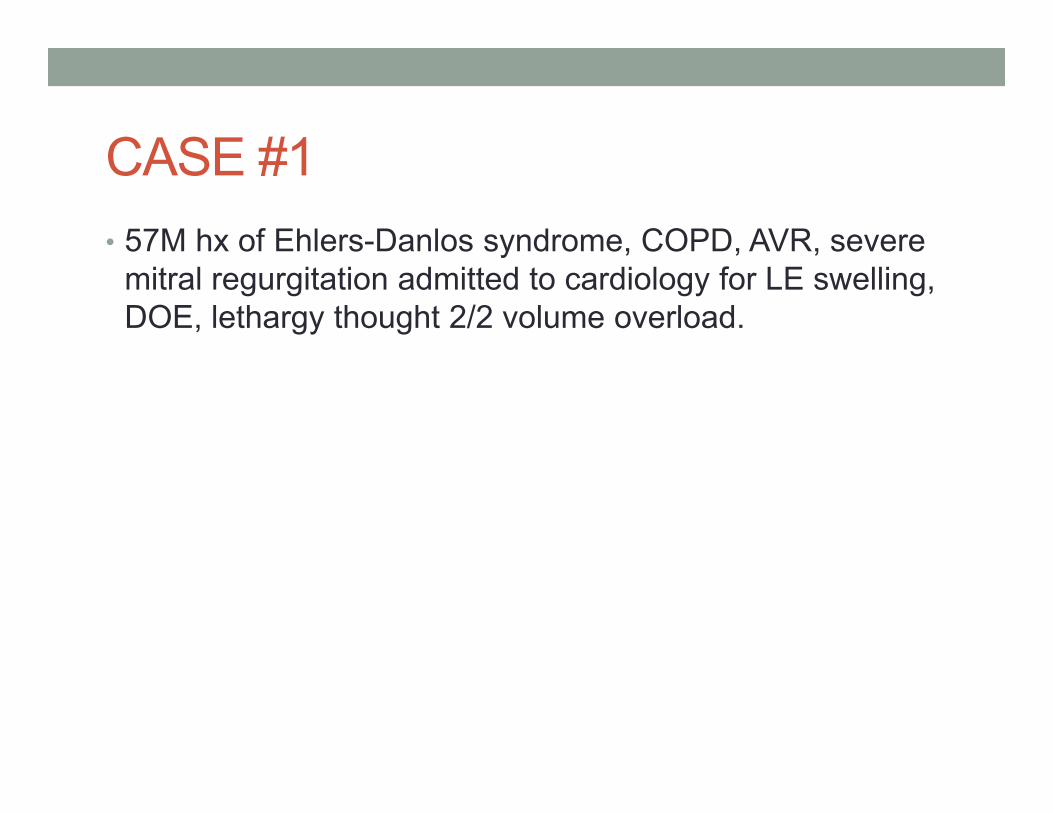
CASE #1• 57M hx of Ehlers-Danlos syndrome, COPD, AVR, severe
mitral regurgitation admitted to cardiology for LE swelling, DOE, lethargy thought 2/2 volume overload.

CASE #1 cont…
HEMATOLOGY CONSULT
“He has AL Amyloidosis”
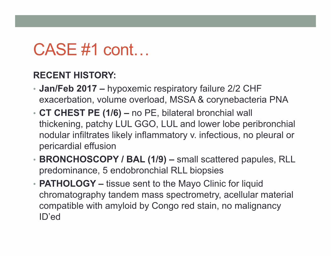
CASE #1 cont…RECENT HISTORY:• Jan/Feb 2017 – hypoxemic respiratory failure 2/2 CHF
exacerbation, volume overload, MSSA & corynebacteria PNA• CT CHEST PE (1/6) – no PE, bilateral bronchial wall
thickening, patchy LUL GGO, LUL and lower lobe peribronchialnodular infiltrates likely inflammatory v. infectious, no pleural or pericardial effusion
• BRONCHOSCOPY / BAL (1/9) – small scattered papules, RLL predominance, 5 endobronchial RLL biopsies
• PATHOLOGY – tissue sent to the Mayo Clinic for liquid chromatography tandem mass spectrometry, acellular material compatible with amyloid by Congo red stain, no malignancy ID’ed
CASE #1 cont…
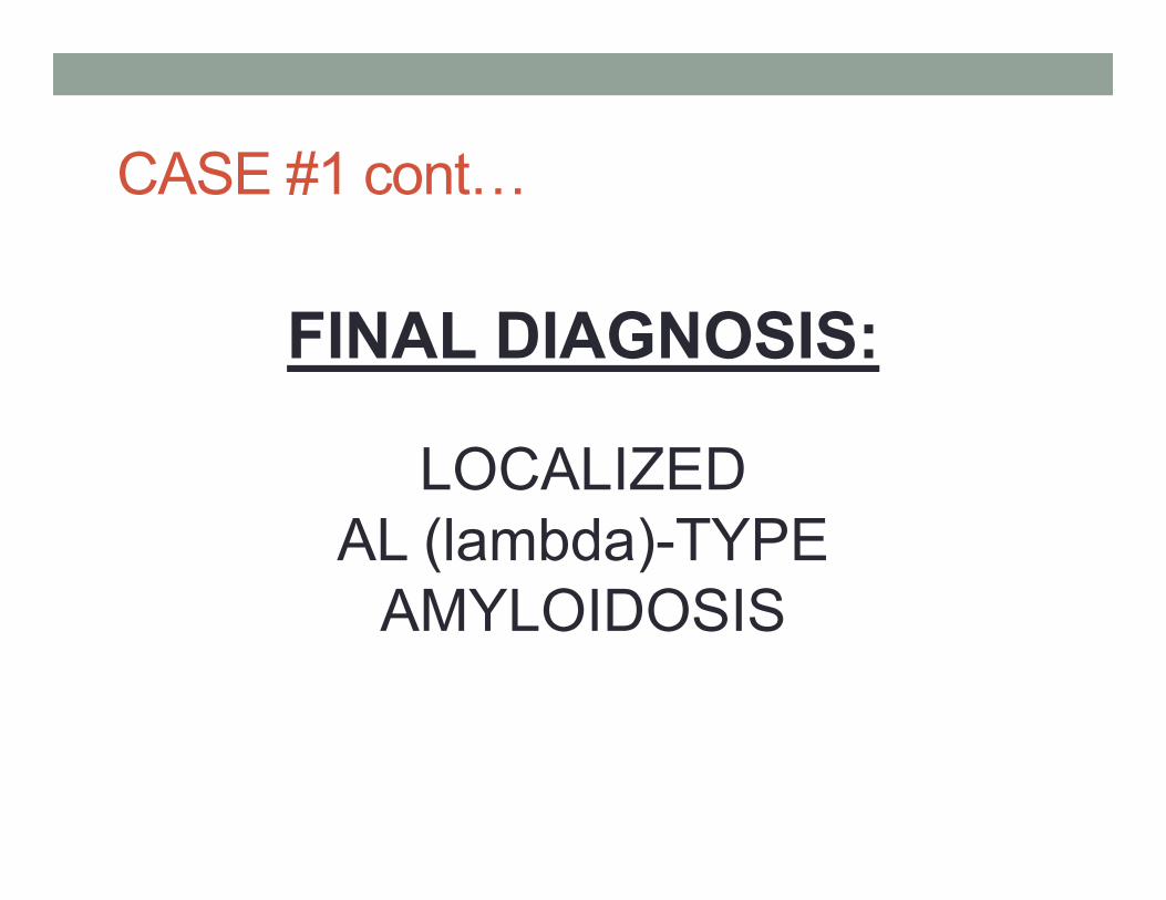
FINAL DIAGNOSIS:
LOCALIZEDAL (lambda)-TYPE
AMYLOIDOSIS
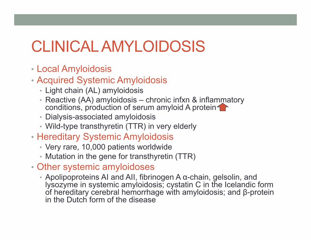
CLINICAL AMYLOIDOSIS• Local Amyloidosis• Acquired Systemic Amyloidosis
• Light chain (AL) amyloidosis• Reactive (AA) amyloidosis – chronic infxn & inflammatory
conditions, production of serum amyloid A protein • Dialysis-associated amyloidosis• Wild-type transthyretin (TTR) in very elderly
• Hereditary Systemic Amyloidosis• Very rare, 10,000 patients worldwide• Mutation in the gene for transthyretin (TTR)
• Other systemic amyloidoses• Apolipoproteins AI and AII, fibrinogen A α-chain, gelsolin, and
lysozyme in systemic amyloidosis; cystatin C in the Icelandic form of hereditary cerebral hemorrhage with amyloidosis; and β-protein in the Dutch form of the disease
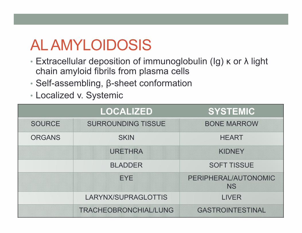
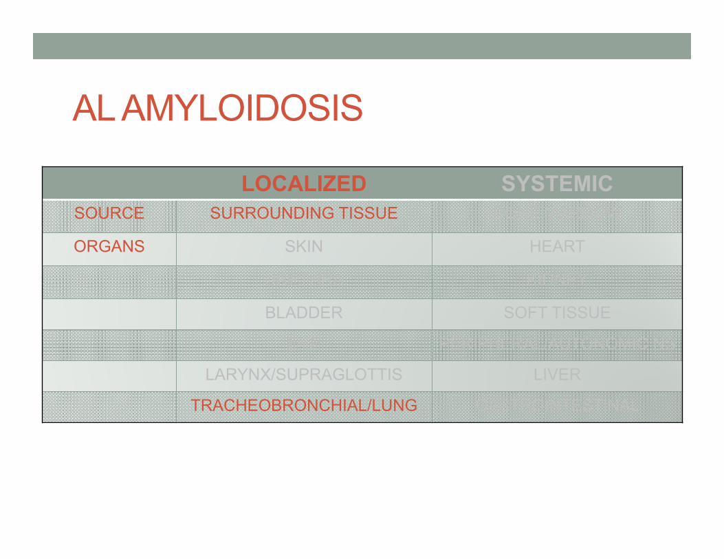
AL AMYLOIDOSIS• Extracellular deposition of immunoglobulin (Ig) κ or λ light
chain amyloid fibrils from plasma cells• Self-assembling, β-sheet conformation• Localized v. Systemic
LOCALIZED SYSTEMICSOURCE SURROUNDING TISSUE BONE MARROW
ORGANS SKIN HEART
URETHRA KIDNEY
BLADDER SOFT TISSUE
EYE PERIPHERAL/AUTONOMIC NS
LARYNX/SUPRAGLOTTIS LIVER
TRACHEOBRONCHIAL/LUNG GASTROINTESTINAL
AL AMYLOIDOSIS
LOCALIZED SYSTEMICSOURCE SURROUNDING TISSUE BONE MARROW
ORGANS SKIN HEART
URETHRA KIDNEY
BLADDER SOFT TISSUE
EYE PERIPHERAL/AUTONOMIC NS
LARYNX/SUPRAGLOTTIS LIVER
TRACHEOBRONCHIAL/LUNG GASTROINTESTINAL

PULMONARY AMYLOIDOSIS
1. Utz et al. Annals of Internal Medicine 1996,124(4):407-4132. Cordier et al. Chest 1986, 90(6):827-313. Hui et al. Arch Pathol Lab Med 1986, 110(3):212-84. Thompson et al. Thorax 1983, 38(2):84-75. Scala et al. Annals of Thoracic Medicine 2015, 10(3):212-16
• 3 forms of pulmonary disease in localized AL amyloid• NOT associated with systemic deposition• NODULAR OPACITIES• DIFFUSE OPACITIES• TRACHEOBRONCHIAL
• Uncommon• Mayo Clinic Experience1 1980-1993, 4 of 55 cases• Cordier & colleagues2 15 yr period, 5 of 21 cases• Armed Forces Institute of Pathology3, 14 of 48 cases• Thompson & Citron4, 1983 review of literature, 67 of 126 cases• Pulmonary Unit of the Hospital of Arezzo5, 5 yr period, 1 of 298 cases
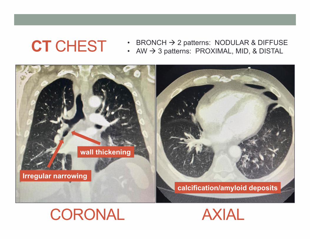
CT CHEST
CORONAL AXIAL
wall thickening
Irregular narrowingcalcification/amyloid deposits
• BRONCH 2 patterns: NODULAR & DIFFUSE• AW 3 patterns: PROXIMAL, MID, & DISTAL
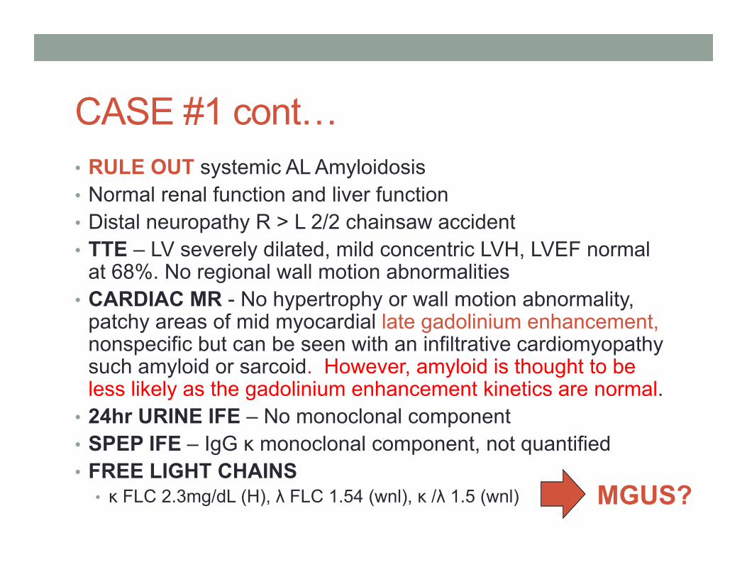
CASE #1 cont…• RULE OUT systemic AL Amyloidosis• Normal renal function and liver function• Distal neuropathy R > L 2/2 chainsaw accident • TTE – LV severely dilated, mild concentric LVH, LVEF normal
at 68%. No regional wall motion abnormalities• CARDIAC MR - No hypertrophy or wall motion abnormality,
patchy areas of mid myocardial late gadolinium enhancement, nonspecific but can be seen with an infiltrative cardiomyopathy such amyloid or sarcoid. However, amyloid is thought to be less likely as the gadolinium enhancement kinetics are normal.
• 24hr URINE IFE – No monoclonal component• SPEP IFE – IgG κ monoclonal component, not quantified• FREE LIGHT CHAINS
• κ FLC 2.3mg/dL (H), λ FLC 1.54 (wnl), κ /λ 1.5 (wnl) MGUS?
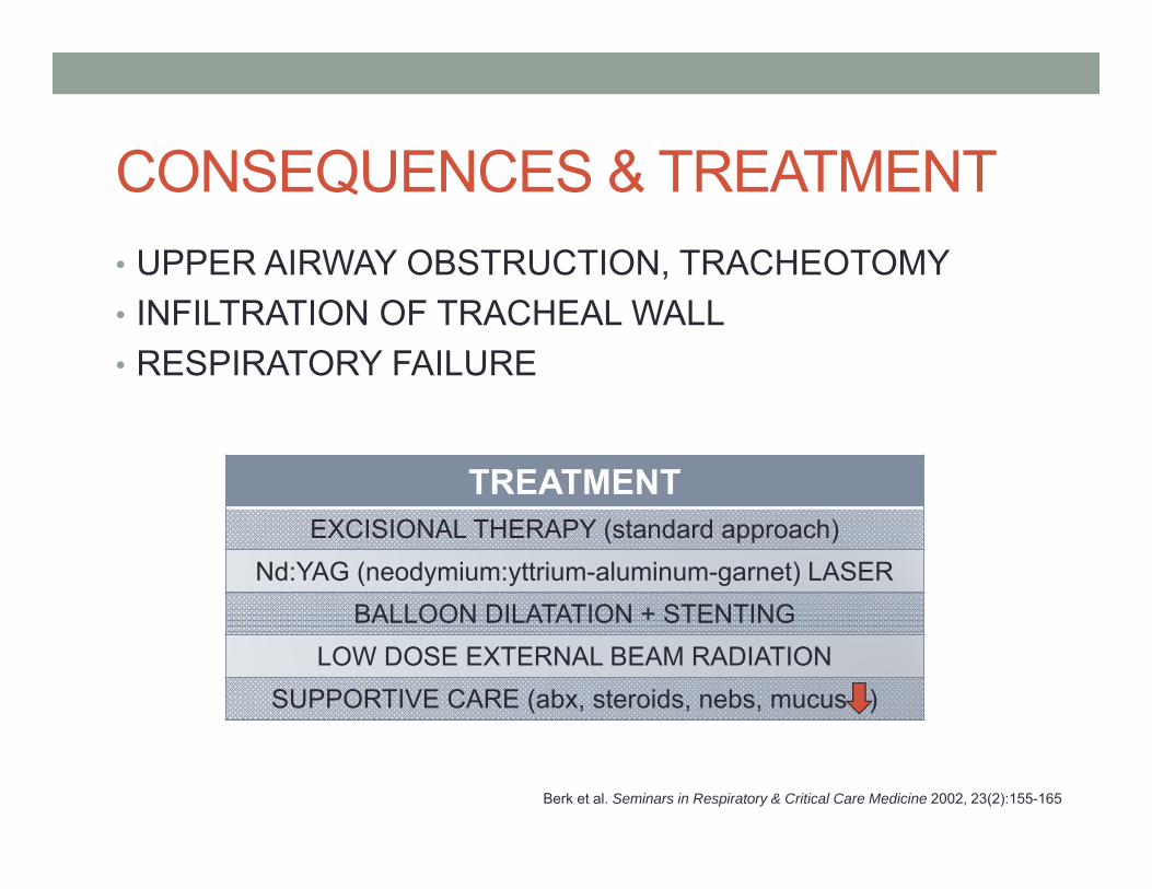
CONSEQUENCES & TREATMENT• UPPER AIRWAY OBSTRUCTION, TRACHEOTOMY• INFILTRATION OF TRACHEAL WALL• RESPIRATORY FAILURE
Berk et al. Seminars in Respiratory & Critical Care Medicine 2002, 23(2):155-165
TREATMENTEXCISIONAL THERAPY (standard approach)
Nd:YAG (neodymium:yttrium-aluminum-garnet) LASERBALLOON DILATATION + STENTING
LOW DOSE EXTERNAL BEAM RADIATIONSUPPORTIVE CARE (abx, steroids, nebs, mucus )
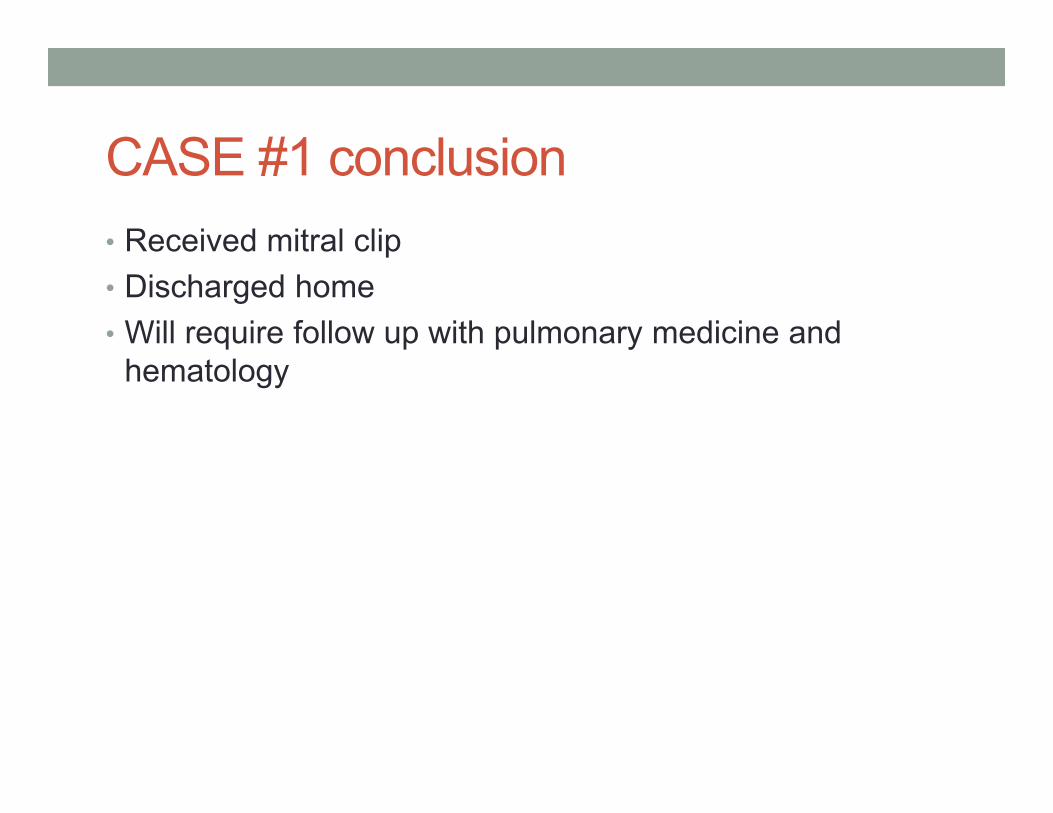
CASE #1 conclusion• Received mitral clip• Discharged home• Will require follow up with pulmonary medicine and
hematology
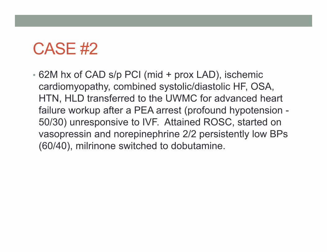
CASE #2• 62M hx of CAD s/p PCI (mid + prox LAD), ischemic
cardiomyopathy, combined systolic/diastolic HF, OSA, HTN, HLD transferred to the UWMC for advanced heart failure workup after a PEA arrest (profound hypotension -50/30) unresponsive to IVF. Attained ROSC, started on vasopressin and norepinephrine 2/2 persistently low BPs (60/40), milrinone switched to dobutamine.
CASE #2 cont…
HEMATOLOGY CONSULT
“He has AL Amyloidosis”

CASE #2 cont…RECENT HISTORY• Summer 2016 – progressive DOE• Nuclear Stress Test (9/22) – partially reversible apical-lateral defect
2/2 diaphragm artifact, distal ischemia could not be excluded• TTE (9/26) – LVEF 60%, filling pressures, bi-atrial enlargement• LHC (11/16) – No obstructive CAD, 45-55% mid RCA narrowing,
elevated LVED pressure, probable cardiomyopathy• Progressive DOE, PND, 15lb +, NYHA III heart failure, R pleural
effusion + thoracentesis 600cc, up-titration of diuresis…• RHC (2/2) – PAP 50/33/25, wedge 25, CI 1.47, admitted A on CHF• TTE (2/2) – LVEF 35%, severe global hypokinesis, infiltrative pattern • CARDIAC MRI (2/3) – diffuse enhancement of the entire LV,
concerning for amyloidosis, large bilateral pleural effusions• RHC / RIJ / ENDOMYOCARDIAL BIOPSY (2/3)
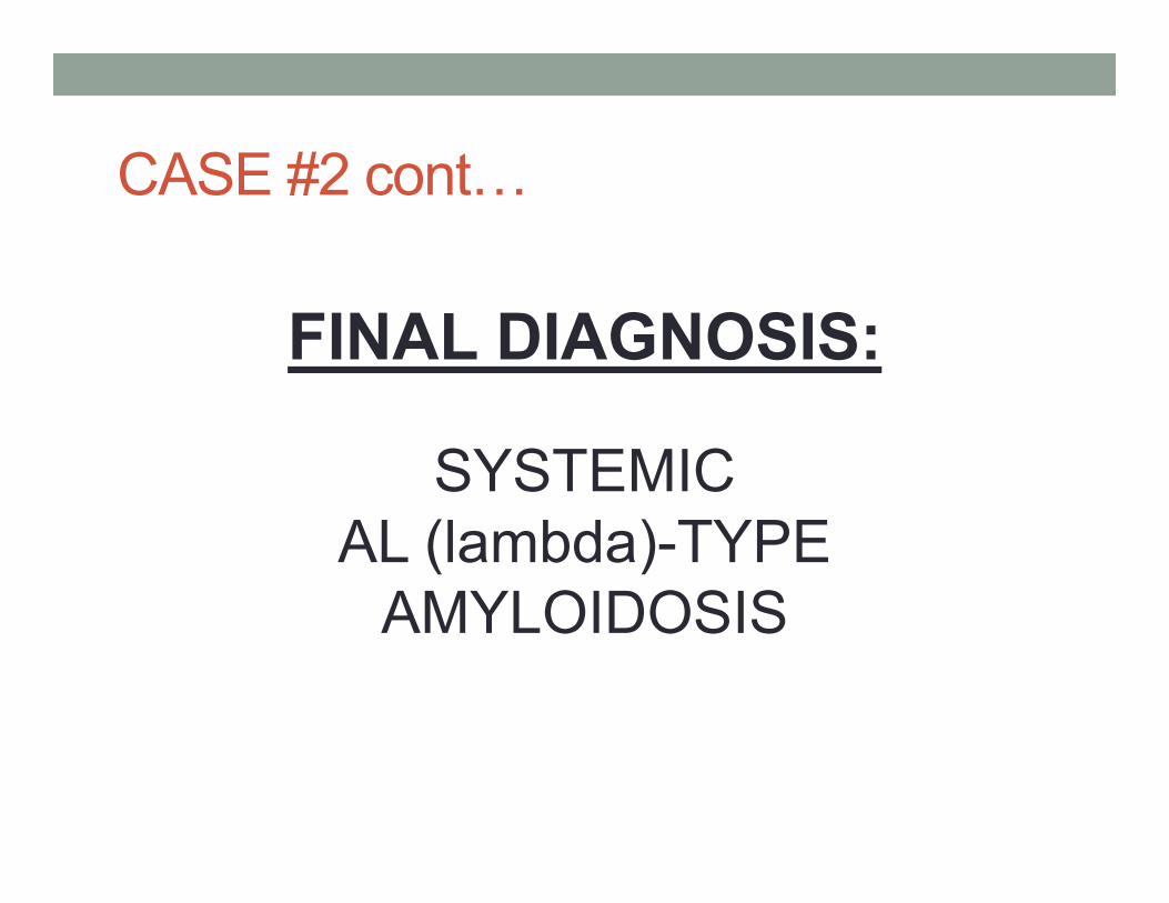
CASE #2 cont…
FINAL DIAGNOSIS:
SYSTEMICAL (lambda)-TYPE
AMYLOIDOSIS
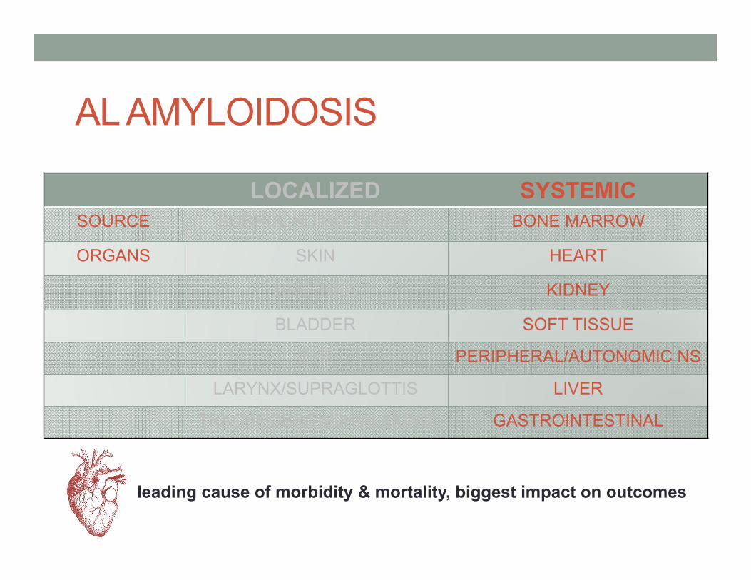
AL AMYLOIDOSIS
LOCALIZED SYSTEMICSOURCE SURROUNDING TISSUE BONE MARROW
ORGANS SKIN HEART
URETHRA KIDNEY
BLADDER SOFT TISSUE
EYE PERIPHERAL/AUTONOMIC NS
LARYNX/SUPRAGLOTTIS LIVER
TRACHEOBRONCHIAL/LUNG GASTROINTESTINAL
leading cause of morbidity & mortality, biggest impact on outcomes
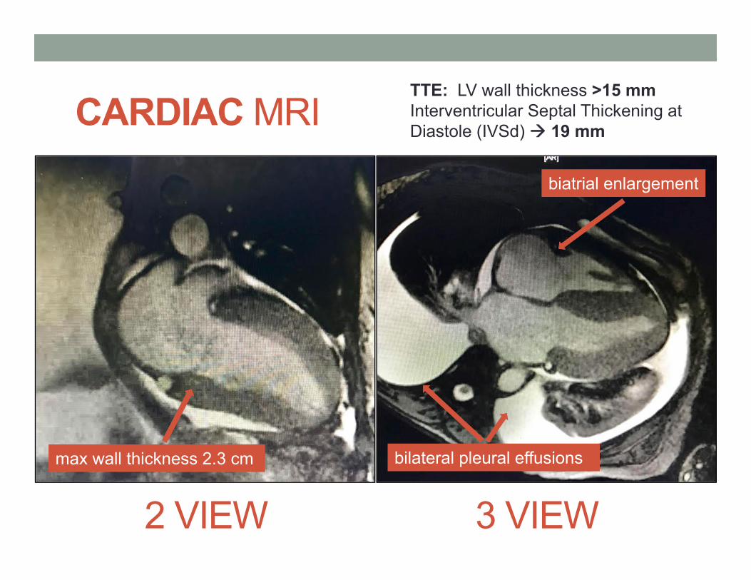
CARDIAC MRI
2 VIEW 3 VIEW
TTE: LV wall thickness >15 mmInterventricular Septal Thickening at Diastole (IVSd) 19 mm
max wall thickness 2.3 cm bilateral pleural effusions
biatrial enlargement
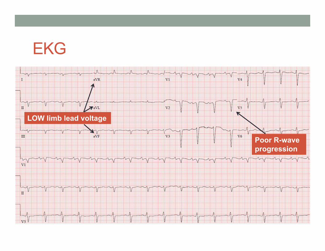
EKG
LOW limb lead voltage
Poor R-wave progression
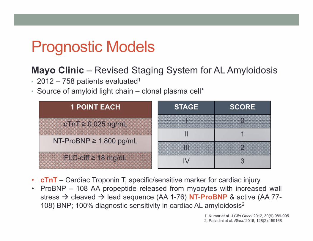
Prognostic ModelsMayo Clinic – Revised Staging System for AL Amyloidosis• 2012 – 758 patients evaluated1
• Source of amyloid light chain – clonal plasma cell*
STAGE SCORE
I 0
II 1
III 2
IV 3
1 POINT EACH
cTnT ≥ 0.025 ng/mL
NT-ProBNP ≥ 1,800 pg/mL
FLC-diff ≥ 18 mg/dL
1. Kumar et al. J Clin Oncol 2012, 30(9):989-9952. Palladini et al. Blood 2016, 128(2):159168
• cTnT – Cardiac Troponin T, specific/sensitive marker for cardiac injury• ProBNP – 108 AA propeptide released from myocytes with increased wall
stress cleaved lead sequence (AA 1-76) NT-ProBNP & active (AA 77-108) BNP; 100% diagnostic sensitivity in cardiac AL amyloidosis2
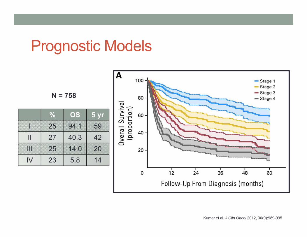
Prognostic Models
Kumar et al. J Clin Oncol 2012, 30(9):989-995
% OS 5 yrI 25 94.1 59II 27 40.3 42III 25 14.0 20IV 23 5.8 14
N = 758
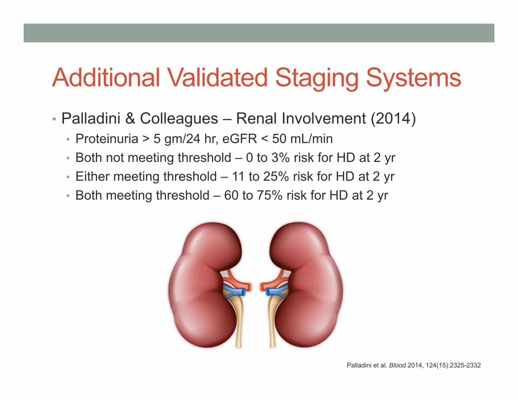
Additional Validated Staging Systems• Palladini & Colleagues – Renal Involvement (2014)
• Proteinuria > 5 gm/24 hr, eGFR < 50 mL/min• Both not meeting threshold – 0 to 3% risk for HD at 2 yr• Either meeting threshold – 11 to 25% risk for HD at 2 yr• Both meeting threshold – 60 to 75% risk for HD at 2 yr
Palladini et al. Blood 2014, 124(15):2325-2332
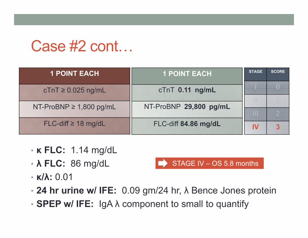
Case #2 cont…
• κ FLC: 1.14 mg/dL• λ FLC: 86 mg/dL• κ/λ: 0.01• 24 hr urine w/ IFE: 0.09 gm/24 hr, λ Bence Jones protein• SPEP w/ IFE: IgA λ component to small to quantify
1 POINT EACH
cTnT ≥ 0.025 ng/mL
NT-ProBNP ≥ 1,800 pg/mL
FLC-diff ≥ 18 mg/dL
1 POINT EACH
cTnT 0.11 ng/mL
NT-ProBNP 29,800 pg/mL
FLC-diff 84.86 mg/dL
STAGE SCORE
I 0
II 1
III 2
IV 3
STAGE IV – OS 5.8 months
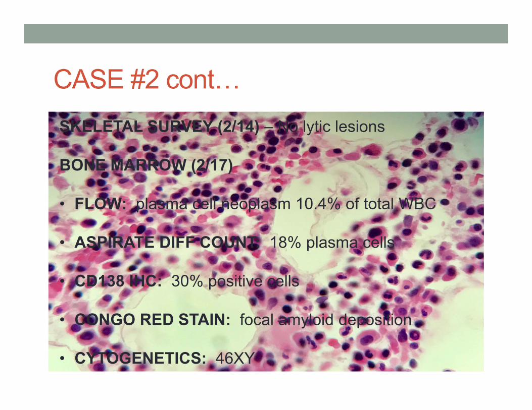
CASE #2 cont…SKELETAL SURVEY (2/14) – No lytic lesions
BONE MARROW (2/17)
• FLOW: plasma cell neoplasm 10.4% of total WBC
• ASPIRATE DIFF COUNT: 18% plasma cells
• CD138 IHC: 30% positive cells
• CONGO RED STAIN: focal amyloid deposition
• CYTOGENETICS: 46XY
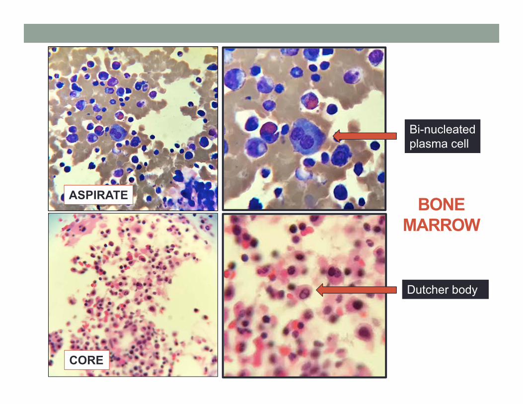
BONE MARROW
ASPIRATE
CORE
Bi-nucleated plasma cell
Dutcher body
Palladini et al. Blood 2016, 128(2):159168
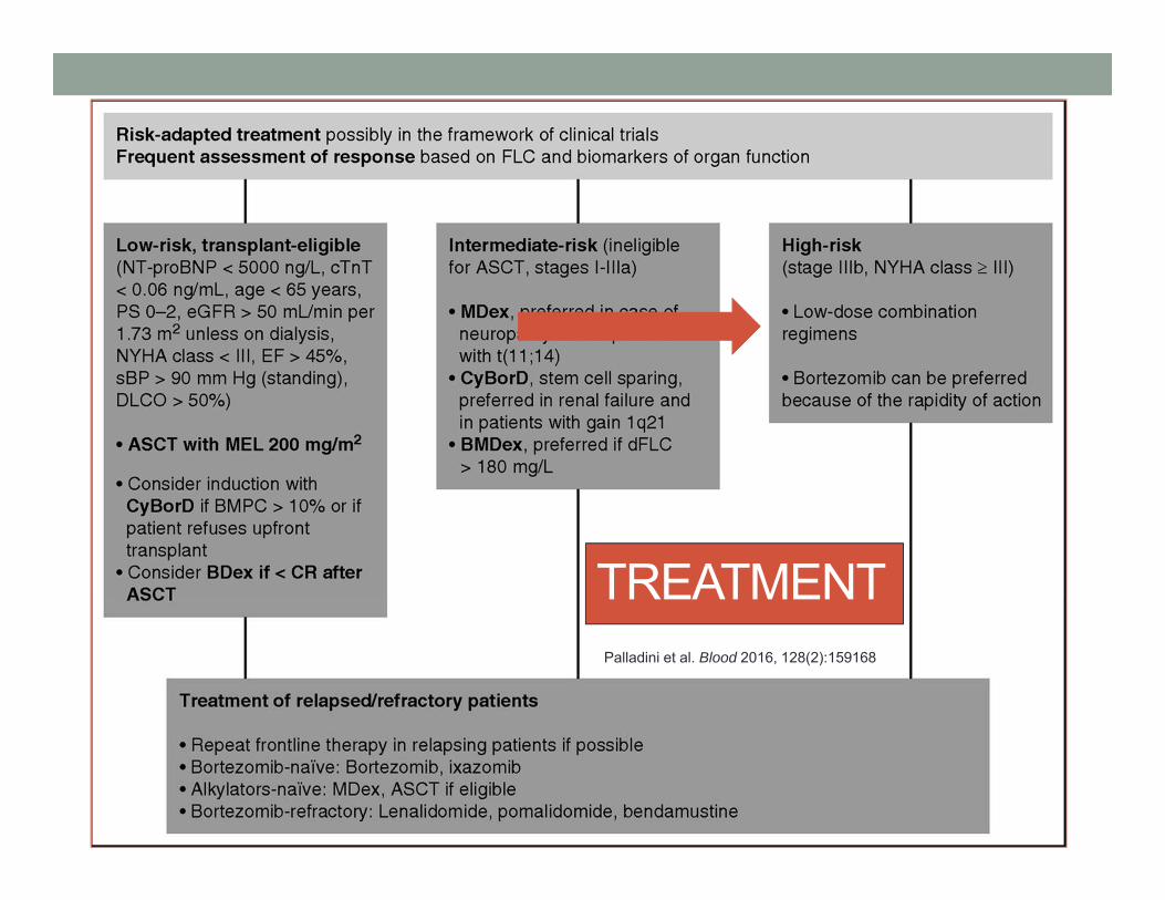
TREATMENT
CASE #2 conclusion • Weaned off bumex gtt• Attempting to wean off dobutamine gtt• Hypotensive to 60/40 PEA arrest CPR ROSC• CyBorD received subq Bortezomib 1mg/m2• Unresponsive during routine turning PEA arrest
CPR intubated significant bloody output from ETT unable to achieve ROSC death pronounced
SUMMARY• AL Amyloidosis – localized v. systemic
• Suspect localized disease – TISSUE + rule out systemic• Treatment based on organ involvement
• Suspect systemic disease – TISSUE + biomarkers• Cardiac involvement significantly impacts outcomes• Staging systems • Treatment based on staging
THANK YOU
EDWARD LIBBY BARBARA KONKLE SIOBAN KEEL