cardiac amyloidosis: the zebra is losing its stripes · cardiac amyloidoses originate from abnormal...
TRANSCRIPT
1
Internal Medicine Grand Rounds University of Texas Southwestern Medical Center
February 1st, 2019
Cardiac Amyloidosis: The Zebra is Losing its Stripes
Justin L. Grodin, MD, MPH, FACC, FHFSA Assistant Professor of Medicine
Texas Health Resources Clinical Scholar Division of Cardiology
Advanced Heart Failure, LVAD, and Cardiac Transplantation
This is to acknowledge that Dr. Grodin has disclosed that he does not have any financial interests or other relationships with commercial concerns related directly or indirectly to this program. Dr. Grodin will not be discussing off-label uses in his
presentation
2
PURPOSE AND OVERVIEW
To discuss the current management strategies for cardiac amyloidosis, including the pathophysiology and prognosis of cardiac amyloidosis, the state-of-the-art diagnostic strategies for cardiac amyloidosis, and the current and new treatments on the horizon for cardiac amyloidosis.
EDUCATIONAL OBJECTIVES
The listener should be able to:
1. Understand the pathophysiologic, prognostic, and clinical differences between light-chain cardiac amyloidosis (AL) and transthyretin cardiac amyloidosis (ATTR).
2. Understand the clinical clues that might be consistent with a diagnosis of cardiac amyloidosis. 3. Identify the appropriate diagnostic strategies to secure the diagnosis of cardiac amyloidosis and
differentiate AL from ATTRm/wt with a focus on 99m technetium pyrophosphate scans and serum free light chain assays plus serum immunofixation.
4. Understand the medical management of heart failure in patients with cardiac amyloidosis and how this differs from more typical heart failure populations.
5. Recognize US FDA approved ATTR pharmacotherapy and newer therapies on the horizon and be able to differentiate categories of ATTR therapies including TTR silencers, TTR stabilizers, and amyloid fibril disruptors.
ABOUT THE AUTHOR
Dr. Grodin was born and raised in El Paso, Texas. He completed a Bachelor of Science in Chemistry at the University of Texas (UT) at Austin and then transitioned to medical school (2005-2009) at the UT Southwestern Medical Center where he also completed his residency training in internal medicine (2009-2012). Following this, he completed fellowships in Cardiovascular Disease and Advanced Heart Failure and Transplant Cardiology at the Cleveland Clinic (2012-2016) and attended the Harvard T.H. Chan School of Public Health where he received the degree of Master of Public Health. He has since returned to UT Southwestern in the Fall of 2016 joining the Division of Cardiology. His clinical focus is in advanced heart failure, cardiac amyloidosis, hypertrophic cardiomyopathy, cardiac transplantation, left-ventricular assist devices, and cardiac critical care. In addition, he splits his time as a clinical epidemiologist focusing on heart failure outcomes research.
3
INTRODUCTION
Historical Perspective
The recognition of "amyloidosis" as an important disease is centuries old. The German physician scientist, Rudolph Virchow, coined the term "amyloid" in 1854 when describing the abnormal macroscopic appearance of abnormal brain tissue. This tissue was similar to prior descriptions (from the 1600s) of livers appearing "lardaceous, waxy, spongy, or like 'white stone'.” He discovered that the corporea amylacea of the brain stained pale blue with iodine and violet upon the addition of sulfuric acid. These observations led him to the conclusion that this unknown substance was cellulose and then gave it the name "amyloid." This term was derived from the Latin term "amylum" and the Greek term "amylon" which are both synonymous with starch. It was later recognized by Freidrich and Kekule (1859) that masses of amyloid substance in tissues were relatively devoid of carbohydrates and rich in nitrogen content leading to the hypothesis that this substance was primarily protein.
The pathophysiologic and structural underpinnings of amyloid evolved along with the available technologies during the 20th century. Key observations from Divry and Florkin (1927) that Congo Red stained sections of tissue with amyloid deposits exhibited what is now known as the pathognomonic "apple-green" birefringence.
What is Amyloidosis?
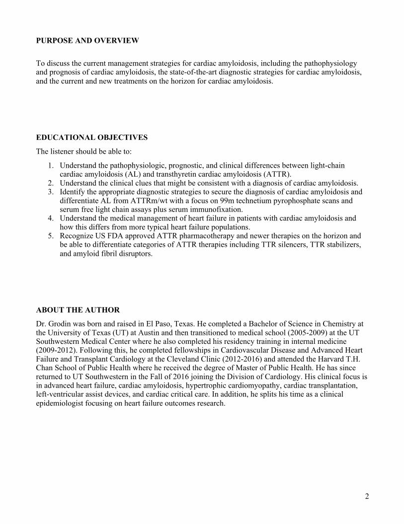
Amyloidosis is a disease of abnormal and often widespread protein deposition. While the origin of these proteins comes from various sources throughout the body, the sine qua none of amyloidosis is that these proteins undergo pathologic misfolding (FIGURE). The normal, physiologic tertiary structures of these proteins transform into more linear structures dominated by β-pleated sheets. These misfolded proteins aggregate into oligomers that eventually form insoluble amyloid fibrils leading to extracellular tissue deposition and organ infiltration. All of the amyloidoses are therefore protein misfolding disorders and this physiologic concept provides the foundation for many amyloidosis-specific therapies described herein.
In addition to the precursor misfolded protein, amyloid fibrils are universally comprised of systemic amyloid P (a chaperone protein), glycosaminoglycans, and calcium. The architecture of amyloid fibrils, visible by electron microscopy, is virtually the same regardless of the amyloid precursor proteins.
In medicine, there are over 30 known precursor proteins with the proclivity to misfold and have the capacity to cause amyloidosis. These various amyloid diseases may be hereditary or acquired and systemic or localized. They vary based on the organ involvement, prognosis, and treatment.
AMYLOIDOSIS TYPES SPECIFIC TO CARDIAC INFILTRATION
The Cardiac Amyloidoses
Amyloidosis is a Protein Misfolding Disorder
Folded Protein Misfolded Protein (Amyloid) Amyloid Fibrils
80-100 Å
4
Although there is a diversity of amyloid-precursor subtypes, approximately 95% of all cases of cardiac amyloidoses originate from abnormal misfolded light chains (AL amyloidosis, formerly "systemic amyloidosis") or misfolded transthyretin (ATTR amyloidosis). The remaining, rare, cardiac amyloidosis subtypes include amyloid A, apolipoprotein A1, apolipoprotein A4, heavy chains, and atrial natriuretic factor[1].
AL Cardiac Amyloidosis
Macroscopically, AL and ATTR amyloidoses are nearly indistinguishable. However, AL and ATTR amyloidoses must be thought of as separate diseases, with different prognosis, and different treatments. As a result, the correct and efficient diagnosis and amyloid typing of cardiac amyloidosis is a clinical necessity.
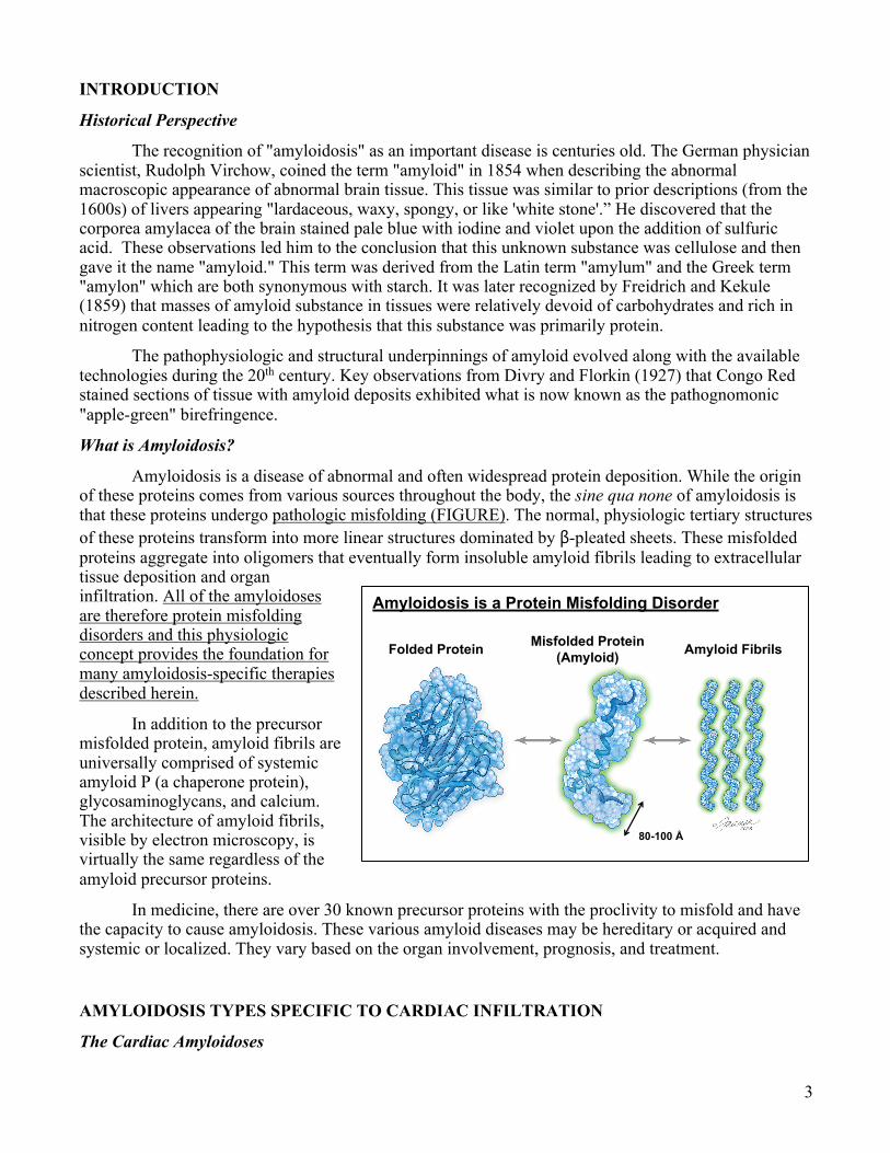
Light chain amyloidosis originates from abnormal plasma cell clones in the bone marrow that overproduce antibody light chain fragments which misfold, aggregate and form protofilaments that then form amyloid fibrils. Estimates from data approximately 30 years old suggest the incidence of AL may be nearly 3,000 new cases per year in the United States. The majority of AL is diagnosed from age 60 onward, but cases have been reported in patients in their 30s and 40s. Therefore, if the clinical suspicion is high, younger age does not rule out AL.
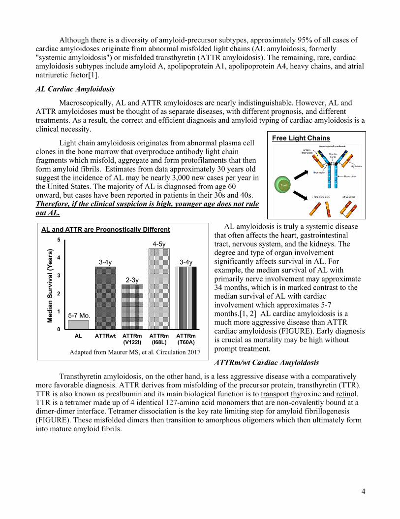
AL amyloidosis is truly a systemic disease that often affects the heart, gastrointestinal tract, nervous system, and the kidneys. The degree and type of organ involvement significantly affects survival in AL. For example, the median survival of AL with primarily nerve involvement may approximate 34 months, which is in marked contrast to the median survival of AL with cardiac involvement which approximates 5-7 months.[1, 2] AL cardiac amyloidosis is a much more aggressive disease than ATTR cardiac amyloidosis (FIGURE). Early diagnosis is crucial as mortality may be high without prompt treatment.
ATTRm/wt Cardiac Amyloidosis
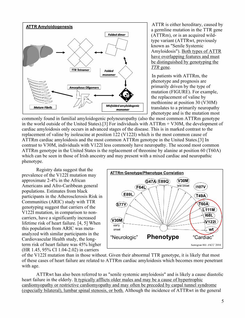
Transthyretin amyloidosis, on the other hand, is a less aggressive disease with a comparatively more favorable diagnosis. ATTR derives from misfolding of the precursor protein, transthyretin (TTR). TTR is also known as prealbumin and its main biological function is to transport thyroxine and retinol. TTR is a tetramer made up of 4 identical 127-amino acid monomers that are non-covalently bound at a dimer-dimer interface. Tetramer dissociation is the key rate limiting step for amyloid fibrillogenesis (FIGURE). These misfolded dimers then transition to amorphous oligomers which then ultimately form into mature amyloid fibrils.
AL and ATTR are Prognostically Different
5-7 Mo.
3-4y
2-3y
4-5y
3-4y
0
1
2
3
4
5
AL ATTRwt ATTRm(V122I)
ATTRm(I68L)
ATTRm(T60A)
Med
ian
Surv
ival
(Yea
rs)
Adapted from Maurer MS, et al. Circulation 2017
5
ATTR is either hereditary, caused by a germline mutation in the TTR gene (ATTRm), or is an acquired wild-type variant (ATTRwt, previously known as "Senile Systemic Amyloidosis"). Both types of ATTR have overlapping features and must be distinguished by genotyping the TTR gene.
In patients with ATTRm, the phenotype and prognosis are primarily driven by the type of mutation (FIGURE). For example, the replacement of valine by methionine at position 30 (V30M) translates to a primarily neuropathy phenotype and is the mutation most
commonly found in familial amyloidogenic polyneuropathy (also the most common ATTRm genotype in the world outside of the United States).[3] For individuals with ATTRm + V30M, the development of cardiac amyloidosis only occurs in advanced stages of the disease. This is in marked contrast to the replacement of valine by isoleucine at position 122 (V122I) which is the most common cause of ATTRm cardiac amyloidosis and the most common ATTRm genotype in the United States.[3] In contrast to V30M, individuals with V122I less commonly have neuropathy. The second most common ATTRm genotype in the United States is the replacement of threonine by alanine at position 60 (T60A) which can be seen in those of Irish ancestry and may present with a mixed cardiac and neuropathic phenotype.
Registry data suggest that the prevalence of the V122I mutation may approximate 2-4% in the African Americans and Afro-Caribbean general populations. Estimates from black participants in the Atherosclerosis Risk in Communities (ARIC) study with TTR genotyping suggest that carriers of the V122I mutation, in comparison to non-carriers, have a significantly increased lifetime risk of heart failure. [4, 5] When this population from ARIC was meta-analyzed with similar participants in the Cardiovascular Health study, the long-term risk of heart failure was 45% higher (HR 1.45, 95% CI 1.04-2.02) in carriers of the V122I mutation than in those without. Given their abnormal TTR genotype, it is likely that most of these cases of heart failure are related to ATTRm cardiac amyloidosis which becomes more penetrant with age.
ATTRwt has also been referred to as "senile systemic amyloidosis" and is likely a cause diastolic heart failure in the elderly. It typically afflicts older males and may be a cause of hypertrophic cardiomyopathy or restrictive cardiomyopathy and may often be preceded by carpal tunnel syndrome (especially bilateral), lumbar spinal stenosis, or both. Although the incidence of ATTRwt in the general

6
population is relatively unknown, autopsy studies suggest that ~25% of individuals 80 years old or older have ATTRwt amyloid deposits in the myocardium. In autopsy studies of individuals 90-105 years old ATTRwt may be present in ~65%. Although, for many of these individuals (44%) the amyloid involvement is minimal (<25%) and likely not enough to cause cardiomyopathy. [6, 7] Altogether, these observations suggest that ATTRwt may deposit in the myocardium over time. However, why some manifest with symptomatic cardiac amyloidosis with marked myocardial infiltration is unknown.
PATHOPHYSIOLOGY OF CARDIAC AMYLOIDOSIS
Degree of Cardiac Involvement.
Diffuse amyloid fibril deposition leads to significant myocardial biventricular thickening (FIGURE, left). It is important to distinguish cardiac amyloidosis from "left ventricular hypertrophy" as there is no true pathologic cardiomyocyte hypertrophy. Rather, the myocardial thickness results from dense extracellular amyloid infiltration (FIGURE, right – the light pink, amorphous material on H&E stain
highlights the diffuse infiltrative nature of amyloidosis) Although there are indeed rare presentations of cardiac amyloidosis without biventricular thickening, its presence should increase the suspicion, in appropriate settings, for cardiac amyloidosis. Macroscopically, AL and ATTR can often be virtually indistinguishable.
Amyloid deposition can lead to atrial and valvular thickening as well, but severely regurgitant valvular lesions are rare. Amyloid infiltration may lead to different conduction delay phenotypes, especially in ATTR, which include atrioventricular blocks and bundle branch blocks. Atrial involvement is nearly universal in all types of amyloidosis and clinically translates into poor atrial function (a critical component of ventricular diastolic function) and atrial fibrillation. Amyloid infiltration of the pericardium is common and can lead to pericardial effusions, but rarely ever leads to large or hemodynamically significant pericardial effusions.
Diastolic Dysfunction in Cardiac Amyloidosis
Patients with cardiac amyloidosis commonly present with signs and symptoms of a restrictive cardiomyopathy. The diffuse amyloid ventricular myocardial deposition and infiltration results in severe, pathological diastolic dysfunction and impaired myocardial relaxation. Increasing degrees of diastolic dysfunction by doppler echocardiography are markers of lower survival and indicated a more advanced amyloid cardiomyopathy. [8, 9] Ventricles with amyloid deposition have very poor compliance and lose the natural physiologic ability to augment stroke volume in the setting of afterload reduction. This fixed stroke volume makes these patients highly dependent on heart rate to augment cardiac output. Furthermore, these stiff non-compliant ventricles are unable to accept incremental filling from atrial contractions, and thus the contribution of atrial contraction is minimal. During diastole, there is early rapid filling of the ventricles resulting from very high atrial pressures which is then immediately
7
followed by filling limited by the stiff myocardium resulting in a prominent “y descent” on atrial pressure curves and “square root sign” on ventricular pressure curves.
The downstream ramifications of these hemodynamic abnormalities translate into signs and symptoms of congestion which include peripheral edema, ascites, pulmonary edema, distended jugular veins, dyspnea, orthopnea, etc.
Systolic Function in Cardiac Amyloidosis
Unlike more traditional dilated or ischemic cardiomyopathies, the left ventricular ejection fraction (LVEF) is a poor marker of systolic function in cardiac amyloidosis and can thus, be misleading. A ventricle with cardiac amyloidosis has a very low end diastolic volume (small chamber size) which then produces low stroke volume that can translate to low cardiac output. For example, a normal heart with an LVEF of 55% may have an end diastolic volume of 120 cc and stroke volume of 66 cc (55%=66/120 x 100) which is in contrast to a heart with amyloidosis with an LVEF of 55% that has an end diastolic volume of 64 cc and a stroke volume of only 35 cc (55%=35/64 x 100).
Atypical Cardiac Manifestations of Cardiac Amyloidosis
Although the most common cardiac amyloidosis phenotype is that of ventricular thickening and restrictive cardiomyopathy, cardiac amyloidosis can present atypically. For example, different patterns of amyloid infiltration and deposition can lead to presentations without ventricular thickening, asymmetric septal thickening (“hypertrophic obstructive cardiomyopathy”, more commonly in ATTRwt than AL), and myocardial ischemia or even infarction (AL more commonly than ATTRm/wt).[10-12] Interestingly, the latter may be the downstream effects from dense coronary microvascular amyloid deposition.
CLINICAL PRESENTATION OF CARDIAC AMYLOIDOSIS
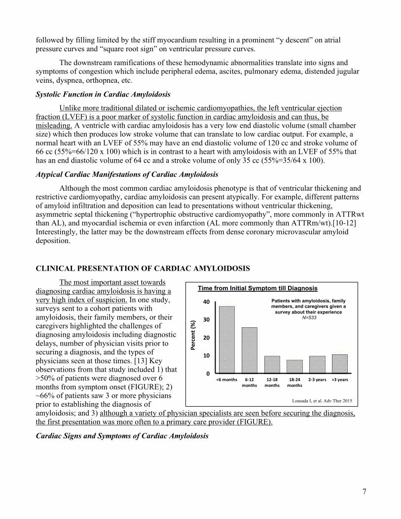
The most important asset towards diagnosing cardiac amyloidosis is having a very high index of suspicion. In one study, surveys sent to a cohort patients with amyloidosis, their family members, or their caregivers highlighted the challenges of diagnosing amyloidosis including diagnostic delays, number of physician visits prior to securing a diagnosis, and the types of physicians seen at those times. [13] Key observations from that study included 1) that >50% of patients were diagnosed over 6 months from symptom onset (FIGURE); 2) ~66% of patients saw 3 or more physicians prior to establishing the diagnosis of amyloidosis; and 3) although a variety of physician specialists are seen before securing the diagnosis, the first presentation was more often to a primary care provider (FIGURE).
Cardiac Signs and Symptoms of Cardiac Amyloidosis
Time from Initial Symptom till Diagnosis
37.3
25.7
9.67.4
9.6 10.5
0
10
20
30
40
<6 months 6-12months
12-18months
18-24months
2-3 years >3 years
Perc
ent (
%)
Lousada I, et al. Adv Ther 2015
Patients with amyloidosis, family members, and caregivers given a
survey about their experienceN=533

8
Cardiac amyloidosis commonly presents with signs and symptoms of congestive heart failure, mainly with right-sided symptoms. Dyspnea, fatigue and weakness are also common. Individuals with cardiac amyloidosis may present with atrial arrhythmia as a result of atrial amyloid infiltration and
atrioventricular blocks or bundle branch bocks as a result of amyloid infiltration of the conduction system. Amyloidosis can infiltrate the coronary macro and microvasculature leading to angina. These individuals typically have “normal coronaries” on coronary angiography.
Three other cardiac phenotypes might be direct manifestations of cardiac amyloidosis and include hypertrophic cardiomyopathy (especially past the 6th decade of life), heart failure with preserved ejection fraction, and low-flow, low-gradient severe aortic stenosis (commonly encountered in the TAVR population - FIGURE). [14, 15]
Non-cardiac Signs and Symptoms of Cardiac Amyloidosis
One of the clinical scenarios that commonly precedes a diagnosis of cardiac amyloidosis is carpal tunnel syndrome – especially when it is bilateral (ATTR>AL). A recent prospective cohort suggested that ~10% of the synovial tissue from carpal tunnel release surgeries may be stains Congo Red positive and precedes the development of heart failure. [16] Other red flags or clues towards the diagnosis of cardiac amyloidosis include (1) unexplained left ventricular hypertrophy by echocardiography (especially with no prior history of hypertension); (2) unexplained restrictive cardiomyopathy; (3) atraumatic biceps tendon rupture, (4) lumbar canal stenosis (ATTRwt), (5) unexplained peripheral or autonomic neuropathy (AL>ATTRm>ATTRwt); and (6) low or normal blood pressure (or intolerance to beta-blockers and calcium channel blockers) in a patient with a prior history of hypertension.
Some signs are more specific to AL amyloidosis. These include proteinuria, which is commonly in the nephrotic range and may indicate renal involvement; jaw claudication; diarrhea that can occur with intestinal amyloid infiltration; and unexplained weight loss. Other clues pointing towards a diagnosis of AL amyloidosis are macroglossia and periorbital purpura. The latter might lead to a patient complaining of a spontaneous “black eye.”
DIAGNOSIS
Electrocardiography
Types of Physicians Visited Before Diagnosis of Amyloidosis
0 100 200 300 400 500
Fourth visit (n=170)
Third visit (n=272)
Second visit (n=383)
First visit (n=433)
Primary care physician CardiologistHematologist/Oncologist NephrologistGastroenterologist Other
Lousada I, et al. Adv Ther 2015
Prevalence of ATTR in Common Clinical Scenarios
Amyloid staining has also been positive
• Lumbar canal stenosis
• Rotator cuff tears
• Atraumatic biceps tendon rupture
Gonzalez-Lopez E, et al. EHJ 2015Longhi S, et al. JACC CV Imaging 2016
Sperry BW, et al. JACC 2018Sueyoshi T, et al. Hum Pathol 2011
~13%~12%
~10%(AL and ATTR)
0%
5%
10%
15%
20%
HospitalizedHFpEF
AorticStenosis(TAVR)
Carpal TunnelRelease
9
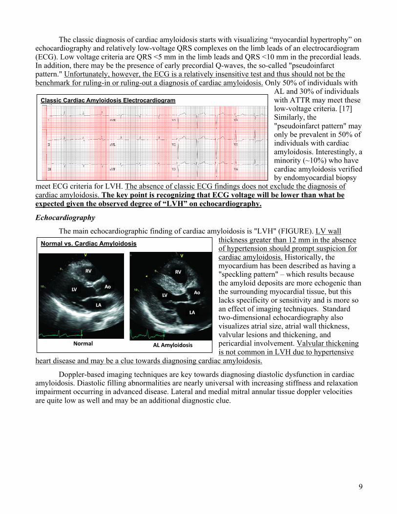
The classic diagnosis of cardiac amyloidosis starts with visualizing “myocardial hypertrophy” on echocardiography and relatively low-voltage QRS complexes on the limb leads of an electrocardiogram (ECG). Low voltage criteria are QRS <5 mm in the limb leads and QRS <10 mm in the precordial leads. In addition, there may be the presence of early precordial Q-waves, the so-called "pseudoinfarct pattern." Unfortunately, however, the ECG is a relatively insensitive test and thus should not be the benchmark for ruling-in or ruling-out a diagnosis of cardiac amyloidosis. Only 50% of individuals with
AL and 30% of individuals with ATTR may meet these low-voltage criteria. [17] Similarly, the "pseudoinfarct pattern" may only be prevalent in 50% of individuals with cardiac amyloidosis. Interestingly, a minority (~10%) who have cardiac amyloidosis verified by endomyocardial biopsy
meet ECG criteria for LVH. The absence of classic ECG findings does not exclude the diagnosis of cardiac amyloidosis. The key point is recognizing that ECG voltage will be lower than what be expected given the observed degree of “LVH” on echocardiography.
Echocardiography
The main echocardiographic finding of cardiac amyloidosis is "LVH" (FIGURE). LV wall thickness greater than 12 mm in the absence of hypertension should prompt suspicion for cardiac amyloidosis. Historically, the myocardium has been described as having a "speckling pattern" – which results because the amyloid deposits are more echogenic than the surrounding myocardial tissue, but this lacks specificity or sensitivity and is more so an effect of imaging techniques. Standard two-dimensional echocardiography also visualizes atrial size, atrial wall thickness, valvular lesions and thickening, and pericardial involvement. Valvular thickening is not common in LVH due to hypertensive
heart disease and may be a clue towards diagnosing cardiac amyloidosis.
Doppler-based imaging techniques are key towards diagnosing diastolic dysfunction in cardiac amyloidosis. Diastolic filling abnormalities are nearly universal with increasing stiffness and relaxation impairment occurring in advanced disease. Lateral and medial mitral annular tissue doppler velocities are quite low as well and may be an additional diagnostic clue.
Classic Cardiac Amyloidosis Electrocardiogram
Normal vs. Cardiac Amyloidosis
Normal AL Amyloidosis
RV
Ao
LA
LV
RV
Ao
LA
LV
10
Speckle tracking techniques via echocardiography can be additionally useful to heighten suspicion for cardiac amyloidosis. These techniques provide information regarding longitudinal strain measurements that represent myocardial deformation. Quantitatively, measurements from this imaging technique can yield a characteristic "apical sparing" (FIGURE) pattern on a polar map that may be specific to amyloidosis.[18]
Cardiac Magnetic Resonance Imaging
Cardiac magnetic resonance imaging (CMR) is useful towards establishing the diagnosis of cardiac amyloidosis. Imaging with gadolinium contrast can yield characteristic late gadolinium enhancement (LGE) patterns (FIGURE) that do not follow a coronary distribution and may be diffuse and subendocardial.[19] In some cases, especially with ATTRwt, the LGE can be patchy or transmural. CMRs are highly sensitive (93%) and relatively specific (70%) for cardiac amyloidosis with a negative predictive value of 84%. Some drawbacks of this imaging modality are renal dysfunction which limits the use of gadolinium and the presence of MRI non-compatible implants or devices. Additional useful CMR modalities include T1-mapping.
99m Technetium Pyrophosphate Scintigraphy
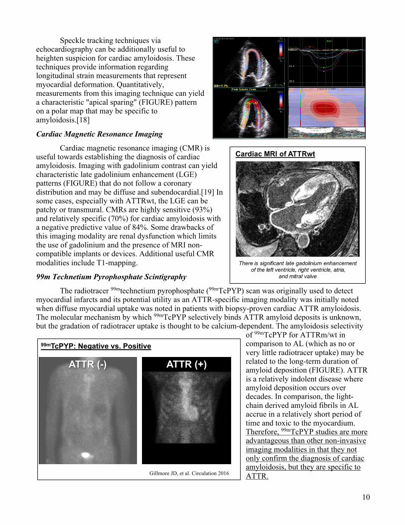
The radiotracer 99mtechnetium pyrophosphate (99mTcPYP) scan was originally used to detect myocardial infarcts and its potential utility as an ATTR-specific imaging modality was initially noted when diffuse myocardial uptake was noted in patients with biopsy-proven cardiac ATTR amyloidosis. The molecular mechanism by which 99mTcPYP selectively binds ATTR amyloid deposits is unknown, but the gradation of radiotracer uptake is thought to be calcium-dependent. The amyloidosis selectivity
of 99mTcPYP for ATTRm/wt in comparison to AL (which as no or very little radiotracer uptake) may be related to the long-term duration of amyloid deposition (FIGURE). ATTR is a relatively indolent disease where amyloid deposition occurs over decades. In comparison, the light-chain derived amyloid fibrils in AL accrue in a relatively short period of time and toxic to the myocardium. Therefore, 99mTcPYP studies are more advantageous than other non-invasive imaging modalities in that they not only confirm the diagnosis of cardiac amyloidosis, but they are specific to ATTR.
99mTcPYP: Negative vs. Positive
ATTR (+)
Gillmore JD, et al. Circulation 2016
ATTR (-)
11
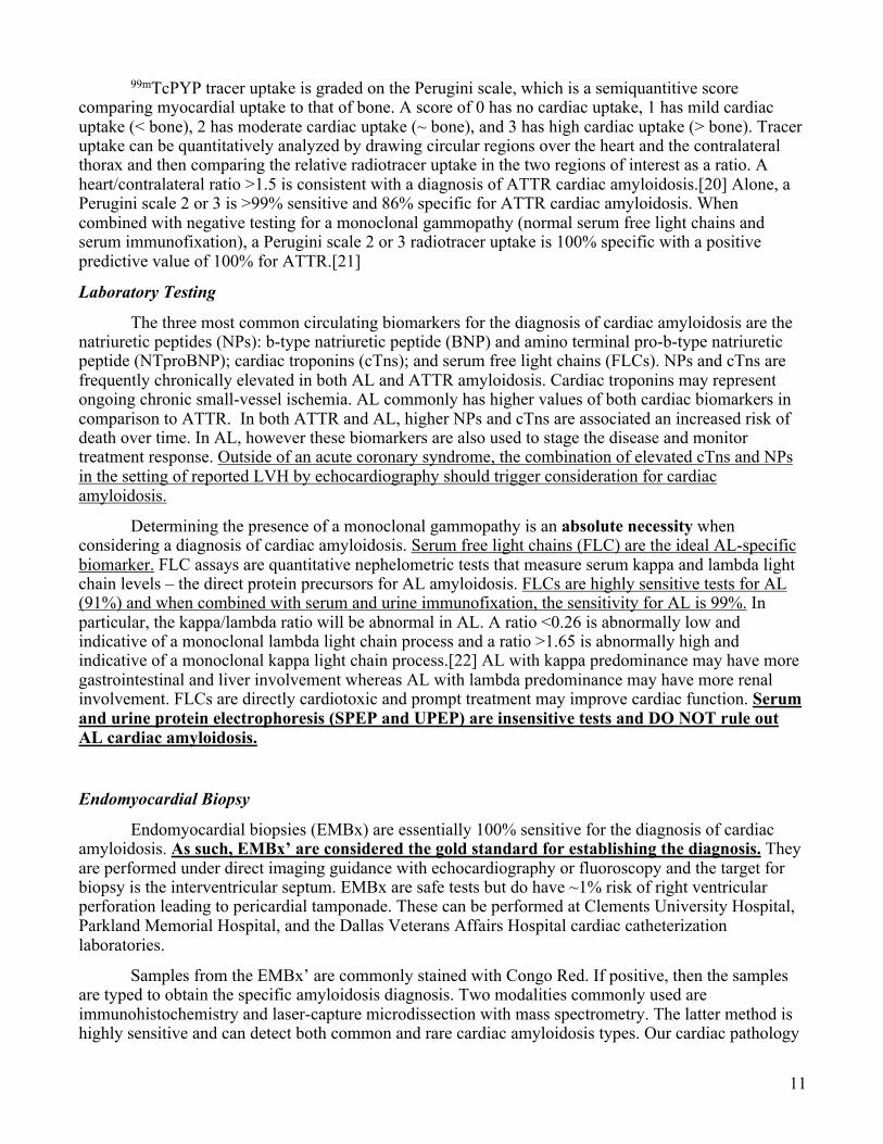
99mTcPYP tracer uptake is graded on the Perugini scale, which is a semiquantitive score comparing myocardial uptake to that of bone. A score of 0 has no cardiac uptake, 1 has mild cardiac uptake (< bone), 2 has moderate cardiac uptake (~ bone), and 3 has high cardiac uptake (> bone). Tracer uptake can be quantitatively analyzed by drawing circular regions over the heart and the contralateral thorax and then comparing the relative radiotracer uptake in the two regions of interest as a ratio. A heart/contralateral ratio >1.5 is consistent with a diagnosis of ATTR cardiac amyloidosis.[20] Alone, a Perugini scale 2 or 3 is >99% sensitive and 86% specific for ATTR cardiac amyloidosis. When combined with negative testing for a monoclonal gammopathy (normal serum free light chains and serum immunofixation), a Perugini scale 2 or 3 radiotracer uptake is 100% specific with a positive predictive value of 100% for ATTR.[21]
Laboratory Testing
The three most common circulating biomarkers for the diagnosis of cardiac amyloidosis are the natriuretic peptides (NPs): b-type natriuretic peptide (BNP) and amino terminal pro-b-type natriuretic peptide (NTproBNP); cardiac troponins (cTns); and serum free light chains (FLCs). NPs and cTns are frequently chronically elevated in both AL and ATTR amyloidosis. Cardiac troponins may represent ongoing chronic small-vessel ischemia. AL commonly has higher values of both cardiac biomarkers in comparison to ATTR. In both ATTR and AL, higher NPs and cTns are associated an increased risk of death over time. In AL, however these biomarkers are also used to stage the disease and monitor treatment response. Outside of an acute coronary syndrome, the combination of elevated cTns and NPs in the setting of reported LVH by echocardiography should trigger consideration for cardiac amyloidosis.
Determining the presence of a monoclonal gammopathy is an absolute necessity when considering a diagnosis of cardiac amyloidosis. Serum free light chains (FLC) are the ideal AL-specific biomarker. FLC assays are quantitative nephelometric tests that measure serum kappa and lambda light chain levels – the direct protein precursors for AL amyloidosis. FLCs are highly sensitive tests for AL (91%) and when combined with serum and urine immunofixation, the sensitivity for AL is 99%. In particular, the kappa/lambda ratio will be abnormal in AL. A ratio <0.26 is abnormally low and indicative of a monoclonal lambda light chain process and a ratio >1.65 is abnormally high and indicative of a monoclonal kappa light chain process.[22] AL with kappa predominance may have more gastrointestinal and liver involvement whereas AL with lambda predominance may have more renal involvement. FLCs are directly cardiotoxic and prompt treatment may improve cardiac function. Serum and urine protein electrophoresis (SPEP and UPEP) are insensitive tests and DO NOT rule out AL cardiac amyloidosis.
Endomyocardial Biopsy
Endomyocardial biopsies (EMBx) are essentially 100% sensitive for the diagnosis of cardiac amyloidosis. As such, EMBx’ are considered the gold standard for establishing the diagnosis. They are performed under direct imaging guidance with echocardiography or fluoroscopy and the target for biopsy is the interventricular septum. EMBx are safe tests but do have ~1% risk of right ventricular perforation leading to pericardial tamponade. These can be performed at Clements University Hospital, Parkland Memorial Hospital, and the Dallas Veterans Affairs Hospital cardiac catheterization laboratories.
Samples from the EMBx’ are commonly stained with Congo Red. If positive, then the samples are typed to obtain the specific amyloidosis diagnosis. Two modalities commonly used are immunohistochemistry and laser-capture microdissection with mass spectrometry. The latter method is highly sensitive and can detect both common and rare cardiac amyloidosis types. Our cardiac pathology
12
division sends the Congo Red (+) samples to the Mayo Clinic for advanced typing with mass spectrometry.
Although 99mTcPYP scans with FLC/IFE assays can non-invasively establish the diagnosis of ATTR or rule out cardiac amyloidosis (if both normal), there are rare instances when these results can be indeterminate necessitating an EMBx. Furthermore, if FLCs are consistent with or there is sufficiently high enough suspicion for AL, establishing end-organ involvement with an EMBx is essential.
Fat Pad Biopsy
Abdominal fat pad biopsies are not reliable tests for cardiac amyloidosis. They are 60-80% sensitive for detecting AL, 65-85% sensitive for detecting ATTRm, and only 14% sensitive for detecting ATTRwt. Fat pad biopsies are highly dependent on the operator performing the biopsy, the amount of tissue acquired, and pathology expertise. Given these diagnostic limitations, a negative fat pad biopsy does not rule out amyloidosis.
Genetic Testing
TTR gene sequencing should be undertaken in ALL cases of TTR-related amyloidosis. Clinical grounds and family history are not sufficient to determine an abnormal TTR genotype. ATTR can have both incomplete and late penetrance. Furthermore, sequencing the TTR gene with genetic counseling is recommended in cases where hereditary TTR is proven by mass spectrometry or suspected with a negative profile on mass spectrometry.
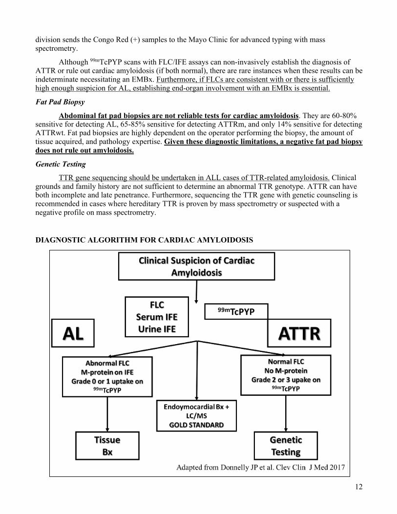
DIAGNOSTIC ALGORITHM FOR CARDIAC AMYLOIDOSIS
13
TREATMENT OF CARDIAC AMYLOIDOSIS
Management of Heart Failure in Cardiac Amyloidosis
Diuretics (mainly loop) and sodium restriction are cornerstones for the management of congestion in patient with cardiac amyloidosis and heart failure. Unlike ischemic or other non-ischemic cardiomyopathies with reduced LVEF, beta-blockers, renin-angiotensin system inhibitors, and angiotensin receptor blocker neprilysin inhibitors are neither effective or well-tolerated in cardiac amyloidosis.
Beta-blockers, however, can be used cautiously for rate control in atrial fibrillation. Calcium channel blockers, however, are contraindicated in amyloidosis. Calcium channel blockers bind amyloid fibrils and promote hypotension and syncope. Historically, digoxin use was frowned upon given observations from in vitro experiments that it binds to amyloid fibrils possibly increasing the risk of digoxin toxicity.[23] However, recent observations from a cohort with cardiac amyloidosis on digoxin therapy support its safety with the understanding that it has a very narrow therapeutic window.[24]
The maintenance of normal sinus rhythm is paramount for the management of atrial fibrillation in cardiac amyloidosis. Atrial fibrillation can lead to further heart failure decompensation. Stroke risk scores like CHADS2 or CHADS2VASC are not applicable to patients with cardiac amyloidosis as they are all considered high-risk of thromboembolism. It is therefore not surprising that intracardiac thrombi are commonly found on autopsy.[25, 26] Atrial function can be so impaired in cardiac amyloidosis, that intracardiac thrombi can form even in sinus rhythm. There should be a lower threshold to anticoagulate in AL as it may be more thrombogenic than ATTR.
Brief Overview of Treatment of AL
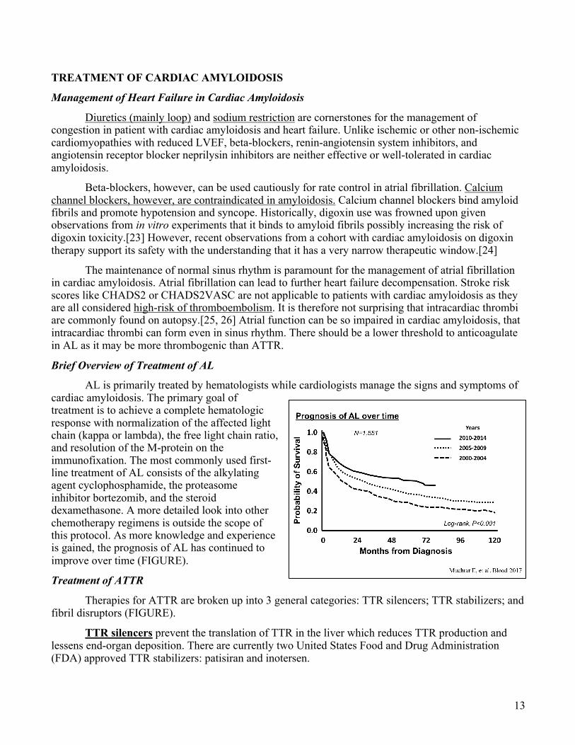
AL is primarily treated by hematologists while cardiologists manage the signs and symptoms of cardiac amyloidosis. The primary goal of treatment is to achieve a complete hematologic response with normalization of the affected light chain (kappa or lambda), the free light chain ratio, and resolution of the M-protein on the immunofixation. The most commonly used first-line treatment of AL consists of the alkylating agent cyclophosphamide, the proteasome inhibitor bortezomib, and the steroid dexamethasone. A more detailed look into other chemotherapy regimens is outside the scope of this protocol. As more knowledge and experience is gained, the prognosis of AL has continued to improve over time (FIGURE).
Treatment of ATTR
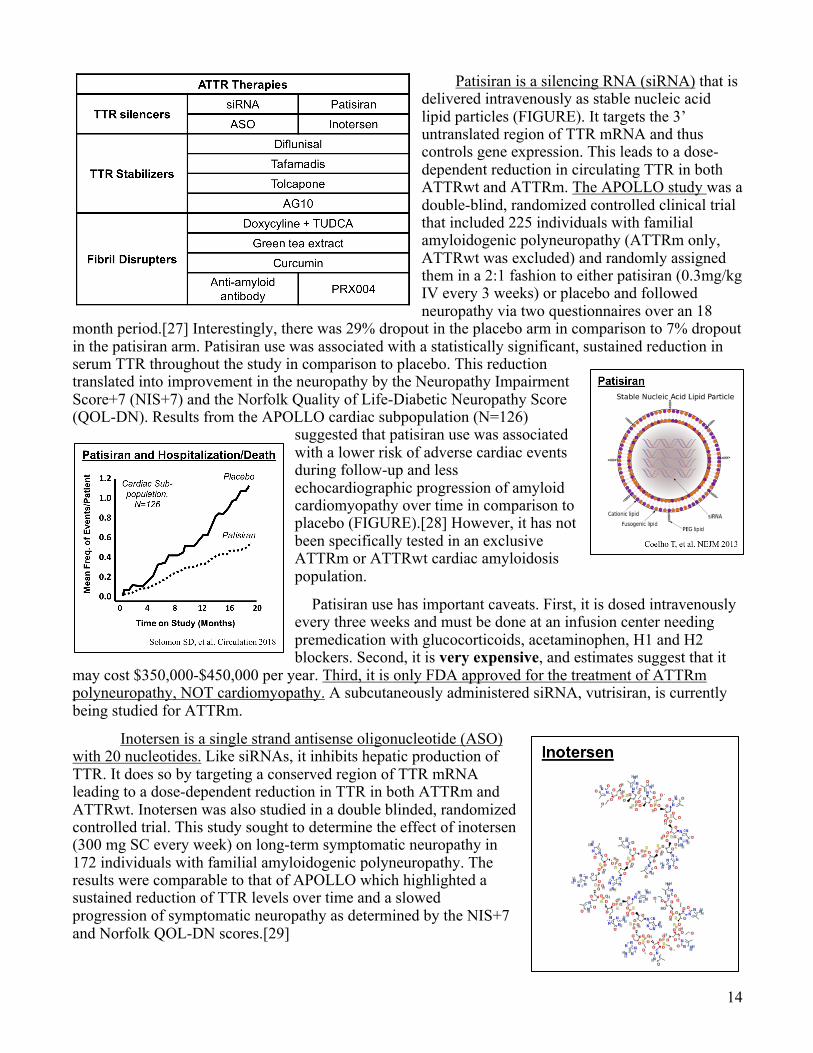
Therapies for ATTR are broken up into 3 general categories: TTR silencers; TTR stabilizers; and fibril disruptors (FIGURE).
TTR silencers prevent the translation of TTR in the liver which reduces TTR production and lessens end-organ deposition. There are currently two United States Food and Drug Administration (FDA) approved TTR stabilizers: patisiran and inotersen.
14
Patisiran is a silencing RNA (siRNA) that is delivered intravenously as stable nucleic acid lipid particles (FIGURE). It targets the 3’ untranslated region of TTR mRNA and thus controls gene expression. This leads to a dose-dependent reduction in circulating TTR in both ATTRwt and ATTRm. The APOLLO study was a double-blind, randomized controlled clinical trial that included 225 individuals with familial amyloidogenic polyneuropathy (ATTRm only, ATTRwt was excluded) and randomly assigned them in a 2:1 fashion to either patisiran (0.3mg/kg IV every 3 weeks) or placebo and followed neuropathy via two questionnaires over an 18
month period.[27] Interestingly, there was 29% dropout in the placebo arm in comparison to 7% dropout in the patisiran arm. Patisiran use was associated with a statistically significant, sustained reduction in serum TTR throughout the study in comparison to placebo. This reduction translated into improvement in the neuropathy by the Neuropathy Impairment Score+7 (NIS+7) and the Norfolk Quality of Life-Diabetic Neuropathy Score (QOL-DN). Results from the APOLLO cardiac subpopulation (N=126)
suggested that patisiran use was associated with a lower risk of adverse cardiac events during follow-up and less echocardiographic progression of amyloid cardiomyopathy over time in comparison to placebo (FIGURE).[28] However, it has not been specifically tested in an exclusive ATTRm or ATTRwt cardiac amyloidosis population.
Patisiran use has important caveats. First, it is dosed intravenously every three weeks and must be done at an infusion center needing premedication with glucocorticoids, acetaminophen, H1 and H2 blockers. Second, it is very expensive, and estimates suggest that it
may cost $350,000-$450,000 per year. Third, it is only FDA approved for the treatment of ATTRm polyneuropathy, NOT cardiomyopathy. A subcutaneously administered siRNA, vutrisiran, is currently being studied for ATTRm.
Inotersen is a single strand antisense oligonucleotide (ASO) with 20 nucleotides. Like siRNAs, it inhibits hepatic production of TTR. It does so by targeting a conserved region of TTR mRNA leading to a dose-dependent reduction in TTR in both ATTRm and ATTRwt. Inotersen was also studied in a double blinded, randomized controlled trial. This study sought to determine the effect of inotersen (300 mg SC every week) on long-term symptomatic neuropathy in 172 individuals with familial amyloidogenic polyneuropathy. The results were comparable to that of APOLLO which highlighted a sustained reduction of TTR levels over time and a slowed progression of symptomatic neuropathy as determined by the NIS+7 and Norfolk QOL-DN scores.[29]
15
Although the SC dosing of inotersen may be attractive in comparison to patisiran, it too has important caveats. First, it has not been tested in an exclusive ATTRm or ATTRwt cardiac amyloidosis population. Although, preliminary studies are ongoing. Second, it is also very expensive with estimates suggesting an annual cost of $350,000-$450,000. Third, it is also only FDA approved for the treatment of ATTRm polyneuropathy, NOT cardiomyopathy. Fourth, serious adverse events include thrombocytopenia and glomerulonephritis.
TTR stabilizers prevent the dissociation of TTR tetramers. Each tetramer has 2 thyroxine-binding pockets, that when bound, prevent dissociation. This is the rate limiting step for ATTR fibrillogenesis. The basis for this mechanism was discovered from rare individuals with ATTRm who are heterozygous for both V30M and T119M. While V30M is a highly penetrant mutation causing familial amyloidogenic polyneuropathy, individuals with concomitant T119M mutations are virtually free of disease. T119M is a trans-suppressor mutation and increases the dissociative transition-state energy effectively preventing tetrameter dissociation.[30] Small molecule binding to the thyroxine-binding pockets has a similar effect. Currently, there are four agents that bind the thyroxine-binding pockets and stabilize TTR dissociation: (1) diflunisal, (2) tolcapone, (3) tafamadis, and (4) AG10.
Diflunisal is a non-steroidal anti-inflammatory drug (NSAID) used to treat arthritis and musculoskeletal pain. Interestingly, it was found to interact with the TTR thyroxine-binding pocket and stabilizes TTR dissociation. However, it is not a well-established therapy for ATTR. While some reports suggest benefit, there are no randomized data to support its use. There are also concerns regarding heightened bleeding risk, adverse effects on renal function, and fluid retention (especially in heart failure).
Tolcapone is a current pharmacotherapy approved by the FDA for the treatment of Parkinson disease. It is a potent TTR stabilizer and binds both TTR thyroxine binding pockets. However, there are no data currently supporting its use in ATTR and has a black box warning for acute fulminant liver failure.
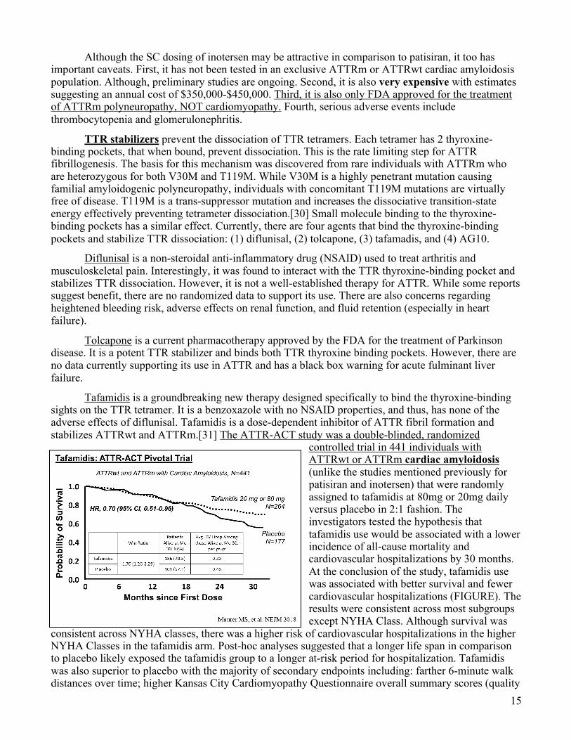
Tafamidis is a groundbreaking new therapy designed specifically to bind the thyroxine-binding sights on the TTR tetramer. It is a benzoxazole with no NSAID properties, and thus, has none of the adverse effects of diflunisal. Tafamidis is a dose-dependent inhibitor of ATTR fibril formation and stabilizes ATTRwt and ATTRm.[31] The ATTR-ACT study was a double-blinded, randomized
controlled trial in 441 individuals with ATTRwt or ATTRm cardiac amyloidosis (unlike the studies mentioned previously for patisiran and inotersen) that were randomly assigned to tafamidis at 80mg or 20mg daily versus placebo in 2:1 fashion. The investigators tested the hypothesis that tafamidis use would be associated with a lower incidence of all-cause mortality and cardiovascular hospitalizations by 30 months. At the conclusion of the study, tafamidis use was associated with better survival and fewer cardiovascular hospitalizations (FIGURE). The results were consistent across most subgroups except NYHA Class. Although survival was
consistent across NYHA classes, there was a higher risk of cardiovascular hospitalizations in the higher NYHA Classes in the tafamidis arm. Post-hoc analyses suggested that a longer life span in comparison to placebo likely exposed the tafamidis group to a longer at-risk period for hospitalization. Tafamidis was also superior to placebo with the majority of secondary endpoints including: farther 6-minute walk distances over time; higher Kansas City Cardiomyopathy Questionnaire overall summary scores (quality
16
of life); lower NTproBNP levels over time, and less reduction in left ventricular stroke volume over time. Importantly, there was little to no difference in side effects or serious adverse events with tafamadis in comparison to placebo. In addition, there was less tafamidis discontinuation than placebo – perhaps related to a reduction in non-cardiac amyloidosis symptoms. Currently, tafamidis is not commercially available and not approved by the United States FDA. It was granted “Breakthrough Therapy” designation, however and regulatory review is currently underway. If approved by the US FDA, this will be the only therapy with approval specifically for ATTRm or ATTRwt cardiomyopathy.
AG10 is the most potent and selective TTR-stabilizing small molecule currently being investigated. It is highly bioavailable with minimal to no toxicity. Like tafamidis, it stabilizes both ATTRm and ATTRwt. Phase 2 and 3 studies are underway and UT Southwestern is a participating site.
There are currently no well-established ATTR fibril disruptors. However, some therapies may have fibril disrupting properties and small, underpowered studies have supported the hypothesis that they may impact ATTR cardiac amyloidosis.
Mouse models demonstrate mature ATTR fibril disruption with doxycycline and tauroursodeoxycholic acid (TUDCA). While this has been studied in one small, open-labeled pilot, there are no large well-designed studies supporting its use and TUDCA is not available in the United States.
Green tea extract contains epigallocatechin-3-gallate (EGCG) which has ATTR fibril-disrupting properties and exhibits TTR stabilization (separate from the thyroxine binding pocket). One pilot study of 9 individuals with ATTR suggested that 500-700 of EGCG per day or 1.5-2L of green tea per day for 12 months was well-tolerated and associated with lower left ventricular mass by CMR over time.
Curcumin is the active ingredient in turmeric, a spice, and is an in vitro TTR stabilizer and amyloid fibril disruptor. Animal studies suggest curcumin may increase macrophage mediated degradation of amyloid fibrils.
A synthetic anti-amyloid antibody, PRX004, designed to prevent ATTR fibril deposition and promote clearance has recently been developed and is currently in Phase 1 investigation.
Anti-Serum Amyloid P Component Antibody
Serum amyloid P (SAP) is a ubiquitous component of all amyloid fibrils, regardless of precursor protein type. Because SAP stabilizes all types of fibrils, an effective monoclonal SAP antibody would be the “Holy Grail” of amyloidosis therapies (i.e. with utility for BOTH AL and ATTR). Recently, a monoclonal IgG1 anti-SAP antibody, desamizumab, has been developed and had promising results in a pilot study (N=16) of individuals (mostly AL) receiving a 6-week treatment.[32] Some had dramatic amyloid reversal and others with significant reduction in amyloid burden. Unfortunately, however, a phase 2 study is currently suspended.
Heart Transplantation and Left Ventricular Assist Devices
Heart transplantation can be available in well-selected individuals with end-stage ATTRm and ATTRwt cardiac amyloidosis and clinically isolated disease. Their post-transplant survival is comparable to individuals with non-amyloidosis cardiomyopathy. Concomitant liver transplant is not needed in ATTRwt and may be considered in ATTRm depending on TTR genotype. For example, ATTRm V122I is a more indolent TTR mutation and these individuals will do well with heart transplantation alone. On the other hand, individuals with ATTRm T60A suffer from both significant polyneuropathy and cardiomyopathy and require combined heart and liver transplantation. Because AL is a much more aggressive and systemically involved disease, heart transplantation can only be offered to very carefully selected patients either with a complete hematological response or who have undergone an autologous stem cell transplantation. The 5-year post-transplant survival in these patients may approach 65%.
17
Left ventricular assist devices (LVADs) are frequently used as a bridge to heart transplant or as destination therapy for individuals with advanced cardiomyopathy not eligible for heart transplantation. While technically feasible in cardiac amyloidosis, LVADs are highly dependent on patient selection. Because the thickened left ventricle chamber size is so small (small end diastolic volume, and small stroke volume), the LVAD inflow cannula (typically inserted into left ventricle apex) has little room for placement.
CONCLUSION
Cardiac amyloidosis is emerging from its characterization as a rare and unusual cause of heart failure. Epidemiological studies have identified at-risk populations for cardiac amyloidosis. In addition, recent technical advances have dramatically altered the diagnostic landscape for both AL and ATTR, with an array of options that offer a highly efficient and a highly detailed cardiomyopathy characterization. This is in parallel to the development of novel, promising treatments for AL and ATTR. Because of this progress, early recognition of cardiac amyloidosis is critical since modern amyloidosis therapies have the potential to alter the course of what was once a disease with little-to-no treatment.
CARDIAC AMYLOIDOSIS IS NOT A “RARE AND HOPELESS” DISEASE!
KEY REFERENCES
1. Kyle, R.A., et al., A trial of three regimens for primary amyloidosis: colchicine alone, melphalan and prednisone, and melphalan, prednisone, and colchicine. N Engl J Med, 1997. 336(17): p. 1202-7.
2. Sperry, B.W., et al., Efficacy of Chemotherapy for Light-Chain Amyloidosis in Patients Presenting With Symptomatic Heart Failure. J Am Coll Cardiol, 2016. 67(25): p. 2941-8.
3. Maurer, M.S., et al., Genotype and Phenotype of Transthyretin Cardiac Amyloidosis: THAOS (Transthyretin Amyloid Outcome Survey). J Am Coll Cardiol, 2016. 68(2): p. 161-72.
4. Quarta, C.C., et al., The amyloidogenic V122I transthyretin variant in elderly black Americans. N Engl J Med, 2015. 372(1): p. 21-9.
5. Jacobson, D.R., et al., Prevalence of the amyloidogenic transthyretin (TTR) V122I allele in 14 333 African-Americans. Amyloid, 2015. 22(3): p. 171-4.
6. Lie, J.T. and P.I. Hammond, Pathology of the senescent heart: anatomic observations on 237 autopsy studies of patients 90 to 105 years old. Mayo Clin Proc, 1988. 63(6): p. 552-64.
7. Grogan, M., et al., Natural History of Wild-Type Transthyretin Cardiac Amyloidosis and Risk Stratification Using a Novel Staging System. J Am Coll Cardiol, 2016. 68(10): p. 1014-20.
8. Klein, A.L., et al., Doppler characterization of left ventricular diastolic function in cardiac amyloidosis. J Am Coll Cardiol, 1989. 13(5): p. 1017-26.
9. Klein, A.L., et al., Prognostic significance of Doppler measures of diastolic function in cardiac amyloidosis. A Doppler echocardiography study. Circulation, 1991. 83(3): p. 808-16.
10. Gertz, M.A., et al., Endomyocardial biopsy-proven light chain amyloidosis (AL) without echocardiographic features of infiltrative cardiomyopathy. Am J Cardiol, 1997. 80(1): p. 93-5.
11. Helder, M.R., et al., Impact of incidental amyloidosis on the prognosis of patients with hypertrophic cardiomyopathy undergoing septal myectomy for left ventricular outflow tract obstruction. Am J Cardiol, 2014. 114(9): p. 1396-9.
12. Neben-Wittich, M.A., et al., Obstructive intramural coronary amyloidosis and myocardial ischemia are common in primary amyloidosis. Am J Med, 2005. 118(11): p. 1287.
18
13. Lousada, I., et al., Light Chain Amyloidosis: Patient Experience Survey from the Amyloidosis Research Consortium. Adv Ther, 2015. 32(10): p. 920-8.
14. Gonzalez-Lopez, E., et al., Wild-type transthyretin amyloidosis as a cause of heart failure with preserved ejection fraction. Eur Heart J, 2015. 36(38): p. 2585-94.
15. Longhi, S., et al., Coexistence of Degenerative Aortic Stenosis and Wild-Type Transthyretin-Related Cardiac Amyloidosis. JACC Cardiovasc Imaging, 2016. 9(3): p. 325-7.
16. Sperry, B.W., et al., Tenosynovial and Cardiac Amyloidosis in Patients Undergoing Carpal Tunnel Release. J Am Coll Cardiol, 2018. 72(17): p. 2040-2050.
17. Sperry, B.W., et al., Are classic predictors of voltage valid in cardiac amyloidosis? A contemporary analysis of electrocardiographic findings. Int J Cardiol, 2016. 214: p. 477-81.
18. Phelan, D., et al., Relative apical sparing of longitudinal strain using two-dimensional speckle-tracking echocardiography is both sensitive and specific for the diagnosis of cardiac amyloidosis. Heart, 2012. 98(19): p. 1442-8.
19. Syed, I.S., et al., Role of cardiac magnetic resonance imaging in the detection of cardiac amyloidosis. JACC Cardiovasc Imaging, 2010. 3(2): p. 155-64.
20. Bokhari, S., et al., (99m)Tc-pyrophosphate scintigraphy for differentiating light-chain cardiac amyloidosis from the transthyretin-related familial and senile cardiac amyloidoses. Circ Cardiovasc Imaging, 2013. 6(2): p. 195-201.
21. Gillmore, J.D., et al., Nonbiopsy Diagnosis of Cardiac Transthyretin Amyloidosis. Circulation, 2016. 133(24): p. 2404-12.
22. Katzmann, J.A., et al., Diagnostic performance of quantitative kappa and lambda free light chain assays in clinical practice. Clin Chem, 2005. 51(5): p. 878-81.
23. Rubinow, A., M. Skinner, and A.S. Cohen, Digoxin sensitivity in amyloid cardiomyopathy. Circulation, 1981. 63(6): p. 1285-8.
24. Muchtar, E., et al., Digoxin use in systemic light-chain (AL) amyloidosis: contra-indicated or cautious use? Amyloid, 2018. 25(2): p. 86-92.
25. Feng, D., et al., Intracardiac thrombosis and embolism in patients with cardiac amyloidosis. Circulation, 2007. 116(21): p. 2420-6.
26. Feng, D., et al., Intracardiac thrombosis and anticoagulation therapy in cardiac amyloidosis. Circulation, 2009. 119(18): p. 2490-7.
27. Adams, D., et al., Patisiran, an RNAi Therapeutic, for Hereditary Transthyretin Amyloidosis. N Engl J Med, 2018. 379(1): p. 11-21.
28. Solomon, S.D., et al., Effects of Patisiran, an RNA Interference Therapeutic, on Cardiac Parameters in Patients With Hereditary Transthyretin-Mediated Amyloidosis. Circulation, 2019. 139(4): p. 431-443.
29. Benson, M.D., et al., Inotersen Treatment for Patients with Hereditary Transthyretin Amyloidosis. N Engl J Med, 2018. 379(1): p. 22-31.
30. Hammarstrom, P., et al., Prevention of transthyretin amyloid disease by changing protein misfolding energetics. Science, 2003. 299(5607): p. 713-6.
31. Bulawa, C.E., et al., Tafamidis, a potent and selective transthyretin kinetic stabilizer that inhibits the amyloid cascade. Proc Natl Acad Sci U S A, 2012. 109(24): p. 9629-34.
32. Richards, D.B., et al., Therapeutic Clearance of Amyloid by Antibodies to Serum Amyloid P Component. N Engl J Med, 2015. 373(12): p. 1106-14.