lmd protocol guide
TRANSCRIPT
1
Version 1.0, August 2011
Living up to Life
Sample Preparation For Laser Microdissection
Protocol Guide for Leica Microsystems Laser Microdissection Systems
2
Version 1.0, August 2011
Living up to Life
Table of Content
Table of Content .................................................................................................................................... 2
Introduction............................................................................................................................................ 5
Preparing Biological Specimens for Laser Microdissection ........................................................... 7
Section 1. Accessories and Consumables for Leica Microsystems LMD Systems ................ 8
Objectives ............................................................................................................................................ 8
Specimen Holders and Collection Devices .................................................................................... 9
Slides .................................................................................................................................................. 10
Sterilizing MembraneSlides ............................................................................................................ 13
Autoclavin .......................................................................................................................................... 13
Chemical Treatment ......................................................................................................................... 13
UV-Treatment .................................................................................................................................... 13
Microcentrifuge Tubes for Specimen Collection ....................................................................... 14
Section 2. How to Prepare Tissue Sections ................................................................................. 15
Preparing Frozen Sections .............................................................................................................. 15
Precautions for working with RNA ........................................................................................... 15
Workflow for Preparation of Frozen Sections ........................................................................ 17
Flash-freezing Methods .............................................................................................................. 17
Sectioning ...................................................................................................................................... 18
Preparing Paraffin Sections ........................................................................................................... 19
Tissue Fixation .............................................................................................................................. 19
Paraffin embedding and Sectioning ......................................................................................... 21
Preparation of archived slides for LMD ................................................................................... 22
Section 3. Preparation of Chromosome Spreads ........................................................................ 23
Section 4. Typical Staining Protocols ............................................................................................ 24
Quick Toluidine Blue Staining ........................................................................................................ 24
H&E (Hematoxylin and Eosin) Staining (method 1) ..................................................................... 25
H&E Staining (method 2) ................................................................................................................. 26
Quick Cresyl Violet Staining ............................................................................................................ 27
Modified Cresyl Violet Staining for RNA-Research ................................................................... 27
Quick Thionin Staining ..................................................................................................................... 28
Direct Visualization of GFP .............................................................................................................. 29
Rapid Immunohistochemistry using Vectastain® ABC-AP Kit and Vector® Blue .............. 30
Rapid immunohistochemistry using Streptavidin-HRP Conjugate .......................................... 31
General LMD Guidelines using Leica Microsystems Microdissection Systems ......................... 32
Section 5. Placement of LMD Systems ......................................................................................... 32

3
Version 1.0, August 2011
Living up to Life
Section 6. Benchmark Data on Cell Number and Section Thickness for LMD ...................... 32
Section 7. Application Software ..................................................................................................... 33
Live Cell Cutting (LCC) ...................................................................................................................... 33
Single-slide LCC ........................................................................................................................... 34
Stack-slide LCC ............................................................................................................................ 35
Classical LCC with Petri dishes ................................................................................................. 36
Auto Vision Control (AVC) ............................................................................................................... 36
Nucleic Acids and Protein Preparation for Downstream Analysis ............................................... 37
Section 8. Preparation of Nucleic Acids ....................................................................................... 37
Section 9. Total Protein Preparation .............................................................................................. 38
Other Information ................................................................................................................................ 38
Section 10. Ordering Information ................................................................................................. 38
Specimen Preparation Instrumentation ....................................................................................... 38
QIAGEN Kits for Nucleic Acids Preparation ................................................................................ 39
Section 11. General References .................................................................................................. 39
Section 12. Useful Websites ......................................................................................................... 39
Section 13. Protocol Form ............................................................................................................. 39
Sample images of tissue sections ..................................................................................................... 42
At the forefront of laser microdissection technology, only the Leica LMD6500 and Leica LMD7000 offer:
Laser beam movement via optics – fast and precise cuts Specimen collection by gravity – contact- and contamination-free Dedicated objectives for LMD – highest possible laser power Adjustable laser – for thick, thin, soft, and hard tissue
Copyright © by Leica Microsystems CMS GmbH, Wetzlar, Germany, 2011 Leica Microsystems CMS GmbH Ernst-Leitz-Strasse 17–37 D-35578 Wetzlar (Germany) www.leica-microsystems.com/LMD Contact your local Leica supplier for further informations:
4
Version 1.0, August 2011
Living up to Life
http://www.leica-microsystems.com/contact-support/local-addresses/
5
Version 1.0, August 2011
Living up to Life
Introduction
Laser microdissection (LMD) is the ultimate tool for perfection. LMD makes it possible to obtain homogenous, ultrapure samples from heterogenous starting material. A researcher can selectively and routinely analyze regions of interest down to single cells and chromosomes to obtain results that are reproducible, and specific. LMD is commonly used for the following investigations:
Absolute identification of mutations within cells of interest Gene expression profiling of a specific cell type within a tissue Identification of cell proteins during a specific physiological event Isolation and further cultivation of transgenic cells from a culture
The application of the laser for cell microdissection was introduced commercially in the mid-nineties. The new technology quickly became a routine application in:
pathology cancer research neuroscience cell biology forensics prenatal diagnostics plant science, and reproduction science.
During the last decade the application of the laser microdissection technology expanded. Today the laser microdissection technology is used e.g. for manipulation of living cell culture (LCC), climate research and micro engraving. This manual is designed to guide researchers through the current specimen preparation methods for laser microdissection and offer helpful hints on nucleic acids and protein preparation for downstream analyses. All protocols discussed in this guide serve as a starting point to establish and perfect your own protocols.

6
Version 1.0, August 2011
Living up to Life
Important: Read the Instruction Manual for your laser microdissection system and for all other products used in your work before following a protocol. Your lab may have specific protocols that differ from those in the guide. Leica Microsystems’ protocols do not supersede your protocols. This guide does not replace the Instruction Manual. Leica Microsystems does not guarantee safety or performance of another company’s products that it recommends, nor does it guarantee the safety or successful outcome of experiments using the suggested protocols for your specific work.
You are welcome to contribute to future editions of this guide by sharing your protocols with Leica Microsystems. To do this, please go to Section 13 where you will find the form for submitting the protocol or contact us: http://www.leica-microsystems.com/contact-support/local-addresses/ We would also appreciate if you could provide us with your peer-reviewed publications. We will reward your input with a small gift.

7
Version 1.0, August 2011
Living up to Life
Preparing Biological Specimens for Laser Microdissection A fundamental step for LMD is to develop a specimen preparation technique that perfectly balances the dissection of specimens and downstream analysis, e.g. mRNA quantitation. Generally, fresh, frozen tissues are preferred for isolating RNA and proteins. Frozen sections can be obtained quickly on a cryostat. High quality RNA is achieved from tissues frozen immediately after surgery, which minimizes RNA degradation over time by ubiquitous RNases or heat. DNA can be prepared from both frozen or formalin-fixed, paraffin-embedded (FFPE) material. The advantage of working with paraffin-embedded sections is superior morphology preservation as compared to frozen sections. Preserving morphology can be critical for diagnostics and for final selection of specific cells for further analyses. However, preparing RNA from fixed paraffin sections is difficult due to RNA fragmentation by formalin, which is the common fixative used for preparing paraffin sections. Recently, alternative fixatives such as methacarn, DSP or HOPE were described as successfully enabling reproducible isolation of high molecular weight DNA and RNA, as well as native proteins, from paraffin-embedded sections (see page 19). Samples for LMD can also be obtained from archived slides. The protocol for preparation is given on page 22.
Important: All protocols serve only as a starting point and may need modification to obtain the best results from your microdissected specimens. Once successfully established, protocols must be strictly followed to assure reproducibility of experiments. Any changes in tissue handling, such as different fixation times or storage, may lead to different laser cutting parameters. For example, longer fixation times can change the hardness of the tissue.
8
Version 1.0, August 2011
Living up to Life
Section 1. Accessories and Consumables for Leica Microsystems LMD
Systems Objectives
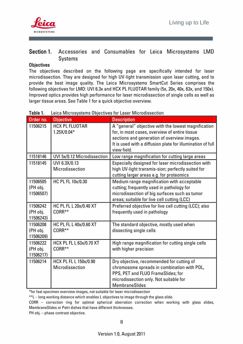
The objectives described on the following page are specifically intended for laser microdissection. They are designed for high UV-light transmission upon laser cutting, and to provide the best image quality. The Leica Microsystems SmartCut Series comprises the following objectives for LMD: UVI 6.3x and HCX PL FLUOTAR family (5x, 20x, 40x, 63x, and 150x). Improved optics provides high performance for laser microdissection of single cells as well as larger tissue areas. See Table 1 for a quick objective overview. Table 1. Leica Microsystems Objectives for Laser Microdissection Order no. Objective Description
11506215 HCX PL FLUOTAR
1.25X/0.04*
A “general” objective with the lowest magnification
for, in most cases, overview of entire tissue
sections and generation of overview images.
It is used with a diffusion plate for illumination of full
view field.
11518146 UVI 5x/0.12 Microdissection Low range magnification for cutting large areas
11518145 UVI 6.3X/0.13
Microdissection
Especially designed for laser microdissection with
high UV-light transmis-sion; perfectly suited for
cutting larger areas e.g. for proteomics
11506505
(PH obj.
11506507)
HC PL FL 10x/0.30 Medium range magnification with acceptable
cutting; frequently used in pathology for
microdissection of big surfaces such as tumor
areas; suitable for live cell cutting (LCC)
11506242
(PH obj.
11506243)
HC PL FL L 20x/0.40 XT
CORR**
Preferred objective for live cell cutting (LCC); also
frequently used in pathology
11506208
(PH obj.
11506209)
HC PL FL L 40x/0.60 XT
CORR**
The standard objective, mostly used when
dissecting single cells
11506222
(PH obj.
11506217)
HCX PL FL L 63x/0.70 XT
CORR**
High range magnification for cutting single cells
with higher precision
11506214 HCX PL FL L 150x/0.90
Microdissection
Dry objective, recommended for cutting of
chromosome spreads in combination with POL,
PPS, PET and FLUO FrameSlides; for
microdissection only. Not suitable for
MembraneSlides
*for fast specimen overview images, not suitable for laser microdissection
**L – long working distance which enables L objectives to image through the glass slide.
CORR – correction ring for optimal spherical aberration correction when working with glass slides,
MembraneSlides or Petri dishes that have different thicknesses.
PH obj. – phase contrast objective.

9
Version 1.0, August 2011
Living up to Life
Specimen Holders and Collection Devices
Leica Microsystems LMD systems support the following holders for mounting samples onto the microscope stage. These are:
1. Standard holder for a single slide (motor stage configurations) 2. The holder for three regular size slides for parallel working with serial sections
(available for scanning stage only) 3. Holder for a single large neuroscience slide or Petri dish (for all configurations)
Table 2. Order numbers of specimen holders for slides & Petri dish. Holder Motor Stage Scanning Stage Slide holder 11 XXX XXX (included in the
entire LMD system) 11 505 226 (included in the entire LMD system)
Petri dish (Ø 5cm) holder 11 505 214 (needs LCC module 11 505 164)
11 505 227 (+ Big Slide, needs LCC module 11 505 241)
Holder for 18-well double stack ibidi slide
11 505 255
For laser microdissection Leica Microsystems LMD systems support following devices for sample collection:
1. Standard collection device for 0.2 ml or 0.5 ml tubes 2. Collection device for 8-well strips 3. Adjustable collection device for multi-well arrays and Lab on a Chip (LOC)
applications (available for scanning stage only) Table 3. Order number of collection devices for dissectates. Collector Motor Stage Scanning Stage for 0.2 ml PCR tubes 11 505 131 (set of 4 levers;
(included in the entire LMD system)
11 505 229 (included in the entire LMD system)
for 0.5 ml PCR tubes 11 505 130 (set of 4 levers; (included in the entire LMD system)
11 505 228 (included in the entire LMD system)
for 8-well strips 11 505 258 11 505 230 for lab on a chip (LOC) and ibidi slides
11 505 256
for petri dish (Ø 5cm) included included Specimen microdissected out of tissue slides are regularly collected into the PCR tube caps of either 0.2 or 0.5 ml, however collection direct into 48 wells of LOC (lab on a chip) is possible as

10
Version 1.0, August 2011
Living up to Life
well. Dissected cultured cells are collected into 8-well strips, in Petri dishes or in 18-well ibidi slides, respectively. The latter option is available with scanning stage only. Table 2 summarizes the holder devices and Table 3 summarizes the collection devices. Please refer to the Instruction Manual or contact your local Leica Microsystems representative for more detailed information. Slides
LMD applications require specifically designed slides, to free the dissectate from the section. The tissue sections are placed on a UV-absorbing membrane. Upon the laser action, the membrane and the tissue in the cut line are vaporized. Since the membrane is not adherent to the slide, the dissectate (with the attached specimen) drops into the cap of the microcentrifuge tube.
Leica Microsystems offers high-quality MembraneSlides that provide high collection rates for laser microdissection. There are five different MembraneSlides to choose from:
PPS (polyphenylene sulfide) PEN (polyethylene naphthalate) PET (polyethylene terephthalate) POL (polyester) FLUO (fluoropolymer)
Membranes are mounted on regular glass slides (PPS/PEN), on steel frames (PPS/PET/POL/FLUO) (Fig. 1), or on the bottom of special Petri dishes (Fig. 2) or µ-Slides (Fig. 3) for cell cultivation. Depending on tissue type, thickness, and inspection method, one slide type may work better for a certain application. Therefore it is advisable to test different combinations of membranes and slide supports for the best results. Membrane combinations are summarized in Table 4. To test and to find best choice fitting the experimental design needs prior the start of a new project, Leica offers a suitable LMD DemoSlide Kit (11600264) containing all membrane slides. Tissue sections must be mounted onto the membrane prior to laser microdissection. Mounting of tissue sections onto glass slides is relatively easy since the glass provides solid support. Only difference between handling of glass MembraneSlides and common glass slides is that any membrane damage must be avoid. However, mounting tissue sections on the membrane of a steel FrameSlide can be more challenging. To simplify this procedure, a Plexiglass support (11532325) can be used to provide a rigid mounting surface (Fig. 3). Slides may be sterilized by autoclaving or UV-treatment. However, RNase-free certified slides are also available (see Table 4).

11
Version 1.0, August 2011
Living up to Life
Fig 1. Glass MembraneSlide (on top) and the FrameSlide (bottom).
The membrane is mounted on the upper side of the slide (the side with the frosted end
for marking). The membrane is glued on the glass at the edges in the central part of the
slide. There is a space of 1 µm between the glass and membrane. The slides must be
handled with care to prevent damage to the membrane. If moisture collects between
the glass and the membrane, this will impede the laser and collection action. See the
description of the FrameSlide (Fig. 3).
Fig 2. Petri Dish for Cell Cultivation for LMD.
The Petri dish is placed on the holder for firm mounting on the microscope stage. Since
the PEN membrane is perforated, gas exchange occurs. For better attachment and
growth of cells, the membrane can be coated with poly-L-lysine or collagen prior to
seeding (see more information on page 32).
Fig 3. Sterile 18-well μ-
Slide for cell
cultivation for LMD.
Fig 4. FrameSlide (on Top) and the FrameSupport (Bottom).
FrameSlide: The front side of the slide contains the engraved Leica Microsystems logo.
Please note that only the back side of the slide (the side opposite the Leica
Microsystems logo) is foil mounted, and only this side can be used for mounting tissue
sections.
FrameSupport: The Plexiglass FrameSupport contains a lifted surface (clear area) that
fits exactly into the cavity or the front side of the FrameSlide. The MembraneSlide must
be turned over to be mounted onto the FrameSupport (not shown).
12
Version 1.0, August 2011
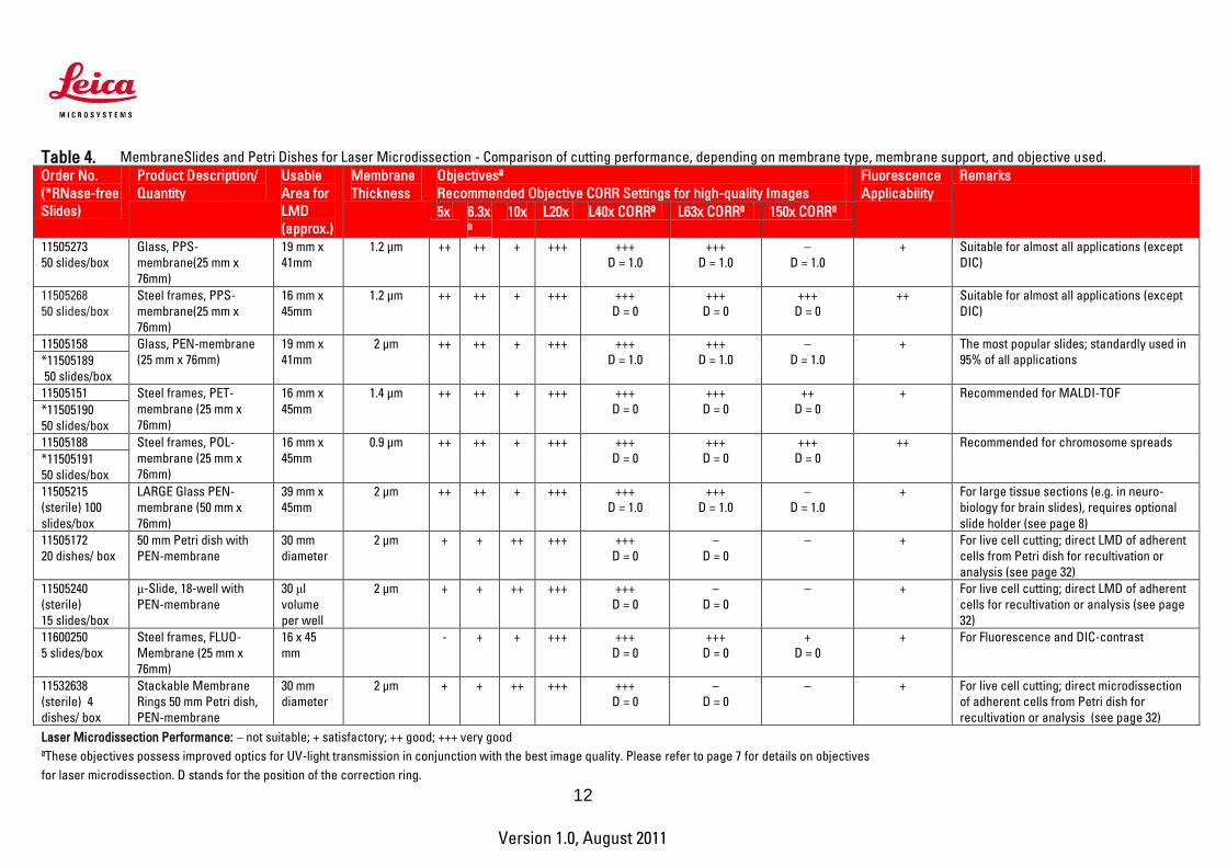
Table 4. MembraneSlides and Petri Dishes for Laser Microdissection - Comparison of cutting performance, depending on membrane type, membrane support, and objective used. Order No.
(*RNase-free
Slides)
Product Description/
Quantity
Usable
Area for
LMD
(approx.)
Membrane
Thickness
Objectivesª
Recommended Objective CORR Settings for high-quality Images
Fluorescence
Applicability
Remarks
5x 6.3x
ª
10x L20x L40x CORRª L63x CORRª 150x CORRª
11505273
50 slides/box
Glass, PPS-
membrane(25 mm x
76mm)
19 mm x
41mm
1.2 µm ++ ++ + +++ +++
D = 1.0
+++
D = 1.0
–
D = 1.0
+ Suitable for almost all applications (except
DIC)
11505268
50 slides/box
Steel frames, PPS-
membrane(25 mm x
76mm)
16 mm x
45mm
1.2 µm ++ ++ + +++ +++
D = 0
+++
D = 0
+++
D = 0
++ Suitable for almost all applications (except
DIC)
11505158 Glass, PEN-membrane
(25 mm x 76mm)
19 mm x
41mm
2 µm ++ ++ + +++ +++
D = 1.0
+++
D = 1.0
–
D = 1.0
+ The most popular slides; standardly used in
95% of all applications *11505189
50 slides/box
11505151 Steel frames, PET-
membrane (25 mm x
76mm)
16 mm x
45mm
1.4 µm ++ ++ + +++ +++
D = 0
+++
D = 0
++
D = 0
+ Recommended for MALDI-TOF
*11505190
50 slides/box
11505188 Steel frames, POL-
membrane (25 mm x
76mm)
16 mm x
45mm
0.9 µm ++ ++ + +++ +++
D = 0
+++
D = 0
+++
D = 0
++ Recommended for chromosome spreads
*11505191
50 slides/box
11505215
(sterile) 100
slides/box
LARGE Glass PEN-
membrane (50 mm x
76mm)
39 mm x
45mm
2 µm ++ ++ + +++ +++
D = 1.0
+++
D = 1.0
–
D = 1.0
+ For large tissue sections (e.g. in neuro-
biology for brain slides), requires optional
slide holder (see page 8)
11505172
20 dishes/ box
50 mm Petri dish with
PEN-membrane
30 mm
diameter
2 µm + + ++ +++ +++
D = 0
–
D = 0
– + For live cell cutting; direct LMD of adherent
cells from Petri dish for recultivation or
analysis (see page 32)
11505240
(sterile)
15 slides/box
μ-Slide, 18-well with
PEN-membrane
30 μl
volume
per well
2 µm + + ++ +++ +++
D = 0
–
D = 0
– + For live cell cutting; direct LMD of adherent
cells for recultivation or analysis (see page
32)
11600250
5 slides/box
Steel frames, FLUO-
Membrane (25 mm x
76mm)
16 x 45
mm
- + + +++ +++
D = 0
+++
D = 0
+
D = 0
+ For Fluorescence and DIC-contrast
11532638
(sterile) 4
dishes/ box
Stackable Membrane
Rings 50 mm Petri dish,
PEN-membrane
30 mm
diameter
2 µm + + ++ +++ +++
D = 0
–
D = 0
– + For live cell cutting; direct microdissection
of adherent cells from Petri dish for
recultivation or analysis (see page 32)
Laser Microdissection Performance: – not suitable; + satisfactory; ++ good; +++ very good
ªThese objectives possess improved optics for UV-light transmission in conjunction with the best image quality. Please refer to page 7 for details on objectives
for laser microdissection. D stands for the position of the correction ring.

13
Version 1.0, August 2011
Living up to Life
Sterilizing MembraneSlides
Important: Please note that sterilizing slides by autoclaving or UV-treatment does not guarantee complete destruction of RNases. For RNA preparation, especially from a single cell (corresponds in average to 10–15 pg of total RNA) or from a small number of cells, RNase-free certified slides are recommended (see Table 4). Alternatively, regular MembraneSlides can be pretreated with reagents such as RNaseZap® (Ambion).
Autoclavin
1. Place slides into a steel basket such as a slide holder for paraffinization 2. Place the basket into a beaker or jar and cover tightly 3. Autoclave at 121°C for 20 minutes. These conditions correspond to a standard
autoclaving set-up.
Chemical Treatment
Treat Membrane Slide with RNase decontamination solution according manufacturer’s instructions. Always rinse membranes and slides in RNase-free water after chemical treatment to remove remaining chemicals.
UV-Treatment
Incubate slides in a UV crosslink chamber and deliver at least 1 joule of energy (maximum power at 30 – 45 minutes). Please refer to the manufacturer’s information when using other UV-light sources.
Note: Sterilizing methods can be combined and should be done shortly before MembraneSlides are used.

14
Version 1.0, August 2011
Living up to Life
Microcentrifuge Tubes for Specimen Collection
During LMD, the specimen falls via gravity into the cap of the selected microcentrifuge tube. The cap may be used dry, but can also be filled with buffer. In this way, degradation of nucleic acids can be prevented, or proteins can be directly rescued for electrophoresis or chromatography. This might be crucial when cutting out hundreds or thousands of individual cells. Regular 0.2 ml and 0.5 ml microcentrifuge tubes are mounted into the collection devices. Please note that microcentrifuge tubes of some suppliers may not fit into the collection device because of the shape or the size of a particular cap. Suitable tubes are supplied by Greiner Bio-One. In addition, it is possible to accommodate particular types or brands of tubes with specially fabricated tube holders. The specimen might also be collected directly into 8-well strips. This option is regularly used for the Live Cell Cutting (LCC, see page 33). Compatible 8-well strips are from Greiner Bio-One (e.g. 762071). For more detailed information on collection devices, please refer to the Instruction Manual or contact your local Leica Microsystems representative.
Greiner PCR Tubes Order no. Leica Microsystems Size Order no. Greiner 11 090 615 027 006 0.5 ml 682 201 11 090 615 028 006 0.2 ml 683 201

15
Version 1.0, August 2011
Living up to Life
Section 2. How to Prepare Tissue Sections Handling tissues and preparing slides for laser microdissection does not differ much from standard procedures for regular glass slides. However, some protocol deviations and certain precautions are necessary to obtain intact nucleic acids and proteins for downstream analyses. A workflow comparison for preparing frozen and paraffin sections is summarized in Table 5.
Table 5. Workflows for Preparing Frozen and Paraffin Sections
Frozen sections – fast method preserving DNA, RNA & proteins, but the morphology
might be diminished
Paraffin sections – time consuming, but morphology is superb; limitations concerning high molecular weight RNA, DNA, and native
proteins may occur surgery
surgery
embed tissue in freezing medium
tissue fixation
flash-freeze
paraffin infiltration and preparation of paraffin block
section in cryostat
section with microtome
stain
stain
LMD
LMD
isolate DNA, RNA or proteins
isolate DNA, RNA or proteins
downstream analysis downstream analysis
Preparing Frozen Sections
Precautions for working with RNA
Frozen sections are the first choice when analyzing DNA, RNA or proteins. When working with RNA, precautions must be taken to avoid RNA cleavage by the ubiquitous RNases. RNases are also present on fingertips and are easily transferred to glassware and consumables, so gloves must be worn at all stages of work with RNA. Typically, labs designate specific areas for RNA work.

16
Version 1.0, August 2011
Living up to Life
Special pretreatment of water or solutions is strongly recommended since RNases survive autoclaving. Water or solutions should be treated with 0.1% solution of DEPC (diethylpyrocarbonate) for at least 12 hours at 37°C and then either heated to 100°C for 15 minutes or autoclaved for 15 minutes to break down the DEPC into carbon dioxide and ethanol. Trace amounts of DEPC can modify purine residues in the RNA by carboxymethylation. Carboxymethylated RNA is an inefficient template in in vitro translation systems, but is not a problem for hybridization experiments.
Important: DEPC is toxic and a suspected carcinogen and must be handled with caution. DEPC cannot be used to treat Tris buffers since it reacts with amino groups present in Tris base and is inactivated. Typically Tris buffers are prepared in DEPC pretreated water.
Glassware is typically baked at high temperatures, such as 180°C for 8 hours or more. Reusable plasticware and electrophoresis tanks melt at high temperatures and do not tolerate organic chemicals such as chloroform that can remove RNases. Instead, treat plasticware as follows:
1. Dip in 3% hydrogen peroxide (H2O2) for 10 minutes 2. Rinse in RNase-free water or dip in a 0.1% solution of DEPC for 2 hours at 37°C 3. Rinse in RNase-free water 4. Heat to 100°C for 15 minutes or autoclave for 15 minutes (traces of DEPC)
Important: Unopened bags of disposable plastic microcentrifuge pipettes are considered not to be contaminated by RNases without further treatment. One caveat is that trace amounts have been reported to still exist even in these sources of plasticware.
Alternatively, reagents for RNases decontamination such as RNaseZap® (Ambion) can be used to clean the gel tray, spatulas, slides, and other surfaces. For more hints on safe work with RNA, please refer to Ausubel at al., Sambrook et al. or RNeasy Micro Kit Handbook (Qiagen).
Note: Most reagents and plasticware can be obtained certified RNase- and DNase-free.

17
Version 1.0, August 2011
Living up to Life
Workflow for Preparation of Frozen Sections
In addition to RNase-free conditions when working with RNA, the whole procedure of tissue handling should possibly be carried out cold and quickly. Therefore, tissue or organs should be flash-frozen (snap-frozen) after surgery followed by cryo-sectioning. Sections should be stained within a very short time, not exceeding in total 30 minutes. The stained tissues should be used directly for laser microdissection, or alternatively, the dried sections can be stored in tightly closed bags, slide boxes or other containers containing desiccant at –80°C even for a few months or longer. Just before laser microdissection, the container with the slides should be taken out of the freezer and slowly adjusted to room temperature to avoid formation of water condensation inside the container, e.g. allow for at least 15 min each step to equilibrate at -20°C, 4°C and room temperature. This precaution is critical for RNA quality, especially when working with small numbers of cells, since water (moisture) activates RNases.
Note: All listed methods are exemplary suggestions. Other methods (e.g. for direct tissue freezing in the cryotome) can be established as well.
Flash-freezing Methods
Tissues are embedded in a freezing medium such as Jung Tissue Freezing Medium™ (Leica Microsystems) or OCT (Tissue-Tek®) and immediately flash-frozen.
Important: Tissue freezing medium can interfere with laser microdissection and must be completely removed with 70% alcohol (e.g. ethanol). For thicker sections, keep the tissue in 70% alcohol longer.
Method 1: Flash-freezing using 2-methyl-butane (synonym: isopentane)
1. Precool 2-methyl-butane in a beaker surrounded by dry ice. This prevents the 2-methyl-butane from bubbling over when the dry ice is added.
2. In a beaker or specimen container, add crushed dry ice to the 2-methyl-butane to make a slurry mixture (work in a hood).
3. When bubbling stops, the 2-methyl-butane is at the correct freezing temperature of approximately -90°C.
4. Immerse the embedded tissue slowly; eventually it will sink to the bottom of the 2-methyl-butane.
Safety Note: Be sure to fully evaporate the 2-methyl-butane after freezing to prevent the possibility of explosion in the freezer.

18
Version 1.0, August 2011
Living up to Life
Method 2: Flash-freezing in liquid nitrogen
1. Place liquid nitrogen in a styrofoam container. 2. Place the styrofoam container inside a Petri dish lid; a support rack may be needed to
hold the Petri dish lid. 3. Place the tissue into a disposable mold and embed it in the tissue freezing medium; or
alternatively, place the tissue (embedded in the tissue freezing medium) on a coverslip and place into the liquid nitrogen.
Method 3: Flash-freezing using 2-methyl-butane and liquid nitrogen
1. Place a beaker with 2-methyl-butane into liquid nitrogen and wait until the 2-methyl-
butane cools down to –80°C. This is the point when the wall of the beaker turns white,
the 2-methyl-butane is now solid.
2. Insert the tissue into 2-methyl-butane and let it freeze. Alternatively, the tissue can be
put onto a floating platform made of cork.
Tissues that are flash frozen without embedding medium can be post embedded in cryostat embedding media. However, the tissue will thaw somewhat in the process. This can compromise RNA preservation and introduce undesirable histological artifacts.
Sectioning
Preparing Cryo Sections
Cryo blocks can be directly sectioned (recommended for RNA preparation) or stored at –80°C. The temperature of the cryo blocks should be adjusted to –20°C in the cryostat before sectioning.
1. Clean the cryostat before sectioning to avoid contamination
2. Mount the cryo block onto the specimen clamp
3. Trim the sample to get a plane surface and an approach to the tissue
4. Cut the block into 5–25 µm sections and immediately place them on the slides for LMD
Important: If you are using FrameSlides, the FrameSupport is strongly recommended. Sections can be fixed with ice-cold acetone for 2–3 minutes, 70% or 100% ethanol for 20 seconds or mixture of ethanol : acetic acid (19:1) to increase the adhesion of the tissue to the PPS-, PEN-, PET-, POL- or FLUO-membranes.
Typical sectioning temperatures are shown in Table 6, page 19. Section thickness depends on the tissue type and the size of the dissected area. Keep the slides in the cryostat or on dry ice, if LMD is performed on the same day. Alternatively, they may be stored in sealed slide boxes with a desiccant at –80°C until needed.

19
Version 1.0, August 2011
Living up to Life
Important: Dry slides at room temperature for 15 minutes using a drying chamber with desiccant or at 40°C for 10 minutes. This is a critical step for successful LMD. Sample slides should be stained just prior to microdissection. Some staining protocols are given in Section 4.
For routine research and frequent cryo sectioning, the Leica CM3050 S cryostat is recommended. Versatility, such as the ability to cool down to –40°C, is an important advantage of this system. This is important for sectioning tissues such as mamma. For less frequent usage a cryostat such as the Leica CM1850 UV with UVC disinfection and the Precision Cryoembedding System can be used (contact your local Leica Microsystems representative for more information).
Table 6. Knife Edge Temperature for Cryo Sectioning
Knife Temperature Tissues –10°C to –15°C adrenals, bone marrow, brain, cartilage, spleen or bloody
tissue, testicular –15°C to –17°C bladder –15°C to –18°C breast - less fatty, cervix, intestine, liver, thyroid –15°C to –20°C kidney, lung, pancreas, prostate, ovary, rectal,
skin without fat, uterus –18°C to –22°C heart and vessel –20°C to –25°C muscular –25°C to –30°C skin with fat, breast - fatty
Preparing Paraffin Sections
Paraffin embedding is a process in which fixed tissue (utilizing neutral buffered formalin or other fixatives) is infiltrated by paraffin to stabilize it for long term storage, easy sectioning and for histological examination, it also affects macromolecules such as RNA. However, other fixatives have been reported to be gentle enough for RNA, DNA or protein downstream analyses (see below).
Please be aware that the methods, tempratures and protocols may need to be optimized depending on the investigated tissue and research objectives.
Tissue Fixation
Fixatives should penetrate the tissue quickly and thoroughly. Incomplete fixation causes many artifacts and can cause shrinkage. The most common fixative is 10% formalin buffered with PBS (phosphate buffered saline) to neutral pH (10% formalin corresponds to 4% formaldehyde). However, formalin breaks nucleic acids and therefore is not suitable for subsequent RNA and DNA analyses, if high molecular weight of nucleic acids are needed or

20
Version 1.0, August 2011
Living up to Life
quantitative analyses are planned. Other well-known fixatives are:
5% acetic acid 4–6% glutaraldehyde 100% ethanol 100% ice-cold acetone Bouin’s solution (contains picrinic acid and it is not compatible with RNA
analysis) Paraffin sections are advantageous over frozen sections in preserving morphology. However, typical fixatives such as formalin degrade RNA. Methacam can also be used as a fixative for paraffin-embedded sections. Methacam has been reported to provide better RNA quality enabling quantitative PCR. Methacam is a mixture of 60% (v/v) absolute methanol, 30% chloroform and 10% glacial acetic acid. Tissue was fixed at 4°C for two hours. (Takagi, et al. J Histochem. 2004 Jul; 52(7):903; Shibutani, et al., Lab Invest. 2000 Feb; 80(2):199–208) Another successfully used fixative in conjunction with RNA, DNA, and protein analyses is HOPE (HEPES-glutamic acid buffer mediated organic solvent protection effect). This patented solution is commercially available from Innovative Diagnostik Systeme GmbH (www.dcs-diagnostics.de).

21
Version 1.0, August 2011
Living up to Life
Paraffin embedding and Sectioning
After fixation, water must be removed from the tissue and replaced by an organic solvent before paraffin infiltration.
1. Wash the tissue with ascending concentrations of alcohol (such as ethanol) as follows: 70% ethanol 30 minutes 2x 95% ethanol 30 minutes 2x 100% ethanol 30 minutes 2x
During these steps the tissue will harden.
2. The alcohol is then replaced by an organic solvent such as xylene, which is miscible with alcohol and paraffin wax. The tissue is cleared twice for 1 hour each time.
3. Paraffin infiltration is carried out two to four times for 2–4 hours. Incubation time depends on the size of the tissue. Normally, embedding larger tissue pieces takes 22 hours, smaller tissue samples such as biopsies are incubated for 6 hours, and tissues for quick sections may require only 1 1/2 hours. The temperature of the liquid paraffin is 55–60°C, which corresponds to 2–4°C above the melting point.
Important: In general, paraffin hardness corresponds to the hardness of the tissue to be embedded. The best microtome sectioning results are obtained from paraffin blocks of the same or nearly the same hardness. There are two types of paraffin:
Low melting point paraffin is softer. Sectioning can be more difficult, but ribboning is easier. Tissue embedded in this type of paraffin may give better results in PCR.
High melting point paraffin is harder. It gives better support for hard tissues. Cutting thinner sections is possible, but ribboning is more difficult.
The entire procedure of fixation and embedding can be done in a tissue processor, which is standard equipment in a histopathology lab. Recommended is the Leica ASP300 S automated tissue processor (contact your local Leica Microsystems representative for more information).
4. Paraffin infiltrated tissues are embedded in a mold with liquid paraffin to form a block for better handling during microtome sectioning. This is easily done using a paraffin embedding station such as the Leica EG1150 H.
5. The tissue block can be stored at room temperature or below. For better cutting
results with the microtome, cooling the paraffin block down to –20°C is recommended.
22
Version 1.0, August 2011
Living up to Life
6. Section the block Clean the cryostat before sectioning to avoid contamination Mount the tissue block onto the microtome Trim the sample to obtain a plane surface and an approach to the tissue Cut the tissue into 5–15 µm sections
Place the paraffin ribbons on the surface of the water in a waterbath at 37°C– 0°C; in this way, the floating ribbons are easier to stretch and subsequently can be placed without folds on the slides. Please note that the water must be pure for PCR experiments or RNase-free, if RNA analysis is intended.
7. The paraffin must be removed prior to staining the paraffin-embedded sections.
This is achieved by washing the slides with xylene followed by a series of descending concentrations of ethanol as follows: xylene 20 seconds 3x (three separate containers) 100% ethanol 30 seconds 2x (two separate containers) 95% ethanol 30 seconds 2x 70% ethanol 30 seconds 2x
For typical staining protocols, see Section 3. Preparation of archived slides for LMD
As written above, regular glass slides for tissue sections cannot be used for laser microdissection. However, there is a possibility to process the regular archived slides to obtain the tissue for the microdissection. The protocol describes all steps of the procedure. a. Removal of the cover slip:
1. Archived slide is placed in a vertical position in a closed container with a solvent such as xylene to dissolve the cover slip seal and the mounting medium. Heating the container from 40°C up to 70°C speeds up the process.
2. Wait until the cover slip falls off. This may take hours to days. 3. Do not use a tool to pry off the cover slip. 4. The tissue section has to be rehydrated in xylene and washed through alcohol
series 95%, 75%, 50% and finally rinsed with PBS or pure water, and air-dried.
23
Version 1.0, August 2011
Living up to Life
b. Removal of the tissue from the slide: 1. Place “Quick Mount” mounting medium on the archived slide. Thickness of the
medium should be about 1–2 mm. 2. Allow the “Quick Mount” to cure overnight at 37°C or for 2 h at 70°C. “Quick
Mount” must remain pliable. 3. Trim excess “Quick Mount” around tissue section. 4. Place in a distilled water bath at 50–60°C for around 1 h. 5. Use a fine razor to lift the section and peel off.
c. Transfer tissue onto the Leica Microsystems MembraneSlide
1. Lay “Quick Mount” with tissue onto membrane. 2. Flatten lightly with finger tip pressure. 3. Place in oven in a horizontal position at 56°C for 3 h or until “Quick Mount” is
completely cured. d. Removal of “Quick Mount” from the Leica Microsystems slide
1. Place slide in xylene until “Quick Mount” dissolves. 2. Rehydrate with xylene, and then dehydrate with the series of 100%, 95%, 75%
alcohol, then wash with PBS water and air dry. 3. The slide is ready for the laser microdissection.
Please note: Depending on the conditions under which archived slides have been stored, and duration of storage, the macromolecules (DNA) may be of poor quality and quantity. It is recommended to utilize the archived paraffin block to cut new sections onto MembraneSlides for laser microdissection, than working with archived slides. However, direct ablation of tissue without coverslip from glass is also possible (draw and scan mode), but not comparable regarding the much better results of downstream analysis with laser microdissected samples derived from MembraneSlides.
Section 3. Preparation of Chromosome Spreads Preparation of chromosomes of white blood cells from human peripheral blood. Cells were cultured for 72 to 96 hours, then treated with colcemid (to arrest cells in metaphase) for 1 hour. After this step cells are treated with a hypotonic solution (KCl in Milli Q water). The white blood cells will swell and the erythrocytes become lysed. This step will take 10–12 minutes. After a washing step to remove lysed erythrocytes the cells are fixed in methanol/acetic acid (3 + 1, v/v). Fixed cells were dropped on the MembraneSlides and were air dried overnight and stained with Giemsa staining solution (1 : 20 diluted in Gurr buffer, which is a standard buffer used for Giemsa staining made by dissolving commercially available tablets) for 3 minutes. After washing in water they were air dried. This protocol was kindly provided by Dr. Henry Dijkman, Department of Pathology, Radboud University Nijmegen Medical Center, The Netherlands

24
Version 1.0, August 2011
Living up to Life
Section 4. Typical Staining Protocols Before staining, the paraffin should be removed from the tissue section. Please see page 21 for the typical protocol. For all types of sections, both frozen and paraffin-embedded, tissue can be fixed with various organic solvents to ensure better adhesion to the foil slide (see Page 17). This is the last step prior to staining. In this section, typical staining procedures are described. You will also find some “quick” procedures that may be useful when investigating RNA. The rough rule of thumb says that the entire staining procedure for subsequent RNA isolation should not exceed a total of 30 minutes.
Please be aware that protocols may need adjustment depending on the investigated tissue and your research objectives.
Quick Toluidine Blue Staining
Toluidine blue is a basic stain, which can also be used as a quick stain for light microscopy. Sections are permanently stained bluish-purple, similar to H&E. Toluidine blue may be used to stain both frozen and paraffin sections. It can stain mast cells and it is successfully used for staining frozen brain sections. Suitable for subsegment RNA preparation. You will need
Toluidine blue DEPC-treated water is used for subsequent RNA preparation, otherwise regular
distilled water can be used (see page 14) 75% ethanol 0.22 µm sterile syringe filter
1% toluidine blue solution: Dissolve 0.1 g toluidine blue in 10 ml DEPC water and filter through a sterile filter. Procedure
1. Apply toluidine blue solution and incubate for 3 minutes 2. Rinse twice in DEPC water for 15 seconds 3. Rinse once in 75% ethanol in DEPC water for 3 minutes 4. Air-dry or dry on a heater at 40°C for 10 minutes
25
Version 1.0, August 2011
Living up to Life
H&E (Hematoxylin and Eosin) Staining (method 1)
H&E is a common histology stain. Hematoxylin stains the nuclei, and eosin stains the cytoplasm. H&E is suitable for both paraffin and frozen sections. Ready-to-use reagents are also commercially available. Reagents
70% ethanol 95% ethanol 100% ethanol Hematoxylin (Hematoxylin Solution, Harris modified, Sigma Diagnostics,
Order no. HHS-16) Eosin (Eosin Y solution, alcoholic with phloxine B, Sigma Diagnostics,
Order no. HT110-3-32) 0,1 % NH4OH (blueing reagent): 200 µl NH4OH 100% + 200 ml H2O or Scot´s tape
water substitute Procedure Place the slide in the following solutions for the designated time frames:
1. Distilled H2O 30 seconds 2. Hematoxylin 1 minute 3. H2O 30 seconds 4. Blueing reagent 30 seconds 5. Eosin solution 10 seconds 6. 70% ethanol 30 seconds 7. 95% ethanol 30 seconds 8. 100% ethanol 30 seconds or air-dry at room temperature
Results
Nuclei - blue-black Cytoplasm - varying shades of pink Muscle fibers - magenta Fibrin - deep pink Red blood cells - orange/red

26
Version 1.0, August 2011
Living up to Life
H&E Staining (method 2)
See the above application range and typical results of staining. Reagents
Gills Hematoxylin Hematoxylin 6.0 g Aluminium Sulphate 4.2 g Citric Acid 1.4 g Sodium Iodate 0.6 g Ethylene Glycol 269 ml Distilled Water 680 ml
Eosin Eosin Yellowish 1.0 g Distilled Water 100 ml
Lithium Carbonate 1 % Lithium Carbonate 1 g Distilled Water 100 g
Acid Alcohol 1 % 70% Alcohol 99 ml conc. Hydrochloric Acid 1 ml
Scott's tap water substitute Before you begin, add 20 g sodium bicarbonate and 3.5 g magnesium sulphate to a beaker containing 1 L distilled water. Mix thoroughly with a magnetic stirrer to dissolve the salts, then pour into a storage container. Procedure
1. Wash the slide with distilled water for 30 seconds 2. Place sections in the hematoxylin solution for 30 seconds 3. Wash in tap water 4. “Blue” sections in Scott's tap water 5. Wash in tap water 6. Place sections in 1% acid alcohol for a few seconds 7. Wash in tap water 8. Place sections in eosin for 15 ~ 30 seconds 9. Wash in tap water 10. Dehydrate with 95% ethanol for 30 seconds, 100% ethanol for 30 seconds, and air-
dry 5 ~ 10 minutes

27
Version 1.0, August 2011
Living up to Life
Quick Cresyl Violet Staining
Cresyl violet stain is commonly used for neuronal tissue. It is a basic stain that binds to acidic molecules of neuronal cytoplasm, such as RNA-rich ribosomes. Cresyl violet permanently stains a section and is suitable for both paraffin and frozen sections and subsequent RNA preparation. Cresyl violet staining solution
1. Add 1 g Cresyl violet into 200 ml distilled water 2. Add 1 ml 10% acetic acid 3. Boil until completely dissolved 4. Filter the solution though filter paper 5. Add H2O up to 250 ml
Procedure
1. Wash once in PBS for 30 seconds 2. Apply Cresyl violet staining solution for 30 seconds 3. Rinse in PBS for 30 seconds 4. Rinse twice in DEPC water for 15 seconds each time 5. Rinse once in 75% ethanol for 1 minute, 95% ethanol for 30 seconds, and 100%
ethanol for 30 seconds 6. Air-dry for 10 minutes
Modified Cresyl Violet Staining for RNA-Research
For RNA protection Cresyl violet can be prepared and applied without PBS or water steps, preventing degradation of RNA by humidity activated RNases. Modified Cresyl violet staining solution
1. Prepare Cresyl violet staining solution at least one week prior usage
2. Add 0.5 g Cresyl violet into 50 ml 100% ethanol
3. Mix solution and store at 4°C sealed air-tight and dark
Procedure
1. Mount section and fix in cold (-20°C) 75% ethanol for 2 minutes 2. Apply Cresyl violet staining solution directly with syringe and sterile filter to the
section and incubate for 1minute, swivel gently 3. Dip for 5 seconds in 75%, 90% and 100% ethanole 4. Final fixation is done in fresh 100% ethanol for 1 minute 5. Dry sample in drying chamber with desiccant
Adapted from Gründemann et al., NAR, 2008

28
Version 1.0, August 2011
Living up to Life
Quick Thionin Staining
This stain is specific for DNA and Nissl substance, which is primarily ribosomal RNA. Thionin binds to acidic proteins and nucleic acids with a specificity determined by the pH of the final staining solution. This stain is suitable for both paraffin and frozen sections and subsequent RNA preparation. Reagents
Thionin (acetate) Sodium Acetate DEPC-treated water is used for subsequent RNA preparation, otherwise regular
distilled water can be used (see page 15) 75%, 95%, 100% ethanol 0.22 µm sterile syringe filter
0.5% Thionin staining solution
1. Dissolve 1.36 g of sodium acetate (anhydrous) or alternatively 2.26 g sodium acetate (trihydrate) in 100 ml H2O and fill up to 250 ml
2. Add 0.5 g thionin to 100 ml of the sodium acetate solution. 3. Stir and heat gently for 1 hour. Filter the solution after the dye has dissolved. Store
in a closed bottle. Procedure
1. Apply thionin staining solution and incubate for 1 minute 2. Rinse twice in DEPC water for 15 seconds 3. Rinse once in 75% ethanol for 1 minute, 95% ethanol for 30 seconds, and 100%
ethanol 30 seconds 4. Air-dry for 10 minutes
29
Version 1.0, August 2011
Living up to Life
Direct Visualization of GFP
GFP is an abbreviation for green fluorescent protein. Following exemplary protocol needs to be optimized for the investigated tissue. Reagents
Formaldehyde based fixative (4% PAF, 10% NBF, PLP) 1x PBS 18% Sucrose in 1x PBS Freezing medium
Procedure
1. Harvest tissue as quickly as possible and trim for correct fixation (4 mm thick slices).
2. Immediately place into fixative (use a tissue to fixative ratio of at least 1:10). 3. Incubate for 8–24 hours; keep out of direct light to avoid bleaching the GFP. 4. Place tissues in 1x PBS for 10–15 minutes. Use a larger volume of buffer to
completely remove fixative from the tissue. 5. Repeat this washing step once. 6. Transfer tissues into 18%–20% sucrose (dissolved in 1x PBS). Use a larger volume
to allow tissue to float freely. 7. Place in the refrigerator at 4°C overnight. 8. The tissue will sink to the bottom when completely infiltrated with sucrose; if the
tissue is still floating after 24 hours, incubate for one more night. 9. Remove the tissue from the sucrose and dab off any excess sucrose. 10. Prepare a standard frozen block. 11. Store the block at –70 °C until needed. 12. Cut the sections on a cryostat (usually 4–10 µm) and thaw the mountant. 13. Wash away the mountant for LMD.

30
Version 1.0, August 2011
Living up to Life
Rapid Immunohistochemistry using Vectastain® ABC-AP Kit and Vector® Blue
In this protocol an unlabelled primary antibody is used, followed by biotinylated secondary antibody. The secondary antibody is conjugated to the alkaline phosphatase (AP) complex. The detection reaction is trigged by Vector®Blue substrate (Vector Laboratories). You will need to prepare
0x PBS: add 80 g NaCl, 2 g KCl, 18 g Na2HPO4 x 2 H2O, 2.4 g KH2PO4 to 800 g distilled H2O. Adjust to pH 7. Fill up with H2O up to 1 L and autoclave.
Other reagents: Acetone Normal Goat Serum Vector Blue Substrate Kit (Vector Laboratories, Alexis) Vectastain ABC-AP-Kit (Vector Laboratories, Alexis)
Procedure
1. Prepare frozen sections of 8 µm on PET or PEN-slides. 2. Fix the sections with cold acetone at 4°C for 10 minutes. 3. Dry the sections at room temperature for 10 minutes. 4. Wash briefly in 1x PBS. 5. Apply 500 µl of primary antibody dilution onto the section and incubate at room
temperature for 10 minutes. The dilution must be adjusted individually for each antibody.
Important: A good starting point may be a dilution of 1:100. In this case, prepare the dilution as follows: Add 20 µl of normal goat serum as the blocking reagent and 5 µl of primary antibody to 500 µl 1x PBS. Depending on the species in which the antibody was raised and the tissue used, it may be necessary to use a different blocking reagent.
6. Wash briefly once in PBS 7. Apply 500 µl of secondary antibody dilution onto the section and incubate for 15
minutes at room temperature. The dilution has to be adjusted individually for each antibody.
Important: A good starting point may be a dilution of 1:100. Add 4 µl normal goat serum as the blocking reagent and 5 µl biotinylated secondary antibody to 500 µl 1x PBS. Depending on the species in which the antibody was raised and the tissue used, it may be necessary to use a different blocking reagent. 8. Wash briefly once in PBS at room temperature. 9. The staining reaction is carried out with the Vectastain® ABC-AP Kit and Vector®
Blue from Vector Laboratories (see manufacturer’s instructions). 10. Incubate with Avidin-Alkaline phosphatase complex. 11. Apply ABC reagents from the Vectastain ABC-AP-Kit for 10 minutes. 12. Wash briefly once in PBS. 13. Incubate with Vector® Blue AP substrate solution for 10 minutes. 14. Wash briefly with RNase-free water. 15. Leave MembraneSlides to dry in the dark.

31
Version 1.0, August 2011
Living up to Life
Rapid immunohistochemistry using Streptavidin-HRP Conjugate
In this protocol an unlabelled primary antibody is used, followed by a biotinylated secondary antibody. The secondary antibody is conjugated to horseradish peroxidase (HRP or POD). The detection reaction is trigged by diamino benzidine (DAB)/H2O2 as a substrate. Prepare solutions as stated in the previous protocol Other reagents:
Acetone
Normal Goat Serum DAB plus substrate solution (DAKO Cytomation) Streptavidin HRP (DAKO Cytomation)
Procedure Follow steps 1 to 8 from the protocol above.
9. Apply 500 µl of HRP-conjugated streptavidin and incubate at room temperature for 10 minutes. The dilution of the streptavidin has to be determined experimentally and might range from 1:300–1:1000 (see manufacturer’s instructions). For example, the 1:300 dilution is prepared by adding 1.7 µl streptavidin to 500 µl 1x PBS.
10. Wash briefly once with PBS. 11. Apply 500 µl of DAB staining solution onto the slide for 10 min at room temperature.
The DAB staining solution is prepared as follows: A drop of DAB plus solution is added per ml buffered substrate solution (both from the DAKO DAB plus solution kit).
12. Wash briefly once with PBS. 13. Wash briefly with RNase-free water. 14. Air-dry the slides in the dark.

32
Version 1.0, August 2011
Living up to Life
General LMD Guidelines using Leica Microsystems Microdissection Systems
Section 5. Placement of LMD Systems A laser microdissection system should be located away from doors and windows that can be opened and air conditioning. Air draft should be avoided, especially during cutting. Scientists who work with RNA should ensure that the LMD is placed in an RNase-free zone in the lab.
Important: All working steps and technical data on LMD are included in the Instruction Manual. Please read the Instruction Manual entirely.
Section 6. Benchmark Data on Cell Number and Section Thickness for LMD Standard numbers for laser microdissection are given in Table 7. Table 7. Average Number of Cells used for LMD. Analyzed Molecules Average Number of Cells DNA Number of cells is not critical because DNA is easily amplified by PCR.
Usually up to 5000 cells can be dissected. RNA 30% of all researchers dissect between 100 –1000 cells
30% or all researchers dissect over 1000 cells However, gene expression profiling by dissecting a single cell or as few as 10-30 cells has been reported.
Proteins Thousands to tens of thousands of cells are required. The collection of cells can be facilitated by Leica Auto Vision Control (AVC) software (see page 36), which enables automatic cell recognition and automatic microdissection from a tissue section(s). The region of interest can also be cut from several consecutive slides and pooled. In this case, regular, thinner sections with the best morphology are used. Alternatively, large tissue areas of 500 µm2 may contain enough cells.
The average section thickness used for LMD is between 4–20 µm. In some cases, where more material is needed and tissue morphology is sufficient, thicker sections of 15–30 µm can be used for LMD. For plant Research, even unfixed fresh tissues above 100 µm can be used.

33
Version 1.0, August 2011
Living up to Life
Section 7. Application Software Live Cell Cutting (LCC)
Microdissecting cells from a cell culture is termed live cell cutting (LCC). Since the laser beam does not come into contact with the live cells, it does not injure them. Hence, this technique is also safe, and it is possible to recultivate the microdissected cells. Leica Microsystems offers an optional LCC module including additional software and hardware, which allows special Petri dishes to be used for LMD. This optional module lowers the stage every time the objective is changed in order to avoid collision with the Petri dish, and then brings the specimen back into focus. This software option is included within the basic software version. Either membrane bottom Petri dish, stackable MembraneRings or 18-well µ-Slides can be used for cell cultivation (see pages 9–12). The protocol below describes LCC when using the Petri dish. Procedure:
1. The membrane of the Petri dish can be coated with poly-L-lysine or collagen prior to inoculation. This may improve cell attachment and growth.
2. The cells are inoculated in the Petri dish. 3. The seeded membrane Petri dish can be inserted into a larger Petri dish and
incubated under usual conditions for 24–48 hours. The cells should not grow to confluence, in order to distinguish single cells for microdissection. Cells can be inspected with the LMD system using brightfield (BF), phase contrast (PH), differential interference contrast (DIC) or fluorescence.
4. Just prior to microdissection, the PBS, base medium (without serum), and complete culture medium (with serum and antibiotics) should be warmed up to 37°C in a waterbath.
5. The stage, substage, holder, and UV shield should be cleaned with alcohol. 300 µl of complete culture medium are added to each well of the 8-well strip (see Instruction Manual).
6. Next, the cells are carefully washed three times with 2 ml PBS. Then 150 µl – 1 ml of PBS or base medium is added to the membrane Petri dish to just cover the cells that have to stay alive.
Important: The amount of liquid in the dish is critical because if there is too much medium (more than just covering the cells), the laser causes “cavitation” (bubbles), which reduces the efficiency of cutting! 7. The Petri dish without cover and the collector with the 8-well strip are mounted
onto the LMD stage. 8. Cells of interest are selected, followed by microdissection

34
Version 1.0, August 2011
Living up to Life
Tips: Be careful to leave wide margins around the cells. Typically, nearly rounded cells are easier to dissect. The selected area should not be too large. Single cells or groups of two to three cells fall down the best. The laser settings are critical to release the dissected pieces during the first
cut. The best settings for the investigated cells have to be defined empirically.
9. The microdissected cells can be inspected in the wells, although the presence of phenol red makes this difficult. Another hindrance: When the specimen drops to the bottom of the well, focusing is not possible.
10. To re-culture the cells, the 8-well strip is removed from the sub-stage after laser microdissection. 50 µl more of the medium is added on top of each well and spun down for a short time (up to 1 minute). The microdissected cells are inoculated in the regular cell culture vessels and incubated as usual.
LCC Applications using membrane µ-Slides and Petri dishes Single-slide LCC
A solution for live cell cutting. Single membrane µ-Slide is mounted on the regular slide holder. µ-Slide can be used with the cover that is UV translucent up to 20x objective. This increases sterility of the experiment. Cells are collected in 8-well strips (see Fig. 5).
Fig 5. Single-slide LCC with the scanning stage and 8-well stripe for LCC collection

35
Version 1.0, August 2011
Living up to Life
Stack-slide LCC
Completely sterile solution for live cell cutting that is available for scanning stage. Membrane µ-Slide with grown cell culture and regular plastic bottom µ-Slide (e.g. Plasma coated, Leica Microsystems order no. 11532520) are fixed in the slide-stack holder under the clean bench. Then the holder is mounted on the regular three-slide holder. The stack can be used with up to 20x objectives (see Fig. 6 and 7).
1. Cover 2. Slide with membrane bottom for LMD 3. Slide with regular plastic bottom 4. Slide-stack holder
Fig 6. Scheme of the stack-slide
A
B C
Fig 7. A View on the mounted stack-slide in the holder. B Stack-slide holder C View on
the stackslide on the scanning stage.
36
Version 1.0, August 2011
Living up to Life
Classical LCC with Petri dishes
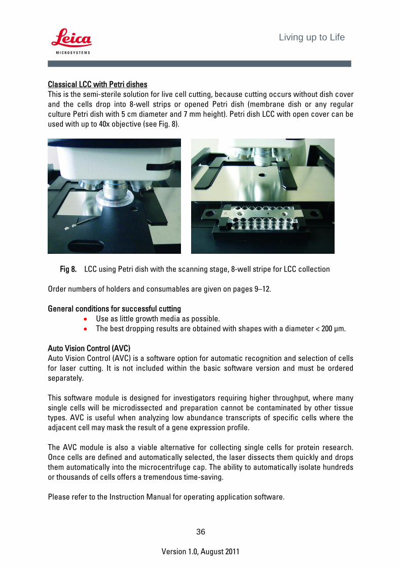
This is the semi-sterile solution for live cell cutting, because cutting occurs without dish cover and the cells drop into 8-well strips or opened Petri dish (membrane dish or any regular culture Petri dish with 5 cm diameter and 7 mm height). Petri dish LCC with open cover can be used with up to 40x objective (see Fig. 8).
Fig 8. LCC using Petri dish with the scanning stage, 8-well stripe for LCC collection
Order numbers of holders and consumables are given on pages 9–12. General conditions for successful cutting
Use as little growth media as possible. The best dropping results are obtained with shapes with a diameter < 200 µm.
Auto Vision Control (AVC)
Auto Vision Control (AVC) is a software option for automatic recognition and selection of cells for laser cutting. It is not included within the basic software version and must be ordered separately. This software module is designed for investigators requiring higher throughput, where many single cells will be microdissected and preparation cannot be contaminated by other tissue types. AVC is useful when analyzing low abundance transcripts of specific cells where the adjacent cell may mask the result of a gene expression profile. The AVC module is also a viable alternative for collecting single cells for protein research. Once cells are defined and automatically selected, the laser dissects them quickly and drops them automatically into the microcentrifuge cap. The ability to automatically isolate hundreds or thousands of cells offers a tremendous time-saving. Please refer to the Instruction Manual for operating application software.

37
Version 1.0, August 2011
Living up to Life
Nucleic Acids and Protein Preparation for Downstream Analysis
Section 8. Preparation of Nucleic Acids Laser microdissected tissue specimens present a particular challenge for molecular analysis, as nucleic acids must be purified from very small samples. It is a particular issue for RNA, which cannot be directly amplified in PCR like DNA. In addition, fixation and staining steps may compromise the integrity of nucleic acids. If the yields and quality of the nucleic acids are not satisfactory, tissue section preparation has to be optimized (see Section 2), and the method of RNA or DNA preparation must be verified. There are many “home-brewed” techniques that may work for larger amount of starting material. However the less material to start with, the more risk of loosing the precious sample. Leica Microsystems recommends the high-quality QIAGEN kits for preparation of nucleic acids. These kits have been tested with the Leica Microsystems LMD systems. These “Micro Kits” have been designed for fast, reproducible isolation of high-quality genomic DNA (QIAamp® DNA Micro Kit) or total RNA (RNeasy® Micro Kit) from laser microdissected tissues. The preparations are free of proteins, nucleases, and other impurities. They can be immediately used in downstream applications such as PCR, sequencing, quantitative, real-time PCR, or can be stored at –20°C until needed. Please refer to www.qiagen.com for details.

38
Version 1.0, August 2011
Living up to Life
Section 9. Total Protein Preparation Proteomics is an emerging application for laser microdissection. The challenge of retrieving a sufficient amount of proteins for downstream experiments can be overcome in several ways:
The entire area of interest can be microdissected using low magnification objectives such as the 5x, 6.3x or 10x.
Cut regions or cells of interest from several consecutive tissue sections. Hundreds of single cells can be cut out from the tissue using the Leica AVC
module (page 36) The following techniques for protein detection have been successfully tested with the proteins gained from LMD:
SDS-PAGE followed by Silver staining SDS-PAGE followed by Western Blotting 2DGE (two dimensional gel electrophoresis) 2D-DIGE (two-dimensional differential fluorescence gel electrophoresis with
labeling of proteins with Cy2, Cy3 and Cy5 dyes) HPLC MALDI SELDI
Other Information
Section 10. Ordering Information Specimen Preparation Instrumentation
Please have a look on: www.leica-microsystems.com
Please note that these are typical products from a broad range of Leica Microsystems specimen preparation portfolio. Please contact your local Leica Microsystems representative to select the best solution for your needs.

39
Version 1.0, August 2011
Living up to Life
QIAGEN Kits for Nucleic Acids Preparation
Order No. Product Description Remarks 56304 QIAamp® DNA Micro Kit
for 50 DNA preparations Kit for genomic DNA preparation from microdissected specimens
74004 RNeasy® Micro Kit for 50 RNA preparations
Kit for total RNA preparation from microdissected specimens
Please visit www.qiagen.com for more information on additional kits.
Section 11. General References
1. Ausubel, F.M., Brent, R., Kingston, R.E., Moore, D.D., Seidman, J.G. and Smith, J.A. (eds.) (1995) Current Protocols in Molecular Biology, Wiley
2. RNeasy® Micro Handbook, available at www.qiagen.com 3. Sambrook, J., Russel D.W. (1991) Molecular Cloning, A Laboratory Manual, 3rd
ed., Cold Spring Harbor (N.Y.) Laboratory Press 4. QIAamp® DNA Micro Handbook, available at www.qiagen.com 5. Sambrook, J.,Fritsch, E.F. and Maniatis, T., Cold Spring Harbor (N.Y.) Laboratory
Press
Section 12. Useful Websites Leica Microsystems LMD system: www.leica-microsystems.com/LMD Leica Microsystems Specimen Preparation: www.leica-microsystems.com QIAGEN www.qiagen.com
Section 13. Protocol Form Please fill in the form and fax to +49 6441 29 2255. You can also request the form as a hard copy or electronic copy from your local Leica support.

40
Version 1.0, August 2011
Living up to Life
Please briefly describe your current workflow for downstream applications:
_________________________________________________________________________________________ _________________________________________________________________________________________ 1. What type of specimen(s) do you work with? a. Tissue _______________________ Cell type _______________________ Species _____________________ b. Cultured cells (please specify) __________________________ c. Stem cells: __________________________ 2. What is the starting material for laser microdissection? (indicate all that apply)
� Frozen sections � Smears
� Paraffin-embedded sections � Cytospins
� Cultured cells � Other, please specify _____________________
3. Do you use a tissue fixative? If yes, which one (indicate all that apply) � Formalin � Methacarn � HOPE � Other, please specify _____________________
4. How thick are the tissue sections?
From ________µm up to ________ µm
5. Do you store paraffin-embedded or frozen tissue blocks before sectioning and LMD?
� Yes � No a. If yes, how long and at what temperature? _________________________ b. Do you observe decreased quality of the preparation (nucleic acids and proteins) over time? � Yes � No c. Do you know, how to overcome this? ______________________________________________________
6. Do you store sections before LMD?
a. If yes, please describe how long and at what temperature: _____________________________________ b. Do you observe decrease of the quality of the preparation (nucleic acids and proteins) over time? � Yes � No c. Do you know, how to overcome this? ______________________________________________________
7. What is your staining protocol? Please indicate reagents and the incubation time as well as precautions
necessary for successful RNA, DNA, and protein isolation after LMD: _________________________________________________________________________________________ _________________________________________________________________________________________ _________________________________________________________________________________________ _________________________________________________________________________________________

41
Version 1.0, August 2011
Living up to Life
8. How many cells do you dissect for downstream experiments? (indicate all that apply) � Single cell � 101 – 1000 cells � 2 - 50 cells � Over 1000 cells � 51 –100 cells
9. What do you analyze after LMD?
� DNA � RNA � Proteins � Live cells � Other (please specify) _________________________________________________________________
10. If you isolate nucleic acids (A) or proteins (B), what analyses do you perform? (indicate all that apply)
a. � PCR b. � Western Blotting � RT-PCR � 2DGE � Real-time RT-PCR � HPLC � Microarrays � MALDI � in vitro transcription � SELDI � Whole genome amplification (WGA) � Other (please specify)___________________________ � Other (please specify)_________________
11. Do you use a commercial kit, or “home-brewed“ procedures? Please indicate the kit(s) or briefly describe
your protocol(s): _________________________________________________________________________________________ _________________________________________________________________________________________ _________________________________________________________________________________________ 12. Do you have any comments about Leica Microsystems’s LMD systems or service? _________________________________________________________________________________________ _________________________________________________________________________________________ _________________________________________________________________________________________ We would like to acknowledge your feedback with a gift. Please tell us about you … Title: _______________________________________________
Name: ______________________________________________
University/Company:____________________________________
Institute/Department: __________________________________
Address: ____________________________________________
City:_________________________________________________
ZipCode:_____________________________________________
Country: _____________________________________________
Phone: ______________________________________________
Fax: ________________________________________________
Email: _______________________________________________
42
Version 1.0, August 2011
Living up to Life
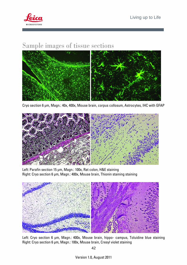
Sample images of tissue sections
Cryo section 6 µm, Magn.: 40x, 400x, Mouse brain, corpus collosum, Astrocytes, IHC with GFAP
Left: Parafin section 15 µm, Magn.: 100x, Rat colon, H&E staining Right: Cryo section 6 µm, Magn.: 400x, Mouse brain, Thionin staining staining
Left: Cryo section 6 µm, Magn.: 400x, Mouse brain, hippo- campus, Toluidine blue staining Right: Cryo section 6 µm, Magn.: 100x, Mouse brain, Cresyl violet staining

43
Version 1.0, August 2011
Living up to Life
Cryo section, Mag.: 630x, Mouse brain, IHC with GFAP
Cryo section 6 µm, Magn.: 40x, 400x, Mouse brain, hippocampus, IHC (DAB as a substrate)
Methaphase chromosomes cut with 150x HCX Fluotar dry objective