liver cirrhosis cured by pericardiectomy–a rare case of ... · constrictive pericarditis usually...
TRANSCRIPT
Research Article Open Access
Clinical & Experimental
CardiologyMeszaros et al., J Clin Exp Cardiolog 2013, 4:4http://dx.doi.org/10.4172/2155-9880.1000242
Volume 4 • Issue 4 • 1000242J Clin Exp Cardiolog
ISSN:2155-9880 JCEC, an open access journal
*Corresponding author: Katharina Meszaros, MD, Medical University of Graz, Department for Cardiac Surgery, Auenbruggerplatz 29, 8036 Graz, Tel: 0043 316 385 80030; Fax: 0043 316 385 14672; E-mail: [email protected]
Received March 08, 2013; Accepted April 16, 2013; Published April 18, 2013
Citation: Meszaros K, Wagner D, Müller H, Iberer F, Schmidt A, et al. (2013) Liver Cirrhosis Cured by Pericardiectomy–A Rare Case of Constrictive, Non-Calcifying Pericarditis. J Clin Exp Cardiolog 4: 242. doi:10.4172/2155-9880.1000242
Copyright: © 2013 Meszaros K, et al. This is an open-access article distributed under the terms of the Creative Commons Attribution License, which permits unrestricted use, distribution, and reproduction in any medium, provided the original author and source are credited.
AbstractConstrictive pericarditis usually leads to heart failure but can also cause extra cardiacdiseases. We report
a patient who presented with dyspnea, recurrent pericardial and pleural effusions as well as ascites. An initial cardiologic examination revealed a pericardial effusion without severe hemodynamic impairment, but without signs of additional pathologies. Abdominal sonography showed liver cirrhosis, which was laboratory classified as Child grade B. The patient was referred to a transplantation center for liver transplant evaluation. During the liver transplant evaluation process, Cardiac Magnetic Resonance Imaging (MRI) and Computed Tomography (CT) finally revealed a constrictive non-calcifying pericarditis as the origin of the cardiac cirrhosis and the patient was scheduled for peri- and partial epicardiectomy.
Two years later, clinical and biochemical liver parameters were completely restored. There was no recurrence of ascites or pleural effusions. At follow-up, cardiac CT and MRI proved the absence of a pericardial constriction while liver sonography showed normal hepatic morphology.
This case presents a highly rare cause for liver cirrhosis and underlines the importance of a complete cardiac evaluation in case of a present liver cirrhosis of unknown causes.
Liver Cirrhosis Cured by Pericardiectomy–A Rare Case of Constrictive, Non-Calcifying PericarditisKatharina Meszaros1*, Doris Wagner2, Helmut Müller2, Florian Iberer2, Albrecht Schmidt3, Rainer R Rienmüller4, Rudolf Stauber5, Peter Kornprat6 and Heinrich Mächler1
1Department for Cardiac Surgery, Medical University of Graz, Graz, Austria 2Department for Transplantation Surgery, Medical University of Graz, Graz, Austria 3Division of Cardiology, Department of Internal Medicine, Medical University of Graz, Graz, Austria4Department of Radiology, Medical University of Graz, Graz, Austria5Division of Gastroenterology and Hepatology, Department of Internal Medicine, Medical University of Graz, Graz, Austria 6Department for General Surgery, Medical University of Graz, Graz, Austria
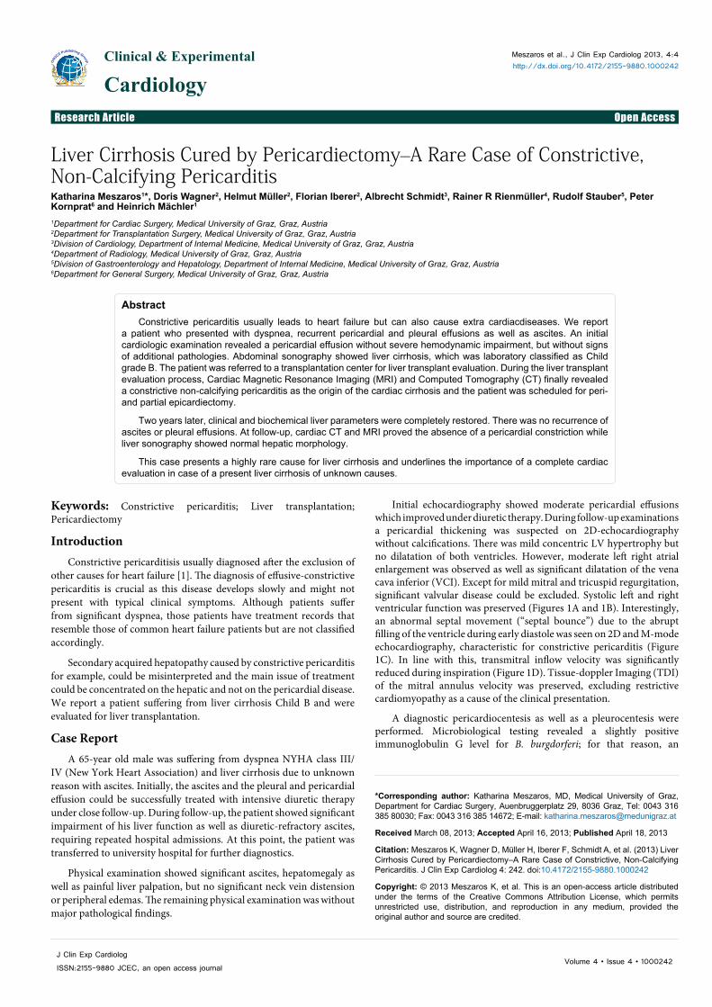
Initial echocardiography showed moderate pericardial effusions which improved under diuretic therapy. During follow-up examinations a pericardial thickening was suspected on 2D-echocardiography without calcifications. There was mild concentric LV hypertrophy but no dilatation of both ventricles. However, moderate left right atrial enlargement was observed as well as significant dilatation of the vena cava inferior (VCI). Except for mild mitral and tricuspid regurgitation, significant valvular disease could be excluded. Systolic left and right ventricular function was preserved (Figures 1A and 1B). Interestingly, an abnormal septal movement (“septal bounce”) due to the abrupt filling of the ventricle during early diastole was seen on 2D and M-mode echocardiography, characteristic for constrictive pericarditis (Figure 1C). In line with this, transmitral inflow velocity was significantly reduced during inspiration (Figure 1D). Tissue-doppler Imaging (TDI) of the mitral annulus velocity was preserved, excluding restrictive cardiomyopathy as a cause of the clinical presentation.
A diagnostic pericardiocentesis as well as a pleurocentesis were performed. Microbiological testing revealed a slightly positive immunoglobulin G level for B. burgdorferi; for that reason, an
Keywords: Constrictive pericarditis; Liver transplantation; Pericardiectomy
IntroductionConstrictive pericarditisis usually diagnosed after the exclusion of
other causes for heart failure [1]. The diagnosis of effusive-constrictive pericarditis is crucial as this disease develops slowly and might not present with typical clinical symptoms. Although patients suffer from significant dyspnea, those patients have treatment records that resemble those of common heart failure patients but are not classified accordingly.
Secondary acquired hepatopathy caused by constrictive pericarditis for example, could be misinterpreted and the main issue of treatment could be concentrated on the hepatic and not on the pericardial disease. We report a patient suffering from liver cirrhosis Child B and were evaluated for liver transplantation.
Case ReportA 65-year old male was suffering from dyspnea NYHA class III/
IV (New York Heart Association) and liver cirrhosis due to unknown reason with ascites. Initially, the ascites and the pleural and pericardial effusion could be successfully treated with intensive diuretic therapy under close follow-up. During follow-up, the patient showed significant impairment of his liver function as well as diuretic-refractory ascites, requiring repeated hospital admissions. At this point, the patient was transferred to university hospital for further diagnostics.
Physical examination showed significant ascites, hepatomegaly as well as painful liver palpation, but no significant neck vein distension or peripheral edemas. The remaining physical examination was without major pathological findings.
Citation: Meszaros K, Wagner D, Müller H, Iberer F, Schmidt A, et al. (2013) Liver Cirrhosis Cured by Pericardiectomy–A Rare Case of Constrictive, Non-Calcifying Pericarditis. J Clin Exp Cardiolog 4: 242. doi:10.4172/2155-9880.1000242
Page 2 of 5
Volume 4 • Issue 4 • 1000242J Clin Exp Cardiolog
ISSN:2155-9880 JCEC, an open access journal
Surgical peri- and epicardiectomy were indicated. Pericardiectomy was performed via median sternotomy without the use of extra-corporal circulation. The two layers of the visceral (visceral pericardium and
A B
C D
Figure 1 A – D.
oral antibiotic therapy with doxycycline was administered. Other potential infective causes including tuberculosis could be excluded. The autoimmune serologic testing did not show any evidence for underlying autoimmune disease.
As a first suspicion, the patient was examined for potential malignancy. For that reason, an endoscopy of the upper and the lower gastrointestinal tract, urologic assessment and haematological analysis as well as a whole-body-CT-scan were performed and proved the absence of any malignant diseases.
Liver ultrasound revealed cirrhosis with perihepatic and perisplenic ascites. The cirrhosis was classified as cryptogenic cirrhosis Child B; the preoperative laboratory values as well as MELD (Model of end stage liver disease) and Child Pugh Score were presented in detail in Table 1. Due to the presence of advanced liver disease, the patient was admitted for liver transplant evaluation.
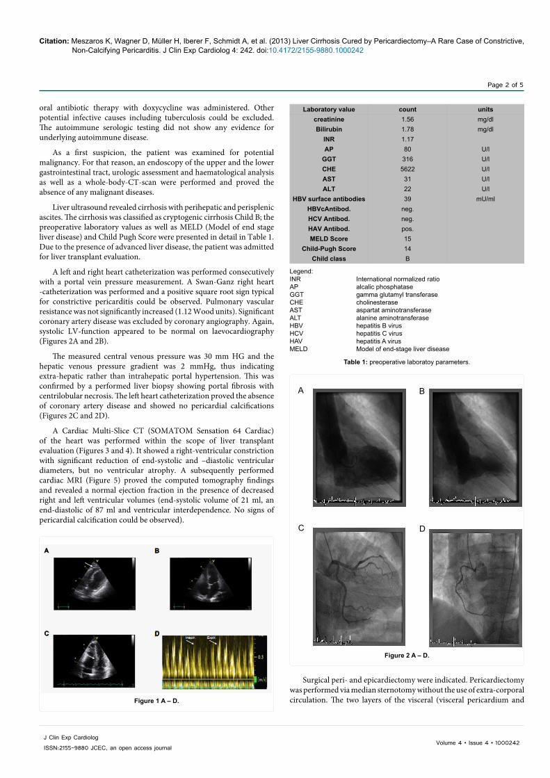
A left and right heart catheterization was performed consecutively with a portal vein pressure measurement. A Swan-Ganz right heart -catheterization was performed and a positive square root sign typical for constrictive pericarditis could be observed. Pulmonary vascular resistance was not significantly increased (1.12 Wood units). Significant coronary artery disease was excluded by coronary angiography. Again, systolic LV-function appeared to be normal on laevocardiography (Figures 2A and 2B).
The measured central venous pressure was 30 mm HG and the hepatic venous pressure gradient was 2 mmHg, thus indicating extra-hepatic rather than intrahepatic portal hypertension. This was confirmed by a performed liver biopsy showing portal fibrosis with centrilobular necrosis. The left heart catheterization proved the absence of coronary artery disease and showed no pericardial calcifications (Figures 2C and 2D).
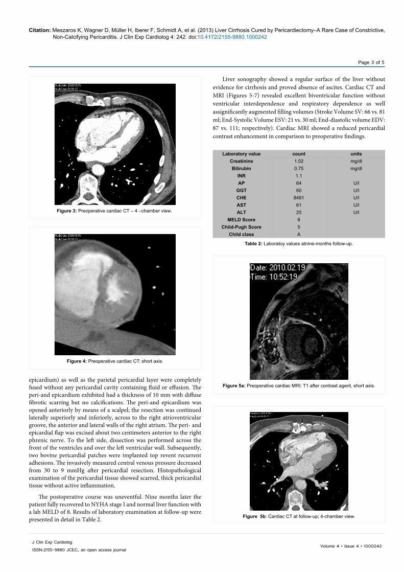
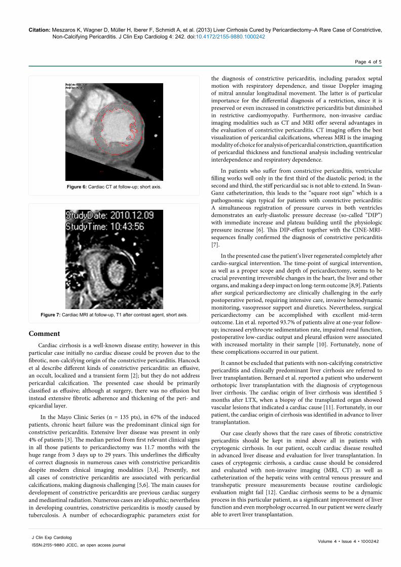
A Cardiac Multi-Slice CT (SOMATOM Sensation 64 Cardiac) of the heart was performed within the scope of liver transplant evaluation (Figures 3 and 4). It showed a right-ventricular constriction with significant reduction of end-systolic and –diastolic ventricular diameters, but no ventricular atrophy. A subsequently performed cardiac MRI (Figure 5) proved the computed tomography findings and revealed a normal ejection fraction in the presence of decreased right and left ventricular volumes (end-systolic volume of 21 ml, an end-diastolic of 87 ml and ventricular interdependence. No signs of pericardial calcification could be observed).
Legend:INR International normalized ratioAP alcalic phosphataseGGT gamma glutamyl transferaseCHE cholinesteraseAST aspartat aminotransferase ALT alanine aminotransferaseHBV hepatitis B virusHCV hepatitis C virusHAV hepatitis A virusMELD Model of end-stage liver disease
Table 1: preoperative laboratoy parameters.
Laboratory value count units creatinine 1.56 mg/dlBilirubin 1.78 mg/dl
INR 1.17 AP 80 U/l
GGT 316 U/lCHE 5622 U/lAST 31 U/lALT 22 U/l
HBV surface antibodies 39 mU/mlHBVcAntibod. neg. HCV Antibod. neg. HAV Antibod. pos. MELD Score 15
Child-Pugh Score 14 Child class B
D C
B A
Figure 2 A – D.
Citation: Meszaros K, Wagner D, Müller H, Iberer F, Schmidt A, et al. (2013) Liver Cirrhosis Cured by Pericardiectomy–A Rare Case of Constrictive, Non-Calcifying Pericarditis. J Clin Exp Cardiolog 4: 242. doi:10.4172/2155-9880.1000242
Page 3 of 5
Volume 4 • Issue 4 • 1000242J Clin Exp Cardiolog
ISSN:2155-9880 JCEC, an open access journal
epicardium) as well as the parietal pericardial layer were completely fused without any pericardial cavity containing fluid or effusion. The peri-and epicardium exhibited had a thickness of 10 mm with diffuse fibrotic scarring but no calcifications. The peri-and epicardium was opened anteriorly by means of a scalpel; the resection was continued laterally superiorly and inferiorly, across to the right atrioventricular groove, the anterior and lateral walls of the right atrium. The peri- and epicardial flap was excised about two centimeters anterior to the right phrenic nerve. To the left side, dissection was performed across the front of the ventricles and over the left ventricular wall. Subsequently, two bovine pericardial patches were implanted top revent recurrent adhesions. The invasively measured central venous pressure decreased from 30 to 9 mmHg after pericardial resection. Histopathological examination of the pericardial tissue showed scarred, thick pericardial tissue without active inflammation.
The postoperative course was uneventful. Nine months later the patient fully recovered to NYHA stage I and normal liver function with a lab MELD of 8. Results of laboratory examination at follow-up were presented in detail in Table 2.
Liver sonography showed a regular surface of the liver without evidence for cirrhosis and proved absence of ascites. Cardiac CT and MRI (Figures 5-7) revealed excellent biventricular function without ventricular interdependence and respiratory dependence as well assignificantly augmented filling volumes (Stroke Volume SV: 66 vs. 81 ml; End-Systolic Volume ESV: 21 vs. 30 ml; End-diastolic volume EDV: 87 vs. 111; respectively). Cardiac MRI showed a reduced pericardial contrast enhancement in comparison to preoperative findings.
Figure 3: Preoperative cardiac CT – 4 –chamber view.
Figure 4: Preoperative cardiac CT: short axis.
Table 2: Laboratoy values atnine-months follow-up.
Laboratory value count units Creatinine 1.02 mg/dlBilirubin 0.75 mg/dl
INR 1.1 AP 64 U/l
GGT 60 U/lCHE 8491 U/lAST 61 U/lALT 25 U/l
MELD Score 8 Child-Pugh Score 5
Child class A
Figure 5a: Preoperative cardiac MRI: T1 after contrast agent, short axis.
Figure 5b: Cardiac CT at follow-up; 4-chamber view.
Citation: Meszaros K, Wagner D, Müller H, Iberer F, Schmidt A, et al. (2013) Liver Cirrhosis Cured by Pericardiectomy–A Rare Case of Constrictive, Non-Calcifying Pericarditis. J Clin Exp Cardiolog 4: 242. doi:10.4172/2155-9880.1000242
Page 4 of 5
Volume 4 • Issue 4 • 1000242J Clin Exp Cardiolog
ISSN:2155-9880 JCEC, an open access journal
CommentCardiac cirrhosis is a well-known disease entity; however in this
particular case initially no cardiac disease could be proven due to the fibrotic, non-calcifying origin of the constrictive pericarditis. Hancock et al describe different kinds of constrictive pericarditis: an effusive, an occult, localized and a transient form [2]; but they do not address pericardial calcification. The presented case should be primarily classified as effusive; although at surgery, there was no effusion but instead extensive fibrotic adherence and thickening of the peri- and epicardial layer.
In the Mayo Clinic Series (n = 135 pts), in 67% of the induced patients, chronic heart failure was the predominant clinical sign for constrictive pericarditis. Extensive liver disease was present in only 4% of patients [3]. The median period from first relevant clinical signs in all those patients to pericardiectomy was 11.7 months with the huge range from 3 days up to 29 years. This underlines the difficulty of correct diagnosis in numerous cases with constrictive pericarditis despite modern clinical imaging modalities [3,4]. Presently, not all cases of constrictive pericarditis are associated with pericardial calcifications, making diagnosis challenging [5,6]. The main causes for development of constrictive pericarditis are previous cardiac surgery and mediastinal radiation. Numerous cases are idiopathic; nevertheless in developing countries, constrictive pericarditis is mostly caused by tuberculosis. A number of echocardiographic parameters exist for
the diagnosis of constrictive pericarditis, including paradox septal motion with respiratory dependence, and tissue Doppler imaging of mitral annular longitudinal movement. The latter is of particular importance for the differential diagnosis of a restriction, since it is preserved or even increased in constrictive pericarditis but diminished in restrictive cardiomyopathy. Furthermore, non-invasive cardiac imaging modalities such as CT and MRI offer several advantages in the evaluation of constrictive pericarditis. CT imaging offers the best visualization of pericardial calcifications, whereas MRI is the imaging modality of choice for analysis of pericardial constriction, quantification of pericardial thickness and functional analysis including ventricular interdependence and respiratory dependence.
In patients who suffer from constrictive pericarditis, ventricular filling works well only in the first third of the diastolic period; in the second and third, the stiff pericardial sac is not able to extend. In Swan-Ganz catheterization, this leads to the “square root sign” which is a pathognomic sign typical for patients with constrictive pericarditis: A simultaneous registration of pressure curves in both ventricles demonstrates an early-diastolic pressure decrease (so-called “DIP”) with immediate increase and plateau building until the physiologic pressure increase [6]. This DIP-effect together with the CINE-MRI-sequences finally confirmed the diagnosis of constrictive pericarditis [7].
In the presented case the patient’s liver regenerated completely after cardio-surgical intervention. The time-point of surgical intervention, as well as a proper scope and depth of pericardiectomy, seems to be crucial preventing irreversible changes in the heart, the liver and other organs, and making a deep impact on long-term outcome [8,9]. Patients after surgical pericardiectomy are clinically challenging in the early postoperative period, requiring intensive care, invasive hemodynamic monitoring, vasopressor support and diuretics. Nevertheless, surgical pericardiectomy can be accomplished with excellent mid-term outcome. Lin et al. reported 93.7% of patients alive at one-year follow-up; increased erythrocyte sedimentation rate, impaired renal function, postoperative low-cardiac output and pleural effusion were associated with increased mortality in their sample [10]. Fortunately, none of these complications occurred in our patient.
It cannot be excluded that patients with non-calcifying constrictive pericarditis and clinically predominant liver cirrhosis are referred to liver transplantation. Bernard et al. reported a patient who underwent orthotopic liver transplantation with the diagnosis of cryptogenous liver cirrhosis. The cardiac origin of liver cirrhosis was identified 5 months after LTX, when a biopsy of the transplanted organ showed vascular lesions that indicated a cardiac cause [11]. Fortunately, in our patient, the cardiac origin of cirrhosis was identified in advance to liver transplantation.
Our case clearly shows that the rare cases of fibrotic constrictive pericarditis should be kept in mind above all in patients with cryptogenic cirrhosis. In our patient, occult cardiac disease resulted in advanced liver disease and evaluation for liver transplantation. In cases of cryptogenic cirrhosis, a cardiac cause should be considered and evaluated with non-invasive imaging (MRI, CT) as well as catheterization of the hepatic veins with central venous pressure and transhepatic pressure measurements because routine cardiologic evaluation might fail [12]. Cardiac cirrhosis seems to be a dynamic process in this particular patient, as a significant improvement of liver function and even morphology occurred. In our patient we were clearly able to avert liver transplantation.
Figure 6: Cardiac CT at follow-up; short axis.
Figure 7: Cardiac MRI at follow-up, T1 after contrast agent, short axis.
Citation: Meszaros K, Wagner D, Müller H, Iberer F, Schmidt A, et al. (2013) Liver Cirrhosis Cured by Pericardiectomy–A Rare Case of Constrictive, Non-Calcifying Pericarditis. J Clin Exp Cardiolog 4: 242. doi:10.4172/2155-9880.1000242
Page 5 of 5
Volume 4 • Issue 4 • 1000242J Clin Exp Cardiolog
ISSN:2155-9880 JCEC, an open access journal
Acknowledgement
We thank M. Jordan Cacici for English correction.
References
1. Bertog SC, Thambidorai SK, Parakh K, Schoenhagen P, Ozduran V, et al. (2004) Constrictive pericarditis: etiology and cause-specific survival after pericardiectomy. J Am Coll Cardiol 43: 1445-1452.
2. Hancock EW (2001) Differential diagnosis of restrictive cardiomyopathy and constrictive pericarditis. Heart 86: 343-349.
3. Ling LH, Oh JK, Schaff HV, Danielson GK, Mahoney DW, et al. (1999) Constrictive pericarditis in the modern era: evolving clinical spectrum and impact on outcome after pericardiectomy. Circulation 100: 1380-1386.
4. Troughton RW, Asher CR, Klein AL (2004) Pericarditis. Lancet 363: 717-727.
5. Atwood JE, Osterberg L (2000) Images in clinical medicine. Constrictive pericarditis. N Engl J Med 343: 106.
6. Ling LH, Oh JK, Breen JF, Schaff HV, Danielson GK, et al. (2000) Calcific constrictive pericarditis: is it still with us? Ann Intern Med 132: 444-450.
7. Talreja DR, Edwards WD, Danielson GK, Schaff HV, Tajik AJ, et al. (2003) Constrictive pericarditis in 26 patients with histologically normal pericardial thickness. Circulation 108: 1852-1857.
8. Little WC, Freeman GL (2006) Pericardial disease. Circulation 113: 1622-1632.
9. Ariyoshi T, Hashizume K, Taniguchi S, Miura T, Tanigawa K, et al. (2012) Surgical experience with chronic constrictive pericarditis. Gen Thorac Cardiovasc Surg 60: 796-802.
10. Lin Y, Zhou M, Xiao J, Wang B, Wang Z (2012) Treating constrictive pericarditis in a chinese single-center study: a five-year experience. Ann Thorac Surg 94: 1235-1240.
11. Bernard PH, Le Metayer P, Le Bail B, Balabaud C, Saric J, et al. (2001) [Liver transplantation and constrictive pericarditis]. Gastroenterol Clin Biol 25: 316-319.
12. Rienmüller R, Gröll R, Lipton MJ (2004) CT and MR imaging of pericardial disease. Radiol Clin North Am 42: 587-601.
Submit your next manuscript and get advantages of OMICS Group submissions
Unique features:
• Userfriendly/feasiblewebsite-translationofyourpaperto50world’sleadinglanguages• AudioVersionofpublishedpaper• DigitalarticlestoshareandexploreSpecial features:
• 250OpenAccessJournals• 20,000editorialteam• 21daysrapidreviewprocess• Qualityandquickeditorial,reviewandpublicationprocessing• IndexingatPubMed(partial),Scopus,DOAJ,EBSCO,IndexCopernicusandGoogleScholaretc• SharingOption:SocialNetworkingEnabled• Authors,ReviewersandEditorsrewardedwithonlineScientificCredits• Betterdiscountforyoursubsequentarticles
Submityourmanuscriptat:www.editorialmanager.com/clinicalgroup
Citation: Meszaros K, Wagner D, Müller H, Iberer F, Schmidt A, et al. (2013) Liver Cirrhosis Cured by Pericardiectomy–A Rare Case of Constrictive, Non-Calcifying Pericarditis. J Clin Exp Cardiolog 4: 242. doi:10.4172/2155-9880.1000242