constrictive bronchiolitis and ulcerative colitis
TRANSCRIPT

Constrictive bronchiolitisand ulcerative colitis
Heather Ward MD FRCPC1, Kendra L Fisher MD FRCPC2,Ranjit Waghray MBBS FRCPC SCAP3, Jody L Wright BSc MD FRCPC4,
Sharon E Card MD MSc FRCPC5, Donald W Cockcroft MD FRPCC1
1Division of Respiratory Medicine; 2Department of Medical Imaging,Royal University Hospital; 3Department of Pathology, St Paul’s Hospital, Saskatoon,
Saskatchewan; 4Department of Pathology, University of British Columbia, Vancouver,British Columbia; 5Division of Internal Medicine, Royal University Hospital,
Saskatoon, Saskatchewan
Extraintestinal manifestations are well documented oc-
currences in patients with ulcerative colitis; however,
pulmonary complications were only first described in a case
series by Kraft et al (1) in 1976. Case presentations of airway,
interstitial, pleural and vascular involvement have all been
reported since then (2). The estimated incidence of pulmo-
nary complications with inflammatory bowel disease is
0.21% (3), and the majority of complications ccur in patients
with ulcerative colitis (2,4). Large airway disease, such as
tracheobronchitis, chronic bronchitis or bronchiectasis, is
most common, while small airway pathology, such as con-
strictive bronchiolitis and bronchiolitis obliterans with or-
Can Respir J Vol 6 No 2 March/April 1999 197
CASE REPORT
Correspondence and reprints: Dr Donald W Cockcroft, Division of Respiratory Medicine, Department of Medicine,Royal University Hospital, 103 Hospital Drive, Ellis Hall, 5th Floor, Saskatoon, Saskatchewan S7N 0W8. Telephone 306-966-8294,fax 306-966-8694, e-mail [email protected]
H Ward, KL Fisher, R Waghray, JL Wright, SE Card,DW Cockcroft. Constrictive bronchiolitis and ulcerativecolitis. Can Respir J 1999;6(2):197-200.
Pulmonary complications occur in an estimated 0.21% of pa-tients with inflammatory bowel disease. The most commonpresentation of pulmonary manifestations is large airway dis-ease, such as tracheobronchitis, chronic bronchitis or bronchiec-tasis. Small airway disease, such as constrictive bronchiolitis orbronchiolitis obliterans with organizing pneumonia, is less fre-quently reported, and is described as occurring in isolationfrom large airway disease. A case of a postcolectomy ulcera-tive colitis in a patient who has both large airway involve-ment, tracheobronchitis and bronchiectasis, and constrictivebronchiolitis is presented.
Key Words: Inflammatory bowel disease, Large airway disease,
Pulmonary complications, Small airway disease
Bronchiolite constrictive et colite ulcéreuseRÉSUMÉ : Les complications pulmonaires surviennent chezenviron 0,21 % des patients atteints d’une maladie inflammatoire del’intestin. La présentation la plus courante des épisodes pulmonairesest la maladie des grosses voies aériennes, comme latrachéo-bronchite, la bronchite chronique ou les bronchectasies. Lamaladie des petites voies aériennes, comme la bronchioliteconstrictive ou la bronchiolite oblitérante avec pneumonieorganisée, est moins fréquemment rapportée, et est décrite commesurvenant indépendamment de la maladie des grosses voiesaériennes. On décrit le cas d’un patient qui après une colectomiepour colite ulcéreuse présente à la fois une atteinte des grosses voiesaériennes, trachéo-bronchite et bronchectasies, et une bronchioliteconstrictive.
ganizing pneumonia, is infrequently reported (2).
Involvement of the entire bronchial tree is rare. We present
the case of a patient with constrictive bronchiolitis and large
airway disease, consisting of tracheobronchitis and bron-
chiectasis, complicating ulcerative colitis.
CASE PRESENTATIONA 44-year-old, lifetime nonsmoking farmer with a past
history of thalassemia minor presented in 1987 at age 35
years with clinically diagnosed and biopsy-proven ulcerative
colitis. Gastrointestinal symptoms progressed despite treat-
ment with prednisone, salazopyrin and imodium, and, after
nine months, a total colectomy was done. All medications
were discontinued following the colectomy. Joint symptoms
consisting of morning stiffness, migratory polyarthralgias,
and polyarthritis involving hands, knees, hips and ankles be-
gan before his colectomy, and persisted postoperatively; a di-
agnosis of enteropathic arthritis was made. Treatment with
nonsteriodal anti-inflammatory drugs was unsuccessful, and
salazopyrin 1000 mg twice daily and prednisone 10 mg daily
was commenced in 1994. Before resuming prednisone and
salazopyrin, the patient developed a productive cough with
exertional dyspnea, and, on pulmonary function testing,
forced expiratory volume in 1 s (FEV1) was 2.90 L/min (68%
predicted), (FEV1/forced vital capacity ratio of 56%), and
the carbon monoxide diffusing capacity of the lungs was
66% predicted. Bronchoscopy, done in 1993, demonstrated
tracheal and bronchial inflammation with thick secretions
that were culture negative. Medical treatment included in-
haled beclomethasone dipropionate 250 g two puffs bid and
inhaled salbutamol as needed. Exertional dyspnea, cough
with sputum (125 mL per day), occasional hemoptysis and
weight loss persisted despite salazopyrin 1000 mg bid,
prednisone 10 to 20 mg per day, inhaled corticosteroids and
ventolin.
In 1997, three-and-a-half years after the onset of respira-
tory symptoms, FEV1 had declined to 1.67 L/min (44% pre-
dicted), and the patient was admitted to hospital for further
investigations. He continued to have asymmetric migratory
polyarthralgias and a history compatible with dactylitis,
again suggestive of enteropathic arthritis. Physical examina-
tion was normal except for scattered inspiratory and expira-
tory crackles, and there were no wheezes on auscultation of
the chest. Oxygen saturation was 94% on room air. Labora-
tory results included a white blood cell count of 13×109/L,
hemoglobin 120 g/L with a mean corpuscular volume 64.6 fl,
rheumatoid factor 1/493 KU/L, an anti-nuclear antigen
(ANA) titre 1/320, anti-dsDNA 1/32 KU/L and a polyclonal
gammopathy. Sweat chloride was 24 mmol/L. There was
bronchial and bronchiolar wall thickening, and a suggestion
of multiple peripheral ‘nodules’ on chest radiograph having
the appearance of bronchiolar wall thickening with possible
mucoid impaction. There was a small 1 cm nodule in the left
upper lobe with apparent cavitation. A standard and high
resolution computed tomography (CT) scan showed both
central and peripheral bronchial and bronchiolar wall thick-
ening with associated bronchiolectasis. There was a ‘tree-in-
bud’ appearance consistent with distal small airway mucoid
198 Can Respir J Vol 6 No 2 March/April 1999
Ward et al
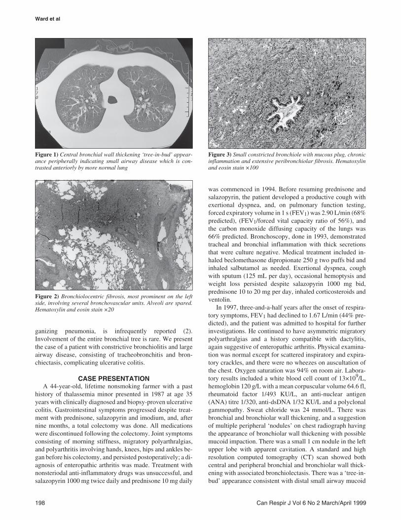
Figure 1) Central bronchial wall thickening ‘tree-in-bud’ appear-ance peripherally indicating small airway disease which is con-trasted anteriorly by more normal lung
Figure 2) Bronchiolocentric fibrosis, most prominent on the leftside, involving several bronchovascular units. Alveoli are spared.Hematoxylin and eosin stain ×20
Figure 3) Small constricted bronchiole with mucous plug, chronicinflammation and extensive peribronchiolar fibrosis. Hematoxylinand eosin stain ×100

impaction (Figure 1). Mediastinal and bilateral hilar adeno-
pathy were seen, and CT scan provided no evidence of inter-
stitial lung disease. Diffuse inflammation of the trachea and
mainstem bronchi with exudate bilaterally was seen on bron-
choscopy. An open lung biopsy was completed, and histo-
logical examination showed large areas of rounded fibrosis
that were situated adjacent to muscular pulmonary arteries
(Figure 2). On elastic stain, a fragmented elastic lamina
proved that the areas were obliterated bronchioles. In some
airways, a partial lumen remained but was distorted and ir-
regular. The fibrous tissue had largely replaced the muscu-
laris and had markedly increased the volume of the
adventitial compartment of the airway. There were no granu-
lation tissue polyps or organizing pneumonia, indicating that
the airway lesions were the result of a rather marked degree
of constrictive bronchiolitis (Figure 3).
Prednisone 20 mg and azathioprine 150 mg per day were
started. At an eight-month follow-up clinic visit, the patient
had symptomatic improvement of exertional dyspnea, cough
and sputum production. FEV1 had improved from 1.67 to
2.85, and chest radiograph had improved (Table 1). A repeat
CT scan completed 15 months after initiating treatment
showed significant improvement in the ‘tree-in-bud’ appear-
ance, with a marked decrease in the central and peripheral
bronchial and bronchiolar wall thickening; however, moder-
ate predominantly bronchial wall thickening and mild bron-
chiolectasis persisted in both lower lobes. Mediastinal and
hilar adenopathy had also largely regressed.
DISCUSSIONConstrictive bronchiolitis is an infrequently reported
complication of ulcerative colitis, and each case report has
unique characteristics that expand our knowledge of clinical
presentation, pathology and treatment. Only two of 33 pa-
tients in the largest reported case series by Camus et al (2)
had small airway disease. The three case reports in the litera-
ture of constrictive bronchiolitis had different clinical pres-
entations to ours (3,5,6). One patient had ulcerative colitis
and no cough or sputum production, but also had exertional
dyspnea, severe airflow obstruction, a negative rheumatoid
factor and ANA, and biopsy-proven constrictive bronchioli-
tis (2,5). The second patient had dyspnea, productive cough,
moderate airflow obstruction and bronchoscopy changes
suggesting chronic bronchitis beginning before the diagnosis
of colitis. This patient also had a negative bronchography for
bronchiectasis and a lung biopsy demonstrating diffuse pan-
bronchiolitis (3). A third had exertional dyspnea and a non-
productive cough, with a normal bronchoscopy and a lung
biopsy showing obliterative bronchiolitis (6). None of these
patients had any other extraintestinal manifestations of ul-
cerative colitis. Our patient had systemic symptoms of
weight loss and articular involvement compatible with entro-
pathic arthritis but not clinically suggestive of rheumatoid ar-
thritis despite an elevated rheumatoid factor. He had similar
respiratory symptoms to the previously reported cases, in-
cluding exertional dyspnea, productive cough and moderate
airflow obstruction on pulmonary function testing. Both
chest radiograph and CT scan were suggestive of large and
small airway pathology. He had marked improvement in
clinical and radiological findings with prednisone and
azathioprine.
Sulfasalazine-associated lung disease is a rare occurrence,
and is described most frequently as patchy bilateral lung in-
filtrates in the mid to upper lung fields with transient mild
eosinophilia in a patient presenting with fever and dry cough.
The symptoms and chest radiograph changes usually resolve
by discontinuing sulfasalazine and starting therapy with
prednisone (7). One case of bronchiolitis obliterans with
organizing pneumonia has been described in association
with sulfasalazine; however, no cases of constrictive bron-
chiolitis have been described in association with sulfasa-
lazine (8,9). Our patient’s respiratory symptoms began
before the resumption of sulfasalzine for his musculoskeletal
symptoms.
The pathogenesis of pulmonary manifestations in in-
flammatory bowel disease remains under speculation.
Nearly half of the cases of airway involvement occur post-
colectomy, and one suggestion has been that the focus of
inflammation switches from the gastrointestinal tract to
the lung after surgery (2,10). The embryological gut source
of both the lung and gastrointestinal tract may account for a
common target of inflammation at both epithelial sites (6).
There is no predictive serology for the development of lung
pathology. Rheumatoid factor is secreted by mucosa of the
gastrointestinal tract (11) and can be present in the serum of
patients with inflammatory bowel disease, but there is no cor-
relation between titres and disease activity or extraintestinal
manifestations (12). ANA is present in 25% and peri anti-
Can Respir J Vol 6 No 2 March/April 1999 199
Constrictive bronchiolitis and ulcerative colitis
TABLE 1Pulmonary function tests of patient with constrictive bronchiolitis and ulcerative colitis
October 1997 October 1998Actual % predicted Actual % predicted
Forced vital capacity (FVC) 3.14 63 4.34 87Forced expiraotry volume in 1 s (FEV1 ) 1.72 46 2.73 73FEV1/FVC 55/75 63/75Total lung capacity 5.35 78 6.89 100Carbon monoxide diffusion capacity of the lungs 28.55 75 26.75 71Prescription prednisone (mg) 0 20Azathioprine (mg) 0 150

nuclear cytoplasmic antibody in 68% to 86% of ulcerative
colitis patients, again with unknown etiological or clinical
implications (13,14). The positive serology results suggest
an autoimmune etiology for inflammatory bowel disease
and its systemic manifestations, but the mechanism is un-
known. There is an insufficient number of case descrip-
tions of pulmonary involvement in ulcerative colitis to
postulate the disease process or the extent of potential lung
involvement.
Case series of large airway involvement in ulcerative coli-
tis describe tracheobronchitis, chronic “suppurative” bron-
chitis or bronchiectasis as being anatomically localized, with
no continuous involvement of the large and small airways
(2,5). Constrictive bronchiolitis is usually a lesion of the
bronchioles with luminal narrowing by scarring (15).
Chronic productive cough was defined by chronic bronchi-
tis; bronchiectasis was characterized by typical radiographi-
cal findings (chest radiograph, bronchography and CT scan);
and tracheobronchitis was defined by the bronchoscopy find-
ings of inflammation and edema. The localized upper airway
pathology explained the clinical presentations in these case
series and did not indicate a need for a lung biopsy to look for
small airway pathology. The patients with bronchiolitis had
nonspecific radiographical findings and a lack of large air-
way pathology to explain their respiratory symptoms, which
lead to their open lung biopsy. Only one patient with bron-
chiolitis had chronic bronchitis found on bronchoscopy (3).
Our patient had tracheobronchitis identified during his bron-
choscopy and bronchiectasis on repeat CT scan following
therapy.
This case is a unique presentation of anatomic involve-
ment of both small and large airways, with constrictive bron-
chiolitis, tracheobronchitis and bronchiectasis associated
with enteropathic articular manifestations in a postcolectomy
ulcerative colitis patient with a positive rheumatoid factor
and ANA. The clinical presentation and pathology results ex-
pand the possible spectrum of ulcerative colitis-associated
pulmonary complications.
REFERENCES1. Kraft SC, Earle RH, Roesler M, Esterly JK. Unexplained
bronchopulmonary disease with inflammatory bowel disease.Arch Intern Med 1976;136:454-9.
2. Camus P, Piard F, Ashcroft T, Gal AA, Colby TV. The lung ininflammatory bowel disease. Medicine 1993;72:151-83.
3. Desai SJ, Gephardt GN, Stoler JK. Diffuse panbronchiolitis precedingulcerative colitis. Chest 1989;95:1342-4.
4. Spira A, Grossman R, Balter M. Large airway disease associated withinflammatory bowel disease. Chest 1998;113:1723-6.
5. Wilcox P, Miller R, Miller G, et al. Airway involvement in ulcerativecolitis. Chest 1987;92:18-22.
6. Hilling GAL, Robertson DAF, Chalmers AH, Ruby HS. Unusualpulmonary complications of ulcerative colitis with a rapid response tocorticosteroids: case report. Gut 1994;35:847-8.
7. Yaffe BH, Korelitz BI. Sulfasalazine pneumonitis. Am J Gastroenterol1983;78:493-4.
8. Reinoso M, Schroeder K, Pisani R. Lung disease associated withorally administered mesalamine for ulcerative colitis. Chest1992;101:1469-71.
9. Schwartzman KJ, Bowie DM, Yeadon C, Fraser R, Sutton ED,Levy RD. Constrictive bronchiolitis obliterans following gold therapyfor psoriatic arthritis. Eur Respir J 1995;8:2191-3.
10. Tzanakis N, Bouros D, Samiou M, et al. Lung function in patients withinflammatory bowel disease. Respir Med 1998;92:516-22.
11. MacDermott RP, Schreiber S, Nash GS, Koopman WJ. Increasedspontaneous secretion of rheumatoid factor by intestinal lamina propriamononuclear cells from Crohn’s disease but not ulcerative colitispatients. Clin Exp Immunol 1993;92:152-7.
12. Nielsen H, Binder V, Daugharty H, Svehag SE. Circulating immunecomplexes in ulcerative colitis: correlation to disease activity.Clin Exp Immunol 1978;11:72-80.
13. Zauli D, Crespi C, Dall’Amore P, Bianchi FB, Pisi E. Antibodies to thecytoskeleton components and other autoantibodies in inflammatorybowel disease. Digestion 1985;32:140-4.
14. Habeeb MA, Rajalingam R, Dhar A, Kumar A, Sharma MP,Mehra NK. HLA associated and occurrence of autoantibodies inAsian-Indian patients with ulcerative colitis. Am J Gastroenterol1997;92:772-6.
15. Colby TV. Bronchiolitis: pathologic considerations. Am J Clin Pathol1998;109:101-9.
200 Can Respir J Vol 6 No 2 March/April 1999
Ward et al

Submit your manuscripts athttp://www.hindawi.com
Stem CellsInternational
Hindawi Publishing Corporationhttp://www.hindawi.com Volume 2014
Hindawi Publishing Corporationhttp://www.hindawi.com Volume 2014
MEDIATORSINFLAMMATION
of
Hindawi Publishing Corporationhttp://www.hindawi.com Volume 2014
Behavioural Neurology
EndocrinologyInternational Journal of
Hindawi Publishing Corporationhttp://www.hindawi.com Volume 2014
Hindawi Publishing Corporationhttp://www.hindawi.com Volume 2014
Disease Markers
Hindawi Publishing Corporationhttp://www.hindawi.com Volume 2014
BioMed Research International
OncologyJournal of
Hindawi Publishing Corporationhttp://www.hindawi.com Volume 2014
Hindawi Publishing Corporationhttp://www.hindawi.com Volume 2014
Oxidative Medicine and Cellular Longevity
Hindawi Publishing Corporationhttp://www.hindawi.com Volume 2014
PPAR Research
The Scientific World JournalHindawi Publishing Corporation http://www.hindawi.com Volume 2014
Immunology ResearchHindawi Publishing Corporationhttp://www.hindawi.com Volume 2014
Journal of
ObesityJournal of
Hindawi Publishing Corporationhttp://www.hindawi.com Volume 2014
Hindawi Publishing Corporationhttp://www.hindawi.com Volume 2014
Computational and Mathematical Methods in Medicine
OphthalmologyJournal of
Hindawi Publishing Corporationhttp://www.hindawi.com Volume 2014
Diabetes ResearchJournal of
Hindawi Publishing Corporationhttp://www.hindawi.com Volume 2014
Hindawi Publishing Corporationhttp://www.hindawi.com Volume 2014
Research and TreatmentAIDS
Hindawi Publishing Corporationhttp://www.hindawi.com Volume 2014
Gastroenterology Research and Practice
Hindawi Publishing Corporationhttp://www.hindawi.com Volume 2014
Parkinson’s Disease
Evidence-Based Complementary and Alternative Medicine
Volume 2014Hindawi Publishing Corporationhttp://www.hindawi.com