lipid accumulation impairs natural killer cell
TRANSCRIPT
RESEARCH ARTICLE Open Access
Lipid accumulation impairs natural killercell cytotoxicity and tumor control in thepostoperative periodSeyedeh Raheleh Niavarani1, Christine Lawson1, Orneala Bakos1, Marie Boudaud2, Cory Batenchuk3,Samuel Rouleau1 and Lee-Hwa Tai1,4*
Abstract
Background: Natural killer (NK) cell dysfunction following cancer surgery has been shown to promote metastases.Recent studies demonstrate an emerging role for lipids in the modulation of NK cell innate responses. However, themechanisms involved in lipid modulation of NK cell postoperative anti-tumor function are unknown. This currentstudy will determine whether the lipid accumulation via scavenger receptors on NK cells is responsible for theincrease in postoperative metastasis.
Methods: Lipid content in mouse and human NK cells was evaluated by flow cytometry. NK cell scavenger receptor(SR) expression was measured by microarray analysis, validated by qRT-PCR and flow cytometry. NK cell ex vivo and invivo tumor killing was measured by chromium-release and adoptive transfer assays, respectively. The mediating role ofsurgery-expanded granulocytic myeloid derived suppressor cells (gMDSC) in SR induction on NK cells was evaluatedusing co-culture assays.
Results: NK cells in surgery-treated mice demonstrated increased lipid accumulation, which occurred via up-regulationof MSR1, CD36 and CD68. NK cells with high lipid content had diminished ability to lyse tumor targets ex vivo. Adoptivetransfer of lipid-laden NK cells into NK cell-deficient mice were unable to protect against a lung tumor challenge.Granulocytic MDSC from surgery-treated mice increased SR expression on NK cells. Colorectal cancer surgicalpatients showed increased NK cell lipid content, higher CD36 expression, decreased granzyme B and perforinproduction in addition to reduced cytotoxicity in the postoperative period.
Conclusions: Postoperative lipid accumulation promotes the formation of metastases by impairing NK cell function inboth preclinical surgical models and human surgical colorectal cancer patient samples. Understanding and targeting themechanisms underlying lipid accumulation in innate immune NK cells can improve prognosis in cancer surgical patients.
Keywords: Fatty natural killer cells, Perioperative immunosuppression, Immunometabolism
BackgroundSurgical resection is the cornerstone of treatment forsolid tumors. Despite complete resection with curativeintent, many patients die from recurrent or metastaticdisease because minimal residual disease and micro-metastases are present at the time of surgery [1–4]. Our
research [5–7] and others [1, 2, 8–10] have demonstratedthat major cancer surgery initiates a series of physiologicalresponses to promote tissue repair and wound healing.However, the proangiogenic, growth factor replete andimmunosuppressed state in the surgically stressed hostcreates a prometastatic environment that can be exploitedby micrometastatic tumors [3].Natural killer (NK) cells play a key role in the innate
elimination of malignant cells. The defective function ofNK cells in cancer contributes greatly to tumor escapeand metastatic disease [5, 11–14]. Impaired NK cellactivity following cancer surgery has been shown in
© The Author(s). 2019 Open Access This article is distributed under the terms of the Creative Commons Attribution 4.0International License (http://creativecommons.org/licenses/by/4.0/), which permits unrestricted use, distribution, andreproduction in any medium, provided you give appropriate credit to the original author(s) and the source, provide a link tothe Creative Commons license, and indicate if changes were made. The Creative Commons Public Domain Dedication waiver(http://creativecommons.org/publicdomain/zero/1.0/) applies to the data made available in this article, unless otherwise stated.
* Correspondence: [email protected] of Anatomy and Cell Biology, Université de Sherbrooke,Pavillon sur la Recherche Appliqué du Cancer at 3201 rue Jean-Mignault,Sherbrooke, QC J1E 4K8, Canada4Centre de Recherche Clinique de Centre Hospitalier de l’Universite deSherbrooke, Sherbrooke, QC, CanadaFull list of author information is available at the end of the article
Niavarani et al. BMC Cancer (2019) 19:823 https://doi.org/10.1186/s12885-019-6045-y
human patients [1, 3, 5, 15] and preclinical studies[5, 16, 17]. Postoperative NK cell suppression is associ-ated with increased metastases in mouse models of spon-taneous [5, 18] metastases, while in translational humanstudies, impaired NK cell function is associated with ahigher rate of cancer recurrence and mortality [1, 9, 10, 19].There is a growing body of literature in the emerging fieldof perioperative immunology that is characterizing themechanisms of cancer surgery-induced immunosuppres-sion and immunotherapeutic strategies to prevent cancerrecurrence [7, 9, 20, 21].We confirmed the in vivo role of NK cells in mediating
tumor removal following cancer surgery in mouse modelsof melanoma [5], colorectal [22] and breast carcinomas[5, 7]. Using an adoptive transfer strategy of surgicallystressed and control donor NK cells into NK cell-deficientrecipient mice, we demonstrated that surgically stressedNK cells cannot protect from a subsequent lung tumorchallenge in these recipient mice. Additionally, we ensuredthat perioperative anesthesia and analgesics did not con-tribute to immunosuppression [21]. In colorectal cancersurgical patients, functional profiling of peripheral bloodmononuclear cells (PBMC) revealed that NK cell cytotox-icity was significantly reduced following primary tumorresection [5]. We recently reported that postoperative NKcell dysfunction is linked to granulocytic myeloid derivedsuppressor cell (gMDSC) accumulation and Arginase I(ARG1) production [7]. MDSCs have been shown in manydisease contexts (cancer, burn injury, chronic inflam-mation, etc.) to impair immune effector cells throughimmune suppressive molecules such as ARG1 and nitricoxide synthase [20, 23–26]. During our flow cytometricinvestigations into the effect of surgical stress on NK cellfunction, we inadvertently observed accumulation of lipidsin NK cells isolated from surgery-treated mice. Furtherinvestigations revealed that this lipid-laden phenotype wasassociated with postoperative NK cell dysfunction. Thesefindings prompted us to investigate the potential role oflipid accumulation on NK cell dysfunction in cancer.Lipids are a diverse group of low molecular weight
molecules that contain fatty acid groups. They representa major structural component of cellular membranes,participate in inter- and intra-cellular signaling, andserve as important energy sources [27]. Previous animaland human studies have shown the negative effect ofdietary lipids on innate immune cell function [27, 28].These data suggest that lipids could be important media-tors of immune cell function. For example, excess lipidaccumulation in the liver has been shown to negativelyimpact macrophage function leading to inflammation,tissue damage and chronic liver disease [29]. The abilityof lipids to participate in the innate immune inflammatoryresponse is an emerging field of study, mainly focused onthe treatment of inflammatory diseases [29, 30]. However,
the mechanisms involved in lipid accumulation and modu-lation of innate immune function are poorly understood.The detrimental effect of lipid accumulation in conven-
tional Dendritic Cells (cDC) has been reported [31, 32].The authors demonstrated that cDC in tumor bearingmice and in cancer patients had increased levels of lipidcontent. Mechanistically, it was reported that upregulationof scavenger receptors was responsible for lipid accumula-tion in cDC. Importantly, cDCs with high lipid contentwere not able to effectively stimulate allogeneic T cells orpresent tumor-associated antigens [31]. In lipid rich envi-ronments, NK cells have been recently reported to rapidlyaccumulate intracellular lipid droplets and undergo meta-bolic reprograming towards lipid metabolism. Mechanistic-ally, these NK cells displayed suppressed effector functionthrough downregulation of perforin and granzyme mRNAtranscripts and significantly reduced IFNγ production.Moreover, lipid bearing NK cells failed to eradicate tumorgrowth in vivo, suggesting that lipid accumulation inducesNK cell metabolic paralysis and impaired antitumor re-sponses [33]. In another recent study focused on character-izing NK cells for adoptive immunotherapy, ex vivo IL15treated NK cells displayed an exhausted phenotype with re-duced IFN-γ production, reduced CD107a degranulationand significant upregulation of fatty acid metabolism [34].An increase in lipid accumulation and fatty acid oxidation(FAO) metabolism in tumor-infiltrating myeloid-derivedsuppressor cells (T-MDSCs) were found to enhance inhibi-tory activity towards T-cell proliferation, supporting tumorgrowth and microenvironment development [35]. Asidefrom these reports, there are no studies on NK cell associ-ation with lipids and the impact of this interaction on NKcell antitumor activity. While the role of lipid accumulationand metabolism has been recently outlined in NK cells, themetabolic state of NK cells under cancer-surgery inducedimmunosuppression is unknown. Here, we report thenovel finding that following primary tumor resection, post-operative NK cells express up-regulation of three scavengerreceptors (SR) and increase lipid accumulation with nega-tive effects on NK cell-mediated tumor recognition andremoval.
MethodsMiceC57BL/6 (B6), BALB/c and NK cell-deficient mice(IL2γR-KO) were purchased from The Jackson Labora-tory. Female mice aged 6–8 weeks (20-25 g) were usedfor all experiments, where 4–6 mice were used perexperimental group according to previous publications[36]. Animals were housed in pathogen-free conditionsat the Central Animal Care Facility of the Université deSherbrooke (Quebec) and the Animal Care VeterinaryService facility of the University of Ottawa (Ontario)with access to food and water ad libitum. Animal were
Niavarani et al. BMC Cancer (2019) 19:823 Page 2 of 14
euthanized by cervical dislocation under anesthesia (isoflur-ane). All studies performed on animals were conducted inaccordance with university guidelines and the CanadianCouncil on Animal Care. The protocols were approved byAnimal Care Committees at both universities.
Cell lines and virusesMurine B16F10LacZ melanoma cell line was obtainedfrom Dr. K Graham (London, Ontario) and maintainedin complete DMEM (cDMEM). Cells were resuspendedin media without serum for intravenous (iv) injectionthrough the lateral tail vein. 3 × 10⁵ cells at > 98% viabilitywere injected in 0.1 ml per mouse. Murine CT26lacZcolorectal carcinoma, murine 4 T1 breast carcinoma,murine YAC-1 lymphoma and human K562 leukemia cellline were purchased from ATCC and maintained incomplete RPMI (cRPMI). All cell lines were verified tobe mycoplasma free and show appropriate microscopicmorphology at time of use.
Establishment of murine surgical stress modelThe murine surgical stress model was conducted as pre-viously described [5, 22]. Routine perioperative care formice was conducted as per standard protocols includingthe use of Buprenorphine (0.05 mg/kg) for pain manage-ment and isoflurane for induction and maintenance(2.0–2.5%). Surgical stress was induced in mice by anabdominal laparotomy (4 cm long midline incision) andleft nephrectomy preceded by an iv challenge of 3 × 10⁵
B16lacZ cells or CT26lacZ to establish pulmonary metasta-ses. Animals were euthanized at 3 days following tumor cellinjection and their harvested lungs were stained with X-gal(Bioshop) as described previously [22]. Total number ofsurface visible metastases was determined on the largestlung lobe (left lobe) using a stereomicroscope (Leica Micro-systems) by a research technician blinded to treatmentgroups.
Antibodies and flow cytometric analysisTo analyze splenic lymphocyte populations, organs wereremoved from mice and RBCs lysed using ACK lysisbuffer. Fc block was added prior to antibody staining.The following mAbs were used: anti-TCRβ (H57597),anti-CD122 (TM-beta1), anti-NK1.1 (clone), anti-MSR1(REA148), anti-CD36 (HM36) and anti-CD68 (FA-11)were purchased from eBiosciences. Isotype controls werepurchased from BD Biosciences. BODIPY 495/503 waspurchased from Life Technologies (D3922). The follow-ing human antibodies were used: anti-CD3 (SK7) anti-CD56 (NCAM), CD36 (CB38), granzyme B (GB11) andperforin (δG9), from BD Biosciences. Flow cytometryacquisitions were performed on a CytoFLEX 30 Summitinstrument (Beckman Coulter). Data was analyzed with
CytExpert software. Bioimaging was performed using afluorescence microscope (Leica).
Microarray and qPCRNK cells were sorted on a FACS Aria (BD) by the FlowCytometry Core of the University of Ottawa. Cells weredouble sorted to > 95% purity. RNA was extracted fromthe sorted and pooled NK cells using an RNeasy MiniKit (Qiagen #74106) used in conjunction with QIAshredderColumns (Qiagen #79656). The quality of all samples wasvalidated by using a Bio-analyzer 2100 (Agilent Technolo-gies Inc.). 100 ng of purified RNA was loaded onto Gene-Chip Mouse Gene 1.0 ST arrays according to manufacturerinstructions. CEL files were later processed by AltAnalyzeV2.0 under default parameters. A detection above back-ground score > 70 and a pV < 0.05 were used to filter probesets. Gene expression was evaluated using constitutiveprobe sets shared across splice variants. Gene enrichmentanalyses were performed on the subset of genes induced(n = 223) or repressed (n = 109) over 2 fold by surgicalstress. All p values reported have been corrected for mul-tiple hypothesis testing using the Benjamini–Hochbergmethod.The following primers were used for validation of
microarray results by qPCR.Msr1 Forward: GACAGAGAATCAGAGGCTCT.Msr1 Reverse: AAGGACTTCAACTTCTCCTG.CD36 Forward: GATGACGTGGCAAAGAACAG.CD36 Reverse: TCCTCGGGGTCCTGAGTTAT.CD68 Forward: AGTCTACCTGGACTACATGG.CD68 Reverse: TGCATTTCCACAGCAGAAGC.The following primers were used for validation of NK
cell purity results by qPCR.Eomes Forward: CGGTGTGGAGGACTTGAATGA.Eomes Reverse: AATCCGTGGGAGATGGAGTT.Gata3 Forward: TCCTCTACGCTCCTTGCTACT.Gata3 Reverse: AGGAGGGTTTAGGGAGGAAAGA.Tbet Forward: CTGGAGCCCACAAGCCATTA.Tbet Reverse: TTGGAAGCCCCCTTGTTGTT.RORc Forward: AACCAGTATCCTGTTCCCAGC.RORc Reverse: TGTCGCCACTGGAAGGATAG.ID2 Forward: CGGTGAGGTCCGTTAGGAAAA.ID2 Reverse: TGACGATAGTGGGATGCGAG.ID3 Forward: TGTGGGGACAAGTCATCTGG.ID3 Reverse: TGGTAGCTGCCCATCTGAGAG.
Fatty acid phagocytosis assay5 × 106 purified NK cells (NK cell isolation kit II, Stem-cell) were resuspended in 500 μL of 10 μg/mL fluores-cently labeled (Bodipy FL) palmitic fatty acid (MolecularProbes) at 37 °C for 15–30min. Controls consisted ofunloaded NK cells and NK cells loaded at 4 °C. Flow cy-tometry acquisitions were performed on a CytoFLEX 30
Niavarani et al. BMC Cancer (2019) 19:823 Page 3 of 14
Summit software (Beckman Coulter). Data was analyzedwith CytExpert software.
NK cell adoptive transfer experimentsFor NK cell transfer experiments, splenocytes were iso-lated from no surgery control or 18 post-surgery fromB6 mice, enriched for NK cells with the NK cell isolationkit II using a Robosep cell sorter (Stemcell). These enrichedNK cells were further sorted for BODIPYhigh NK cells byflow cytometry sorting (FACS Aria, BD). 3 × 10⁶ BODIPY-high NK cells as determined by flow cytometry were injectedvia the lateral tail vein into NK-deficient mice. For all trans-fers, 3 × 10⁵ B16lacZ tumor cells were injected iv 1 h postimmune cell transfer. 3 days post immune and tumor cellinjection, lungs of NK-deficient mice were isolated andquantified with Xgal (as described above).
NK cell cytotoxicity assaysThe chromium release assay was performed as previouslydescribed [37]. Briefly, splenocytes were isolated from sur-gically stressed and control mice at 18 h post-surgery fromMSR1-deficient mice. Pooled and sorted NK cells were re-suspended at a concentration of 2.5 × 10⁶ cells/ml andthen mixed with chromium labelled target cells (Yac-1),which were re-suspended at a concentration of 3 × 10⁴
cells/ml at different effector to target ratios (E:T) (50:1, 25:1, 12:1, 6:1). The cell mixture was incubated for 4 h priorto analysis of 51Cr release in the supernatant using agamma counter (Perkin Elmer).
Granulocytic MDSC:NK cell coculture assaygMDSC were purified using the Mouse MDSC isolationkit (Miltenyi). Purified gMDSC were seeded in triplicatesin 96 V-bottom well plates at 1:1 ratio with purified NKcells. Following 24 h of co-culture in the presence ofabsence of a transwell insert (5.0 μm, Corning), NK cellswere harvested and stained with anti-MSR1 (REA148),anti-CD36 (HM36) and anti-CD68 (FA-11) for flowcytometric analysis.
Human PBMC for flow cytometry and cytotoxicity assayHuman whole blood was collected (CIPO study, 2017–1506 approved by the ethics board of CIUSSS de l’EstrieCHUS) and processed immediately for PBMC usingFicoll-Paque (Stemcell). PBMC were resuspended at aconcentration of 5 × 10⁶ in freezing media (RPMI with12.5% Human Serum Albumin and 10% DMSO). 1 mLaliquots were frozen in cryovials overnight at − 80 °Cand transferred to liquid N2 for long-term storage.Following batch thawing of viably frozen cells, PBMCwere stained with anti-human CD3, CD56, granzyme B,perforin and BODIPY 493/503 flow cytometry analysis.For cytotoxicity assessment, PBMC were rested for 20 hwith recombinant IL2 (250 U). 51Cr-labelled K562 cells
were then added and assessment of target cell killingwas determined as above.
Statistical analysisStatistical significance other than microarray work wasdetermined by student t test with a cutoff P value of0.05. Data is presented as +/− SEM. Prism v.7 was usedfor all statistical tests.
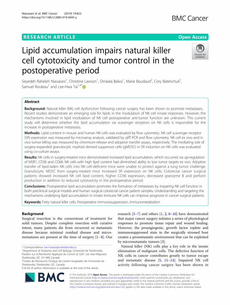
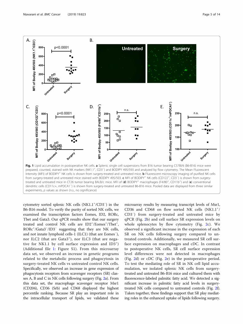
ResultsNK cells accumulate lipids following surgeryWe previously demonstrated that NK cell antitumorcytotoxic function is critically impaired following cancersurgery and significantly contributes to the growth oflung tumor metastases in the B16 melanoma [5], CT26colorectal tumor [22] and 4 T1 breast tumor metastasismodels [7, 38]. During flow cytometric investigationsinto the mechanisms of NK cell impairment followingsurgery, we observed accumulation of lipids in splenicNK cells (NK1.1+/CD3−) isolated from surgery-treated(abdominal nephrectomy) B16F10lacZ-tumor bearingC57Bl/6 mice (B6-B16) as compared to NK cells fromuntreated control mice using the lipophilic fluorescentdye Bodipy 493/503 (Fig. 1a). From both flow cytometryand microscopy, we observed increased lipid accumulationin surgery-treated NK cells over controls. We verifiedthese results using fluorescent microscopy to visualizeBodipy+ flow cytometry sorted NK cells (NK1.1+/CD3−)from surgery treated and untreated B6-B16 mice (Fig. 1b).To further support our observations of fatty acid accumu-lation in NK cells in the B6-B16 model, we assessed fattyacid levels in NK cells from the BALB/c-CT26 model ofexperimental colorectal cancer and surgery, which wehave previously established to study the prometastaticeffects of major surgery [5, 18]. In this model, we also ob-served increased lipid levels in NK cells (CD122+/CD3−)from surgery-treated mice compared to controls (Fig. 1c).The presence of lipids in innate NK cells prompted us toinvestigate whether other innate myeloid subsets mightdisplay a similar phenotype in the postoperative period.Therefore, we measured lipid content in macrophages andconventional dendritic cells (cDC), comparing surgery-treated and untreated controls in the B6-B16 model. Incontrast to postoperative NK cells, no differences in lipidlevels as measured by Bodipy 493/503 were observed inmacrophages (Fig. 1d) or cDC (Fig. 1e). Taken together,these results suggest surgical stress increases lipidaccumulation in NK cells.
Scavenger receptors are upregulated on NK cellsfollowing surgeryTo investigate the mechanism of lipid accumulation inpostoperative NK cells, we performed an unbiased micro-array analysis of genes induced by surgery from flow
Niavarani et al. BMC Cancer (2019) 19:823 Page 4 of 14
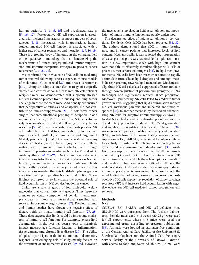
cytometry sorted splenic NK cells (NK1.1+/CD3−) in theB6-B16 model. To verify the purity of sorted NK cells, weexamined the transcription factors Eomes, ID2, RORc,Tbet and Gata3. Our qPCR results show that our surgerytreated and control NK cells are ID2+/Eomes+/Tbet+,RORc+/Gata3−/ID3− suggesting that they are NK cells,and not innate lymphoid cells-1 (ILC1) (that are Eomes−),nor ILC2 (that are Gata3+), nor ILC3 (that are nega-tive for NK1.1 by cell surface expression and ID3+)(Additional file 1: Figure S1). From this microarraydata set, we observed an increase in genetic programsrelated to the metabolic process and phagocytosis insurgery-treated NK cells vs untreated control NK cells.Specifically, we observed an increase in gene expression ofphagocytosis receptors from scavenger receptors (SR) clas-ses A, B and C in NK cells following surgery (Fig. 2a). Fromthis data set, the macrophage scavenger receptor Msr1(CD204), CD36 (Srb) and CD68 displayed the highestpercentile ranking. Because SR play an important role inthe intracellular transport of lipids, we validated these
microarray results by measuring transcript levels of Msr1,CD36 and CD68 on flow sorted NK cells (NK1.1+/CD3−) from surgery-treated and untreated mice byqPCR (Fig. 2b) and cell surface SR expression levels onwhole splenocytes by flow cytometry (Fig. 2c). Weobserved a significant increase in the expression of eachSR on NK cells following surgery compared to un-treated controls. Additionally, we measured SR cell sur-face expression on macrophages and cDC. In contrastto postoperative NK cells, SR cell surface expressionlevel differences were not detected in macrophages(Fig. 2d) or cDC (Fig. 2e) in the postoperative period.To test the mediating role of SR in NK cell lipid accu-mulation, we isolated splenic NK cells from surgery-treated and untreated B6-B16 mice and cultured them withfluorescence-labeled palmitic fatty acid. We detected a sig-nificant increase in palmitic fatty acid levels in surgery-treated NK cells compared to untreated controls (Fig. 2f).Taken together, these findings support that SR play mediat-ing roles in the enhanced uptake of lipids following surgery.
Fig. 1 Lipid accumulation in postoperative NK cells. a Splenic single cell suspensions from B16 tumor bearing C57Bl/6 (B6-B16) mice wereprepared, counted, stained with NK markers (NK1.1+, CD3−) and BODIPY 495/593 and analyzed by flow cytometry. The Mean FluorescentIntensity (MFI) of BODIPY+ NK cells is shown from surgery-treated and untreated mice. b Fluorescent microscopy imaging of purified NK cellsfrom surgery-treated and untreated mice stained with BODIPY 495/503. c MFI of BODIPY+ NK cells (CD122+, CD3−) is shown from surgery-treated and untreated mice in CT26 tumor bearing BALB/c mice. MFI of (d) BODIPY+ macrophages (F4/80+, CD11b+) and (e) conventionaldendritic cells (CD11c+, mPDCA1−) is shown from surgery-treated and untreated B6-B16 mice. Pooled data are displayed from three similarexperiments, p values as shown (n.s., no significance)
Niavarani et al. BMC Cancer (2019) 19:823 Page 5 of 14
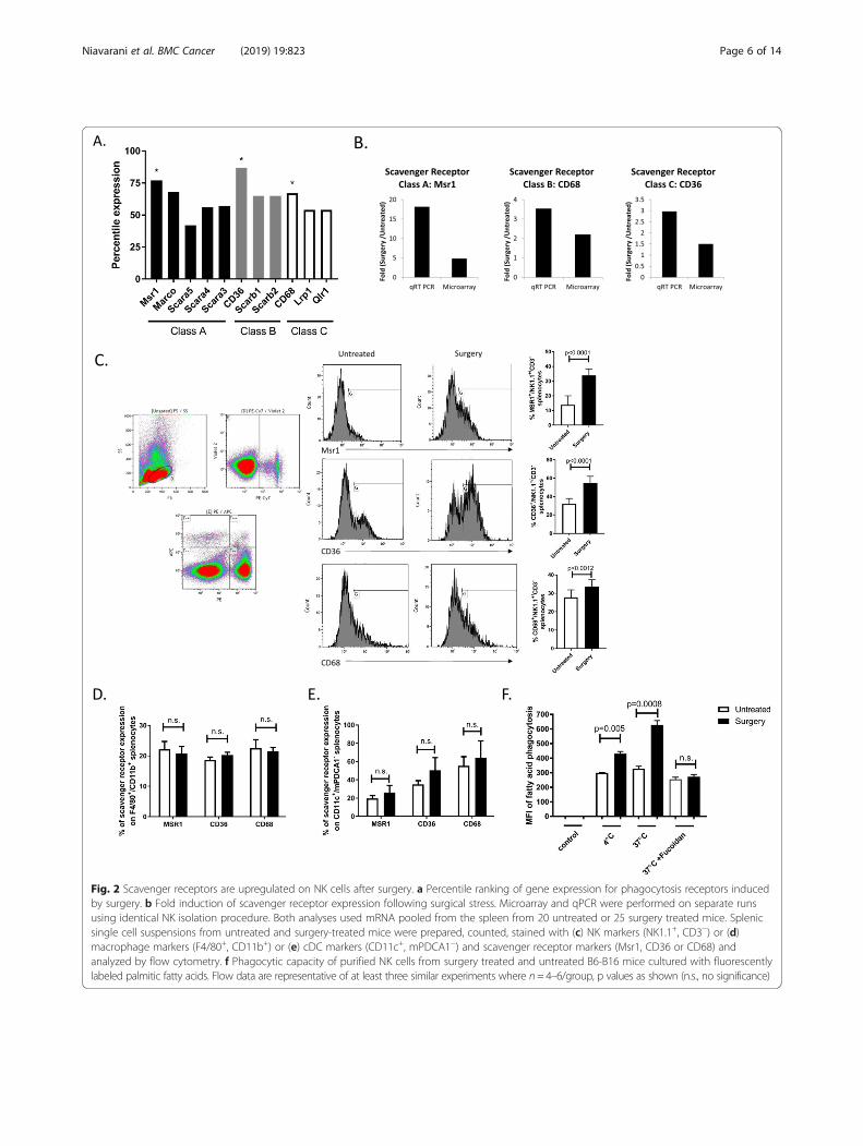
Fig. 2 Scavenger receptors are upregulated on NK cells after surgery. a Percentile ranking of gene expression for phagocytosis receptors inducedby surgery. b Fold induction of scavenger receptor expression following surgical stress. Microarray and qPCR were performed on separate runsusing identical NK isolation procedure. Both analyses used mRNA pooled from the spleen from 20 untreated or 25 surgery treated mice. Splenicsingle cell suspensions from untreated and surgery-treated mice were prepared, counted, stained with (c) NK markers (NK1.1+, CD3−) or (d)macrophage markers (F4/80+, CD11b+) or (e) cDC markers (CD11c+, mPDCA1−) and scavenger receptor markers (Msr1, CD36 or CD68) andanalyzed by flow cytometry. f Phagocytic capacity of purified NK cells from surgery treated and untreated B6-B16 mice cultured with fluorescentlylabeled palmitic fatty acids. Flow data are representative of at least three similar experiments where n= 4–6/group, p values as shown (n.s., no significance)
Niavarani et al. BMC Cancer (2019) 19:823 Page 6 of 14
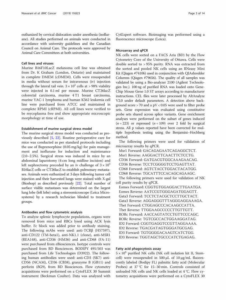
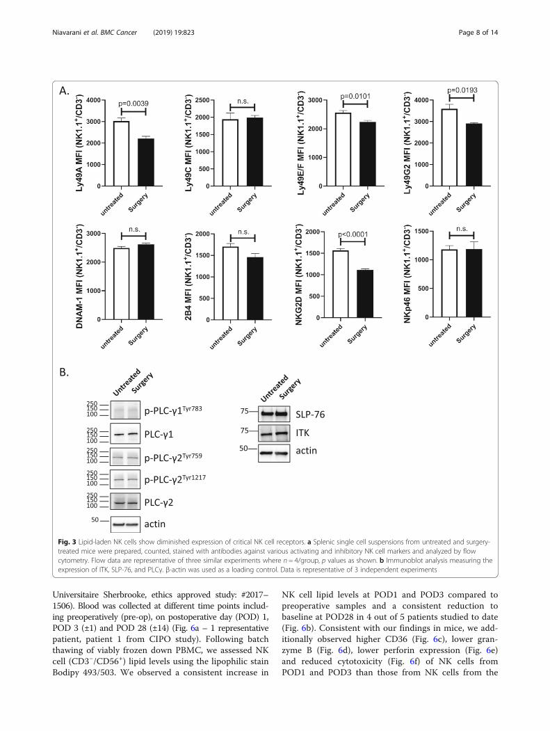
Lipid-laden NK cells show diminished expression ofcritical NK cell receptorsMultiple groups have established that NK cells are regu-lated by the integration of signals derived from activatingand MHC-specific inhibitory receptors on their surface.The responsiveness of each NK cell is quantitativelyadjusted to ensure self-tolerance while at the same timeguaranteeing useful reactivity against potential threats,such as transformed tumor cells [13, 39, 40]. Therefore,we examined the effect of postoperative lipid accumula-tion on MHC-specific receptors Ly49A, Ly49C, Ly49E/F,and Ly49G2; the signaling lymphocytic activation mo-lecule (SLAM) family molecule 2B4 (CD244), the DNAXaccessory molecule (DNAM-1, CD226), the mousenatural cytotoxicity receptors NKp46 and the criticalactivating receptor NKG2D (Fig. 3a). Comparing NKcells from untreated and surgery-treated B6-B16 mice,we detected a downregulation in NK cell surface expres-sion of Ly49A, Ly49E/F, Ly49G2 and NKG2D, but notin the other receptors. Next, we examined the effect oflipid on NK cell signaling. Specifically, we examinedSLP76 (a critical adapter molecule downstream of ITAM-containing surface receptors), ITK (IL-2-inducible T cellkinase), and PLCγ (phospholipase Cγ) (Fig. 3b). Comparingacross these 3 important signalling molecules, we did notdetect any differences in protein expression in untreated vs.surgery-treated NK cells. These results suggest key NK cellinhibitory (Ly49A, Ly49E/F and Ly49G2) receptors and acritical activating (NKG2D) receptor are affected bysurgery.
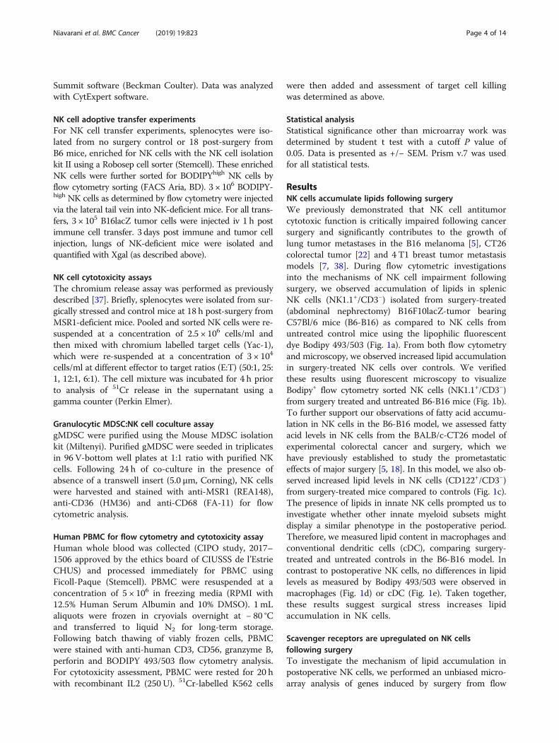
Lipid-laden NK cells are impaired in their ability to lysetumorsTo investigate whether lipid accumulation in NK cellshave functional consequences, Msr1+/Bodipy+ vs. Msr1−/Bodipy−, CD36+/Bodipy+ vs. CD36−/Bodipy−, and CD68+/Bodipy+ vs. CD68−/Bodipy− NK cells (NK1.1+/CD3−) wereflow sorted from spleens of surgery-treated B6-B16 miceand used as effector cells to lyse chromium-labelled YAC-1 tumor targets in an ex vivo NK cell cytotoxicity assay.We observed that all 3 sets of SR+/Bodipy+ NK cells fromsurgery treated mice were responsible for NK cell cyto-toxic dysfunction following surgery, while SR−/Bodipy−
NK cells retained normal cytotoxic activity postoperatively(Figs. 4a-c). These results suggest that SR expression, ingeneral, negatively affects postoperative NK cell cytotoxicfunction ex vivo. To establish the important role of lipidaccumulation in defective NK cell function in cancer invivo, we adoptively transferred flow sorted Bodipy+ orBodipy− NK cells from donor surgery-treated B6 mice intorecipient NK cell-deficient mice (IL2γR-KO). One hourfollowing adoptive transfer of NK cells, we challengedthese recipients with intravenous injection of B16F10lacZlung tumors (Fig. 4d timeline). We have used this model
previously to establish the mediating role of NK cells inthe increase of cancer metastases following surgical stress[4]. At 3 days post treatment, we found significantly in-creased lung tumor burden in NK-deficient mice thatreceived Bodipy+ surgery-treated NK cells compared tothose that received Bodipy− NK cells (Fig. 4e). By trans-ferring Bodipy+ surgery-treated NK cells and recreatingthe effect of surgery on the formation of metastases,our results suggest that the prometastatic effect ofsurgery is mediated by lipid-laden NK cells.
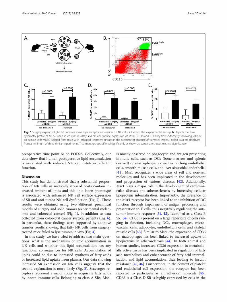
Surgery-expanded gMDSC induces scavenger receptorexpression on NK cellsIn our previous studies, we have assessed for surgery-in-duced tissue signals that might be responsible for thesuppression of NK cells. Specifically, we observed an in-crease in serum IL5, IL6 and TGFβ in surgery-treatedmice compared to controls [5]. We, therefore, questionedwhether these cytokines could induce SR expression onNK cells. Following treatment of purified NK cells with re-combinant IL5, IL6 and TGFβ, we did not detect any dif-ferences in SR expression levels (Msr1, CD36, CD68) inthe presence of these cytokines (Additional file 2: FigureS2). Next, we questioned whether surgery-expanded mye-loid derived suppressor cells (MDSC), which we have re-cently documented to impair NK cells [7] could induce SRexpression on NK cells. We purified granulocytic MDSC(Ly6Ghigh gMDSC) from surgery-treated and controlmice and co-cultured them with naïve purified NK cells(Fig. 5a, b). In the presence of surgery-treated gMDSC,we observed significant increases in all 3 SR expressionon NK cells (Fig. 5c-e). Moreover, we repeated thegMDSC:NK cell co-culture experiment in the presenceof a transwell insert to evaluate whether the observedupregulation of SR on NK cells by gMDSC wasdependent on direct cell contact. In the presence of thetranswell, we observed that gMDSC from surgery-treated mice still induced the upregulation of SR onNK cells, suggesting that cell-to-cell contact is not re-quired for this effect (Fig. 5c-e). These results furthersupport our previously published studies that surgery-expanded myeloid regulatory cells impair NK cellfunction. Importantly, they demonstrate that gMDSCimpair NK cells through upregulation of SR expression.
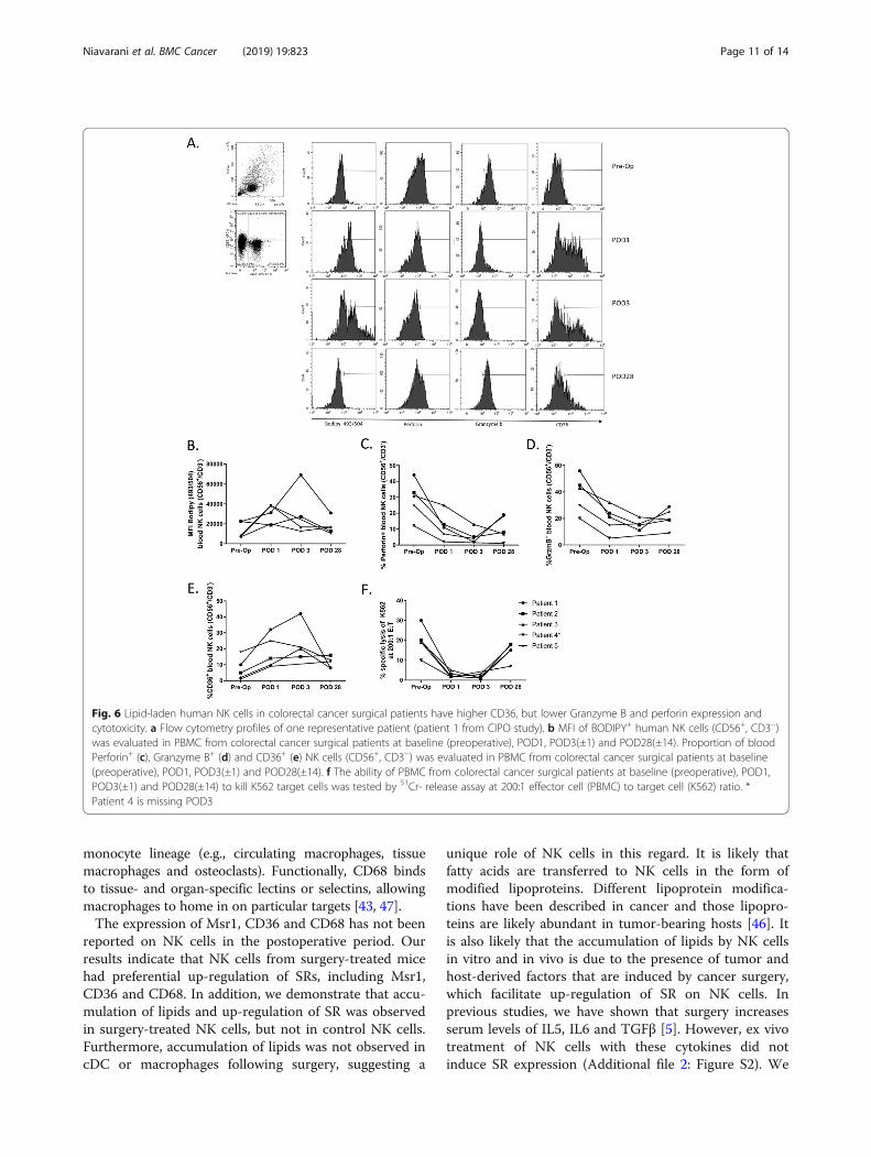
Lipid-laden NK cells from colorectal cancer surgicalpatients have higher CD36, but lower Granzyme BexpressionTo investigate whether human cancer surgery has thesame effect as mouse cancer surgery on NK cells, wemeasured bodipy levels in NK cells from 5 surgical patientswith colorectal cancer. We collected blood from patientsenrolled in the “Characterization of Immunosuppression inthe Postoperative Period - CIPO” study (Centre Hospitalier
Niavarani et al. BMC Cancer (2019) 19:823 Page 7 of 14
Universitaire Sherbrooke, ethics approved study: #2017–1506). Blood was collected at different time points includ-ing preoperatively (pre-op), on postoperative day (POD) 1,POD 3 (±1) and POD 28 (±14) (Fig. 6a – 1 representativepatient, patient 1 from CIPO study). Following batchthawing of viably frozen down PBMC, we assessed NKcell (CD3−/CD56+) lipid levels using the lipophilic stainBodipy 493/503. We observed a consistent increase in
NK cell lipid levels at POD1 and POD3 compared topreoperative samples and a consistent reduction tobaseline at POD28 in 4 out of 5 patients studied to date(Fig. 6b). Consistent with our findings in mice, we add-itionally observed higher CD36 (Fig. 6c), lower gran-zyme B (Fig. 6d), lower perforin expression (Fig. 6e)and reduced cytotoxicity (Fig. 6f) of NK cells fromPOD1 and POD3 than those from NK cells from the
Fig. 3 Lipid-laden NK cells show diminished expression of critical NK cell receptors. a Splenic single cell suspensions from untreated and surgery-treated mice were prepared, counted, stained with antibodies against various activating and inhibitory NK cell markers and analyzed by flowcytometry. Flow data are representative of three similar experiments where n = 4/group, p values as shown. b Immunoblot analysis measuring theexpression of ITK, SLP-76, and PLCγ. β-actin was used as a loading control. Data is representative of 3 independent experiments
Niavarani et al. BMC Cancer (2019) 19:823 Page 8 of 14
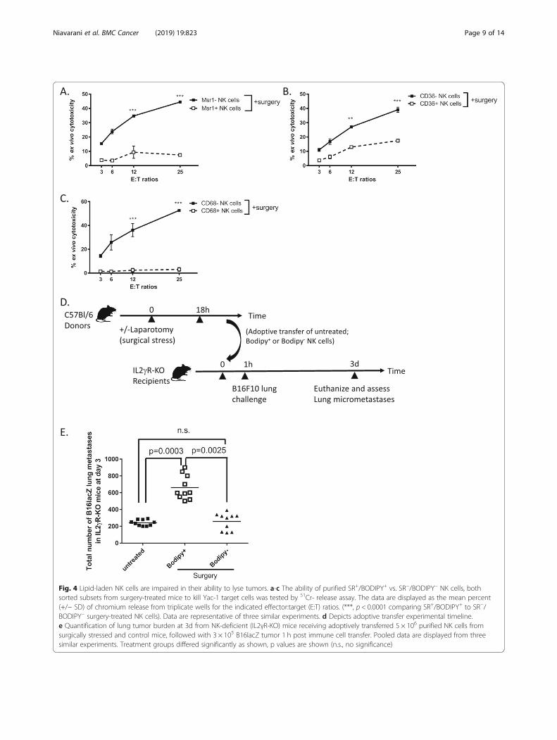
Fig. 4 Lipid-laden NK cells are impaired in their ability to lyse tumors. a-c The ability of purified SR+/BODIPY+ vs. SR−/BODIPY− NK cells, bothsorted subsets from surgery-treated mice to kill Yac-1 target cells was tested by 51Cr- release assay. The data are displayed as the mean percent(+/− SD) of chromium release from triplicate wells for the indicated effector:target (E:T) ratios. (***, p < 0.0001 comparing SR+/BODIPY+ to SR−/BODIPY− surgery-treated NK cells). Data are representative of three similar experiments. d Depicts adoptive transfer experimental timeline.e Quantification of lung tumor burden at 3d from NK-deficient (IL2γR-KO) mice receiving adoptively transferred 5 × 10⁶ purified NK cells fromsurgically stressed and control mice, followed with 3 × 10⁵ B16lacZ tumor 1 h post immune cell transfer. Pooled data are displayed from threesimilar experiments. Treatment groups differed significantly as shown, p values are shown (n.s., no significance)
Niavarani et al. BMC Cancer (2019) 19:823 Page 9 of 14
preoperative time point or on POD28. Collectively, ourdata show that human postoperative lipid accumulationis associated with reduced NK cell cytotoxic effectorfunction.
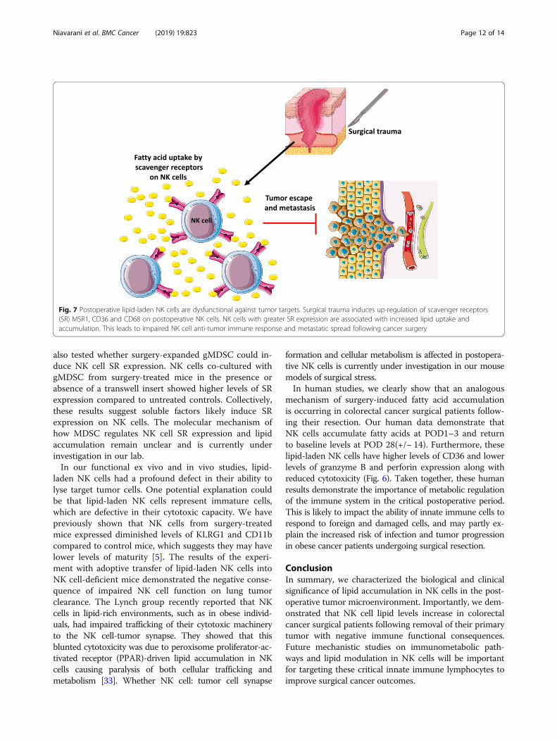
DiscussionThis study has demonstrated that a substantial propor-tion of NK cells in surgically stressed hosts contain in-creased amount of lipids and this lipid-laden phenotypeis associated with enhanced NK cell surface expressionof SR and anti-tumor NK cell dysfunction (Fig. 7). Theseresults were obtained using two different preclinicalmodels of surgery and solid tumors (experimental melan-oma and colorectal cancer) (Fig. 1), in addition to datacollected from colorectal cancer surgical patients (Fig. 6).In particular, these findings were supported by adoptivetransfer results showing that fatty NK cells from surgery-treated mice failed to lyse tumors in vivo (Fig. 4).In this study, we have tried to address two main ques-
tions: what is the mechanism of lipid accumulation inNK cells and whether this lipid accumulation has anyfunctional consequences for NK cells. Accumulation oflipids could be due to increased synthesis of fatty acidsor increased lipid uptake from plasma. Our data showingincreased SR expression on NK cells suggests that thesecond explanation is more likely (Fig. 2). Scavenger re-ceptors represent a major route in acquiring fatty acidsby innate immune cells. Belonging to class A SRs, Msr1
is mostly observed on phagocytic and antigen presentingimmune cells, such as DCs (bone marrow and splenicderived) or macrophages, as well as on lung endothelialcells, smooth muscle cells, and liver sinusoidal endothelial[41]. Msr1 recognizes a wide array of self and non-selfmolecules and has been implicated in the developmentand progression of various diseases [42]. Additionally,Msr1 plays a major role in the development of cardiovas-cular diseases and atherosclerosis by increasing cellularlipoprotein internalization. Importantly, the presence ofthe Msr1 receptor has been linked to the inhibition of DCfunction through impairment of antigen processing andpresentation to T cells, thus negatively regulating the anti-tumor immune response [31, 43]. Identified as a Class BSR [34], CD36 is present on a large repertoire of cells ran-ging in function, including DCs, macrophages, micro-vascular cells, adipocytes, endothelium cells, and skeletalmuscle cells [43]. Similar to Msr1, the expression of CD36on macrophages has been linked to increased uptake oflipoproteins in atherosclerosis [44]. In both animal andhuman studies, increased CD36 expression in metabolic-ally active tissue has been implicated in regulation of fattyacid metabolism and enhancement of fatty acid internal-ization and lipid accumulation, thus leading to insulinresistance [45, 46]. Furthermore, in the context of plateletand endothelial cell expression, the receptor has beenreported to participate as an adhesion molecule [46].CD68 is a Class D SR is highly expressed by cells in the
Fig. 5 Surgery-expanded gMDSC induces scavenger receptor expression on NK cells. a Depicts the experimental set up. b Depicts the flowcytometry profile of MDSC used in co-culture assay. c-e NK cell surface expression of MSR1, CD36 and CD68 by flow cytometry following 20 h ofco-culture with MDSC isolated from mice with indicated treatment groups in the presence or absence of transwell inserts. Pooled data are displayedfrom a minimum of three similar experiments. Treatment groups differed significantly as shown, p values are shown (n.s., no significance)
Niavarani et al. BMC Cancer (2019) 19:823 Page 10 of 14
monocyte lineage (e.g., circulating macrophages, tissuemacrophages and osteoclasts). Functionally, CD68 bindsto tissue- and organ-specific lectins or selectins, allowingmacrophages to home in on particular targets [43, 47].The expression of Msr1, CD36 and CD68 has not been
reported on NK cells in the postoperative period. Ourresults indicate that NK cells from surgery-treated micehad preferential up-regulation of SRs, including Msr1,CD36 and CD68. In addition, we demonstrate that accu-mulation of lipids and up-regulation of SR was observedin surgery-treated NK cells, but not in control NK cells.Furthermore, accumulation of lipids was not observed incDC or macrophages following surgery, suggesting a
unique role of NK cells in this regard. It is likely thatfatty acids are transferred to NK cells in the form ofmodified lipoproteins. Different lipoprotein modifica-tions have been described in cancer and those lipopro-teins are likely abundant in tumor-bearing hosts [46]. Itis also likely that the accumulation of lipids by NK cellsin vitro and in vivo is due to the presence of tumor andhost-derived factors that are induced by cancer surgery,which facilitate up-regulation of SR on NK cells. Inprevious studies, we have shown that surgery increasesserum levels of IL5, IL6 and TGFβ [5]. However, ex vivotreatment of NK cells with these cytokines did notinduce SR expression (Additional file 2: Figure S2). We
Fig. 6 Lipid-laden human NK cells in colorectal cancer surgical patients have higher CD36, but lower Granzyme B and perforin expression andcytotoxicity. a Flow cytometry profiles of one representative patient (patient 1 from CIPO study). b MFI of BODIPY+ human NK cells (CD56+, CD3−)was evaluated in PBMC from colorectal cancer surgical patients at baseline (preoperative), POD1, POD3(±1) and POD28(±14). Proportion of bloodPerforin+ (c), Granzyme B+ (d) and CD36+ (e) NK cells (CD56+, CD3−) was evaluated in PBMC from colorectal cancer surgical patients at baseline(preoperative), POD1, POD3(±1) and POD28(±14). f The ability of PBMC from colorectal cancer surgical patients at baseline (preoperative), POD1,POD3(±1) and POD28(±14) to kill K562 target cells was tested by 51Cr- release assay at 200:1 effector cell (PBMC) to target cell (K562) ratio. *Patient 4 is missing POD3
Niavarani et al. BMC Cancer (2019) 19:823 Page 11 of 14
also tested whether surgery-expanded gMDSC could in-duce NK cell SR expression. NK cells co-cultured withgMDSC from surgery-treated mice in the presence orabsence of a transwell insert showed higher levels of SRexpression compared to untreated controls. Collectively,these results suggest soluble factors likely induce SRexpression on NK cells. The molecular mechanism ofhow MDSC regulates NK cell SR expression and lipidaccumulation remain unclear and is currently underinvestigation in our lab.In our functional ex vivo and in vivo studies, lipid-
laden NK cells had a profound defect in their ability tolyse target tumor cells. One potential explanation couldbe that lipid-laden NK cells represent immature cells,which are defective in their cytotoxic capacity. We havepreviously shown that NK cells from surgery-treatedmice expressed diminished levels of KLRG1 and CD11bcompared to control mice, which suggests they may havelower levels of maturity [5]. The results of the experi-ment with adoptive transfer of lipid-laden NK cells intoNK cell-deficient mice demonstrated the negative conse-quence of impaired NK cell function on lung tumorclearance. The Lynch group recently reported that NKcells in lipid-rich environments, such as in obese individ-uals, had impaired trafficking of their cytotoxic machineryto the NK cell-tumor synapse. They showed that thisblunted cytotoxicity was due to peroxisome proliferator-ac-tivated receptor (PPAR)-driven lipid accumulation in NKcells causing paralysis of both cellular trafficking andmetabolism [33]. Whether NK cell: tumor cell synapse
formation and cellular metabolism is affected in postopera-tive NK cells is currently under investigation in our mousemodels of surgical stress.In human studies, we clearly show that an analogous
mechanism of surgery-induced fatty acid accumulationis occurring in colorectal cancer surgical patients follow-ing their resection. Our human data demonstrate thatNK cells accumulate fatty acids at POD1–3 and returnto baseline levels at POD 28(+/− 14). Furthermore, theselipid-laden NK cells have higher levels of CD36 and lowerlevels of granzyme B and perforin expression along withreduced cytotoxicity (Fig. 6). Taken together, these humanresults demonstrate the importance of metabolic regulationof the immune system in the critical postoperative period.This is likely to impact the ability of innate immune cells torespond to foreign and damaged cells, and may partly ex-plain the increased risk of infection and tumor progressionin obese cancer patients undergoing surgical resection.
ConclusionIn summary, we characterized the biological and clinicalsignificance of lipid accumulation in NK cells in the post-operative tumor microenvironment. Importantly, we dem-onstrated that NK cell lipid levels increase in colorectalcancer surgical patients following removal of their primarytumor with negative immune functional consequences.Future mechanistic studies on immunometabolic path-ways and lipid modulation in NK cells will be importantfor targeting these critical innate immune lymphocytes toimprove surgical cancer outcomes.
Fig. 7 Postoperative lipid-laden NK cells are dysfunctional against tumor targets. Surgical trauma induces up-regulation of scavenger receptors(SR) MSR1, CD36 and CD68 on postoperative NK cells. NK cells with greater SR expression are associated with increased lipid uptake andaccumulation. This leads to impaired NK cell anti-tumor immune response and metastatic spread following cancer surgery
Niavarani et al. BMC Cancer (2019) 19:823 Page 12 of 14
Additional files
Additional file 1: Figure S1. Verification of the purity of sorted NK cells.The purity of sorted NK cells was tested by qPCR using the transcriptionfactors Eomes, ID2, RORc, Tbet and Gata3 for qPCR analyses. (PPTX 505 kb)
Additional file 2: Figure S2. SR expression on NK cells followingtreatment with IL5, IL6 and TGFβ. NK cells were treated ex vivo with therecombinant cytokines IL5, IL6 or TGFβ followed by assessment of SRexpression on NK cells by flow cytometry. (PPTX 277 kb)
AbbreviationsNK: Cells natural killer cells; SR: Scavenger receptors; gMDSC: granulocyticmyeloid derived suppressor cells; PBMC: Peripheral blood mononuclear cells;ARG1: Arginase 1; cDC: conventional dendritic cells; FAO: Fatty acidoxidation; t-MDSC: tumor infiltrating myeloid derived suppressor cells;B6: C57Bl/6 mice; ILC: Innate lymphoid cells; ITK IL-2: Inducible T cell kinase;PLCγ: Phospholipase Cγ; CIPO: Characterization of postoperativeimmunosuppression study; POD: Postoperative day; PPAR: Peroxisomeproliferator-activated receptor
AcknowledgementsThe authors would like to thank Dr. Rebecca Auer and Christiano Tanese DeSouza (Ottawa Hospital Research Institute) for their expertise and guidanceon mouse surgical models and experiments. Additionally, the authors wouldlike to thank Vera Tang and Leonid Volkov for their flow cytometry expertise.
Authors’ contributionsSN, CL, OB, MB, CB, SR and LT executed experiments, read and approved themanuscript; SR critically revised the manuscript; LT conceived, designed andexecuted experiments, was a major contributor in writing the manuscript,and supervised the study. All authors have read and approved themanuscript.
FundingCancer Research Society, Scholarship for the Next Generation of Scientists(LT) – provided operating funds for this study; Natural Sciences andEngineering Research Council of Canada, Discovery Grant (LT) – providedoperating funds for this study; CIHR New Investigator Award and FRQS Jr. 1Salary Awards (LT) – provided salary for the principal investigator; CIHRProject Scheme Grant (LT) – provided operating funds for this study;Universite de Sherbrooke, Faculty of Medicine and Health Sciences GraduateScholarship (SN) – provided the student scholarship for the first author. Thefunding bodies did not play a role in the design of the study and collection,analysis, and interpretation of data and in writing the manuscript.
Availability of data and materialsThe datasets used and/or analyzed during the current study are availablefrom the corresponding author on reasonable request.
Ethics approvalMice were housed in pathogen-free conditions at the Central Animal CareFacility of the Université de Sherbrooke (Quebec) and the Animal CareVeterinary Service facility of the University of Ottawa (Ontario). All studiesand manipulations performed on animals were conducted in accordancewith university guidelines and approved by the Animal Care Committee atboth universities. Human whole blood was collected (CIPO study, #2017–1506 approved by the ethics board of CIUSSS de l’Estrie CHUS. Informed andwritten consent to participate in this study was obtained from allparticipants.
Consent for publicationInformed consent for publication was obtained from all participants in the study.
Competing interestsThe authors declare that they have no competing interests.
Author details1Department of Anatomy and Cell Biology, Université de Sherbrooke,Pavillon sur la Recherche Appliqué du Cancer at 3201 rue Jean-Mignault,Sherbrooke, QC J1E 4K8, Canada. 2Department of Pediatrics, Division of
Immunology, Université de Sherbrooke, Sherbrooke, QC, Canada.3Department of Biochemistry, Microbiology and Immunology, University ofOttawa, Ottawa, ON, Canada. 4Centre de Recherche Clinique de CentreHospitalier de l’Universite de Sherbrooke, Sherbrooke, QC, Canada.
Received: 15 April 2019 Accepted: 16 August 2019
References1. Bartal I, Melamed R, Greenfeld K, Atzil S, Glasner A, Domankevich V, et al.
Immune perturbations in patients along the perioperative period:alterations in cell surface markers and leukocyte subtypes before and aftersurgery. Brain Behav Immun. 2010;24(3):376–86.
2. Ben-Eliyahu S, Page GG, Yirmiya R, Shakhar G. Evidence that stress andsurgical interventions promote tumor development by suppressing naturalkiller cell activity. Int J Cancer. 1999;80(6):880–8.
3. Coffey JC, Wang JH, Smith MJ, Bouchier-Hayes D, Cotter TG, Redmond HP.Excisional surgery for cancer cure: therapy at a cost. Lancet Oncol. 2003;4(12):760–8.
4. Bakos O, Lawson C, Rouleau S, Tai LH. Combining surgery andimmunotherapy: turning an immunosuppressive effect into a therapeuticopportunity. J Immunother Cancer. 2018;6(1):86.
5. Tai LH, de Souza CT, Belanger S, Ly L, Alkayyal AA, Zhang J, et al. Preventingpostoperative metastatic disease by inhibiting surgery-induced dysfunctionin natural killer cells. Cancer Res. 2013;73(1):97–107.
6. Tai LH, Zhang J, Scott KJ, de Souza CT, Alkayyal AA, Ananth AA, et al.Perioperative influenza vaccination reduces postoperative metastatic diseaseby reversing surgery-induced dysfunction in natural killer cells. Clin CancerRes. 2013;19(18):5104–15.
7. Tai LH, Alkayyal AA, Leslie AL, Sahi S, Bennett S, Tanese de Souza C, et al.Phosphodiesterase-5 inhibition reduces postoperative metastatic disease bytargeting surgery-induced myeloid derived suppressor cell-dependent inhibitionof natural killer cell cytotoxicity. Oncoimmunology. 2018;7(6):e1431082.
8. Page GG, Blakely WP, Ben-Eliyahu S. Evidence that postoperative pain isa mediator of the tumor-promoting effects of surgery in rats. Pain.2001;90(1–2):191–9.
9. Shaashua L, Shabat-Simon M, Haldar R, Matzner P, Zmora O, Shabtai M, etal. Perioperative COX-2 and beta-adrenergic blockade improves metastaticbiomarkers in breast Cancer patients in a phase-II randomized trial. ClinCancer Res. 2017;23(16):4651–61.
10. Benish M, Bartal I, Goldfarb Y, Levi B, Avraham R, Raz A, et al. Perioperativeuse of beta-blockers and COX-2 inhibitors may improve immunecompetence and reduce the risk of tumor metastasis. Ann Surg Oncol.2008;15(7):2042–52.
11. Malmberg KJ, Bryceson YT, Carlsten M, Andersson S, Bjorklund A, BjorkstromNK, et al. NK cell-mediated targeting of human cancer and possibilities fornew means of immunotherapy. Cancer Immunol Immunother. 2008;57(10):1541–52.
12. Albertsson PA, Basse PH, Hokland M, Goldfarb RH, Nagelkerke JF, NannmarkU, et al. NK cells and the tumour microenvironment: implications for NK-cellfunction and anti-tumour activity. Trends Immunol. 2003;24(11):603–9.
13. Marcus A, Gowen BG, Thompson TW, Iannello A, Ardolino M, Deng W, et al.Recognition of tumors by the innate immune system and natural killer cells.Adv Immunol. 2014;122:91–128.
14. Iannello A, Thompson TW, Ardolino M, Marcus A, Raulet DH.Immunosurveillance and immunotherapy of tumors by innate immunecells. Curr Opin Immunol. 2016;38:52–8.
15. Pollock RE, Lotzova E, Stanford SD. Mechanism of surgical stress impairmentof human perioperative natural killer cell cytotoxicity. Arch Surg. 1991;126(3):338–42.
16. Wang J, Yang L, Yu L, Wang YY, Chen R, Qian J, et al. Surgery-inducedmonocytic myeloid-derived suppressor cells expand regulatory T cells inlung cancer. Oncotarget. 2017;8(10):17050–8.
17. Glasner A, Avraham R, Rosenne E, Benish M, Zmora O, Shemer S, et al.Improving survival rates in two models of spontaneous postoperativemetastasis in mice by combined administration of a beta-adrenergicantagonist and a cyclooxygenase-2 inhibitor. J Immunol. 2010;184(5):2449–57.
18. Tai LH, Tanese de Souza C, Sahi S, Zhang J, Alkayyal AA, Ananth AA, et al. Amouse tumor model of surgical stress to explore the mechanisms ofpostoperative immunosuppression and evaluate novel perioperativeimmunotherapies. J Vis Exp. 2014;85.
Niavarani et al. BMC Cancer (2019) 19:823 Page 13 of 14
19. Shakhar G, Ben-Eliyahu S. Potential prophylactic measures againstpostoperative immunosuppression: could they reduce recurrence rates inoncological patients? Ann Surg Oncol. 2003;10(8):972–92.
20. Wang J, Su X, Yang L, Qiao F, Fang Y, Yu L, et al. The influence of myeloid-derived suppressor cells on angiogenesis and tumor growth after cancersurgery. Int J Cancer. 2016;138(11):2688–99.
21. Zhu X, Pribis JP, Rodriguez PC, Morris SM Jr, Vodovotz Y, Billiar TR, et al. Thecentral role of arginine catabolism in T-cell dysfunction and increasedsusceptibility to infection after physical injury. Ann Surg. 2014;259(1):171–8.
22. Seth R, Tai LH, Falls T, de Souza CT, Bell JC, Carrier M, et al. Surgical stresspromotes the development of cancer metastases by a coagulation-dependent mechanism involving natural killer cells in a murine model. AnnSurg. 2013;258(1):158–68.
23. Li H, Han Y, Guo Q, Zhang M, Cao X. Cancer-expanded myeloid-derivedsuppressor cells induce anergy of NK cells through membrane-bound TGF-beta 1. J Immunol. 2009;182(1):240–9.
24. Mauti LA, Le Bitoux MA, Baumer K, Stehle JC, Golshayan D, Provero P, et al.Myeloid-derived suppressor cells are implicated in regulating permissivenessfor tumor metastasis during mouse gestation. J Clin Invest. 2011;121(7):2794–807.
25. Gabrilovich DI, Nagaraj S. Myeloid-derived suppressor cells as regulators ofthe immune system. Nat Rev Immunol. 2009;9(3):162–74.
26. Khaled YS, Ammori BJ, Elkord E. Myeloid-derived suppressor cells in cancer:recent progress and prospects. Immunol Cell Biol. 2013;91(8):493–502.
27. de Pablo MA, Angeles Puertollano M, Alvarez de Cienfuegos G. Immune cellfunctions, lipids and host natural resistance. FEMS Immunol Med Microbiol.2000;29(4):323–8.
28. Yaqoob P, Newsholme EA, Calder PC. Inhibition of natural killer cell activityby dietary lipids. Immunol Lett. 1994;41(2–3):241–7.
29. de Pablo MA, Alvarez de Cienfuegos G. Modulatory effects of dietary lipidson immune system functions. Immunol Cell Biol. 2000;78(1):31–9.
30. Kelley DS, Taylor PC, Nelson GJ, Schmidt PC, Ferretti A, Erickson KL, et al.Docosahexaenoic acid ingestion inhibits natural killer cell activity andproduction of inflammatory mediators in young healthy men. Lipids.1999;34(4):317–24.
31. Herber DL, Cao W, Nefedova Y, Novitskiy SV, Nagaraj S, Tyurin VA, et al.Lipid accumulation and dendritic cell dysfunction in cancer. Nat Med.2010;16(8):880–6.
32. Ramakrishnan R, Tyurin VA, Veglia F, Condamine T, Amoscato A,Mohammadyani D, et al. Oxidized lipids block antigen cross-presentation bydendritic cells in cancer. J Immunol. 2014;192(6):2920–31.
33. Michelet X, Dyck L, Hogan A, Loftus RM, Duquette D, Wei K, et al. Metabolicreprogramming of natural killer cells in obesity limits antitumor responses.Nat Immunol. 2018;19(12):1330–40.
34. Mah AY, Rashidi A, Keppel MP, Saucier N, Moore EK, Alinger JB, et al.Glycolytic requirement for NK cell cytotoxicity and cytomegalovirus control.JCI Insight. 2017;2(23).
35. Hossain F, Al-Khami AA, Wyczechowska D, Hernandez C, Zheng L, Reiss K, etal. Inhibition of fatty acid oxidation modulates immunosuppressivefunctions of myeloid-derived suppressor cells and enhances Cancertherapies. Cancer Immunol Res. 2015;3(11):1236–47.
36. Alkayyal AA, Tai LH, Kennedy MA, de Souza CT, Zhang J, Lefebvre C, et al.NK-cell recruitment is necessary for eradication of peritoneal Carcinomatosiswith an IL12-expressing Maraba virus cellular vaccine. Cancer Immunol Res.2017;5(3):211–21.
37. Patel R, Belanger S, Tai LH, Troke AD, Makrigiannis AP. Effect of Ly49haplotype variance on NK cell function and education. J Immunol. 2010;185(8):4783–92.
38. Zhang J, Tai LH, Ilkow CS, Alkayyal AA, Ananth AA, de Souza CT, et al.Maraba MG1 virus enhances natural killer cell function via conventionaldendritic cells to reduce postoperative metastatic disease. Mol Ther. 2014;22(7):1320–32.
39. Brodin P, Karre K, Hoglund P. NK cell education: not an on-off switch but atunable rheostat. Trends Immunol. 2009;30(4):143–9.
40. Raulet DH, Marcus A, Coscoy L. Dysregulated cellular functions and cell stresspathways provide critical cues for activating and targeting natural killer cells totransformed and infected cells. Immunol Rev. 2017;280(1):93–101.
41. Peiser L, De Winther MP, Makepeace K, Hollinshead M, Coull P, Plested J, etal. The class a macrophage scavenger receptor is a major patternrecognition receptor for Neisseria meningitidis which is independent oflipopolysaccharide and not required for secretory responses. Infect Immun.2002;70(10):5346–54.
42. Kunjathoor VV, Febbraio M, Podrez EA, Moore KJ, Andersson L, Koehn S, etal. Scavenger receptors class A-I/II and CD36 are the principal receptorsresponsible for the uptake of modified low density lipoprotein leading tolipid loading in macrophages. J Biol Chem. 2002;277(51):49982–8.
43. Peiser L, Mukhopadhyay S, Gordon S. Scavenger receptors in innateimmunity. Curr Opin Immunol. 2002;14(1):123–8.
44. Thorne RF, Mhaidat NM, Ralston KJ, Burns GF. CD36 is a receptor foroxidized high density lipoprotein: implications for the development ofatherosclerosis. FEBS Lett. 2007;581(6):1227–32.
45. Glatz JF, Angin Y, Steinbusch LK, Schwenk RW, Luiken JJ. CD36 as a targetto prevent cardiac lipotoxicity and insulin resistance. Prostaglandins LeukotEssent Fatty Acids. 2013;88(1):71–7.
46. Goldberg IJ, Eckel RH, Abumrad NA. Regulation of fatty acid uptake intotissues: lipoprotein lipase- and CD36-mediated pathways. J Lipid Res.2009;50(Suppl):S86–90.
47. Song L, Lee C, Schindler C. Deletion of the murine scavenger receptorCD68. J Lipid Res. 2011;52(8):1542–50.
Publisher’s NoteSpringer Nature remains neutral with regard to jurisdictional claims inpublished maps and institutional affiliations.
Niavarani et al. BMC Cancer (2019) 19:823 Page 14 of 14