guanine nucleotide pool imbalance impairs multiple steps
TRANSCRIPT

Copyright � 2011 by the Genetics Society of AmericaDOI: 10.1534/genetics.110.122135
Guanine Nucleotide Pool Imbalance Impairs Multiple Steps of ProteinSynthesis and Disrupts GCN4 Translational Control
in Saccharomyces cerevisiae
Diego Iglesias-Gato,* Pilar Martın-Marcos,*,† Marıa A. Santos,*Alan G. Hinnebusch† and Mercedes Tamame*,1
*Instituto de Biologıa Funcional y Genomica, Consejo Superior de Investigaciones Cientıficas/Universidad de Salamanca, EdificioDepartamental, Campus Miguel de Unamuno, 37007 Salamanca, Spain and †Laboratory of Gene Regulation and Development,
Eunice Kennedy Shriver National Institute of Child Health and Human Development, Bethesda, Maryland 20892
Manuscript received August 12, 2010Accepted for publication October 14, 2010
ABSTRACT
Purine nucleotides are structural components of the genetic material, function as phosphate donors,participate in cellular signaling, are cofactors in enzymatic reactions, and constitute the main carriers ofcellular energy. Thus, imbalances in A/G nucleotide biosynthesis affect nearly the whole cellular me-tabolism and must be tightly regulated. We have identified a substitution mutation (G388D) that reducesthe activity of the GMP synthase Gua1 in budding yeast and the total G-nucleotide pool, leading toprecipitous reductions in the GDP/GTP ratio and ATP level in vivo. gua1–G388D strongly reduces the rateof growth, impairs general protein synthesis, and derepresses translation of GCN4 mRNA, encoding atranscriptional activator of diverse amino acid biosynthetic enzymes. Although processing of pre-tRNAi
Met
and other tRNA precursors, and the aminoacylation of tRNAiMet are also strongly impaired in gua1–G388D
cells, tRNAiMet-containing complexes with the macromolecular composition of the eIF2�tRNAi
Met.GTPcomplex (TC) and the multifactor complex (MFC) required for translation initiation accumulate �10-fold ingua1–G388D cells and, to a lesser extent, in wild-type (WT) cells treated with 6-azauracil (6AU). Consistently,addition of an external supply of guanine reverts all the phenotypes of gua1–G388D cells, but not those ofgua1–G388D Dhpt1 mutants unable to refill the internal GMP pool through the salvage pathway. These andother findings suggest that a defect in guanine nucleotide biosynthesis evokes a reduction in the rate ofgeneral protein synthesis by impairing multiple steps of the process, disrupts the gene-specific reinitiationmechanism for translation of GCN4 mRNA and has far-reaching effects in cell biology and metabolism.
IMBALANCE in purine nucleotide biosynthesis af-fects nearly the whole of cellular metabolism and,
hence, this process must be tightly regulated depend-ing on nutrient availability and the conditions of growth.In the yeast Saccharomyces cerevisiae, regulation of purinenucleotide biosynthesis occurs at the enzymatic and ge-netic levels (Daignan-Fornier and Fink 1992; Rolfes
2006) and is finely coordinated with phosphate con-sumption (Pinson et al. 2009). The GMP is synthesizedfrom IMP through a de novo synthesis pathway in a two-step reaction (Figure 1). First, IMP is converted intoXMP by the IMP dehydrogenase (Impdh) (Hyle et al.2003) and second, the GMP-synthase Gua1 mediatesthe amination of XMP to GMP (Dujardin et al. 1994).Transcription of the IMD genes is strongly repressed byguanine, but activated by treatment with the Impdhinhibitors 6AU or mycophenolate (MPA) (Exinger andLacroute 1992; Bremer et al. 2009). Partial guanine
deprivation results in a drastic reduction of the internalGTP pool in a yeast gua1 mutant, inducing meiosis andsporulation in homothallic strains (Varma et al. 1985)and leads to concomitant reductions of the methionineand S-adenosyl-methionine (SAM) pools (Freese
et al. 1984). Recently the effects on the yeast tran-scriptome of a marked depletion of guanine nucleo-tides have been reported (Saint-Marc et al. 2009).Remarkably, 71 genes are affected under conditionsthat lead to GDP/GTP shortage, most of which are in-volved in amino acid metabolism and regulated byGcn4 (Natarajan et al. 2001).
In addition to the de novo synthesis, the salvagepathway for GMP production is physiologically impor-tant because it allows cells to reutilize nucleosidesand nucleotides produced by degradation of nucleicacids and permits the interconversion of adenine withguanine-containing nucleotides (Figure 1). Deletion ofHPT1 impairs guanine uptake from the medium andleads to deregulation of the purine de novo pathway(Guetsova et al. 1997). Strikingly, accumulation ofguanine nucleotides in deregulated hpt1 mutants has
1Corresponding author: Instituto de Biologıa Funcional y Genomica,CSIC/Universidad de Salamanca. Edificio Departamental, CampusMiguel de Unamuno, 37007 Salamanca, Spain. E-mail: [email protected]
Genetics 187: 105–122 ( January 2011)

deleterious consequences for yeast cells, indicating thatan overabundance of GMP derivatives can be as detri-mental as starvation (Breton et al. 2008).
Protein synthesis requires great amounts of cellularGTP, mainly during the elongation phase. However, theinitiation process is the most tightly regulated step andconsumes two GTP molecules. The translation initiationfactor 2 (eIF2) bound to GTP recruits the methionylinitiator tRNA (tRNAi
Met) in a ternary complex (TC) anddelivers it to the 40S ribosome. This process also involvesthe formation of an intermediate multi factor complex(MFC) by the addition of other eIFs (eIF1, eIF3, andeIF5) that can exist unbound to 40S in the yeast cells(Asano et al. 2000). After the recognition of the
initiation AUG codon, GTP hydrolysis promotestRNAi
Met release and the eIF2�GDP complex dissociationfrom the ribosome prior to 60S ribosomal subunitjoining, which requires the hydrolysis of another GTPmolecule bound to eIF5B (Lee et al. 2002). Next, eIF2Bmediates the recycling of eIF2�GDP to eIF2�GTP, in-creasing by �10-fold the affinity of eIF2 for Met–tRNAi
Met and stimulating the beginning of a new roundof initiation (Kapp and Lorsch 2004). This process isinhibited by the phosphorylation of eIF2a in serine 51, akey event in translational regulation under starvationand stress in higher eukaryotes (Pestova et al. 2007). Inyeast, the protein kinase Gcn2 phosphorylates eIF2a inresponse to single amino acid starvation, leading to ageneral inhibition of translation initiation due to im-paired eIF2B function and the attendant reduced ratesof TC formation (Hinnebusch 1985; Rolfes andHinnebusch 1993). By contrast, small defects in TCformation or its recruitment to the 40S ribosome that donot appreciably affect global protein synthesis specificallypromotes the translation of the mRNA encoding tran-scription factor Gcn4 (Dever et al. 1995). Gcn4 activatesthe expression of many amino acid biosynthetic geneswhen cells are starved for a single amino acid, a responseknown as general amino acid control (GAAC). Morerecently Gcn4 was shown to be required for full inductionof at least 539 genes under histidine starvation conditions(�1/10 of the yeast genome), including 77 amino acidbiosynthetic genes (Natarajan et al. 2001).
GCN4 translation is regulated through a mechanism ofreinitiation that depends on the presence of four shortupstream open reading frames (uORFs �1 to �4) in theGCN4 mRNA leader and on the action of positive (Gcn)and negative (Gcd) trans-acting factors (Hinnebusch andFink 1983; Harashima and Hinnebusch 1986). Underamino acid replete conditions, �50% of the ribosomesthat translate uORF1 remain bound to the leader and areable to resume scanning and reacquire the TC in time toreinitiate translation at the inhibitory uORFs �3 and �4downstream, which elicits high rates of dissociation of 40Sribosomal subunits from the GCN4 mRNA leader andattendant low levels of Gcn4 synthesis. However, low ratesof TC formation under amino acid starvation conditionsreduce the likelihood that ribosomes reacquire the TC intime to reinitiate at uORFs �3 and �4, favoring reinitia-tion at the GCN4 AUG instead. The resulting increase inGcn4 levels allows the transcriptional activation of aminoacid biosynthetic genes under its control.
Mutations in GCD genes (Harashima and Hinnebusch
1986; Niederberger et al. 1986; Cuesta et al. 1998;Calvo et al. 1999) or in the recently identified GCD17/RPL33A gene (Martin-Marcos et al. 2007), lead toconstitutive derepression of GCN4 translation inde-pendent of the positive regulators Gcn2 and Gcn3 andof amino acid availability. In this article, we reportthe identification of a gua1–G388D mutation in a GMPsynthase that provokes constitutive derepression of GCN4
Figure 1.—Pathways of purine nucleotide biosynthesis inSaccharomyces cerevisiae. Cytosolic reactions are shown over arectangle representing the plasmatic membrane. The de novopathway to synthesize IMP from PRPP involves 10 enzymaticreactions that are catalyzed by the products of the ADE 1–10genes. The gene products of ADE12 and ADE13 catalyzedthe production of AMP from IMP, and the products ofIMD2/3/4 and GUA1 the synthesis of GMP from IMP. Thesalvage pathway involves three reactions that produce IMP,GMP, and AMP directly from their nonphosphorylated pre-cursors, and adenine is converted to hypoxanthine by theAah1 enzyme. The reaction products are indicated in boldfont: ADP, adenosine-59-diphosphate; AMP, adenosine-59-monophosphate; ATP, adenosine-59-triphosphate; GDP,guanosine-59-diphosphate; GMP, guanosine-59-monophos-phate; GTP, guanosine-59-triphosphate; IMP, inosine-59-monophosphate; PRPP, 5-phosphoribosyl-1-pyrophosphate;SAMP, S-adenosine-59-monophosphate; XMP, xanthosine-59-monophosphate. Genes indicated in italics encode the follow-ing enzymatic activities: ADE12, adenylosuccinate synthetase;ADE13, adenylosuccinate lyase; ADK1, AMP kinase; AMD1,AMP deaminase; APT1, adenine phosphoribosyltransferase;FCY2, purine cytosine permease; GUK1, GMP kinase; HPT1,hypoxanthine-guanine phosphoribosyltransferase; and IMD2,IMD3, and IMD4, IMP dehydrogenase. GUA1, GMP synthetase.
106 D. Iglesias-Gato et al.

translation (Gcd� phenotype), unconditional slow growth(Slg�), thermosensitivity at 37� (Tsm�) and altered poly-some profiles. In addition, gua1–G388D cells exhibitdefects in mature ribosome levels, processing and accu-mulation of pre-tRNA and reductions of mature tRNAspecies, including tRNAi
Met, a component of the TC.Surprisingly, whereas the total guanine nucleotide level isreduced, and GDP is undetectable, GTP is elevated ingua1–G388D vs. WT cells. Moreover, we obtained evi-dence that TC and MFC accumulate to high levels in thegua1–G388D mutant. We propose that imbalance in theguanine nucleotide pool evokes a reduction in the rate ofprotein synthesis by impairing multiple steps in theprocess, including one or more reactions involved inthe reinitiation mechanism governing GCN4 translation.
MATERIALS AND METHODS
Plasmids: Cloning of GCD18 and gcd18-1 alleles: PlasmidpDI2 bearing a �17-kb DNA fragment of yeast chromosomeXIII was isolated from a yeast genomic library in YCp50 (Rose
et al. 1987). Subclones of the genomic insert in pDI2were constructed in the shuttle-vector pRS316 to definethe boundaries of GCD18. Plasmid pDI3 bears a 4619-bpHindIII–EcoRV genomic fragment from pDI2 cloned into theunique SmaI site of pRS316. The 4.6-kb insert contains theGUA1 ORF flanked by 614-bp belonging to the 59 region ofthe SKY1 ORF and 1456 bp belonging to the 39 end of theTRS130 ORFs. A 6649-bp BamHI–HindIII fragment from pDI3was cloned into pRS426 to produce the high-copy numberplasmid pDI12. The gcd18-1 mutation (a transition from G toA at nucleotide 1163) was identified by independent PCRamplifications of the corresponding mutant allele fromgenomic DNAs of strains H166 and Hm458 (Table 1) usingas primers oligonucleotides 1 and 2 (Table 2), and followed byautomatic sequencing of the amplification products on thetwo DNA strands. The gua1–G388D mutant allele was clonedas a 2498-bp fragment into pGEM-T Easy (Promega) toproduce pgcd18. A 2516-kb EcoRI fragment from pgcd18 wascloned into the EcoRI site of pRS316 yielding pDI9.
Construction of a gua1 null allele: A 1.3-kb fragment contain-ing sequences belonging to the 59-upstream region of GUA1was amplified by PCR using genomic DNA of H117 as templateand primers 6 and 7 (Table 2). The 1.3-kb PCR product wascloned at the XhoI/EcoRI sites of pRS316 to obtain pDI31.A 0.9-kb fragment containing sequences belonging to the39-downstream region of GUA1 was amplified by PCR using
TABLE 1
Yeast strains
Strain Genotype Source or reference
H96 MATa gcn2-101 gcn3-101 his1-29 ura3-52 (HIS4TlacZ ura3-52) Harashima and Hinnebusch (1986)H117 MATa gcn2-101 gcn3-101 his1-29 ino1 ura3-52 (HIS4TlacZ URA3) Harashima and Hinnebusch (1986)H166 MATa gcn2-101 gcn3-101 his1-29 ino1 ura3-52 (HIS4TlacZ URA3)
gua1-G388DHarashima and Hinnebusch (1986)
Hm455 MATa gcn2-101 gcn3-101 his1-29 ino1 ura3-52 (HIS4TlacZ ura3-52)gua1-G388D
This study
Hm458 MATa gcn2-101 gcn3-101 his1-29 ino1 ura3-52 (HIS4TlacZ ura3-52)gua1-G388D
This study
Hm500 MATa gcn2-101 gcn3-101 his1-29 ino1 ura3-52 leu2ThisG-URA3-hisG(HIS4TlacZ ura3) gua1-G388D
This study
Hm510 MATa gcn2-101 gcn3-101 his1-29 ino1 ura3-52 leu2ThisG(HIS4TlacZ ura3-52) gua1ThisG
This study
Hm512 MATa gcn2-101 gcn3-101 his1-29 ino1 ura3-52 leu2ThisG-URA3-hisG(HIS4TlacZ ura3) GUA1
This study
Hm534 MATa gcn2-101 gcn3-101 his1-29 ino1 ura3-52 (HIS4TlacZ ura3-52)hpt1TKan-MX4 gua1-G388D
This study
Hm535 MATa gcn2-101 gcn3-101 his1-29 ino1 ura3-52 (HIS4TlacZ URA3)hpt1TKan-MX4
This study
H2894 (KAY35) MATa gcn2D, ura3-52, leu2-3, -122, trp1-D63, Dtif5THisG p(TIF5-FL, LEU2)
Asano et al. (1999)
Hm520 MATa gcn2D, ura3-52, leu2-3, -122, trp1-D63, Dtif5THisG p(TIF5-FL, LEU2) GUA1
This study
Hm521 MATa gcn2D, ura3-52, leu2-3, -122, trp1-D63, Dtif5THisG p(TIF5-FL, LEU2) gua1-G388D
This study
H2888 (KAY25) MATa Dgcn2ThisG, ura3-52, leu2-3, -122, ino1, Dsui3(HIS4-lacZ ura3-52) YCpSUI3(SUI3-FL, LEU2)
Asano et al. (1999)
Hm522 MATa Dgcn2ThisG, ura3-53, leu2-3,-122, ino1, Dsui3YCpSUI3(SUI3-FL,LEU2) GUA1
This study
Hm523 MATa Dgcn2ThisG, ura3-53, leu2-3, -122, ino1, Dsui3YCpSUI3(SUI3-FL, LEU2) gua1-G388D
This study
F35 MATa, ura3-52, ino1, can1, (HIS4TlacZ URA3) Lucchini et al. (1984)MY10 MATa, ura3-52, leu2-3, leu2-112, gcn4TLEU2 Ramirez et al. (1992)H4 MATa, ura3-52, leu2-3, leu2-112 Harashima et al. (1987)
Translational Defects of gua1–G388D Mutants 107

DNA of H117 as template and primers 9 and 10 (Table 2), andcloned at the EcorI/NotI sites of pDI31 to obtain pDI32. Finally,the hisG –URA3–hisG cassette (Alani et al.1987), cloned at theEcoRI site of pDI32, in between the 59 (1.3-kb) and 39 (0.9-kb)flanking sequences of GUA1, yielded pDI33. The 5.5-kb nullallele gua1ThisG–URA3–hisG was excised from pDI33 bydigestion with KpnI. This allele was subcloned at the uniqueEcoRI site of pRS425 to obtain pDI34.
Other vectors and plasmids used in this work: The pRS vectorswere previously described (Sikorski and Hieter 1989).Plasmids p180, p226, and p227 contain distinct GCN4–LacZfusions (Mueller and Hinnebusch 1986). Two fragments of890-bp containing sequences belonging to the 39-end codingregions of wild-type GUA1 and of the gua1–G388D allele wereamplified by PCR using as template genomic DNA from strainsH117 and H166, respectively, and primers 1 and 3 (Table 2).The two fragments were independently cloned into theintegrative pRS306 vector, producing, respectively, plasmidspDI30 and pDI22. The GCN2 gene was excised from p585 as a9.1-kb PstI/SalI fragment (Wek et al. 1990) and cloned at thesame restriction sites of pRS314 to produce pDI29.
Yeast strain construction: The S. cerevisiae strains used inthis study are listed in Table 1. The mutant strains Hm455and Hm458 (ura3-52) were selected from the offspring of across between H96 (GUA1 ura3-52) and H166 (gua1–G388D,URA3). Mutant Hm500 is an isogenic leucine auxotroph ofHm458 that was obtained by interrupting the LEU2 gene with aleu2ThisG –URA3–hisG cassette from plasmid PNKY85 (Alani
et al. 1987). The WT strain Hm512 is an isogenic GUA1derivative of Hm500 that was obtained by the replacement ofa 1.2-kb BglII–BstEII fragment internal to gua1–G388D withthe equivalent WT fragment cloned from a GUA1 strain (H96).The WT transformant was selected in SD medium at 37� andthe correct replacement verified by PCR amplification of theresulting GUA1 allele followed by automatic sequencing.Hm510, containing a chromosomal deletion of GUA1 (Dgua1)was generated by integration the gua1ThisG –URA3–hisGcassette into the Hm500 strain followed by the selection ofthe uracile auxotroph clones in 5-FOA medium. Chromo-somal deletions of HPT1 (Dhpt1) were generated by replacinga 820-bp BglII internal fragment of HPT1 with a KanMx4 cas-sette (Wach et al. 1994) in strains Hm458 and H117, obtainingisogenic strains Hm534 (gua1–G388D Dhpt1TkanMX4) andHm535 (GUA1 Dhpt1TkanMX4). Isogenic Hm520 (GUA1 TIF5–FL) and Hm521 (gua1–G388D TIF5–FL) strains bearing a
tagged TIF5–FLAG allele were constructed by transformingstrain KAY35 (Asano et al. 1999) with integrating plasmidspDI30 or pDI22, respectively, previously linearized with Tth111I,followed by selection of a Ura� derivative in 5-FOA plates(Boeke et al. 1984). The same strategy was used to generate fromstrain KAY25 bearing a plasmid-borne SUI3–FLAG-tagged allele(Asano et al. 1999) a pair of isogenic GUA1 and gua1–G388Dstrains, Hm522 (GUA1 SUI3–FL) and Hm523 (gua1–G388DSUI3–FL).
Media: Yeast were grown in rich YPD medium or instandard-dextrose SD medium supplemented as required.Starvation for histidine with 3AT was performed as described(Harashima and Hinnebusch 1986). 5-FOA plates wereprepared as described (Boeke et al. 1984).
Biochemical techniques: Assay of HIS4–lacZ and GCN4–lacZfusions: b-Galactosidase assays were conducted as previouslydescribed (Burke and Kwast 2000) on �1 OD600 of cellsgrown in SD medium that contained only the required sup-plements. For repressing conditions, cultures were harvestedin midlogarithmic phase after 8 hr of growth. For derepressingconditions, cells were grown for 3 hr under repressingconditions and then for 6 hr after 3AT was added to 10 mm.The values shown in Figure 2, D and F are the averages ofthree independent determinations. b-Galactosidase activi-ties are expressed in b-galactosidase units at OD420 (nmolesof o-nitrophenyl-b-d-galactopyranoside cleaved per minuteper�100 mg of protein). For 6AU treatment, cells were grownto early logarithmic phase (DO600 �0.2) in SD medium andthen incubated for 12 hr after 6AU was added to 100 mg/ml, orin 6AU absence (final OD600 �0.8).
Northern analysis: Yeast total RNAs to quantify the steady-statelevels of several mRNA transcripts were purified and analyzedby Northern exactly as described previously (Hinnebusch
1985). A 3.1-kb PCR amplification product containing HIS4sequences was used as radiolabeled probe for the HIS4 mRNA;a 6.7-kb HindIII fragment containing the entire pyruvate kinase-coding sequence (PYK1) was used as the probe for PYK1 mRNA;a 1.24-kb BglII/BstEII fragment internal to the GUA1 gene wasused as probe for the GUA1 mRNA; and a 0.45-kb KpnI–MluIfragment internal to the GCN4 gene was used as probe for theGCN4 mRNA.
Yeast total RNAs to quantify the steady-state levels of pre-tRNA, tRNA and small 5S and 5.8S rRNAs were purified by thephenol-acid method (Schmitt et al. 1990) and RNAs wereseparated by electrophoresis on a 10% polyacrylamide, pH 8.0,8 m urea gels. Aminoacylated tRNAs were prepared underacidic conditions (0.3 m NaOAc, pH 4.5, and 10 mm EDTA) viaglass bead lysis (Sarkar et al. 1999) and RNAs were separatedby electrophoresis on a 10% polyacrylamide, pH 4.5, 8 m ureagel. Cells were grown in liquid SD medium at 28� to OD600�1(t ¼ 0) and then transferred for 3–8 hr to 37�. Samplescontaining 15 mg of total RNA were electrophoresed inpolyacrylamide/urea gels, electroblotted to positively chargednylon membranes (Roche) and immobilized by UV cross-linking with an UV Stratalinker 2400 (Stratagene). The blotswere sequentially hybridized with oligonucleotides 59 endlabeled with [g-32P] ATP (3000 Ci/mmol), and direct quan-tification of the corresponding hybridization signals wasperformed by phosphorimage analysis, using the MacBasv2.5 software and a BAS-1500 PhosphorImager. Oligonucleo-tides 4, 5, and 10–13 (Table 2) were used as probes fortRNAi
Met, tRNAeMet, tRNALeu
ðUAUÞ, 5S rRNA, 5.8S rRNA, andU4 RNA species, respectively.
Western analysis: These analyses were carried out exactly asdescribed (Nielsen et al. 2004). Briefly, immunoprecipitatesand whole cell extracts (WCEs) were boiled in SDS-loadingbuffer for 10 min and separated by SDS–PAGE and transferredto a PVDF membrane (Roche). Antibodies against Nip1, Prt1
TABLE 2
Oligonucleotides used in this study
Oligonumber Sequence
1 59 gcgcaagcttcagaaagctgtatgcagacg 39
2 59 gcgcggatccctcttcaacctcaaaagggg 39
3 59 gcgcggatccgtgtcactgacccagaaaag 39
4 59 tgctccaggggaggttcgaactctcgacc 39
5 59 tcggtttcgatcccgaggacatcaagggttatga 39
6 59 gcgcctcgagctttgtcgccccgaacaatc 39
7 59 cgcggaattctaccaaatgacccagggaag 39
8 59 gcgcgaattcgcaggagaaatgtaagtagg 39
9 59 cgcggcggccgcattgctacgaaatcacagcc 39
10 59 tgctcgaggtggggttgaacccacgacgg 39
11 59 cagttgatcggacgggaaac 39
12 59 tgcgttcttcatcgatgcgagaacc 39
13 _59 a_g_g_t_a_t_t_c_c_a_a_a_a_a_t_t_c_c_c_t_ _39
108 D. Iglesias-Gato et al.

Sui3, and eiF5 (Nielsen et al. 2004) and monoclonal anti-FlagM2-(HRP) antibody (Sigma) were used for protein detection.
Immunoprecipitation assays: Co-immunoprecipitation assaysof Sui3–FL and eIF5–FL proteins were performed using anti-FLAG antibodies as described (Asano et al. 1999, 2000).Briefly, cells were cultured in 100 ml of the appropriate me-dium, harvested at an OD600 of�0.8, washed, concentrated in0.6 ml of buffer A (20 mm Tris, pH 7.5; 100 mm KCl; 5 mm
MgCl2; 0.1 mm EDTA; 7 mm b-mercaptoethanol, 5 mm NaF;1 mm phenylmethylsulfonyl fluoride, PMSF) with proteaseinhibitors and broken with glass beads by three 15-sec pulses ina Fastprep, with 30 sec of cooling between pulses. Lysates wereclarified by 10 min of centrifugation at 16,000 3 g at 4� and
used as WCEs. Approximately 0.5 mg of each WCE wasincubated with 15 ml of FLAG affinity resin (Sigma) at 4� for4 hr. After washing the samples four times with 1 ml of bufferA both the immunoprecipitates and �4% of WCEs wereresolved by SDS–PAGE electrophoresis and analyzed by Wes-tern as indicated above. For tRNA analysis, the immunopre-cipitates were resuspended in water, total RNA purified byorganic extraction and precipitated with ethanol (adding 50ng of total wheat tRNA as carrier). Samples of total RNA weredenatured in 15 ml of buffer D (1 m Glyoxal, 10 mm NaH2PO4,50% DMSO) for 1 hr at 50� and analyzed by slot blot usingradiolabeled oligonucleotides specific for tRNAi
Met (4) andtRNAe
Met (5) (Table 2).
Figure 2.—Gcd� (3ATR), slow growth (Slg�),cell morphology defects, and antibiotic sensitivityphenotypes of the H166 mutant (gcd18-1). (A)3ATR phenotype of H166 (gcn2-101 gcn3-101gcd18-1) relative to the 3ATs of the isogenic wild-type strain H117 (gcn2-101 gcn3-101 GCD18).Isolated colonies of each strain were grown onminimally supplemented SD plates for 2 daysand replica printed to plates containing 10 mm
3AT and to new SD plates, which were incubatedat 28� for 3 days. (B) Slow growth and aberrantcell morphologies of the gcd18 mutant. Cells ofH117 and of the H166 mutant were streakedfor single colonies on yeast extract-peptone dex-trose medium plates (YPD) that were incubatedat 28� or 37� for 3 to 7 days (top). Cells weregrown for 12 hr in liquid YPD at 28� or 37� fixedwith 4% formaldehyde and stained with Calco-fluor White (Ufano et al. 2004). Pictures weretaken under a phase-contrast microscope usingvisible or an UV filter (340). (C) Increased sen-sitivity of gcd18 mutants to compounds thatinhibit mRNA translation. Serial dilutions (105–10) of cells from the same strains as above grow-ing exponentially were plotted on YPD plates andon YPD containing 25 ng/ml of cycloheximide(CYH) or 500 mg/ml of paromomycin (PAR).(D and E) The gcd18-1 mutation increases ex-pression of the HIS4 gene. (D) Cells of isogenicstrains H117 (gcn GCD18) and H166 (gcn gcd18-1)were grown under repressing (R), amino acid re-plete conditions (SD medium), and under dere-pressing (DR) conditions of histidine starvationinduced by 3-aminotriazole (10 mm 3AT) during6 hr at 28�. The b-galactosidase activity synthe-sized from an integrated HIS4–lacZ allele wasmeasured in the corresponding WCE (materi-
als and methods). Results are the average ofthree independent determinations. (E) TotalRNA was extracted from strains F35 (GCNGCD), H117 (gcn GCD18), and H166 (gcngcd18) grown as indicated above, and 10 mg ana-lyzed by Northern using radiolabeled probes spe-cific to visualize HIS4, GCN4, and PYK1 mRNAs
(materials and methods). The hybridization signals were quantified with a phosphorimager and values, normalized relativeto PYK1 mRNA, are given in percentages below each panel relative to the corresponding values in F35, which were set to100%. (F) The gcd18-1G388D mutation leads to constitutive derepression of GCN4–lacZ independently of the positive Gcn factors.GCN4–lacZ fusions were introduced into isogenic strains Hm520 (Dgcn2 GCD18) and Hm521(Dgcn2 gcd18-1) on low-copy numberplasmids p180, p226, and p227. The four uORFs in the leader sequence of p180 are shown as small vertical lanes, and point mu-tations that remove the AUG codons of uORFs 1–3 (p226) or 1–4 (p227) are shown as Xs. b-Galactosidase activity was measured inWCE of cells grown to midlogarithmic phase under nonstarvation, repressing (R) conditions or derepressing (DR) conditions ofhistidine starvation induced by 3AT. Values are average of results obtained in three independent determinations with two inde-pendent transformants.
Translational Defects of gua1–G388D Mutants 109

Polysome analysis: Polysomes analysis by sucrose gradientcentrifugation was done basically as previously described(Foiani et al. 1991). Cells growing in SD at 28� were harvestedat OD600 �1 and cycloheximide was added at a final concen-tration of 100 mg/ml. WCEs were obtained as described(Foiani et al. 1991), and resolved onto 7–50% gradients,which were scanned at OD254. For ribosomal subunit quanti-fication, low-Mg21 sucrose gradients and WCEs were preparedin the absence of cycloheximide. Translation elongation rateswere evaluated by polysome analysis of WCE obtained afterincubate the cells in glucose-free SD media, as previouslydescribed (Ashe et al. 2000). Briefly, cells growing in SD at 28�were harvested at OD600 �0.5 and resuspended in 200 ml ofmedium either with or without glucose as carbon source. Aftera specific time (1–5 min in the majority of experiments), theculture was added to cold 200-ml centrifuge bottles containing2 ml of 10 mg/ml cycloheximide. WCEs were obtained andpolysome analysis performed as indicated above.
Analysis of purine nucleosides and nucleotides by HPLC: WCEswere essentially prepared as described by Gonzalez et al.(1997), a reliable and reproducible method that combines arapid quenching using cold methanol to arrest cellular activitywith a boiling buffered ethanol step to extract major metab-olites of the intermediary metabolism. Briefly, cells growing inSD at 28� were harvested at OD600�0.3 from cultures, and 2 3107 cells (�7 ml) were mixed immediately with 25 ml 60%(v/v) methanol/10 mm Hepes, pH 7.1 solution at �40� in50 ml Nalgene tubes. The tubes were then centrifuged at3000 3 g for 5 min in a Beckman centrifuge at �20�. Theextraction of metabolites according to Gonzalez et al. (1997)consisted of adding 3 ml of boiling buffered ethanol (BE: 75%ethanol in 10 mm Hepes, pH 7.1) to the cell pellet andincubation of the suspension for 3 min at 80�. The ethanolsolution was eliminated using a rotavapor apparatus. Theresidue was resuspended in 120 ml of sterile water. After elimi-nating the insoluble particles by centrifugation at 5000 3 g,10 min at 4�, purine nucleotides on the supernatant was de-termined by high-pressure liquid chromatography, (HPLC).Samples of 5 ml were mixed with 45 ml of mobile phase (20 mm
sodium pyrophosphate buffered at pH 5.79 with phosphoricacid (Breton et al. 2008) and injected on a C18 reverse-phasecolumn (Gemini-NX 110A; 5 mm; 250 mm 3 4.6 mm), ob-tained from Phenomex (Torrance, CA). Separation of purinenucleotides was performed at a constant flow rate (1 ml/min)of mobile phase. The eluent was monitored at 260 nm andpurine nucleotides identified by UV absorption spectrum witha Waters 996 photodiode array detector (Waters, Milford, MA)in accordance with absorption spectra of purified purinenucleotide standards (Sigma, St. Louis). The amount of thenucleotides in the samples was determined from the areas ofthe corresponding peaks, using the absorption coefficientsobtained from standard curves with the Millenium32 quantifi-cation software (Waters).
RESULTS
The gcd18-1 mutation elicits constitutive derepres-sion of GCN4 translation: All gcn mutations confersensitivity to 3-aminotriazole (3ATS), impairing dere-pression of GCN4 and of histidine biosynthetic genesregulated by Gcn4 (Hinnebusch and Fink 1983). TheGcd� mutant H166 was originally isolated as a sponta-neous 3ATR revertant of the Gcn� (3ATS) phenotype ofH117 (gcn2-101 gcn3-101) (Harashima and Hinnebusch
1986) (Figure 2A), carries a monogenic gcd mutation that
is recessive in heterozygous gcd/GCD diploids (Cuesta
et al. 1998) and, as demonstrated below, defines a newcomplementation group of Gcd� mutants, designatedas gcd18. The 3ATR phenotype conferred by gcd18-1suggests that it derepresses GCN4 translation. In addi-tion, at 28� mutant H166 exhibits a severe slow-growthphenotype (Slg�) on rich YPD medium (Figure 2B, top),and on minimally supplemented SD medium (Figure4A), and is unable to form colonies at 37�. The doublingtime of mutant H166 in liquid SD at 28� (�7.5 hr) wasthreefold greater than that of the isogenic wild-typestrain H117 (�2.2 hr). Furthermore, �20% of the mu-tant cells grown at 28� had two buds and�80% formedchains after 12 hr incubation at 37� in liquid YPD.Staining with calcofluor white revealed a defect inmother–daughter cell separation (Figure 2B, bottom).The Gcd� and Slg� phenotypes, and the increasedsensitivity to drugs that affect protein synthesis of gcd18mutants (Figure 2C), all suggest that Gcd18 has anessential function in the initiation of protein synthesis,as with all other known Gcd factors.
Under amino acid starvation conditions, Gcn4 acti-vates transcription of a large number of amino acid bio-synthetic genes, including the HIS4 gene (Natarajan
et al. 2001). Gcd� mutants exhibit constitutive highlevels of HIS4 transcription under conditions of aminoacid sufficiency owing to constitutive derepression ofGCN4 expression. We found that gcd18-1 elicits highlevels of b-galactosidase activity from a HIS4–lacZ re-porter under both nonstarvation conditions of growthin minimal medium (SD) and under histidine starvationimposed by 3AT (Figure 2D). The steady-state levels ofthe authentic HIS4 mRNA were approximately seven-fold higher in the gcn gcd18 mutant than in the isogenicgcn GCD18 strain under both conditions (Figure 2E).Whereas levels of GCN4 mRNA increased by approxi-mately threefold in the mutant, levels of HIS4–mRNAincreased by a larger factor, consistent with translationalderepression of GCN4 mRNA in gcd18 cells (Figure 2E).
To obtain direct evidence that gcd18-1 dere-presses translation of GCN4 we first measured levelsof b-galactosidase expressed from a GCN4–lacZ fusionon plasmid p180 in isogenic strains Hm520 (Dgcn2GCD18) and Hm521 (Dgcn2 gcd18-1). This fusion con-tains the wild-type GCN4 mRNA leader with the fouruORFs and thus, exhibits efficient translational regula-tion of GCN4 expression (Mueller and Hinnebusch
1986). As expected, low-level expression of GCN4–lacZfrom p180 was observed in the Dgcn2 GCD18 strainunder nonstarvation (R) or histidine starvation con-ditions (DR), because the Dgcn2 allele present in thisstrain impairs derepression of GCN4 translation (Gcn�
phenotype) (Figure 2F). Levels of b-galactosidase were8- to 10-fold greater in Dgcn2 gcd18-1 than in Dgcn2GCD18 cells under R and DR conditions, showing thatgcd18-1 produces a strong, constitutive derepression ofGCN4–lacZ expression. These data indicate that Gcd18
110 D. Iglesias-Gato et al.

is required for repression of GCN4 expression underconditions of amino acid sufficiency.
Derepression of GCN4 translation under amino acidstarvation requires uORF1 whereas efficient repressionrequires the presence of the negative elements uORFs�3or �4. We found that gcd18-1 had little effect on ex-pression of a GCN4–lacZ construct lacking all four uORFs(p227), whereas the presence of uORF4 alone impairsderepression of GCN4–lacZ expression to nearly the sameextent as in GCD18 cells, under repressing and derepress-ing conditions (p226) (Figure 2F). Together, these dataindicate that Gcd18 is a novel repressor of GCN4 trans-lation under conditions of amino acid sufficiency.
Cloning of GCD18 and the gcd18-1 mutant allele:The wild-type allele of GCD18 was cloned in plasmidpDI2 from a yeast genomic library (materials and
methods) by complementing the recessive Slg� phe-notype at 28� of the gcd18-1 ura3-52 mutant Hm458(whose complete genotype is listed in Table 1). Analysisof subclones of the genomic insert contained in pDI2identified the YMR217W/GUA1 open reading frame asthe gene responsible for the complementation. Todistinguish between complementation by GUA1 anddosage-dependent suppression of gcd18-1, an 895-bpfragment of the pDI2 insert corresponding to the 39
end of the GUA1 gene (http://www.yeastgenome.org/
cgi-bin/locus.fpl?locus¼GUA1) was subcloned into anonreplicating URA3 plasmid (pDI30) and shown todirect plasmid integration to the GUA1 locus in Hm458(data not shown). As the integration event restoredwild-type growth to Hm458, this analysis also verified bymarker rescue that GUA1 is the wild-type allele ofGCD18, and mapped the gcd18-1 mutation to the 695-bp sequences at the 39 end of the gua1-1 ORF. Wedetermined that gcd18-1 is a single-point mutation(materials and methods), consisting of a transitionfrom G to A at nucleotide 1163 of the GUA1 openreading frame, replacing a glycine codon (GGU) withan aspartic acid codon (GAU) at amino acid 388 of theGua1 protein (Figure 3A). Henceforth, gcd18-1 isreferred to as gua1–G388D.
Gua1 is a highly conserved protein of 524 amino acidswith orthologs in bacteria and eukaryotes. The gua1–G388D mutation maps to the GMPase domain of Gua1,defined by homology with the equivalent enzymes fromother organisms, replacing the highly conserved glycinelocated 43 residues upstream of the catalytic center ofthis enzyme (amino acid residues 431–523) (Figure 3A).Bioinformatic predictions suggest that the G388Dmutation maps to a a-helix near to a turn and wouldnot drastically modify the secondary structure of theGua1 protein (http://www.predictprotein.org).
Figure 3.—Evidence that the gua1–G388D enzyme is partially functional.(A) Schematic representation of the twofunctional domains of Gua1, glutamine-amido-transferase (GATase) and GMP-synthetase (GMPSase), and location ofthe G388/D mutation relative to theGMPSase catalytic domain (CD). Align-ment of 119 amino acid residues of theGua1 primary sequence with those ofEscherichia coli, Caenorhabditis elegans,and human GMP synthetases, and sec-ondary-structure predictions for Gua1(H, a-helix; L, loop; E, b-sheet). TheG388/D mutation is indicated in bold-face type. (B) Transformants of Hm458(gua1–G388D) carrying empty vector(pRS426), low-copy number plasmidsbearing GUA1 (pDI3) and (pDI9), anda high-copy number plasmid bearing(pDI10), were streaked for single colonieson minimally supplemented SD mediumand incubated at 37� for 3 days.
Translational Defects of gua1–G388D Mutants 111

The gua1–G338D mutation severely impairs GMPsynthesis: Unlike other GCD genes, GUA1 is nonessen-tial because the redundant salvage pathway synthesizesGMP when an external supply of guanine is available(Figure 1). Because the GMP-synthetase activity of Gua1is required for the synthesis de novo of GMP, nullmutations in GUA1 confer auxotrophy for guanine(Gardner and Woods 1979). Consistently, addition of130 mm guanine to minimally supplemented SD me-dium allows gua1–G388D mutants to grow almostidentically to an isogenic GUA1 strain at 28� and togrow at a reduced rate at 37� (Figure 4A), and it fullysuppresses the Gcd� (3-ATR) phenotype of gcn gua1–G388D cells (Figure 4B, columns 1 and 2). The recoveryof the Gcd1 (3-ATS) phenotype depends on the sal-vage pathway and therefore, the Gcd� phenotype of agcn gua1–G388D Dhpt1 mutant was not corrected byguanine (Figure 4B, columns 3 and 4)—nor was theSlg�—and Dgua1 and Dhpt1 mutations are syntheticallylethal (data not shown). This last result implies that thegua1–G388D enzyme provides enough GMPase activityto permit biosynthesis of guanine at a level sufficient forviability in the absence of the salvage pathway. Consis-tent with this, the growth phenotype of the gua1–G388Dmutant was partially complemented by extra copies ofthe mutant gua1–G388D allele on low- (pDI9) or high-copy number (pDI10) plasmids (Figure 3B).
The findings above suggest that the constitutivederepression of GCN4 translation in this mutant resultsfrom low levels of guanine nucleotides. To test thisconclusion further, we asked whether a pharmacologi-cal reduction in guanine nucleotide pools is sufficient to
derepress GCN4 translation. To this end, transformantsof Dgcn2 GUA1 cells harboring the WT GCN4–lacZ re-porter with all four uORFs (p180) or the reporter lack-ing uORFs (p227) were treated for 12 hr with 100 mg/ml6AU, a strong inhibitor of the IMP dehydrogenaseactivity (Figure 1) (Exinger and Lacroute 1992). Theresults showed that treatment with 6AU evoked a nine-fold increase in b-galactosidase expression from the WTreporter, but only a twofold increase from the reporterlacking uORFs (data not shown). These findings indicatethat reducing GTP/guanine nucleotide levels is suffi-cient to derepress GCN4 translation.
To provide direct evidence that gua1–G388D reduceslevels of guanine nucleotides, we analyzed by HPLC thelevels of purine nucleosides and nucleotides present inWCEs of gua1–G388D and WTcells. The technique usedallowed us to quantify free nucleotides and those boundto cell components by salt bridges and hydrogen bonds,but not by covalent bonds. WCEs were prepared fromcells grown in liquid SD medium at 28� to early loga-rithmic phase (A600 of �0.3) in the absence of guanine.The growth phenotypes of the isogenic strains em-ployed for these experiments are shown in Figure 4C,and their doubling times are given in Table 3. As shownin Table 3, the total guanine nucleotide pool (GTP 1
GDP 1 GMP) is �40% lower in the gua1–G388D andgua1–G388D Dhpt1 cells than in the isogenic GUA1 cells.Whereas GDP was below the level of detection (ND), theGMP pool was either reduced by only �30% (HPT1cells) or unchanged (Dhpt1 cells), and the levels of GTPwere actually 40–60% higher in the gua1–G388D mu-tants vs. the cognate GUA1 strains. As discussed further
Figure 4.—Guanine reverts the pleiotropicphenotype of the gua1–G388D mutant. (A) Re-version of the Slg� phenotype of gua1–G388Dby guanine. Strains Hm458 (gua1–G388D) andH117 (GUA1) were streaked for single colonieson minimally supplemented SD plates (right),and on SD plates containing 130 mm guanine(1G, left) that were incubated for 3 days at theindicated temperatures. (B) Reversion of theGcd phenotype by guanine requires the integrityof the salvage pathway. Strains H117 (GUA1) andHm535 (GUA1 Dhpt1), and transformants ofHm458 (gua1–G388D) and Hm534 (gua1–G388D Dhpt1) with scGUA1 (pDI3) or with emptyvector (pRS426), were replica printed to 10 mm
3AT plates (top) and to 3AT plates containing130 mm guanine (1G, bottom) that were incu-bated for 4 days at 28�. (C) Growth phenotypesof the five strains used to obtain WCE for HPLCassays (Table 3 and materials and methods).Cells of the five strains with the relevant geno-types indicated were streaked for single colonieson minimally supplemented SD plates and incu-bated for 8 days at 25�.
112 D. Iglesias-Gato et al.
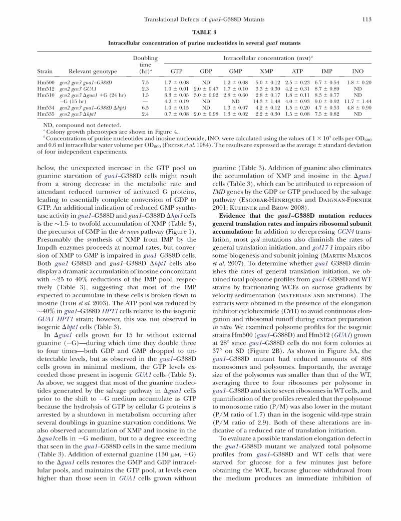
below, the unexpected increase in the GTP pool onguanine starvation of gua1–G388D cells might resultfrom a strong decrease in the metabolic rate andattendant reduced turnover of activated G proteins,leading to essentially complete conversion of GDP toGTP. An additional indication of reduced GMP synthe-tase activity in gua1–G388D and gua1–G388D Dhpt1 cellsis the �1.5- to twofold accumulation of XMP (Table 3),the precursor of GMP in the de novo pathway (Figure 1).Presumably the synthesis of XMP from IMP by theImpdh enzymes proceeds at normal rates, but conver-sion of XMP to GMP is impaired in gua1–G388D cells.Both gua1–G388D and gua1–G388D Dhpt1 cells alsodisplay a dramatic accumulation of inosine concomitantwith �25 to 40% reductions of the IMP pool, respec-tively (Table 3), suggesting that most of the IMPexpected to accumulate in these cells is broken down toinosine (Itoh et al. 2003). The ATP pool was reduced by�40% in gua1–G388D HPT1 cells relative to the isogenicGUA1 HPT1 strain; however, this was not observed inisogenic Dhpt1 cells (Table 3).
In Dgua1 cells grown for 15 hr without externalguanine (�G)—during which time they double threeto four times—both GDP and GMP dropped to un-detectable levels, but as observed in the gua1–G388Dcells grown in minimal medium, the GTP levels ex-ceeded those present in isogenic GUA1 cells (Table 3).As above, we suggest that most of the guanine nucleo-tides generated by the salvage pathway in Dgua1 cellsprior to the shift to �G medium accumulate as GTPbecause the hydrolysis of GTP by cellular G proteins isarrested by a shutdown in metabolism occurring afterseveral doublings in guanine starvation conditions. Wealso observed accumulation of XMP and inosine in theDgua1cells in �G medium, but to a degree exceedingthat seen in the gua1–G388D cells in the same medium(Table 3). Addition of external guanine (130 mm, 1G)to the Dgua1 cells restores the GMP and GDP intracel-lular pools, and maintains the GTP pool, at levels evenhigher than those seen in GUA1 cells grown without
guanine (Table 3). Addition of guanine also eliminatesthe accumulation of XMP and inosine in the Dgua1cells (Table 3), which can be attributed to repression ofIMD genes by the GDP or GTP produced by the salvagepathway (Escobar-Henriques and Daignan-Fornier
2001; Kuehner and Brow 2008).Evidence that the gua1–G388D mutation reduces
general translation rates and impairs ribosomal subunitaccumulation: In addition to derepressing GCN4 trans-lation, most gcd mutations also diminish the rates ofgeneral translation initiation, and gcd17-1 impairs ribo-some biogenesis and subunit joining (Martin-Marcos
et al. 2007). To determine whether gua1–G388D dimin-ishes the rates of general translation initiation, we ob-tained total polysome profiles from gua1–G388D and WTstrains by fractionating WCEs on sucrose gradients byvelocity sedimentation (materials and methods). Theextracts were obtained in the presence of the elongationinhibitor cycloheximide (CYH) to avoid continuous elon-gation and ribosomal runoff during extract preparationin vitro. We examined polysome profiles for the isogenicstrains Hm500 (gua1–G388D) and Hm512 (GUA1) grownat 28� since gua1–G388D cells do not form colonies at37� on SD (Figure 2B). As shown in Figure 5A, thegua1–G388D mutant had reduced amounts of 80Smonosomes and polysomes. Importantly, the averagesize of the polysomes was smaller than that of the WT,averaging three to four ribosomes per polysome ingua1–G388D and six to seven ribosomes in WTcells, andquantification of the profiles revealed that the polysometo monosome ratio (P/M) was also lower in the mutant(P/M ratio of 1.7) than in the isogenic wild-type strain(P/M ratio of 2.9). Both of these alterations are in-dicative of a reduced rate of translation initiation.
To evaluate a possible translation elongation defect inthe gua1–G388D mutant we analyzed total polysomeprofiles from gua1–G388D and WT cells that werestarved for glucose for a few minutes just beforeobtaining the WCE, because glucose withdrawal fromthe medium produces an immediate inhibition of
TABLE 3
Intracellular concentration of purine nucleotides in several gua1 mutants
Doublingtime(hr)a
Intracellular concentration (mm)b
Strain Relevant genotype GTP GDP GMP XMP ATP IMP INO
Hm500 gcn2 gcn3 gua1–G388D 7.5 1.7 6 0.08 ND 1.2 6 0.08 5.0 6 0.12 2.5 6 0.23 6.7 6 0.54 1.8 6 0.20Hm512 gcn2 gcn3 GUA1 2.3 1.0 6 0.01 2.0 6 0.47 1.7 6 0.10 3.3 6 0.30 4.2 6 0.31 8.7 6 0.89 NDHm510 gcn2 gcn3 Dgua1 1G (24 hr) 1.5 3.3 6 0.05 3.0 6 0.92 2.8 6 0.60 2.8 6 0.17 1.8 6 0.11 8.3 6 0.77 ND
�G (15 hr) — 4.2 6 0.19 ND ND 14.3 6 1.48 4.0 6 0.93 9.0 6 0.92 11.7 6 1.44Hm534 gcn2 gcn3 gua1–G388D Dhpt1 6.5 1.0 6 0.15 ND 1.3 6 0.07 4.2 6 0.12 1.5 6 0.20 4.7 6 0.53 4.8 6 0.90Hm535 gcn2 gcn3 Dhpt1 2.4 0.7 6 0.08 2.0 6 0.98 1.3 6 0.02 2.2 6 0.30 1.5 6 0.08 7.5 6 0.82 ND
ND, compound not detected.a Colony growth phenotypes are shown in Figure 4.b Concentrations of purine nucleotides and inosine nucleoside, INO, were calculated using the values of 1 3 107 cells per OD600
and 0.6 ml intracellular water volume per OD600 (Freese et al. 1984). The results are expressed as the average 6 standard deviationof four independent experiments.
Translational Defects of gua1–G388D Mutants 113

translation initiation (Ashe et al. 2000). This approachallows the detection of an effect on ribosomal transitwithout the added complication of de novo translationinitiation (Shenton et al. 2006). WT and gua1–G388Dcells were grown at 28� for 2 or 5 min after glucosewithdrawal, cycloheximide was added for 5 min to fixthe positions of elongating ribosomes, and the P/Mratios were quantified from polysome profiles. As shownin Figure 5B, gua1–G388D caused a slower rate ofpolysomal runoff under these conditions. Despite areduced polysome content in gua1–G388D cells prior toglucose withdrawal, the amounts of polysomes werehigher in the mutant vs. WT cells after glucose with-drawal. Because we obtained polysome profiles for thesame cultures prior to glucose withdrawal, we couldcalculate the P/M ratios after glucose withdrawal as apercentage of the values observed with glucose present,
as indicated in parenthesis beneath the P/M ratios ofFigure 5B. Thus, the P/M ratio dropped approximatelytwo- to threefold faster in WT than in gua1–G388D cellsafter glucose withdrawal, a phenotype that indicates aninhibition of translation elongation or termination inthe mutant. A reduced rate of elongation alone isexpected to increase the average size of the polysomes.However, as noted above, the average size of polysomes inglucose-replete medium was smaller in gua1–G388D thanin WT cells (Figure 5, A and B, 0 min). We conclude thatthe decreased rate of elongation or termination is over-powered by an even stronger defect in translation ini-tiation due to reduced rates of GMP synthesis in thegua1–G388D mutant. Consistent with this conclusion,the rate of incorporation of radioactive methionine intoacid-insoluble material was reduced by �45% at 28� ingua1–G388D compared to isogenic GUA1 cells (Table 4).
Figure 5.—Translational defects of gua1–G388D mutants. (A) Total polysome profiles ofisogenic strains Hm500 (gua1–G388D) andHm512 (GUA1) grown in liquid SD to midlogar-ithmic phase at 28� (OD600 �0.8). Cyclohexi-mide was added at 100 mg/ml beforeharvesting cells, and whole-cell extracts (WCE)containing ribosomes and polyribosomes pre-pared in the presence of 10 mm Mg12 and sepa-rated by velocity sedimentation on 7–50%sucrose gradients. Peaks representing free-ribosomal 40S and 60S subunits and 80S mono-somes are indicated. The polysome to monosomeratio (P/M) was estimated, and the average of val-ues from three independent determinations indi-cated below the A254 tracings. (B) Polysomeprofiles of the same strains as in A were obtainedbefore and after subjecting cells to glucose starva-tion to inhibit translation initiation (Ashe et al.2000). Aliquots were harvested of cells growingin liquid SD to midlogarithmic phase at 28�(0 min) and after being resuspended for 2 or5 min in glucose-free SD medium and incubatedat 28�. Cycloheximide was added at 100 mg/ml5 min before harvesting cells and the rates ofpolysomal runoff estimated relative to the polyso-mal content of each strain before glucose starva-tion (0 min) that was set to 100%. Thepolysome to monosome ratio (P/M) was calcu-lated and the average of values from two indepen-dent determinations indicated below the A254
tracings. (C) The same strains were cultured asdescribed in A, but WCE were prepared in theabsence of cycloheximide and Mg12, and resolvedby velocity sedimentation through 7–50% sucrosegradients. The mean ratios of total 60S/40S subu-nits determined from two replicate experimentsare indicated below the A254 tracings (left). TotalRNAs obtained from Hm512 (WT) and Hm500(mutant, m) grown at 28� to midlogarithmicphase and after being transferred to 37� for3 hr electrophoresed in a native agarose 1.1%gel. Electrophoretograms of the same RNA sam-ples were obtained with an Agilent Technologies2100 Bioanalyzer and the estimated 25S/18Sratios are indicated below (right).
114 D. Iglesias-Gato et al.

It was recently reported that pharmacological de-pletion of guanine nucleotides inhibits preribosomalrRNA synthesis and causes nucleolar disruption inmammalian cells (Huang et al. 2008). Because lowamounts of total ribosomal material were consistentlyrecovered from gua1–G388D cells (Figure 5A), weanalyzed the levels of mature ribosomal subunits pre-sent in cells grown at 28�. Quantification of total ri-bosomal subunits in low-Mg2 sucrose gradients, wherepolysomes and 80S ribosomes dissociate into freesubunits, revealed a deficit in 60S relative to 40Sribosomal subunits in gua1–G388D cells. A markeddecrease in the total amount of the 60S ribosomalsubunits per A260 units of total cell extract (�30%reduction), and a moderate decrease in the amount of40S subunits (�20% reduction) were observed in themutant under these conditions (Figure 5C, left). More-over, whereas a 60S/40S ratio of �1.25 was consistentlyobserved in the WT at 28�, the 60S/40S ratio was only�1.07 in the gua1–G388D mutant.
We also observed a large reduction in the totalamount of mature 25S rRNA, and a more moderatedecrease in the amount of 18S rRNA, per A260 units oftotal RNA, in gua1–G388D compared to the WT cellsgrown at 28� and after 3 hr incubation at 37� (Figure 5C,right). Measurements of the 25S/18S ratios with anAgilent Bionalyzer showed a nearly constant ratio in theWTof 1.0 to 1.1 that was reduced in gua1–G388D cells to0.7 at both temperatures, similar to the reductionsobserved in the 60S/40S subunit ratio (Figure 5C, left).These data reveal a significant shortage of both ribosomalsubunits and an imbalance of 60S relative to 40S subunitsin the gua1–G388D mutant, which most likely also contrib-utes to its decreased rate of general protein synthesis andSlg� phenotype on medium lacking guanine.
Together, our data show that translation of GCN4mRNA is constitutively derepressed, and that generaltranslation is strongly inhibited in gua1–G388D mu-tants, at the level of initiation and elongation, as aconsequence of reduced de novo synthesis of guaninenucleotides. They also suggest that translation initiationis an important rate-limiting step for cell growth anddivision under stressful conditions that reduce theavailability of guanine nucleotides. Our genetic datafurther indicate that the de novo pathway is indeedfunctioning at low levels in gua1–G388D cells.
Evidence that TC and MFC accumulate in gua1–G388D cells: The current model for translation initia-tion proposes that Met–tRNAi
Met is recruited to the 40Sribosomal subunit in the TC it forms with eIF2 �GTP,and that delivery of TC to the 40S ribosome is promotedby its interactions with eIF1, eIF3, and eIF5, constituentsof the MFC, either free in the cytoplasm or when thesefactors are bound to the 40S subunit (Sonenberg andHinnebusch 2009). To determine whether the reduc-tion in guanine nucleotides reduces the rate of TC orMFC formation, we analyzed the amount of tRNAi
Met
associated with Sui3, the b-subunit of eIF2 (TC), andeIF5 (MFC) in gua1–G388D and WT cells (materials
and methods). We employed pairs of isogenic gua1–G388D and GUA1 strains with a chromosomal deletionof SUI3 and a functional plasmid-borne SUI3–FLAG(SUI3–FL) allele, or with a chromosomal deletion of thegene-encoding eIF5 (TIF5) and a functional TIF5–FLAGallele (TIF5–FL) (Asano et al. 1999).
Unexpectedly, the amounts of tRNAiMet that co-
immunoprecipitated with the Sui3–FL and eIF5–FLprotein, relative to the tRNAi
Met levels in WCE, were�10-fold and �11-fold higher, respectively, in extractsof the gua1–G388D mutants than in the isogenic GUA1strains (Figure 6A, top, columns 1 and 2 and middle,columns 1 and 2, rows 1 and 2). Western analysis of thebait proteins confirmed that relatively similar amountsof Sui3–FL and eIF5–FL were immunoprecipitatedfrom the gua1–G388D and WT WCEs (Figure 6A, bottomof the SUI3–FL and eIF5–FL sections), thus indicatingthat gua1–G388D cells exhibit markedly higher levels oftRNAi
Met associated with eIF2 and in complexes contain-ing eIF5.
The increased association with Sui3–FL and eIF5–FLin gua1–G388D cells was specific for tRNAi
Met, as bothWT and gua1–G388D cells showed similar low levels ofelongator tRNAe
Met in the immunoprecipitates (Figure6A, top and middle, columns 3 and 4). The specificity ofthe Sui3–FL � tRNAi
Met and eIF5–FL � tRNAiMet interac-
tions was further verified in strains bearing untaggedSUI3 and TIF5 alleles (Figure 6A, bottom, columns 1 and2, rows 1 and 2), or when anti-Flag resin alone and�50 ng of wheat–tRNA (used as carrier in the purifica-tion of total RNAs from the immunoprecipitates) werehybridized with the probe for tRNAi
Met (Figure 6A,bottom, row 3). As expected, eIF5 did not immunopre-cipitate with anti-Flag from WCEs of untagged gua1–G388D TIF5 and GUA1 TIF5 isogenic strains (Figure 6A,bottom of the eIF5, Sui3 section).
As mentioned before, addition of guanine refills theinternal pool of GMP restoring the growth rate of thegua1–G388D cells (Figure 4A). Consistently, guanineaddition also reduced the amount of tRNAi
Met that co-immunoprecipitated with eIF5–FL from gua1–G388Dcells nearly to the levels seen in the isogenic WT (Figure6A middle, rows 1–4). Thus, the increased association oftRNAi
Met with eIF2 and eIF5 in the gua1–G388D mutant
TABLE 4
Rates of protein synthesis in gua1–G388D
Strain Relevant genotypel-[35S]methionine
(cpm/OD600)a
H96 gcn2 gcn3 15.540 (652)Hm458 gcn2 gcn3 gua1–G388D 7.400 (695)
a Values are averages of results obtained from assays on twoor three independent experiments (standard errors areshown inside parentheses).
Translational Defects of gua1–G388D Mutants 115

can be attributed to the defective de novo synthesis ofguanine nucleotides.
To confirm that the eIF5–FL � tRNAiMet association
specifically reflects the intracellular levels of MFC thatare formed in vivo, isogenic gua1–G388D and GUA1strains carrying either TIF5–FL or the tif5–7A–FL mutantallele, whose product is defective in forming the MFC(Asano et al. 2000), were analyzed as above in strains
with wild-type chromosomal TIF5. As expected, no de-tectable amounts of tRNAi
Met were associated with themutant eIF5–7A–FL protein (Figure 6B). Thus, theelevated association of tRNAi
Met with eIF5–FL in gua1–G388D cells likely occurs in the context of the MFC. Itwas noticeable that the association of wild-type eIF5–FLand tRNAi
Met was only �6-fold higher in gua1–G388Dthan in WT strains, rather than the �10-fold difference
Figure 6.—Accumulation of initiation factor-tRNAi
Met complexes in gua1–G388D cells andin WT cells treated with 6-azauracil (6AU). (A)Methionyl-initiator tRNA (tRNAi
Met) associatedwith flagged-eIF2 (Sui3–FL) and with eIF5–FLaccumulates in gua1–G388D cells. WCE were pre-pared from the following three pairs of isogenicstrains: (i) Hm522 (WT) and Hm523 (gua1–G388D), bearing a SUI3–FL-tagged allele, (top);(ii) Hm520 (WT) and Hm521 (gua1–G388D),bearing a TIF5–FL allele (middle); and (iii)H117 (WT) and Hm458 (gua1–G388D), lackingFLAG-tagged genes (bottom). All strains weregrown to midlogarithmic phase in liquid SD me-dium at 28�. Strains Hm520 and Hm521 werealso grown in SD containing 130 mm guanine(1G) (middle, lanes 3 and 4). Aliquots of thecorresponding WCE were incubated with ananti-FLAG affinity resin for 4 hr at 4�. After inten-sive washing, RNA from the precipitates (P) andfrom �1% of the immunoprecipitated WCE (I),were extracted and precipitated with ethanol us-ing 50 ng of wheat tRNA as carrier and blottedonto a nylon membrane. Radiolabeled oligonucleo-tides were used as probes for methionyl-initiatortRNA (tRNAi
Met, left) and methionyl-elongatortRNA species (tRNAe
Met, right) (materials and
methods). The affinity resin and wheat tRNAwere independently subjected to the purificationprocess and analyzed with the same probes (bot-tom, lane 3). Western blot detection of the Flag-tagged proteins showed similar precipitationefficiencies (bottom in SUI3–FL and eIF5–FLsections). (B) An eIF5–7A–FL mutant proteindoes not co-immunoprecipitate with tRNAi
Met.WCE were prepared from transformants ofHm512 (WT) and Hm500 (gua1–G388D) with alow-copy number plasmid containing TIF5–FL(p3147) or the mutant tif5–7A–FL allele (p3148)and co-immunoprecipitation assays conductedas in A. Western blot detection of eIF5–FL andeif5–7A–FL proteins with anti eIF5 antibodiesshowed similar precipitation efficiencies (bot-tom). (C) Nip1 and Prt1 subunits of eIF3 co-
immunoprecipitate with eIF5 � FL/tRNAiMet complexes in WCE from gua1–G388D cells. WCEs from Hm520 (GUA1, WT) and
Hm521 (gua1–G388D, m) were immunoprecipitated as in A. After extensive washing, precipitates (P, lanes 1–4) and �5% of thecrude extract (I, lanes 5–8) were analyzed by Western using anti-Nip1 and anti-Prt1 antibodies (materials and methods). Growth inminimally supplemented SD containing 130 mm guanine (lanes 3, 4, 7, and 8) is indicated as 1G. (D) Accumulation of eIF5 � FL/tRNAi
Met
complexes in cells treated with 6AU. WCE were obtained from cells of the WT strain Hm520 grown in SD with or without 100 mg/mlof 6-azauracil, and complexes containing eIF5 � FL/tRNAi
Met were immunoprecipitated and analyzed by slot blot as above. Westernblot detection of eIF5–FL showed similar precipitation efficiencies with and without 6AU (bottom). (E and F) Accumulation ofeIF5 � FL/tRNAi
Met complexes that contain subunits of eIF3 in WT cells treated with cycloheximide. (E) Detection of tRNAiMet in
immunocomplexes with eIF5–FL from WCE of the same strains as in A, middle panels, without and with added guanine (1G),except that 100 mg/ml of cycloheximide (CYH) was added to the cultures prior to the obtention of WCE. (F) The same immu-noprecipitates as in E (P, lanes 1–4) and �5% of the crude extract (I, lanes 5–8) were analyzed by Western using anti-Nip1, anti-Prt1, and anti-Flag antibodies as in C. Growth in minimally supplemented SD containing 130 mm guanine (lanes 3, 4, 7, and 8) isindicated as 1G (top).
116 D. Iglesias-Gato et al.

seen in Figure 6A. This difference can be due to thepresence of chromosomal untagged eIF5 in thesestrains, which can form MFC that is not precipitatedwith anti-Flag antibodies.
In an effort to confirm that the MFC accumulates ingua1–G388D cells, we measured the amounts of eIF3that are associated with eIF5–FL. To that end, immuno-precipitates from WCEs of gua1–G388D TIF5–FL andGUA1 TIF5–FL isogenic strains were analyzed by Westernblotting using antisera against the Nip1 and Prt1 sub-units of eIF3 (materials and methods). As shown inFigure 6C, a much greater amount of Nip1 and Prt1were present in the immunoprecipitates of gua1–G388D(lane 2) relative to WT cells (lane 1), but a similaramount was detected when mutant cells were grown inthe presence of guanine (lanes 3 and 4). These dataindicate that the MFC accumulates when guanine is notprovided to gua1–G388D cells.
The accumulation of eIF2 � tRNAiMet and eIF5 � tRNAi
Met
complexes in gua1–G388D cells suggests that TC andMFC are assembled but not utilized in translationinitiation when guanine nucleotide synthesis is sub-stantially impaired. Consistent with this interpretation,treatment of WT cells with 100 mg/ml of 6AU leads to asignificant�2.5-fold accumulation of tRNAi
Met bound toeIF5–FL (Figure 6D). This result indicates that theinhibition of the IMPD dehydrogenase by 6AU (Figure1) has the same qualitative effect as the gua1–G388Dmutation leading to an in vivo accumulation of MFC(Figure 6A, middle). Presumably, the reduction ofguanine nucleotide synthesis by gua1–G388D is greaterthan that in WT cells treated with 6AU, because theaccumulation of MFC was less pronounced in the lastcase.
We wished to determine to what extent accumulationof the MFC merely reflects the reduced rate of proteinsynthesis that occurs in gua1–G388D cells, leading to abuildup of initiation intermediates. To address this, weanalyzed whether completely inhibiting both the laststep of initiation and elongation of translation withcycloheximide (Hartwell and McLaughlin 1968)produces an accumulation of MFC similar to thatobserved in gua1–G388D cells. As shown in Figure 6E,addition of cycloheximide to WTcells at a concentrationthat impairs polysome runoff during preparation of theWCE (100 mg/ml, Figure 5A) resulted in a smallaccumulation (approximately twofold) of tRNAi
Met as-sociated with eIF5–FL relative to the untreated WTcontrol (Figure 6A, middle). These immunocomplexescontain tRNAi
Met, eIF5, and the Nip1 and Prt1 compo-nents of eIF3 (Figures 6, C and F) and thus likelycorrespond to the MFC. Addition of guanine reducesthe yield of MFC in the gua1–G388D mutant, but not inthe WT cells treated with cycloheximide, confirmingthat low rates of guanine nucleotide synthesis in gua1–G388D cells leads to a greater accumulation of MFCthan occurs with a complete block of translation. Thus,
the reduction in the rate of elongation probablyaccounts for only a small part of the accumulation ofthe MFC that occurs in gua1–G388D cells.
The gua1–G388D cells are defective in biogenesisand aminoacylation of tRNAi
Met: When we quantifiedthe amount of tRNAi
Met that immunoprecipitates withthe Flag-tagged forms of Sui3 or eIF5 in the aforemen-tioned experiments, we often observed that the amountof tRNAi
Met present in WCEs of gua1–G388D cells(input) was lower than that seen in WTcells (e.g., Figure6A, top, column 2, rows 1 and 2). This observation pro-mpted us to analyze the steady-state levels of tRNAi
Met
and of other Pol III transcripts in gua1–G388D mutants.Total RNAs were extracted from gua1–G388D and WTcells growing logarithmically at 28� in SD or aftertransferring to 37� and analyzed by Northern blottingusing radioactive oligonucleotides to probe for severalPol III transcripts (tRNAi
Met, tRNAeMet, tRNA(UAU)
Ile, and5S rRNA), the Pol I transcript 5.8S rRNA and the Pol IItranscript U4 RNA.
As shown in Figure 7A (top two panels), levels of themature initiator tRNAi
Met were diminished by �70% ingua1–G388D cells relative to WTcells, concomitant withaccumulation of immature pre-tRNAi
Met precursorscontaining 59 and 39 extensions, in cells grown at 28�or 37�. Because of the defective accumulation ofribosomal subunits and reduced 25S and 18S rRNAlevels in gua1–G388D cells (Figure 5C), loading equalA260 units of total RNA in each lane led to a relativeincrease in the steady-state level of the Pol II transcriptU4 in the mutant (Figure 7B, bottom), which was usedas an internal standard for normalization of results ontRNA levels. Relative to U4, the 5S and 5.8S rRNAs werealso reduced by as much as �50% in gua1–G388D cellscompared to the WT (Figure 7B). From these data weconclude that gua1–G388D cells exhibit specific defectsin processing and accumulation of tRNAi
Met.An obvious defect in processing of the primary pre-
tRNA (UAU)Ile precursor ‘‘P1’’ was also observed in gua1–
G388D cells, concomitant with reductions of �60% inthe levels of the tRNA (UAU)
Ile mature transcript (Figure7A, middle). The production of an intermediate ‘‘P2’’pre-tRNA (UAU)
Ile precursor requires the splicing of anintron present in the P1 precursor form of this tRNAspecies, and ‘‘P3’’ contains the second exon plus the 39
extension present in the primary precursor (O’Connor
and Peebles 1991). Whereas P1 accumulates, P2 isundetectable in gua1–G388D cells, indicating thattrimming of the P1 extensions is required prior to elim-ination of the intron from this pre-tRNA species.Accumulation of P3 in the mutant also indicates thatprocessing of the 39 extension is still impaired after the59 end has been removed (59 processing would mainlyoccur prior to 39 elimination by endo- or exonucleolyticprocessing). A less severe defect in processing of 59
and 39 extensions and accumulation of the elongatortRNAe
Met (�40%) was detected in gua1–G388D mutants
Translational Defects of gua1–G388D Mutants 117

(Figure 7A, bottom), indicating that the mutationdifferentially impacts the final levels of distinct tRNAspecies, with stronger effects on mature initiator tRNA,which presumably contributes to the severe defect intranslation initiation in gua1–G388D cells.
We next reasoned that reduced rates of guaninenucleotide synthesis in gua1–G388D cells could becoupled with a decrease of S-adenosyl-methionine(SAM) and aminoacylated methionyl-tRNA species,because GTP is a precursor of tetrahydrofolate, whichis required for the methylation of homocysteine to formmethionine (Freese et al. 1984). To determine the levelsof charged Met–tRNAi
Met relative to uncharged tRNAiMet,
total RNAs were extracted at pH 4.5 from gua1–G388Dand WT cells growing logarithmically at 28� and afterbeing transferred to 37� for 5 hr and 8 hr, and analyzedby Northern blot (materials and methods). As shownin Figure 7C, the ratios of aminoacylated to nonami-noacylated tRNAi
Met species dropped �30% in gua1–G388D relative to WT cells, at both 28� and 37�, whichwould be consistent with a reduction in the synthesis ofmethionine in the mutant. Since SAM is the principalmethylating agent for many macromolecules, reducedSAM synthesis might evoke deficient methylation oftRNA species, which could account for defects in tRNA
processing observed in gua1–G388D cells. It is remark-able that the TC and MFC, which presumably containeIF2 bound to mature Met–tRNAi
Met, accumulate byfactors of �10 (Figure 6A) despite the reductions inmature tRNAi
Met abundance (Figure 7A) and amino-acylation in gua1–G388D relative to WT cells.
DISCUSSION
We have identified a substitution mutation (G388D)in the GMP-synthetase Gua1 that impairs guaninenucleotide biosynthesis, leading to slow growth (Slg�)and constitutive derepression of GCN4 translation(Gcd�), which are suppressed by providing the mutantcells with an external supply of guanine. The G388Dmutation maps to the GMPSase domain of the Gua1enzyme and is predicted to reduce but not abolish thisactivity, because extra copies of the hypomorphic gua1–G388D allele revert all of the mutant phenotypes, andDhpt1 gua1–G388D cells (lacking the salvage pathway forGMP synthesis) are viable. We obtained evidence thatG388D reduces the total guanine nucleotide pool(GMP 1 GDP 1 GTP) in vivo by �40%, and leads toaccumulation of XMP, the Gua1 substrate. Interestingly,the levels of GMP, GDP, and GTP were not reduced
Figure 7.—The gua1–G388D mutationimpairs biogenesis and methionine ami-noacylation of tRNAi
Met. (A) Steady-statelevels of several tRNA transcripts ingua1–G388D mutants. Total RNA was puri-fied from isogenic strains Hm520 (GUA1,WT) and Hm521 (gua1–G388D, m) thatwere grown in SD medium to midlogarith-mic phase at 28� or after being shifted for3 hr at 37�, and 15 mg electrophoresed inpolyacrilamide/urea gel and analyzed byNorthern blot. The blot was sequentiallyprobed using specific radiolabeled oligonu-cleotides for each tRNA species indicatedon the left (materials and methods).Probes for tRNAi
Met, tRNA(UAU)Ile, and
tRNAeMet detect precursor (P) and mature
(M) forms of each tRNA. The precursortRNA species (pre-tRNA) are indicated onthe right as P1a: pre-tRNAi
Met containing-59 and -39 extensions encoded by IMT2and IMT3; P1b: pre-tRNAi
Met plus -59 and-39 extensions encoded by IMT1 andIMT4; P1c: pre-tRNAi
Met containing -39 ex-tensions. P1: pre-tRNA(UAU)
Ile containingtwo exons, an intron and -59 and -39 ex-tensions; P2: pre-tRNA(UAU)
Ile containingthe two exons and the intron; P3: pre-tRNA(UAU)
Ile containing a -39 extension.(B) Steady-state levels of several small RNAs in gua1–G388D mutants. The Pol III 5S rRNA, Pol II U4 RNA and Pol I 5.8S rRNAtranscripts were analyzed in the same blot as in (A) with specific oligonucleotides (materials and methods). To control for loading,the blot was probed for U4 rRNA. (C) Aminoacylation of initiator Met–tRNAi
Met is reduced in gua1–G388D cells. Northern blot anal-yses of total RNA isolated under acidic conditions (pH 4.5) from WT (Hm520) and gua1–G338D (Hm521) cells. After polyacrylamidegel electrophoresis and transfer under appropriate conditions, the blot was probed tRNAi
Met (materials and methods). To controlfor loading, the blot was probed for U4 RNA. A black arrow indicates the position of aminoacetylated Met–tRNAi
Met and a whitearrow that of deacylated tRNAi
Met species.
118 D. Iglesias-Gato et al.

equivalently: whereas GMP was moderately reduced,GDP was undetectable, and GTP was actually 1.5- to 1-7-fold higher in gua1–G388D cells than in wild type.
The unexpected high GTP and undetectable GDPlevels observed in gua1–G388D cells might be attribut-able to a massive reduction in the metabolic rate.Consequently, there would be a very low rate of GTPto GDP turnover by the many G proteins that participatein the myriad aspects of cell metabolism, and any GDPthat is generated would be rapidly converted back toGTP. In wild-type cells, GTP is continuously converted toGDP by the turnover of activated G proteins to yield asteady-state level of GDP that exceeds that of GTP. Incontrast to our findings, it was shown previously that theGTP pool drops dramatically by �90% in a leaky gua1mutant subjected to guanine starvation for 24 hr(Freese et al. 1984). At present, we have no explanationfor this discrepancy in results between the two studies.
Similar to all other known mutations conferringSlg� and Gcd� phenotypes, gua1–G388D cells exhibit ageneral defect in the rate of translation initiation; how-ever, an elongation defect was also detected, manifestedby a slower rate of polysome runoff when initiation wasblocked by glucose starvation. The reduction in averagepolysome size and abundance indicates that the initia-tion defect is relatively more severe during guaninestarvation of gua1–G388D cells. In addition, a markedreduction in the amounts of mature 60S and 40S sub-units likely impair all steps of translation in the mutant.Together, these defects provoke a�50% decrease in therate of incorporation of 35S-methionine into total pro-tein (Table 4).
Because GTP is required for all phases of proteinsynthesis, it would be natural to assume that geneticallyimpairing GMP synthesis would affect translation bydecreasing the amount of GTP available for this process.However, because GTP levels are elevated, it is possiblethat the different G proteins that participate in trans-lation occur primarily in the GTP-bound state in gua1–G388D cells, and that the process is impeded by one ormore unknown signal transduction pathways that targetboth the initiation and elongation steps as a response toreduced synthesis of guanine nucleotides. Consistentwith this interpretation, tRNAi
Met-containing complexeswith the protein compositions of TC and MFC accumu-late in excess of�10-fold in gua1–G388D relative to theirlevels in isogenic WT cells (Figure 6). The apparentaccumulation of TC and MFC is even more strikingconsidering that pre-tRNAi
Met processing, and the accu-mulation and aminoacylation of mature tRNAi
Met wereall reduced in the mutant, which should compromiseassembly of TC. These findings suggest that translationinitiation is blocked downstream of the assembly of TCand MFC, and that the underutilization of these com-plexes leads to their abnormal accumulation in gua1–G388D cells.
The fact that the gua1–G388D mutation constitutivelyderepresses GCN4 translation is consistent with thepossibility that guanine nucleotide depletion provokesa reduction in the rate of TC recruitment by thereinitiating 40S ribosomes that remain associated withthe GCN4 mRNA leader after translating uORF1. As aresult, a fraction of these scanning 40S subunits wouldnot reacquire the TC in time to reinitiate translation atuORFs 2–4 (either 60S subunits) but would do so by thetime they reach the GCN4 AUG and reinitiate trans-lation there instead. Because TC levels apparently arenot reduced by gua1–G388D, the Gcd�phenotype wouldarise from a reduced rate of TC binding to the re-initiating 40S subunits, or the failure to recognize theAUG codons at uORFs 2–4 after rebinding of TC, ratherthan a deficit in TC assembly. The reduced con-centration of 60S subunits in the mutant could alsocontribute to the bypass of uORFs 2–4 via impairedsubunit joining, as mutations in 60S subunit proteinsthat reduce 60S subunit levels confer Gcd� phenotypesand constitutively derepress GCN4 translation (Foiani
et al. 1991; Martin-Marcos et al. 2007). One interest-ing, albeit speculative, possibility to explain the Gcd�
phenotype would be that low-level GMP synthesistriggers a signal transduction pathway that reduces TCloading on 40S subunits, by decreasing the affinity ofeIF2 or tRNAi
Met for the 40S, either through modifica-tion of one of these components of the 43S preinitiationcomplex (PIC) or by impairing the function of one ormore other factors eIFs -1, -1A, -3, or -5, that enhanceTC recruitment. This would be akin to the effects ofknown mutations in tRNAi
Met, eIF2 subunits, or in othereIFs that confer Gcd� phenotypes by this mechanism(Hinnebusch 2005). The decrease in SAM expectedin gua1 mutants (Varma et al. 1985), owing to the in-volvement of GTP in SAM biosynthesis, could reducemethylation of tRNAi
Met bases and thereby also impairtRNAi
Met function in 40S binding. All of these reactionsrepresent plausible targets for the hypothetical signal-transduction pathway(s) that would modify TC compo-nents, other eIFs, or the ribosomal subunits in responseto low-level GMP synthesis in gua1–G388D cells. In viewof our finding that ATP levels are also markedly reducedin the gua1–G388D mutant, the hypothetical signal-transduction response could be triggered by reducedATP levels, as a manifestion of energy depletion, ratherthan a response to diminished guanine nucleotides.
Our finding that tRNAiMet-containing complexes ac-
cumulate by�10-fold but the GTP level is only�1.5-foldhigher in gua1–G388D vs. GUA1 cells further suggeststhat at least a proportion of these complexes are notgenuine TC or MFC, and lack GTP bound to eIF2. TheKd for Met–tRNAi
Met binding to apo–eIF2 is only anorder of magnitude higher than for eIF2 �GTP (Kapp
and Lorsch 2004). Hence, eIF2 � tRNAiMet binary com-
plexes might accumulate if eIF2 cannot compete ef-fectively with other G proteins for the available GTP
Translational Defects of gua1–G388D Mutants 119

under conditions of guanine nucleotide limitation.Similarly, the Kd for binding nonaminoacetylatedtRNAi
Met to eIF2 �GTP is only an order of magnitudehigher than for methionyl–tRNAi
Met (Kapp and Lorsch
2004). Thus, the defect in aminoacetylating tRNAiMet
could provoke the formation of defective complexesbetween eIF2 �GTP and uncharged tRNAi
Met in gua1–G388D cells. If these defective complexes compete withauthentic TC for binding to 40S subunits, they would beexpected to reduce the rate of assembling functional43S or 48S PICs, contributing to the decrease in generaltranslation initiation and the bypass of uORFs 2–4during reinitiation on GCN4 mRNA.
Nearly one-half of the eIF2 in yeast cells, presumablyin its GDP-bound state, forms a complex with eIF5,which appears to antagonize GTP exchange on eIF2–GDP to render eIF2 recycling rate limiting for trans-lation initiation (Singh et al. 2006); this complexinsures an effective inhibition of eIF2 recycling by eIF2Bwhen eIF2a is phosphorylated by Gcn2 ( Jennings andPavitt 2010). However, the eIF2 �GDP/eIF5 complexesidentified by Singh et al. (2006) likely differ from thosedescribed here, which contain tRNAi
Met and accumulateunder conditions of guanine nucleotide depletionwhere GDP is undetectable.
Recently it was reported that depletion of the guaninenucleotide pool by three different means led to increasedexpression of a large number of genes, most of whichparticipate in amino acid metabolism and are under thetranscriptional control of Gcn4 (Saint-Marc et al. 2009);however, neither the Gcn4 dependence nor molecularmechanism of the response was investigated. In light ofour data, we suggest that GCN4 mRNA translation wasderepressed by the GDP/GTP shortages evoked by thedifferent conditions examined by Saint-Marc et al.(2009) and it would be interesting to determinewhether tRNAi
Met containing complexes accumulateunder those conditions.
We found that reduced synthesis of G nucleotides ingua1–G388D cells impairs the processing of pre-tRNAi
Met
and precursors of certain elongator tRNA species(Figure 7). The predicted reduction in SAM biosynthe-sis and hypomethylation of pre-tRNAs might impedetheir proper folding and processing, as occurs in gcd10and gcd14 mutants defective in methylation of m1A58
(Anderson et al. 1998; Calvo et al. 1999). It is alsointeresting to note that Ran-dependent nucleocytoplas-mic transport is dependent on both GTP- and GDP-bound forms of the Ran GTPase, and that defects intRNA maturation have been observed in nucleoporinmutants (Gorlich et al. 1996; Sharma et al. 1996;Izaurralde et al. 1997). Hence, the abnormal GTP/GDP ratio in the gua1–G388D mutant could contributeto the observed defects in tRNA maturation by in-terfering with Ran function. Defects in Ran-dependenttransport of ribosomal processing factors or ribosomalsubunit precursors (Thomas and Kutay 2003; Todaka
et al. 2005) could likewise be involved in the reducedlevels of ribosomal subunits seen in gua1–G388D cells.
In summary, our results indicate that geneticallyreducing the activity of the GMP synthase (Gua1) inbudding yeast has myriad effects on cell biology andmetabolism. A reduction in the total G nucleotide pool,with a precipitous drop in the GDP/GTP ratio, plus adecrease in ATP levels, are among the most direct effectsof the gua1–388D mutation. These alterations in nucle-otide levels evoke defects in ribosome and tRNA bio-genesis and decreased rates of both the initiation andelongation phases of protein synthesis, leading to areduced rate of general translation, and a specificderepression of GCN4 mRNA translation. Contrary toour expectations, the defects in general and GCN4-specific translation do not appear to arise from impairedassembly of the eIF2�tRNAi
Met.GTP ternary complex.Rather, the apparent accumulation of TC and MFC inthe gua1–388D mutant argues for a block further down-stream in the initiation pathway that decreases the rate ofTC consumption. Future studies are required to identifythe exact steps in the initiation pathway that are down-regulated on reduction of GMP synthesis and also thehypothetical regulatory mechanisms that mediate thismultifaceted response.
We thank D. Martın-Zanca for helpful suggestions regarding thiswork and J. R. Gutierrez for help with the procedure of yeastmetabolite extractions. D.I.-G. was supported by a postgraduatefellowship (BFI02.95), granted by the Department of Education,Universities, and Research of the Basque government (Spain).M.A.S. was supported by Project #SA098A09, granted by the De-partment of Universities and Research of the Junta de Castilla y LeonGovernment (JCyL) (Spain). M. Tamame was supported by ProjectsPET2008-0283-01, granted by the Spanish Ministerio de Ciencia eInnovacion (MICINN), and CSI007A10-2, granted by JCyL. The workwas supported partly by the Intramural Research Program of theNational Institutes of Health.
LITERATURE CITED
Alani, E., L. Cao and N. Kleckner, 1987 A method for gene disrup-tion that allows repeated use of URA3 selection in the construc-tion of multiply disrupted yeast strains. Genetics 116: 541–545.
Anderson, J., L. Phan, R. Cuesta, B. A. Carlson, M. Pak et al.,1998 The essential Gcd10p-Gcd14p nuclear complex is re-quired for 1-methyladenosine modification and maturation ofinitiator methionyl-tRNA. Genes Dev. 12: 3650–3662.
Asano, K., T. Krishnamoorthy, L. Phan, G. D. Pavitt and A. G.Hinnebusch, 1999 Conserved bipartite motifs in yeast eIF5and eIF2Bepsilon, GTPase-activating and GDP-GTP exchange fac-tors in translation initiation, mediate binding to their commonsubstrate eIF2. EMBO J. 18: 1673–1688.
Asano, K., J. Clayton, A. Shalev and A. G. Hinnebusch, 2000 Amultifactor complex of eukaryotic initiation factors, eIF1, eIF2,eIF3, eIF5, and initiator tRNA(Met) is an important translationinitiation intermediate in vivo. Genes Dev. 14: 2534–2546.
Ashe, M. P., S. K. De Long and A. B. Sachs, 2000 Glucose depletionrapidly inhibits translation initiation in yeast. Mol. Biol. Cell 11:833–848.
Boeke, J. D., F. LaCroute and G. R. Fink, 1984 A positive selectionfor mutants lacking orotidine-59-phosphate decarboxylase activ-ity in yeast: 5-fluoro-orotic acid resistance. Mol. Gen. Genet.197: 345–346.
120 D. Iglesias-Gato et al.

Bremer, S., N. T. Vethe, H. Rootwelt and S. Bergan,2009 Expression of IMPDH1 is regulated in response to myco-phenolate concentration. Int. Immunopharmacol. 9: 173–180.
Breton, A., B. Pinson, F. Coulpier, M. F. Giraud, A. Dautant et al.,2008 Lethal accumulation of guanylic nucleotides in Saccharomy-ces cerevisiae HPT1-deregulated mutants. Genetics 178: 815–824.
Burke, P. V., and K. E. Kwast, 2000 Oxygen dependence of expres-sion of cytochrome C and cytochrome C oxidase genes in S.cerevisiae. Adv. Exp. Med. Biol. 475: 197–208.
Calvo, O., R. Cuesta, J. Anderson, N. Gutierrez, M. T. Garcia-Barrio et al., 1999 GCD14p, a repressor of GCN4 translation,cooperates with Gcd10p and Lhp1p in the maturation of initiatormethionyl-tRNA in Saccharomyces cerevisiae. Mol. Cell. Biol. 19:4167–4181.
Cuesta, R., A. G. Hinnebusch and M. Tamame, 1998 Identificationof GCD14 and GCD15, novel genes required for translational re-pression of GCN4 mRNA in Saccharomyces cerevisiae. Genetics 148:1007–1020.
Daignan-Fornier, B., and G. R. Fink, 1992 Coregulation of purineand histidine biosynthesis by the transcriptional activators BAS1and BAS2. Proc. Natl. Acad. Sci. USA 89: 6746–6750.
Dever, T. E., W. Yang, S. Astrom, A. S. Bystrom and A. G.Hinnebusch, 1995 Modulation of tRNA(iMet), eIF-2, andeIF-2B expression shows that GCN4 translation is inversely cou-pled to the level of eIF-2.GTP.Met-tRNA(iMet) ternary com-plexes. Mol. Cell. Biol. 15: 6351–6363.
Dujardin, G., M. Kermorgant, P. P. Slonimski and H. Boucherie,1994 Cloning and sequencing of the GMP synthetase-encodinggene of Saccharomyces cerevisiae. Gene 139: 127–132.
Escobar-Henriques, M., and B. Daignan-Fornier, 2001 Transcrip-tional regulation of the yeast gmp synthesis pathway by itsend products. J. Biol. Chem. 276: 1523–1530.
Exinger, F., and F. Lacroute, 1992 6-Azauracil inhibition of GTPbiosynthesis in Saccharomyces cerevisiae. Curr. Genet. 22: 9–11.
Foiani, M., A. M. Cigan, C. J. Paddon, S. Harashima and A. G.Hinnebusch, 1991 GCD2, a translational repressor of theGCN4 gene, has a general function in the initiation of protein syn-thesis in Saccharomyces cerevisiae. Mol. Cell. Biol. 11: 3203–3216.
Freese, E. B., Z. Olempska-Beer, A. Hartig and E. Freese,1984 Initiation of meiosis and sporulation of Saccharomycescerevisiae by sulfur or guanine deprivation. Dev. Biol. 102:438–451.
Gardner, W. J., and R. A. Woods, 1979 Isolation and characterisa-tion of guanine auxotrophs in Saccharomyces cerevisiae. Can. J.Microbiol. 25: 380–389.
Gonzalez, B., J. Francois and M. Renaud, 1997 A rapid andreliable method for metabolite extraction in yeast using boilingbuffered ethanol. Yeast 13: 1347–1355.
Gorlich, D., N. Pante, U. Kutay, U. Aebi and F. R. Bischoff,1996 Identification of different roles for RanGDP and RanGTPin nuclear protein import. EMBO J. 15: 5584–5594.
Guetsova, M. L., K. Lecoq and B. Daignan-Fornier, 1997 Theisolation and characterization of Saccharomyces cerevisiae mutantsthat constitutively express purine biosynthetic genes. Genetics147: 383–397.
Harashima, S., and A. G. Hinnebusch, 1986 Multiple GCD genesrequired for repression of GCN4, a transcriptional activator ofamino acid biosynthetic genes in Saccharomyces cerevisiae.Mol. Cell. Biol. 6: 3990–3998.
Harashima, S., E. M. Hannig and A. G. Hinnebusch, 1987 Interactionsbetween positive and negative regulators of GCN4 controllinggene expression and entry into the yeast cell cycle. Genetics 117:409–419.
Hartwell, L. H., and C. S. McLaughlin, 1968 Temperature-sensitivemutants of yeast exhibiting a rapid inhibition of protein synthesis.J. Bacteriol. 96: 1664–1671.
Hinnebusch, A. G., 1985 A hierarchy of trans-acting factors modu-lates translation of an activator of amino acid biosynthetic genesin Saccharomyces cerevisiae. Mol. Cell. Biol. 5: 2349–2360.
Hinnebusch, A. G., 2005 Translational regulation of GCN4 and thegeneral amino acid control of yeast. Annu. Rev. Microbiol. 59:407–450.
Hinnebusch, A. G., and G. R. Fink, 1983 Positive regulation in thegeneral amino acid control of Saccharomyces cerevisiae. Proc. Natl.Acad. Sci. USA 80: 5374–5378.
Huang, M., Y. Ji, K. Itahana, Y. Zhang and B. Mitchell,2008 Guanine nucleotide depletion inhibits pre-ribosomal RNAsynthesis and causes nucleolar disruption. Leuk. Res. 32: 131–141.
Hyle, J. W., R. J. Shaw and D. Reines, 2003 Functional distinctionsbetween IMP dehydrogenase genes in providing mycophenolateresistance and guanine prototrophy to yeast. J. Biol. Chem. 278:28470–28478.
Itoh, R., C. Saint-Marc, S. Chaignepain, R. Katahira, J. M.Schmitter et al., 2003 The yeast ISN1 (YOR155c) gene encodesa new type of IMP-specific 59-nucleotidase. BMC Biochem. 4: 4.
Izaurralde, E., U. Kutay, C. von Kobbe, I. W. Mattaj and D.Gorlich, 1997 The asymmetric distribution of the constituentsof the Ran system is essential for transport into and out of thenucleus. EMBO J. 16: 6535–6547.
Jennings, M. D., and G. D. Pavitt, 2010 eIF5 has GDI activity nec-essary for translational control by eIF2 phosphorylation. Nature465: 378–381.
Kapp, L. D., and J. R. Lorsch, 2004 GTP-dependent recognition ofthe methionine moiety on initiator tRNA by translation factoreIF2. J. Mol. Biol. 335: 923–936.
Kuehner, J. N., and D. A. Brow, 2008 Regulation of a eukaryoticgene by GTP-dependent start site selection and transcription at-tenuation. Mol. Cell 31: 201–211.
Lee, J. H., T. V. Pestova, B. S. Shin, C. Cao, S. K. Choi et al.,2002 Initiation factor eIF5B catalyzes second GTP-dependentstep in eukaryotic translation initiation. Proc. Natl. Acad. Sci.USA99: 16689–16694.
Lucchini, G., A. G. Hinnebusch, C. Chen and G. R. Fink,1984 Positive regulatory interactions of the HIS4 gene of Sac-charomyces cerevisiae. Mol. Cell. Biol. 4: 1326–1333.
Martin-Marcos, P., A. G. Hinnebusch and M. Tamame,2007 Ribosomal protein L33 is required for ribosome biogen-esis, subunit joining, and repression of GCN4 translation. Mol.Cell. Biol. 27: 5968–5985.
Mueller, P. P., and A. G. Hinnebusch, 1986 Multiple upstreamAUG codons mediate translational control of GCN4. Cell 45:201–207.
Natarajan, K., M. R. Meyer, B. M. Jackson, D. Slade, C. Roberts
et al., 2001 Transcriptional profiling shows that Gcn4p is a mas-ter regulator of gene expression during amino acid starvation inyeast. Mol. Cell. Biol. 21: 4347–4368.
Niederberger, P., M. Aebi and R. Hutter, 1986 Identification andcharacterization of four new GCD genes in Saccharomyces cere-visiae. Curr. Genet. 10: 657–664.
Nielsen, K. H., B. Szamecz, L. Valasek, A. Jivotovskaya, B. S. Shin
et al., 2004 Functions of eIF3 downstream of 48S assembly im-pact AUG recognition and GCN4 translational control. EMBO J.23: 1166–1177.
O’Connor, J. P., and C. L. Peebles, 1991 In vivo pre-tRNA process-ing in Saccharomyces cerevisiae. Mol. Cell. Biol. 11: 425–439.
Pestova, T. V., J. R. Lorsch and C. U. T. Hellen, 2007 The mech-anism of translation initiation in eukaryotes, pp. 87–128 in Trans-lational Control in Biology and Medicine, edited by M. B. Mathews,N. Sonenberg and J. W. Hershey. Cold Spring Harbor Labora-tory Press, Cold Spring Harbor, NY.
Pinson, B., S. Vaur, I. Sagot, F. Coulpier, S. Lemoine et al.,2009 Metabolic intermediates selectively stimulate transcrip-tion factor interaction and modulate phosphate and purine path-ways. Genes Dev. 23: 1399–1407.
Ramirez, M., R. C. Wek, C. R. Vazquez de Aldana, B. M. Jackson,B. Freeman et al., 1992 Mutations activating the yeast eIF-2alpha kinase GCN2: isolation of alleles altering the domain re-lated to histidyl-tRNA synthetases. Mol. Cell. Biol. 12: 5801–5815.
Rolfes, R. J., 2006 Regulation of purine nucleotide biosynthesis: inyeast and beyond. Biochem. Soc. Trans. 34: 786–790.
Rolfes, R. J., and A. G. Hinnebusch, 1993 Translation of the yeasttranscriptional activator GCN4 is stimulated by purine limitation:implications for activation of the protein kinase GCN2. Mol. Cell.Biol. 13: 5099–5111.
Rose, M. D., P. Novick, J. H. Thomas, D. Botstein and G. R. Fink,1987 A Saccharomyces cerevisiae genomic plasmid bank basedon a centromere-containing shuttle vector. Gene 60: 237–243.
Saint-Marc, C., B. Pinson, F. Coulpier, L. Jourdren, O. Lisova et al.,2009 Phenotypic consequences of purine nucleotide imbalancein Saccharomyces cerevisiae. Genetics 183: 529–538, 521SI–527SI.
Translational Defects of gua1–G388D Mutants 121

Sarkar, S., A. K. Azad and A. K. Hopper, 1999 Nuclear tRNAaminoacylation and its role in nuclear export of endogenoustRNAs in Saccharomyces cerevisiae. Proc. Natl. Acad. Sci. USA 96:14366–14371.
Schmitt, M. E., T. A. Brown and B. L. Trumpower, 1990 A rapidand simple method for preparation of RNA from Saccharomycescerevisiae. Nucleic Acids Res. 18: 3091–3092.
Sharma, K., E. Fabre, H. Tekotte, E. C. Hurt and D. Tollervey,1996 Yeast nucleoporin mutants are defective in pre-tRNA splic-ing. Mol. Cell. Biol. 16: 294–301.
Shenton, D., J. B. Smirnova, J. N. Selley, K. Carroll, S. J. Hubbard
et al., 2006 Global translational responses to oxidative stress im-pact upon multiple levels of protein synthesis. J. Biol. Chem. 281:29011–29021.
Sikorski, R. S., and P. Hieter, 1989 A system of shuttle vectors andyeast host strains designed for efficient manipulation of DNA inSaccharomyces cerevisiae. Genetics 122: 19–27.
Singh, C. R., B. Lee, T. Udagawa, S. S. Mohammad-Qureshi, Y.Yamamoto et al., 2006 An eIF5/eIF2 complex antagonizes gua-nine nucleotide exchange by eIF2B during translation initiation.EMBO J. 25: 4537–4546.
Sonenberg, N., and A. G. Hinnebusch, 2009 Regulation of trans-lation initiation in eukaryotes: mechanisms and biological tar-gets. Cell 136: 731–745.
Thomas, F., and U. Kutay, 2003 Biogenesis and nuclear export ofribosomal subunits in higher eukaryotes depend on the CRM1export pathway. J. Cell Sci. 116: 2409–2419.
Todaka, Y., Y. Wang, K. Tashiro, N. Nakashima, T. Nishimoto et al.,2005 Association of the GTP-binding protein Gtr1p withRpc19p, a shared subunit of RNA polymerase I and III in yeastSaccharomyces cerevisiae. Genetics 170: 1515–1524.
Ufano, S., F. del Rey and C. R. Vazquez de Aldana, 2004 Swm1p, asubunit of the APC/cyclosome, is required to maintain cell wallintegrity during growth at high temperature in Saccharomycescerevisiae. FEMS Microbiol. Lett. 234: 371–378.
Varma, A., E. B. Freese and E. Freese, 1985 Partial deprivation ofGTP initiates meiosis and sporulation in Saccharomyces cerevi-siae. Mol. Gen. Genet. 201: 1–6.
Wach, A., A. Brachat, R. Pohlmann and P. Philippsen, 1994 Newheterologous modules for classical or PCR-based gene disrup-tions in Saccharomyces cerevisiae. Yeast 10: 1793–1808.
Wek, R. C., M. Ramirez, B. M. Jackson and A. G. Hinnebusch,1990 Identification of positive-acting domains in GCN2 proteinkinase required for translational activation of GCN4 expression.Mol. Cell. Biol. 10: 2820–2831.
Communicating editor: M. Hampsey
122 D. Iglesias-Gato et al.