levodopa modulates functional connectivity in the upper
TRANSCRIPT

ORIGINAL RESEARCHpublished: 02 July 2019
doi: 10.3389/fnhum.2019.00223
Edited by:
Vladimir Litvak,University College London,
United Kingdom
Reviewed by:Jan Hirschmann,
Heinrich Heine University ofDüsseldorf, Germany
Maria Herrojo Ruiz,Goldsmiths University of London,
United Kingdom
*Correspondence:Jens Volkmann
Received: 18 January 2019Accepted: 18 June 2019Published: 02 July 2019
Citation:Ramirez Pasos UE, Steigerwald F,
Reich MM, Matthies C, Volkmann Jand Reese R (2019) Levodopa
Modulates Functional Connectivity inthe Upper Beta Band Between
Subthalamic Nucleus and MuscleActivity in Tonic and Phasic Motor
Activity Patterns inParkinson’s Disease.
Front. Hum. Neurosci. 13:223.doi: 10.3389/fnhum.2019.00223
Levodopa Modulates FunctionalConnectivity in the Upper Beta BandBetween Subthalamic Nucleus andMuscle Activity in Tonic and PhasicMotor Activity Patterns inParkinson’s DiseaseUri E. Ramirez Pasos1, Frank Steigerwald1, Martin M. Reich1, Cordula Matthies2,Jens Volkmann1* and René Reese1,3
1Department of Neurology, University Hospital Würzburg, Würzburg, Germany, 2Department of Neurosurgery, UniversityHospital Würzburg, Würzburg, Germany, 3Department of Neurology, University of Rostock, Rostock, Germany
Introduction: Striatal dopamine depletion disrupts basal ganglia function and causesParkinson’s disease (PD). The pathophysiology of the dopamine-dependent relationshipbetween basal ganglia signaling and motor control, however, is not fully understood. Weobtained simultaneous recordings of local field potentials (LFPs) from the subthalamicnucleus (STN) and electromyograms (EMGs) in patients with PD to investigate theimpact of dopaminergic state and movement on long-range beta functional connectivitybetween basal ganglia and lower motor neurons.
Methods: Eight PD patients were investigated 3 months after implantation of a deepbrain stimulation (DBS)-system capable of recording LFPs via chronically-implantedleads (Medtronic, ACTIVA PC+Sr). We analyzed STN spectral power and its coherencewith EMG in the context of two different movement paradigms (tonic wrist extension vs.alternating wrist extension and flexion) and the effect of levodopa (L-Dopa) intake usingan unbiased data-driven approach to determine regions of interest (ROI).
Results: Two ROIs capturing prominent coherence within a grand average coherogramwere identified. A trend of a dopamine effect was observed for the first ROI(50–150 ms after movement start) with higher STN-EMG coherence in medicatedpatients. Concerning the second ROI (300–500 ms after movement start), an interactioneffect of L-Dopa medication and movement task was observed with higher coherencein the isometric contraction task compared to alternating movements in the medicationON state, a pattern which was reversed in L-Dopa OFF.
Discussion: L-Dopa medication may normalize functional connectivity between remotestructures of the motor system with increased upper beta coherence reflecting aphysiological restriction of the amount of information conveyed between remotestructures. This may be necessary to maintain simple movements like isometriccontraction. Our study adds dynamic properties to the complex interplay between STN
Frontiers in Human Neuroscience | www.frontiersin.org 1 July 2019 | Volume 13 | Article 223

Ramirez Pasos et al. STN-EMG Coherence
spectral beta power and the nucleus’ functional connectivity to remote structures ofthe motor system as a function of movement and dopaminergic state. This may helpto identify markers of neuronal activity relevant for more individualized programming ofDBS therapy.
Keywords: Parkinson’s disease, subthalamic nucleus, deep brain stimulation, local field potentials,dopamine, movement
INTRODUCTION
In vivo recordings of basal ganglia neuronal activity inParkinson’s disease (PD) patients have been facilitated by thechronic implantation of stimulation electrodes for therapeuticdeep brain stimulation (DBS; Benabid et al., 1994). Local fieldpotential (LFP) recordings from the subthalamic nucleus (STN),the most common DBS target for PD (Deuschl et al., 2006;Weaver et al., 2009; Follett et al., 2010; Schuepbach et al., 2013),have revealed abnormally synchronized oscillatory activity inthe beta band (about 13–30 Hz) to be an electrophysiologicalhallmark of the Parkinsonian state (Brown et al., 2001; Brown,2003; Kühn et al., 2006, 2009). STN beta activity is mostprominent in the resting state of PD patients (López-Azcárateet al., 2010), is attenuated by voluntary movement (Cassidy et al.,2002; Levy et al., 2002; Priori et al., 2002; Kühn et al., 2004; Alegreet al., 2005, 2013; Doyle et al., 2005; Foffani et al., 2005; Hebbet al., 2012; Joundi et al., 2012, 2013; Litvak et al., 2012), andincreases again when movement voluntarily ends (Kühn et al.,2004; Alegre et al., 2013). It has been suggested that the encodingof steady motor states might be a physiological role of betarhythms, which become pathologically exaggerated as a resultof striatal dopamine depletion, a pathophysiological hallmarkof PD (Engel and Fries, 2010; Brittain and Brown, 2014). Thismechanism is supported by several studies demonstrating thatoral levodopa (L-Dopa) intake in PD patients desynchronizesSTN beta band activity (Brown et al., 2001; Levy et al., 2002;Priori et al., 2004; Alegre et al., 2005; Kühn et al., 2006;Hirschmann et al., 2013). Beta desynchronization is moreovercorrelated with improved bradykinesia scores (Kühn et al.,2006, 2009; Weinberger et al., 2006; Androulidakis et al.,2008; Ray et al., 2008). Because of the correlative nature ofthese experiments, it remains an open question whether STNbeta rhythms cause bradykinesia (Jenkinson and Brown, 2011),despite ample evidence that beta exaggerations represent avalid electrophysiological marker of the Parkinsonian motorOFF state. Studies which evaluated the effects of dopamineand movement on functional beta oscillatory coupling of STN,motor cortex, and muscle activity have delivered conflictingresults (Marsden et al., 2001; Salenius et al., 2002; Litvak et al.,2012; Hirschmann et al., 2013), possibly due to differencesin recording and analyses techniques as well as movementparadigms. Frequency-specific coding of STN-muscle coherencewas found in the beta band during isometric contractionsand in the 31–45 Hz band during phasic motor activity inPD patients after L-Dopa intake (Marsden et al., 2001). Incontrast, Hirschmann et al. (2013) found reduced STN-musclebeta band coherence during alternating forearm movements
compared to isometric contractions, which was independentof L-Dopa medication. One study found STN-motor cortexbeta coherence in a task of tonic muscle activity to be greatercompared to that of alternating movements in the L-DopaON state of PD patients (Marsden et al., 2001), whereasother studies found this coherence suppressed (Hirschmannet al., 2013) or restored by L-Dopa during movement (Saleniuset al., 2002). With a latency of 2 s from movement onset,STN-motor cortex beta band coherence increased compared tobaseline, which was independent of dopaminergic medicationstate (Litvak et al., 2012). In contrast, STN spectral powerand STN-motor cortex coherence in the beta band decreasedcompared to baseline prior to and during movement inunmedicated PD patients (Talakoub et al., 2016). Only thesetwo studies have explicitly looked at dynamic changes of STNoscillatory activity and STN-cortex coherence to be dependenton movement activity. This is of considerable significancegiven that increased beta oscillations in motor cortex and basalganglia have been associated with immutability and steady statein motor activity while being modulated during movementchange (Brittain and Brown, 2014) Additionally, the analysisof dynamic changes in oscillatory beta band activity andcoherence between relay structures of the motor circuit maybe relevant in the process of defining physiological read-outparameters for on demand stimulation systems as closedloop techniques.
In the above-mentioned studies, recordings in the STNwere performed through externalized leads in the immediatepost-operative period, when a surgical stun effect may mitigatethe Parkinsonian OFF-state. Here, we investigated patientsimplanted with the ACTIVA PC+Sr (Medtronic Inc.,Minneapolis, MN, USA), a DBS device that is capable ofrecording LFP activity from chronically implanted STN leads,and studied the effect of motor task and dopaminergic stateon the modulation of beta band activity using an event-relatedapproach in PD patients. We were interested to test whetherthe STN differentially encodes movements that involve changes(here the voluntary alternating movement) and movementsthat do not involve changes (here the isometric contraction),and if so, where in time-frequency this differential processingis localized.
PATIENTS AND METHODS
Patient Characteristics and DBS DeviceThe study protocol was approved by the ethical reviewboard of the University Hospital Würzburg and patientsprovided written informed consent to participate prior to
Frontiers in Human Neuroscience | www.frontiersin.org 2 July 2019 | Volume 13 | Article 223
Ramirez Pasos et al. STN-EMG Coherence
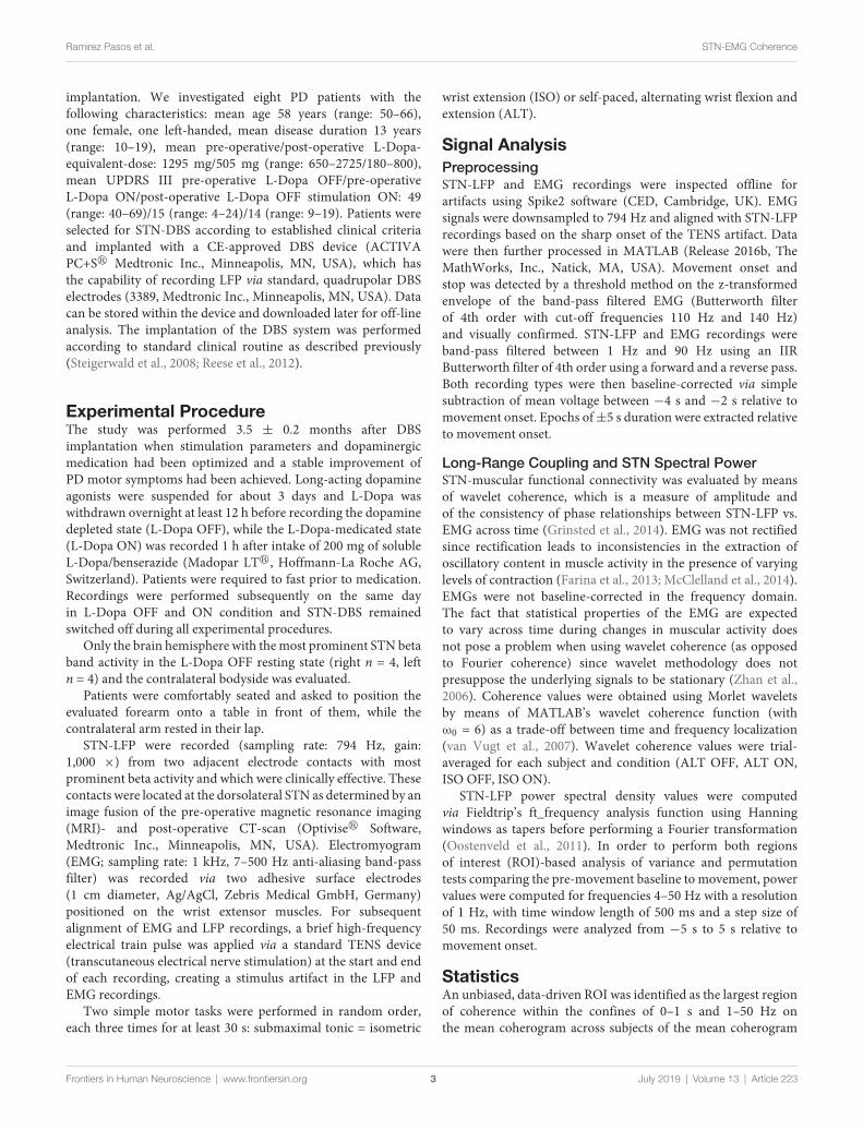
implantation. We investigated eight PD patients with thefollowing characteristics: mean age 58 years (range: 50–66),one female, one left-handed, mean disease duration 13 years(range: 10–19), mean pre-operative/post-operative L-Dopa-equivalent-dose: 1295 mg/505 mg (range: 650–2725/180–800),mean UPDRS III pre-operative L-Dopa OFF/pre-operativeL-Dopa ON/post-operative L-Dopa OFF stimulation ON: 49(range: 40–69)/15 (range: 4–24)/14 (range: 9–19). Patients wereselected for STN-DBS according to established clinical criteriaand implanted with a CE-approved DBS device (ACTIVAPC+Sr Medtronic Inc., Minneapolis, MN, USA), which hasthe capability of recording LFP via standard, quadrupolar DBSelectrodes (3389, Medtronic Inc., Minneapolis, MN, USA). Datacan be stored within the device and downloaded later for off-lineanalysis. The implantation of the DBS system was performedaccording to standard clinical routine as described previously(Steigerwald et al., 2008; Reese et al., 2012).
Experimental ProcedureThe study was performed 3.5 ± 0.2 months after DBSimplantation when stimulation parameters and dopaminergicmedication had been optimized and a stable improvement ofPD motor symptoms had been achieved. Long-acting dopamineagonists were suspended for about 3 days and L-Dopa waswithdrawn overnight at least 12 h before recording the dopaminedepleted state (L-Dopa OFF), while the L-Dopa-medicated state(L-Dopa ON) was recorded 1 h after intake of 200 mg of solubleL-Dopa/benserazide (Madopar LTr, Hoffmann-La Roche AG,Switzerland). Patients were required to fast prior to medication.Recordings were performed subsequently on the same dayin L-Dopa OFF and ON condition and STN-DBS remainedswitched off during all experimental procedures.
Only the brain hemisphere with themost prominent STN betaband activity in the L-Dopa OFF resting state (right n = 4, leftn = 4) and the contralateral bodyside was evaluated.
Patients were comfortably seated and asked to position theevaluated forearm onto a table in front of them, while thecontralateral arm rested in their lap.
STN-LFP were recorded (sampling rate: 794 Hz, gain:1,000 ×) from two adjacent electrode contacts with mostprominent beta activity and which were clinically effective. Thesecontacts were located at the dorsolateral STN as determined by animage fusion of the pre-operative magnetic resonance imaging(MRI)- and post-operative CT-scan (Optiviser Software,Medtronic Inc., Minneapolis, MN, USA). Electromyogram(EMG; sampling rate: 1 kHz, 7–500 Hz anti-aliasing band-passfilter) was recorded via two adhesive surface electrodes(1 cm diameter, Ag/AgCl, Zebris Medical GmbH, Germany)positioned on the wrist extensor muscles. For subsequentalignment of EMG and LFP recordings, a brief high-frequencyelectrical train pulse was applied via a standard TENS device(transcutaneous electrical nerve stimulation) at the start and endof each recording, creating a stimulus artifact in the LFP andEMG recordings.
Two simple motor tasks were performed in random order,each three times for at least 30 s: submaximal tonic = isometric
wrist extension (ISO) or self-paced, alternating wrist flexion andextension (ALT).
Signal AnalysisPreprocessingSTN-LFP and EMG recordings were inspected offline forartifacts using Spike2 software (CED, Cambridge, UK). EMGsignals were downsampled to 794 Hz and aligned with STN-LFPrecordings based on the sharp onset of the TENS artifact. Datawere then further processed in MATLAB (Release 2016b, TheMathWorks, Inc., Natick, MA, USA). Movement onset andstop was detected by a threshold method on the z-transformedenvelope of the band-pass filtered EMG (Butterworth filterof 4th order with cut-off frequencies 110 Hz and 140 Hz)and visually confirmed. STN-LFP and EMG recordings wereband-pass filtered between 1 Hz and 90 Hz using an IIRButterworth filter of 4th order using a forward and a reverse pass.Both recording types were then baseline-corrected via simplesubtraction of mean voltage between −4 s and −2 s relative tomovement onset. Epochs of±5 s duration were extracted relativeto movement onset.
Long-Range Coupling and STN Spectral PowerSTN-muscular functional connectivity was evaluated by meansof wavelet coherence, which is a measure of amplitude andof the consistency of phase relationships between STN-LFP vs.EMG across time (Grinsted et al., 2014). EMG was not rectifiedsince rectification leads to inconsistencies in the extraction ofoscillatory content in muscle activity in the presence of varyinglevels of contraction (Farina et al., 2013; McClelland et al., 2014).EMGs were not baseline-corrected in the frequency domain.The fact that statistical properties of the EMG are expectedto vary across time during changes in muscular activity doesnot pose a problem when using wavelet coherence (as opposedto Fourier coherence) since wavelet methodology does notpresuppose the underlying signals to be stationary (Zhan et al.,2006). Coherence values were obtained using Morlet waveletsby means of MATLAB’s wavelet coherence function (withω0 = 6) as a trade-off between time and frequency localization(van Vugt et al., 2007). Wavelet coherence values were trial-averaged for each subject and condition (ALT OFF, ALT ON,ISO OFF, ISO ON).
STN-LFP power spectral density values were computedvia Fieldtrip’s ft_frequency analysis function using Hanningwindows as tapers before performing a Fourier transformation(Oostenveld et al., 2011). In order to perform both regionsof interest (ROI)-based analysis of variance and permutationtests comparing the pre-movement baseline to movement, powervalues were computed for frequencies 4–50 Hz with a resolutionof 1 Hz, with time window length of 500 ms and a step size of50 ms. Recordings were analyzed from −5 s to 5 s relative tomovement onset.
StatisticsAn unbiased, data-driven ROI was identified as the largest regionof coherence within the confines of 0–1 s and 1–50 Hz onthe mean coherogram across subjects of the mean coherogram
Frontiers in Human Neuroscience | www.frontiersin.org 3 July 2019 | Volume 13 | Article 223

Ramirez Pasos et al. STN-EMG Coherence
across the four conditions. ROIs were selected by drawing aquadrilateral over the prominent regions of coherence visibleon the grand average map after masking away coefficientsunder 0.3. Examining the average across conditions precludesdouble dipping (Kriegeskorte et al., 2009). Mean coherencevalues within the identified ROI were subsequently extractedfor each patient and condition. The effects of dopamine andmovement type on STN-EMG coherence were analyzed using atwo-way analysis of variance (ANOVA) with repeated measuresfor both fixed effects, namely DOPAMINE (with levels OFFand ON) and MOVEMENT (with levels ALT and ISO) thatincluded SUBJECTS as a random effect. In order to examinewhether dopamine and movement type effect on STN-EMGcoherence by parallel, frequency-specific modulation of STNspectral power and EMG activity, the same ROI was employedon trial-averaged power spectral density data and analyzedusing the same factorial design as above. For this, trial-averageswere baseline-corrected by computing the decibel changes frombaseline (−4 to −2 s relative to movement onset). Statisticalsignificance was set at p = 0.05/(number of ROIs) to correct formultiple comparisons.
In addition, movement-induced changes in STN spectralpower were compared to baseline levels via nonparametricpermutation testing (Theiler et al., 1992; Maris and Oostenveld,2007; MATLAB code adapted from Cohen, 2014). The goalwas to create a null hypothesis distribution to compare ourobserved data to. Here, the null hypothesis states that movementinitiation induces no difference in STN spectral power withrespect to baseline, and therefore any temporal reordering ofthe data should possess statistical properties similar to thoseof the observed dataset. Prior to permutation, power mapswere smoothed with a 2-D Gaussian kernel (σ = 2). Thebaseline vs. movement comparison was conducted for eachcondition separately, as well as after averaging across conditionsfor each subject. First, an average map across subjects (the‘‘observed map’’) was computed and was baseline-corrected byobtaining the decibel change from its baseline. Each permutationwas obtained by randomly selecting a timepoint t within theepoch, then dividing each (uncorrected) subject map at t intotwo submaps and placing the earlier submap after the latersubmap, thus essentially randomly shifting the subject mapsalong the time axis. Time-shifted subject maps were thenaveraged and subsequently baseline-corrected by computingthe decibel change from the baseline values of the observedmap. This process was repeated 1,000 times to have a nullhypothesis distribution of power values for each time-frequencybin. To determine statistical significance, a z-map was computedby bin-wise subtracting the mean of the distribution fromthe observed map and dividing by the standard deviation ofthe distribution. The z-map was thresholded such that onlyz-scores corresponding to a p-value of 0.01 or less survived.The p-value threshold was chosen to account for multiplecomparisons (here 4). The ensuing clusters of significantpoints were corrected by eliminating clusters with pixel sizeslower than a cluster size threshold defined by the permuteddata. To derive such threshold, a distribution of maximumcluster sizes was obtained as follows. Each permutation was
standardized by subtracting the mean permutation map anddividing by its standard deviation, yielding z-maps which werethresholded as above, after which the maximum cluster sizewas stored. The cluster size threshold was defined as equalor greater than the 99th percentile of the distribution oflargest clusters.
RESULTS
3.5 months after implantation all patients presented an excellentresponse to STN-DBS reaching at least 80% of the pre-operativeUPDRS-III L-Dopa-ON score. One patient (ID 4) was excludedfrom the analysis since she/he could not complete the wholeexperiment due to severe OFF motor symptoms. Two trialsfrom subject-ID 3 were rejected after visual inspection due toelectrical artifacts. Total number of ISO trials were n = 21 forL-Dopa-OFF and n = 20 for L-Dopa-ON. For ALT trialsn = 20 for L-Dopa-OFF and n = 21 for L-Dopa-ON. Exemplaryraw STN-LFP and EMG epochs and corresponding spectrogramscan be seen in Figure 1.
STN-Muscular CoherenceTwo prominent regions of STN-EMG coherence were identified,namely ‘‘high beta 1’’ in the frequency range 28–44 Hzsoon after movement onset (50–150 ms) and ‘‘high beta 2’’enclosed by the ranges 23–37 Hz and 300–500 ms (Figure 2).Shapiro-Wilk tests on each model’s residuals yielded W = 0.96,p = 0.37 and W = 0.95, p = 0.19, respectively. The results ofthe repeated-measures ANOVAs can be seen in Table 1. Weconducted a two-way repeated-measures ANOVA to comparethe effect of dopamine and movement type on STN-EMGcoherence. Coherence in high beta 1 showed a DOPAMINEtrend (F(1,6) = 5.4, p = 0.059) and no significant MOVEMENT orinteraction effects. In contrast, high beta 2 yielded a significant
FIGURE 1 | Raw recordings and spectrograms. Example raw subthalamicnucleus (STN)-Local Field Potentials (LFPs) and electromyogram (EMG)epochs from subject wue11 during an isometric contraction in medicationstate ON. The corresponding spectrograms display the trial-average powerspectral densities for the same subject and condition for the STN-LFP andthe EMG, respectively. The spectrograms show the decibel change frombaseline (−4 s to −2 s relative to movement onset).
Frontiers in Human Neuroscience | www.frontiersin.org 4 July 2019 | Volume 13 | Article 223
Ramirez Pasos et al. STN-EMG Coherence
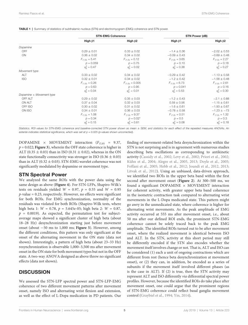
TABLE 1 | Summary of statistics of subthalamic nucleus (STN) and electromyogram (EMG) coherence and STN power.
STN-EMG Coherence STN Power (dB)
High β1 High β2 High β1 High β2
DopamineOFF 0.29 ± 0.01 0.33 ± 0.02 −1.4 ± 0.36 −2.02 ± 0.53ON 0.36 ± 0.02 0.34 ± 0.02 −0.09 ± 0.43 −0.69 ± 0.48
F(1,6) = 5.41 F(1,6) = 0.12 F(1,6) = 3.65 F(1,6) = 2.27p = 0.059 p = 0.75 p = 0.10 p = 0.18η2
p = 0.47 η2p = 0.02 η2
p = 0.38 η2p = 0.27
Movement typeALT 0.33 ± 0.02 0.34 ± 0.02 −0.29 ± 0.42 −1.13 ± 0.58ISO 0.32 ± 0.01 0.34 ± 0.02 −1.2 ± 0.42 −1.58 ± 0.48
F(1,6) = 0.26 F(1,6) = 0.005 F(1,6) = 6.73 F(1,6) = 2.61p = 0.63 p = 0.95 p = 0.041 p = 0.16η2
p = 0.04 η2p < 0.01 η2
p = 0.53 η2p = 0.30
Dopamine × Movement typeOFF ALT 0.29 ± 0.02 0.35 ± 0.03 −1.2 ± 0.43 −2.1 ± 0.88ON ALT 0.37 ± 0.04 0.32 ± 0.03 0.59 ± 0.56 −1.15 ± 0.61OFF ISO 0.30 ± 0.02 0.31 ± 0.02 −1.6 ± 0.61 −1.93 ± 0.67ON ISO 0.34 ± 0.01 0.36 ± 0.03 −0.78 ± 0.58 −1.23 ± −73
F(1,6) = 1.08 F(1,6) = 9.37 F(1,6) = 0.51 F(1,6) = 1.32p = 0.34 p = 0.02∗ p = 0.5 p = 0.3η2
p = 0.15 η2p = 0.61 η2
p = 0.08 η2p = 0.18
Statistics. ROI values for STN-EMG coherence and baseline-corrected STN power shown as mean ± SEM, and statistics for each effect of the repeated measures ANOVAs. Anasterisk indicates statistical significance, which was set at p = 0.025 (p-values shown uncorrected).
DOPAMINE × MOVEMENT interaction (F(1,6) = 9.37,p = 0.022; Figure 3), wherein the OFF state coherence is higher inALT (0.35 ± 0.03) than in ISO (0.31 ± 0.02), whereas in the ONstate functionally connectivity was stronger in ISO (0.36 ± 0.03)than in ALT (0.32± 0.03). STN-EMGwavelet coherence was notsignificantly modulated by dopamine or movement type.
STN Spectral PowerWe analyzed the same ROIs with the power data using thesame design as above (Figure 4). For STN-LFPs, Shapiro-Wilk’stests on residuals yielded W = 0.97, p = 0.55 and W = 0.95p-value = 0.23, respectively. However, no effects were significantfor both ROIs. For EMG synchronization, normality of theresiduals was violated for both ROIs (Shapiro-Wilk tests, wherehigh beta 1: W = 0.78, p = 5.645e-05; high beta 2: W = 0.87,p = 0.0019). As expected, the permutation test for subject-average maps showed a significant cluster of high beta (about18–28 Hz) desynchronization starting just before movementonset (about −50 ms to 1,000 ms; Figure 5). However, amongthe different conditions, this pattern was only significant at theonset of the alternating movement in the ON state (data notshown). Interestingly, a pattern of high beta (about 23–33 Hz)resynchronization is observable 1,000–3,500 ms after movementonset in theON state for bothmovement types but not in theOFFstate. A two-way ANOVA designed as above threw no significanteffects (data not shown).
DISCUSSION
We assessed the STN-LFP spectral power and STN-LFP-EMGcoherence of two different movement patterns after movementonset, namely ISO and alternating wrist flexion and extension,as well as the effect of L-Dopa medication in PD patients. Our
finding of movement-related beta desynchronization within theSTN is not surprising and is in agreement with numerous studiesdescribing beta oscillations as corresponding to antikineticactivity (Cassidy et al., 2002; Levy et al., 2002; Priori et al., 2002;Kühn et al., 2004; Alegre et al., 2005, 2013; Doyle et al., 2005;Foffani et al., 2005; Hebb et al., 2012; Joundi et al., 2012, 2013;Litvak et al., 2012). Using an unbiased, data-driven approach,we identified two ROIs in the upper beta band within the firstsecond after movement onset (Figure 2). At 300–500 ms, wefound a significant DOPAMINE × MOVEMENT interactionfor coherent activity, with greater upper beta band coherencein the isometric contraction task compared to alternating wristmovements in the L-Dopa medicated state. This pattern mightgo awry in the unmedicated state, where coherence is higher foralternating wrist movements. As the peak amplitude of EMGactivity occurred at 555 ms after movement onset, i.e., about50 ms after our defined ROI ends, the prominent STN-EMGcoherence cannot be solely traced back to the peak EMGamplitude. The identified ROIs turned out to be after movementonset, where the realized movement is identical between ISOand ALT. In the STN, activity at this short period may stillbe differently encoded if the STN also encodes whether themovement itself involves change or not. That is, ALT and ISO canbe considered (1) each a unit of ongoing contractions which aredifferent from rest (hence beta desynchronization at movementonset), or (2) they can, in addition, be encoded as a series ofsubunits if the movement itself involved different phases (asis the case in ALT). If (2) is true, then the STN activity mayrepresent ALT and ISO differently via differential spectral powerprofiles. However, because the identified ROIs do take place aftermovement onset, one could argue that the prominent regionsof STN-EMG coherence could reflect basal ganglia movementcontrol (Graybiel et al., 1994; Yin, 2014).
Frontiers in Human Neuroscience | www.frontiersin.org 5 July 2019 | Volume 13 | Article 223

Ramirez Pasos et al. STN-EMG Coherence
FIGURE 2 | Regions of interest (ROIs) extraction. Two ROIs were extractedfrom the grand average map of STN-EMG coherence. The map displays theaverage coherence across all conditions within the ranges 0–1 s and4–50 Hz. Two sizeable ROIs were identified: “high beta 1” within 28–44 Hzand 50–150 ms and “high beta 2” enclosed by the ranges 23–37 Hz and300–500 ms.
FIGURE 3 | Coherence statistics. (A,B) Stronger high beta coherence soonafter movement onset (50–150 ms) was observed in the medication ON state,though this was only marginally significant (F(1,6) = 5.41, uncorrectedp = 0.059, η2
p = 0.47). (C,D) A significant dopamine × movement type effect(F(1,6) = 9.37, uncorrected p = 0.02, η2
p = 0.61) on high beta coherence wasobserved later between 300 and 500 ms after movement onset.
Methodological Considerations andPossible ConfoundersThe recordings were obtained several weeks after implantationof the electrodes and therefore rapid impedance changes are
FIGURE 4 | Spectrograms across conditions. Average spectrograms acrosssubjects depicting first second after movement onset for each condition withROIs. Hard line and dashed line correspond to high beta 1 and 2,correspondingly.
unlikely and should not have affected data quality (Rosa et al.,2010). The micro-lesioning effect due to insertion of theelectrodes into the STN, which often attenuates Parkinsonianmotor symptoms and naturally interferes with stimulation andmedication effects, had abated at the time of the experimentas evidenced by patients reaching their pre-operative symptomseverity after withdrawal of medication and DBS. We recordedLFPs in a bipolar montage from adjacent contacts with mostprominent beta activity during a resting state in L-Dopa-OFF, which may indicate the motor part of the STN (Yoshidaet al., 2010; Accolla et al., 2016; Horn et al., 2017). Thesecontacts were located at the dorsolateral aspect of the STNaccording to an image fusion of the pre-operative MRI andthe post-operative CT-scan (Optiviser software, Medtronic Inc.,Minneapolis, MN, USA) and were of good clinical efficacyof STN-DBS. Nevertheless, this is a pragmatic and mostlyelectrophysiologically-driven delineation of the STN motorregion, which may not correspond to the ‘‘true’’ physiologicalmotor area in individual patients. Likewise, the low number ofpatients and trials may have led to reduced sensitivity, however,we draw this sample given by the restricted availability of thePC + S stimulation systems.
Subthalamic Nucleus-Muscle Coherence(STN-Muscle Coupling)Within the two identified ROIs a trend of the dopamine effectwas observed for the first ROI (50–150ms) with higher coherencein medicated patients (Figures 2, 3). Concerning the secondROI (300–500 ms), an interaction effect of L-Dopa medicationand movement task was significant with higher coherencein the isometric contraction task compared to alternatingmovements, an effect which was reversed in the dopaminergic
Frontiers in Human Neuroscience | www.frontiersin.org 6 July 2019 | Volume 13 | Article 223

Ramirez Pasos et al. STN-EMG Coherence
FIGURE 5 | Movement-induced high beta desynchronization.(A) Baseline-corrected grand average map, where each subject map was theaverage across all four conditions. (B) After cluster correction, a final clustersurvives showing event-related desynchronization and located at about−50 to 1,000 ms and 18–27 Hz.
OFF state. Owing to the fact that EMG recordings reflect lowermotor neuron synchronization only after movement start, wecannot compare our data to the pre-movement period. In astudy employing a comparable movement task, the coherencemagnitude between the motor cortex and the STN in theupper beta band was markedly attenuated during movement(Talakoub et al., 2016).
Movement-Related STN Spectral PowerThe previously mentioned increase in beta desynchronizationin the L-Dopa ON state compared to the OFF state (Doyleet al., 2005; Devos et al., 2006; Androulidakis et al., 2007) didnot reach a level of significance in our study, which couldbe attributed to a low signal-to-noise ratio and sample size.Likewise, no significant influence of the movement task couldbe detected. Joundi et al. (2013) could show that a fingertapping induced suppression of STN spectral power may beindependent of the velocity of these repetitive movementsand an inter-movement rebound of beta synchronization mayonly be suppressed at higher tapping rates. The degree ofdesynchronization has moreover been shown to depend onthe effort of the respective motor task, which may reach afloor effect at higher efforts. The latter strongly correlated with
spectral power in the gamma band (55–90 Hz; Tan et al., 2013,2015). Attenuated cortical post-movement beta synchronizationhas been shown in bradykinetic-rigid PD compared to healthycontrols (Pfurtscheller et al., 1998; Tamás et al., 2003). In ourdata, high beta (about 23–33 Hz) resynchronization in the STNabout 1,000–3,500 ms after movement onset was stronger in theON than in the OFF medication state, although not significantlyso (Figure 4).
Physiological ConsiderationsSynchronized oscillatory activity between remote neuronalpopulations can facilitate information processing over largedistances (Varela et al., 2001; Schnitzler and Gross, 2005).There is good evidence that motor system communication overlarge distances is physiologically realized via the segregationof synchronization in distinct frequency bands. In this view,motor cortical synchronization in the beta band was initiallyregarded as representing ‘‘idleness’’ (Pfurtscheller et al., 1996)but it may instead be conductive to motor constancy (Brittainand Brown, 2014). For instance, studies have shown thatmovement onset is accompanied by desynchronization ofmotor cortical beta activity (event-related desynchronization,ERD) with rebound synchronization around movement stop(Pfurtscheller and Lopes da Silva, 1999). In PD patients,the motor cortical pre-movement ERD is delayed and itshemispheric specificity is reduced compared to healthy controls(Defebvre et al., 1993, 1994, 1996; Magnani et al., 1998).Exaggerated beta synchrony is therefore strongly associated withthe OFF (dopamine depleted)motor state (Jenkinson and Brown,2011). Conversely, post-movement beta synchronization (PMBS)is attenuated in PD (Pfurtscheller et al., 1998; Tamás et al.,2003), which may be a marker of certainty in feedforwardcontrol ofmovements. In healthy subjects, PMBS has been shownto be negatively correlated with uncertainty in feedforwardestimations (Tan et al., 2016).
We used PD as a model disease to further investigate therole of dopamine in the context of motor activity and wereproduced a movement-dependent ERD of STN beta activity(Cassidy et al., 2002; Levy et al., 2002; Priori et al., 2002;Kühn et al., 2004; Alegre et al., 2005, 2013; Doyle et al., 2005;Foffani et al., 2005; Hebb et al., 2012; Joundi et al., 2012,2013; Litvak et al., 2012), whose magnitude, however, wasnot significantly affected by dopamine substitution. Long-rangeSTN-muscle coherence in the upper beta band increased shortlyafter movement onset and a trend of a dopamine effectcould be observed with stronger functional connectivity inthe ON state. Thus, beta spectral power per se might notsimply be a universal marker of bradykinesia. Instead, ourdata suggest STN beta spectral power to be present at motorrest and reduced with changes in motor activity (ERD) whilea subset of neurons, however, functionally connects to thespinal motor neuron pool (measured by EMG activity). Onlyafter substitution of L-Dopa was STN-muscle coherence in theupper beta band stronger in the isometric contraction taskcompared to alternating movements. Assuming that this stateis closer to normal motor function than in PD patients offL-Dopa medication, beta coherence could physiologically limit
Frontiers in Human Neuroscience | www.frontiersin.org 7 July 2019 | Volume 13 | Article 223

Ramirez Pasos et al. STN-EMG Coherence
the amount of information conveyed between remote structures,which may be necessary to maintain simple movements likethe isometric contraction. In this view, a physiologically inverserelationship between neuronal synchrony and information flowhas been hypothesized (Hanslmayr et al., 2012; Oswal et al.,2013). Our data also suggest that higher striatal dopamine levelsmay be essential for an appropriate setting of neuronal synchronyin the STN and information flow between remote structures ofthe motor circuit. Salenius et al. (2002) reported a similar effectof L-Dopa with restorage of cortico-muscular beta coherencein PD patients during the isometric contraction of contralateralforearm muscles.
Muscle activity synchronized in higher frequencies ofabout 40 Hz (called Piper-rhythm, see Piper, 1912) can berecorded from muscles of normal subjects during muscleactivity near maximum contraction force (McAuley et al.,1997). In PD patients, impaired muscle strength in themedication OFF state (Yanagawa et al., 1990; Corcos et al.,1996) is correlated with a disruption of this higher frequencysynchronization patterns, both of which return to normal levelsin patients after L-Dopa substitution (McAuley et al., 2001).Accordingly, the basal ganglia-lower motor neuron couplingdetected in our study may be a physiological phenomenonnecessary for normal motor execution. Consequently,impaired coupling on various relay stations of the motorcircuit in PD may contribute to Parkinsonian OFF motorsymptoms, such as bradykinesia, hypokinesia and reducedmuscle strength.
A simple monosynaptic connection between basal ganglianuclei and spinal motor neurons is unknown (Lanciego et al.,2012), thus anatomical pathways explaining our findings haveto be discussed. Motor cortical beta oscillatory activity has beenshown to drive beta oscillations in the STN (Williams et al., 2002;Fogelson et al., 2006; Litvak et al., 2011), most likely via theso-called ‘‘hyperdirect pathway’’ (HDP; Monakow et al., 1978;Nambu et al., 1996, 1997). Recently, the HDP has also beenuncovered and further characterized in humans (Brunenberget al., 2012; Miocinovic et al., 2018). This pathway may representthe anatomical correlate of clinical efficacy of STN-DBS whichmay act via a retrograde propagation of DBS signals to the motorcortex (Gradinaru et al., 2009; Li et al., 2012; de Hemptinneet al., 2015; Sanders and Jaeger, 2016). Accordingly, a certainanatomical proximity of the DBS electrode may be necessary forgood clinical efficacy of STN-DBS in PD (Akram et al., 2017;Horn et al., 2017; Chen et al., 2018). It remains to be further
investigated whether the activity in the upper beta band werecorded from STN and which was coherent to the EMG maymirror motor cortical activity. This hypothesis is corroboratedby resting state data showing STN-motor cortical coherencein the upper beta band, whereas STN spectral power wasprominent in the lower beta band in patients withdrawn frommedication (Litvak et al., 2011).
Our study adds dynamic properties to the complex interplaybetween STN beta spectral power and its coherence with remotestructures of the motor system as a function of movementand dopaminergic state. This may help to identify markers ofneuronal activity relevant for more individualized paradigms ofDBS therapy.
ETHICS STATEMENT
The study protocol was approved by the ethical review board ofthe University Hospital Würzburg and patients provided writteninformed consent to participate prior to implantation.
AUTHOR CONTRIBUTIONS
URP: data analysis concept, data analysis and interpretationand creation of figures, writing and revision of the manuscript.FS: conduct of experiments, data interpretation, revision ofthe manuscript. MR: conduct of experiments, revision of themanuscript. CM: implantation of the DBS systems, revision ofthe manuscript. JV: study concept, data analysis concept, datainterpretation, writing and revision of the manuscript. RR: studyconcept, data analysis concept, conduct of experiments, datainterpretation, writing and revision of the manuscript.
FUNDING
This publication was funded by the German ResearchFoundation (DFG) and the University of Würzburg in thefunding programme Open Access Publishing.
ACKNOWLEDGMENTS
ACTIVA PC+Sr devices and related hard- and software wereprovided by Medtronic under a research agreement. Medtronichad no influence on patient recruitment, design of the study, dataanalysis or interpretation of the results.
REFERENCES
Accolla, E. A., Herrojo Ruiz, M., Horn, A., Schneider, G.-H., Schmitz-Hübsch, T.,Draganski, B., et al. (2016). Brain networks modulated by subthalamicnucleus deep brain stimulation. Brain 139, 2503–2515. doi: 10.1093/brain/aww182
Akram, H., Sotiropoulos, S. N., Jbabdi, S., Georgiev, D., Mahlknecht, P.,Hyam, J., et al. (2017). Subthalamic deep brain stimulation sweet spots andhyperdirect cortical connectivity in Parkinson’s disease. Neuroimage 158,332–345. doi: 10.1016/j.neuroimage.2017.07.012
Alegre, M., Alonso-Frech, F., Rodríguez-Oroz, M. C., Guridi, J., Zamarbide, I.,Valencia, M., et al. (2005). Movement-related changes in oscillatory activity in
the human subthalamic nucleus: ipsilateral vs. contralateral movements. Eur.J. Neurosci. 22, 2315–2324. doi: 10.1111/j.1460-9568.2005.04409.x
Alegre, M., Lopez-Azcarate, J., Obeso, I., Wilkinson, L., Rodriguez-Oroz, M. C.,Valencia, M., et al. (2013). The subthalamic nucleus is involved in successfulinhibition in the stop-signal task: a local field potential study in Parkinson’sdisease. Exp. Neurol. 239, 1–12. doi: 10.1016/j.expneurol.2012.08.027
Androulidakis, A. G., Brücke, C., Kempf, F., Kupsch, A., Aziz, T., Ashkan, K., et al.(2008). Amplitudemodulation of oscillatory activity in the subthalamic nucleusduring movement. Eur. J. Neurosci. 27, 1277–1284. doi: 10.1111/j.1460-9568.2008.06085.x
Androulidakis, A. G., Kühn, A. A., Chen, C. C., Blomstedt, P., Kempf, F.,Kupsch, A., et al. (2007). Dopaminergic therapy promotes lateralized motor
Frontiers in Human Neuroscience | www.frontiersin.org 8 July 2019 | Volume 13 | Article 223

Ramirez Pasos et al. STN-EMG Coherence
activity in the subthalamic area in Parkinson’s disease. Brain 130, 457–468.doi: 10.1093/brain/awl358
Benabid, A. L., Pollak, P., Gross, C., Hoffmann, D., Benazzouz, A., Gao, D. M.,et al. (1994). Acute and long-term effects of subthalamic nucleusstimulation in Parkinson’s disease. Stereotact. Funct. Neurosurg. 62, 76–84.doi: 10.1159/000098600
Brittain, J. S., and Brown, P. (2014). Oscillations and the basal ganglia: motorcontrol and beyond. Neuroimage 85, 637–647. doi: 10.1016/j.neuroimage.2013.05.084
Brown, P. (2003). Oscillatory nature of human basal ganglia activity: relationshipto the pathophysiology of Parkinson’s disease. Mov. Disord. 18, 357–363.doi: 10.1002/mds.10358
Brown, P., Oliviero, A., Mazzone, P., Insola, A., Tonali, P., and DiLazzaro, V. (2001). Dopamine dependency of oscillations between subthalamicnucleus and pallidum in Parkinson’s disease. J. Neurosci. 21, 1033–1038.doi: 10.1523/jneurosci.21-03-01033.2001
Brunenberg, E. J., Moeskops, P., Backes, W. H., Pollo, C., Cammoun, L.,Vilanova, A., et al. (2012). Structural and resting state functional connectivityof the subthalamic nucleus: identification of motor STN parts and thehyperdirect pathway. PLoS One 7:e39061. doi: 10.1371/journal.pone.0039061
Cassidy, M., Mazzone, P., Oliviero, A., Insola, A., Tonali, P., Di Lazzaro, V.,et al. (2002). Movement-related changes in synchronization in the human basalganglia. Brain 125, 1235–1246. doi: 10.1093/brain/awf135
Chen, Y., Ge, S., Li, Y., Li, N., Wang, J., Wang, X., et al. (2018). The role ofthe cortico-subthalamic hyperdirect pathway in deep brain stimulation forthe treatment of Parkinson’s disease: a diffusion tensor imaging study. WorldNeurosurg. 114, e1079–e1085. doi: 10.1016/j.wneu.2018.03.149
Cohen, M. X. (2014). Analyzing Neural Time Series Data: Theory and Practice.Cambridge, MA, USA: MIT Press.
Corcos, D. M., Chen, C. M., Quinn, N. P., McAuley, J., and Rothwell, J. C. (1996).Strength in Parkinson’s disease: relationship to rate of force generation andclinical status. Ann. Neurol. 39, 79–88. doi: 10.1002/ana.410390112
Defebvre, L., Bourriez, J. L., Destee, A., and Guieu, J. D. (1996). Movementrelated desynchronisation pattern preceding voluntary movement inuntreated Parkinson’s disease. J. Neurol. Neurosurg. Psychiatry 60, 307–312.doi: 10.1136/jnnp.60.3.307
Defebvre, L., Bourriez, J. L., Dujardin, K., Derambure, P., Destée, A., andGuieu, J. D. (1994). Spatiotemporal study of Bereitschaftspotential and event-related desynchronization during voluntary movement in Parkinson’s disease.Brain Topogr. 6, 237–244. doi: 10.1007/bf01187715
Defebvre, L., Derambure, P., Bourriez, J. L., Jacquesson, J. M., Dujardin, K.,Destée, A., et al. (1993). Spatiotemporal study of event-relateddesynchronization in idiopathic Parkinson’s disease. Adv. Neurol. 60,422–428.
de Hemptinne, C., Swann, N. C., Ostrem, J. L., Ryapolova-Webb, E. S., SanLuciano, M., Galifianakis, N. B., et al. (2015). Therapeutic deep brainstimulation reduces cortical phase-amplitude coupling in Parkinson’s disease.Nat. Neurosci. 18, 779–786. doi: 10.1038/nn.3997
Deuschl, G., Schade-Brittinger, C., Krack, P., Volkmann, J., Schafer, H.,Botzel, K., et al. (2006). A randomized trial of deep-brain stimulation forParkinson’s disease. N. Engl. J. Med. 355, 896–908. doi: 10.1056/NEJMoa060281
Devos, D., Szurhaj, W., Reyns, N., Labyt, E., Houdayer, E., Bourriez, J. L., et al.(2006). Predominance of the contralateral movement-related activity in thesubthalamo-cortical loop. Clin. Neurophysiol. 117, 2315–2327. doi: 10.1016/j.clinph.2006.06.719
Doyle, L. M., Kühn, A. A., Hariz, M., Kupsch, A., Schneider, G. H., and Brown, P.(2005). Levodopa-induced modulation of subthalamic beta oscillations duringself-paced movements in patients with Parkinson’s disease. Eur. J. Neurosci. 21,1403–1412. doi: 10.1111/j.1460-9568.2005.03969.x
Engel, A. K., and Fries, P. (2010). Beta-band oscillations–signalling the status quo?Curr. Opin. Neurobiol. 20, 156–165. doi: 10.1016/j.conb.2010.02.015
Farina, D., Negro, F., and Jiang, N. (2013). Identification of common synapticinputs to motor neurons from the rectified electromyogram. J. Physiol. 591,2403–2418. doi: 10.1113/jphysiol.2012.246082
Foffani, G., Bianchi, A. M., Baselli, G., and Priori, A. (2005). Movement-related frequency modulation of beta oscillatory activity in the human
subthalamic nucleus. J. Physiol. 568, 699–711. doi: 10.1113/jphysiol.2005.089722
Fogelson, N., Williams, D., Tijssen, M., van Bruggen, G., Speelman, H.,and Brown, P. (2006). Different functional loops between cerebral cortexand the subthalmic area in Parkinson’s disease. Cereb. Cortex 16, 64–75.doi: 10.1093/cercor/bhi084
Follett, K. A., Weaver, F. M., Stern, M., Hur, K., Harris, C. L., Luo, P.,et al. (2010). Pallidal versus subthalamic deep-brain stimulation forParkinson’s disease. N. Engl. J. Med. 362, 2077–2091. doi: 10.1056/NEJMoa0907083
Gradinaru, V., Mogri, M., Thompson, K. R., Henderson, J. M., and Deisseroth, K.(2009). Optical deconstruction of Parkinsonian neural circuitry. Science 324,354–359. doi: 10.1126/science.1167093
Graybiel, A. M., Aosaki, T., Flaherty, A. W., and Kimura, M. (1994).The basal ganglia and adaptive motor control. Science 265, 1826–1831.doi: 10.1126/science.8091209
Grinsted, A., Moore, J. C., and Jevrejeva, S. (2014). Application of the cross wavelettransform and wavelet coherence to geophysicial time series.Nonlinear Process.Geophys. 11, 561–566. doi: 10.5194/npg-11-561-2004
Hanslmayr, S., Staudigl, T., and Fellner, M. C. (2012). Oscillatory powerdecreases and long-term memory: the information via desynchronizationhypothesis. Front. Hum. Neurosci. 6:74. doi: 10.3389/fnhum.2012.00074
Hebb, A. O., Darvas, F., andMiller, K. J. (2012). Transient and state modulation ofbeta power in human subthalamic nucleus during speech production and fingermovement. Neuroscience 202, 218–233. doi: 10.1016/j.neuroscience.2011.11.072
Hirschmann, J., Özkurt, T. E., Butz,M., Homburger,M., Elben, S., Hartmann, C. J.,et al. (2013). Differential modulation of STN-cortical and cortico-muscularcoherence by movement and levodopa in Parkinson’s disease. Neuroimage 68,203–213. doi: 10.1016/j.neuroimage.2012.11.036
Horn, A., Neumann, W. J., Degen, K., Schneider, G. H., and Kühn, A. A. (2017).Toward an electrophysiological "sweet spot" for deep brain stimulation in thesubthalamic nucleus. Hum. Brain Mapp. 38, 3377–3390. doi: 10.1002/hbm.23594
Jenkinson, N., and Brown, P. (2011). New insights into the relationship betweendopamine, beta oscillations and motor function. Trends Neurosci. 34, 611–618.doi: 10.1016/j.tins.2011.09.003
Joundi, R. A., Brittain, J. S., Green, A. L., Aziz, T. Z., Brown, P., and Jenkinson, N.(2012). Oscillatory activity in the subthalamic nucleus during arm reaching inParkinson’s disease. Exp. Neurol. 236, 319–326. doi: 10.1016/j.expneurol.2012.05.013
Joundi, R. A., Brittain, J. S., Green, A. L., Aziz, T. Z., Brown, P., andJenkinson, N. (2013). Persistent suppression of subthalamic beta-band activityduring rhythmic finger tapping in Parkinson’s disease. Clin. Neurophysiol. 124,565–573. doi: 10.1016/j.clinph.2012.07.029
Kriegeskorte, N., Simmons, W. K., Bellgowan, P. S. F., and Baker, C. I. (2009).Circular analysis in systems neuroscience: the dangers of double dipping. Nat.Neurosci. 12, 535–540. doi: 10.1038/nn.2303
Kühn, A. A., Kupsch, A., Schneider, G. H., and Brown, P. (2006). Reduction insubthalamic 8–35 Hz oscillatory activity correlates with clinical improvementin Parkinson’s disease. Eur. J. Neurosci. 23, 1956–1960. doi: 10.1111/j.1460-9568.2006.04717.x
Kühn, A. A., Tsui, A., Aziz, T., Ray, N., Brucke, C., Kupsch, A., et al. (2009).Pathological synchronisation in the subthalamic nucleus of patients withParkinson’s disease relates to both bradykinesia and rigidity. Exp. Neurol. 215,380–387. doi: 10.1016/j.expneurol.2008.11.008
Kühn, A. A., Williams, D., Kupsch, A., Limousin, P., Hariz, M., Schneider, G. H.,et al. (2004). Event-related beta desynchronization in human subthalamicnucleus correlates with motor performance. Brain 127, 735–746.doi: 10.1093/brain/awh106
Lanciego, J. L., Luquin, N., and Obeso, J. A. (2012). Functional neuroanatomyof the basal ganglia. Cold Spring Harb. Perspect. Med. 2:a009621.doi: 10.1201/9781420048209.supl25
Levy, R., Ashby, P., Hutchison, W. D., Lang, A. E., Lozano, A. M., andDostrovsky, J. O. (2002). Dependence of subthalamic nucleus oscillationson movement and dopamine in Parkinson’s disease. Brain 125, 1196–1209.doi: 10.1093/brain/awf128
Frontiers in Human Neuroscience | www.frontiersin.org 9 July 2019 | Volume 13 | Article 223

Ramirez Pasos et al. STN-EMG Coherence
Li, Q., Ke, Y., Chan, D. C., Qian, Z. M., Yung, K. K., Ko, H., et al. (2012).Therapeutic deep brain stimulation in Parkinsonian rats directly influencesmotor cortex. Neuron 76, 1030–1041. doi: 10.1016/j.neuron.2012.09.032
Litvak, V., Eusebio, A., Jha, A., Oostenveld, R., Barnes, G., Foltynie, T.,et al. (2012). Movement-related changes in local and long-rangesynchronization in Parkinson’s disease revealed by simultaneousmagnetoencephalography and intracranial recordings. J. Neurosci. 32,10541–10553. doi: 10.1523/JNEUROSCI.0767-12.2012
Litvak, V., Jha, A., Eusebio, A., Oostenveld, R., Foltynie, T., Limousin, P., et al.(2011). Resting oscillatory cortico-subthalamic connectivity in patients withParkinson’s disease. Brain 134, 359–374. doi: 10.1093/brain/awq332
López-Azcárate, J., Tainta, M., Rodríguez-Oroz, M. C., Valencia, M., González, R.,Guridi, J., et al. (2010). Coupling between beta and high-frequency activityin the human subthalamic nucleus may be a pathophysiological mechanismin Parkinson’s disease. J. Neurosci. 30, 6667–6677. doi: 10.1523/JNEUROSCI.5459-09.2010
Magnani, G., Cursi, M., Leocani, L., Volonte, M. A., Locatelli, T., Elia, A.,et al. (1998). Event-related desynchronization to contingent negative variationand self-paced movement paradigms in Parkinson’s disease. Mov. Disord. 13,653–660. doi: 10.1002/mds.870130408
Maris, E., andOostenveld, R. (2007). Nonparametric statistical testing of EEG- andMEG-data. J. Neurosci. Methods 164, 177–190. doi: 10.1016/j.jneumeth.2007.03.024
Marsden, J. F., Limousin-Dowsey, P., Ashby, P., Pollak, P., and Brown, P. (2001).Subthalamic nucleus, sensorimotor cortex and muscle interrelationships inParkinson’s disease. Brain 124, 378–388. doi: 10.1093/brain/124.2.378
McAuley, J. H., Corcos, D. M., Rothwell, J. C., Quinn, N. P., and Marsden, C. D.(2001). Levodopa reversible loss of the Piper frequency oscillation componentin Parkinson’s disease. J. Neurol. Neurosurg. Psychiatry 70, 471–476.doi: 10.1136/jnnp.70.4.471
McAuley, J. H., Rothwell, J. C., and Marsden, C. D. (1997). Frequency peaksof tremor, muscle vibration and electromyographic activity at 10 Hz, 20 Hzand 40 Hz during human finger muscle contraction may reflect rhythmicitiesof central neural firing. Exp. Brain Res. 114, 525–541. doi: 10.1007/pl00005662
McClelland, V. M., Cvetkovic, Z., and Mills, K. R. (2014). Inconsistent effectsof EMG rectification on coherence analysis. J. Physiol. 592, 249–250.doi: 10.1113/jphysiol.2013.265181
Miocinovic, S., de Hemptinne, C., Chen, W., Isbaine, F., Willie, J. T.,Ostrem, J. L., et al. (2018). Cortical potentials evoked by subthalamicstimulation demonstrate a short latency hyperdirect pathway in humans.J. Neurosci. 38, 9129–9141. doi: 10.1523/JNEUROSCI.1327-18.2018
Monakow, K. H., Akert, K., and Künzle, H. (1978). Projections of the precentralmotor cortex and other cortical areas of the frontal lobe to the subthalamicnucleus in the monkey. Exp. Brain Res. 33, 395–403. doi: 10.1007/BF00235561
Nambu, A., Takada, M., Inase, M., and Tokuno, H. (1996). Dual somatotopicalrepresentations in the primate subthalamic nucleus: evidence for ordered butreversed body-map transformations from the primary motor cortex and thesupplementary motor area. J. Neurosci. 16, 2671–2683. doi: 10.1523/jneurosci.16-08-02671.1996
Nambu, A., Tokuno, H., Inase, M., and Takada, M. (1997). Corticosubthalamicinput zones from forelimb representations of the dorsal and ventral divisions ofthe premotor cortex in the macaque monkey: comparison with the input zonesfrom the primary motor cortex and the supplementary motor area. Neurosci.Lett. 239, 13–16. doi: 10.1016/s0304-3940(97)00877-x
Oostenveld, R., Fries, P., Maris, E., and Schoffelen, J. M. (2011). FieldTrip:open source software for advanced analysis of MEG, EEG and invasiveelectrophysiological data. Comput. Intell. Neurosci. 2011:156869.doi: 10.1155/2011/156869
Oswal, A., Brown, P., and Litvak, V. (2013). Synchronized neural oscillations andthe pathophysiology of Parkinson’s disease. Curr. Opin. Neurol. 26, 662–670.doi: 10.1097/WCO.0000000000000034
Pfurtscheller, G., and Lopes da Silva, F. H. (1999). Event-related EEG/MEGsynchronization and desynchronization: basic principles. Clin. Neurophysiol.110, 1842–1857. doi: 10.1016/s1388-2457(99)00141-8
Pfurtscheller, G., Pichler-Zalaudek, K., Ortmayr, B., Diez, J., and Reisecker, F.(1998). Postmovement beta synchronization in patients with Parkinson’s
disease. J. Clin. Neurophysiol. 15, 243–250. doi: 10.1097/00004691-199805000-00008
Pfurtscheller, G., Stancák, A. Jr., and Neuper, C. (1996). Post-movement betasynchronization. A correlate of an idling motor area? Electroencephalogr. Clin.Neurophysiol. 98, 281–293. doi: 10.1016/0013-4694(95)00258-8
Piper, H. (1912). Elektrophysiologie Menschlicher Muskeln. Berlin, Heidelberg:Springer-Verlag.
Priori, A., Foffani, G., Pesenti, A., Bianchi, A., Chiesa, V., Baselli, G., et al.(2002). Movement-related modulation of neural activity in human basalganglia and its L-DOPA dependency: recordings from deep brain stimulationelectrodes in patients with Parkinson’s disease. Neurol. Sci. 23, S101–S102.doi: 10.1007/s100720200089
Priori, A., Foffani, G., Pesenti, A., Tamma, F., Bianchi, A. M., Pellegrini, M., et al.(2004). Rhythm-specific pharmacological modulation of subthalamic activityin Parkinson’s disease. Exp. Neurol. 189, 369–379. doi: 10.1016/j.expneurol.2004.06.001
Ray, N. J., Jenkinson, N., Wang, S., Holland, P., Brittain, J. S., Joint, C., et al.(2008). Local field potential beta activity in the subthalamic nucleus of patientswith Parkinson’s disease is associated with improvements in bradykinesiaafter dopamine and deep brain stimulation. Exp. Neurol. 213, 108–113.doi: 10.1016/j.expneurol.2008.05.008
Reese, R., Pinsker, M. O., Herzog, J., Wodarg, F., Steigerwald, F., Pötter-Nerger, M., et al. (2012). The atypical subthalamic nucleus--an anatomicalvariant relevant for stereotactic targeting. Mov. Disord. 27, 544–548.doi: 10.1002/mds.24902
Rosa, M., Marceglia, S., Servello, D., Foffani, G., Rossi, L., Sassi, M., et al. (2010).Time dependent subthalamic local field potential changes after DBS surgery inParkinson’s disease. Exp. Neurol. 222, 184–190. doi: 10.1016/j.expneurol.2009.12.013
Salenius, S., Avikainen, S., Kaakkola, S., Hari, R., and Brown, P. (2002). Defectivecortical drive to muscle in Parkinson’s disease and its improvement withlevodopa. Brain 125, 491–500. doi: 10.1093/brain/awf042
Sanders, T. H., and Jaeger, D. (2016). Optogenetic stimulation of cortico-subthalamic projections is sufficient to ameliorate bradykinesia in 6-ohdalesioned mice. Neurobiol. Dis. 95, 225–237. doi: 10.1016/j.nbd.2016.07.021
Schnitzler, A., and Gross, J. (2005). Normal and pathological oscillatorycommunication in the brain. Nat. Rev. Neurosci. 6, 285–296. doi: 10.1038/nrn1650
Schuepbach, W. M., Rau, J., Knudsen, K., Volkmann, J., Krack, P.,Timmermann, L., et al. (2013). Neurostimulation for Parkinson’sdisease with early motor complications. N. Engl. J. Med. 368, 610–622.doi: 10.1056/NEJMoa1205158
Steigerwald, F., Pötter, M., Herzog, J., Pinsker, M., Kopper, F., Mehdorn, H.,et al. (2008). Neuronal activity of the human subthalamic nucleus in theparkinsonian and nonparkinsonian state. J. Neurophysiol. 100, 2515–2524.doi: 10.1152/jn.90574.2008
Talakoub, O., Neagu, B., Udupa, K., Tsang, E., Chen, R., Popovic, M. R., et al.(2016). Time-course of coherence in the human basal ganglia during voluntarymovements. Sci. Rep. 6:34930. doi: 10.1038/srep34930
Tamás, G., Szirmai, I., Pálvölgyi, L., Takáts, A., and Kamondi, A. (2003).Impairment of post-movement beta synchronisation in parkinson’s diseaseis related to laterality of tremor. Clin. Neurophysiol. 114, 614–623.doi: 10.1016/s1388-2457(02)00424-8
Tan, H., Pogosyan, A., Anzak, A., Ashkan, K., Bogdanovic, M., Green, A. L.,et al. (2013). Complementary roles of different oscillatory activities in thesubthalamic nucleus in coding motor effort in Parkinsonism. Exp. Neurol. 248,187–195. doi: 10.1016/j.expneurol.2013.06.010
Tan, H., Pogosyan, A., Ashkan, K., Cheeran, B., FitzGerald, J. J., Green, A. L.,et al. (2015). Subthalamic nucleus local field potential activity helps encodemotor effort rather than force in Parkinsonism. J. Neurosci. 35, 5941–5949.doi: 10.1523/JNEUROSCI.4609-14.2015
Tan, H., Wade, C., and Brown, P. (2016). Post-movement beta activity insensorimotor cortex indexes confidence in the estimations from internalmodels. J. Neurosci. 36, 1516–1528. doi: 10.1523/JNEUROSCI.3204-15.2016
Theiler, J., Eubank, S., Longtin, A., Galdrikian, B., and Doyne Farmer, J. (1992).Testing for nonlinearity in time series: the method of surrogate data. Physica D58, 77–94. doi: 10.1016/0167-2789(92)90102-S
Frontiers in Human Neuroscience | www.frontiersin.org 10 July 2019 | Volume 13 | Article 223

Ramirez Pasos et al. STN-EMG Coherence
vanVugt,M. K., Sederberg, P. B., and Kahana,M. J. (2007). Comparison of spectralanalysis methods for characterizing brain oscillations. J. Neurosci. Methods 162,49–63. doi: 10.1016/j.jneumeth.2006.12.004
Varela, F., Lachaux, J. P., Rodriguez, E., and Martinerie, J. (2001). The brainweb:phase synchronization and large-scale integration. Nat. Rev. Neurosci. 2,229–239. doi: 10.1038/35067550
Weaver, F. M., Follett, K., Stern, M., Hur, K., Harris, C., Marks, W. J. Jr., et al.(2009). Bilateral deep brain stimulation vs. best medical therapy for patientswith advanced Parkinson’s disease: a randomized controlled trial. JAMA 301,63–73. doi: 10.1001/jama.2008.929
Weinberger, M., Mahant, N., Hutchison, W. D., Lozano, A. M., Moro, E.,Hodaie, M., et al. (2006). Beta oscillatory activity in the subthalamicnucleus and its relation to dopaminergic response in Parkinson’s disease.J. Neurophysiol. 96, 3248–3256. doi: 10.1152/jn.00697.2006
Williams, D., Tijssen, M., Van Bruggen, G., Bosch, A., Insola, A., Di Lazzaro, V.,et al. (2002). Dopamine-dependent changes in the functional connectivitybetween basal ganglia and cerebral cortex in humans. Brain 125, 1558–1569.doi: 10.1093/brain/awf156
Yanagawa, S., Shindo, M., and Yanagisawa, N. (1990). Muscular weakness inParkinson’s disease. Adv. Neurol. 53, 259–269.
Yin, H. H. (2014). Action, time and the basal ganglia. Philos. Trans. R Soc. Lond. BBiol. Sci. 369:20120473. doi: 10.1098/rstb.2012.0473
Yoshida, F., Martinez-Torres, I., Pogosyan, A., Holl, E., Petersen, E., Chen, C. C.,et al. (2010). Value of subthalamic nucleus local field potentials recordings inpredicting stimulation parameters for deep brain stimulation in Parkinson’sdisease. J. Neurol. Neurosurg. Psychiatry 81, 885–889. doi: 10.1136/jnnp.2009.190918
Zhan, Y., Halliday, D., Jiang, P., Liu, X., and Feng, J. (2006). Detectingtime-dependent coherence between non-stationary electrophysiologicalsignals–a combined statistical and time-frequency approach.J. Neurosci. Methods 156, 322–332. doi: 10.1016/j.jneumeth.2006.02.013
Conflict of Interest Statement: FS has received speaker and consultant honorariafrom Boston Scientific and St. Jude Medical = Abbott and performed researchprojects sponsored by Boston Scientific and Medtronic. MR has been a member ofthe advisory board of Medtronic, has received speaking honoraria fromMedtronicand grant support from Boston Scientific, St. Jude and TEVA. JV has servedas a consultant for Boston Scientific and Medtronic, has received honorariafrom Allergan, Merz, UCB, Abbvie, TEVA, Zambon, Bial. RR has receivedspeaking honoraria from Medtronic and travel grants from Medtronic andBoston Scientific.
The remaining authors declare that the research was conducted in the absence ofany commercial or financial relationships that could be construed as a potentialconflict of interest.
Copyright © 2019 Ramirez Pasos, Steigerwald, Reich, Matthies, Volkmann andReese. This is an open-access article distributed under the terms of the CreativeCommons Attribution License (CC BY). The use, distribution or reproduction inother forums is permitted, provided the original author(s) and the copyright owner(s)are credited and that the original publication in this journal is cited, in accordancewith accepted academic practice. No use, distribution or reproduction is permittedwhich does not comply with these terms.
Frontiers in Human Neuroscience | www.frontiersin.org 11 July 2019 | Volume 13 | Article 223