leukocyte immunoglobulin-like receptor b1 is critical for
TRANSCRIPT
Leukocyte immunoglobulin-like receptor B1 is criticalfor antibody-dependent dengueKuan Rong Chana,b,1, Eugenia Z. Onga,1, Hwee Cheng Tana, Summer Li-Xin Zhangc, Qian Zhanga,d, Kin Fai Tanga,Nivashini Kaliaperumale, Angeline Pei Chiew Limc, Martin L. Hibberdf, Soh Ha Chang, John E. Connollye,Manoj N. Krishnana, Shee Mei Loka,d, Brendon J. Hansonc,g, Chao-Nan Lina,h,2, and Eng Eong Ooia,c,g,i,3
aProgram in Emerging Infectious Diseases, Duke-NUS Graduate Medical School, Singapore 169857; bGraduate School for Integrative Sciences and Engineering,National University of Singapore, Singapore 117456; cBiological Defense Program, DSO National Laboratories, Singapore 117510; dCenter for BioimagingSciences, Department of Biological Sciences, National University of Singapore, Singapore 117557; eSingapore Immunology Network, Agency for Science,Technology and Research (A*STAR), Singapore 138648; fGenome Institute of Singapore, Agency for Science, Technology and Research (A*STAR), Singapore138672; gDepartment of Microbiology, Yong Loo Lin School of Medicine, National University of Singapore, Singapore 117545; hDepartment of VeterinaryMedicine, National Pingtung University of Science and Technology, Pingtung 912, Taiwan; and iSaw Swee Hock School of Public Health, National Universityof Singapore, Singapore 117597
Edited by Rafi Ahmed, Emory University, Atlanta, GA, and approved December 20, 2013 (received for review September 16, 2013)
Viruses must evade the host innate defenses for replication anddengue is no exception. During secondary infection with a heter-ologous dengue virus (DENV) serotype, DENV is opsonized withsub- or nonneutralizing antibodies that enhance infection ofmonocytes, macrophages, and dendritic cells via the Fc-gammareceptor (FcγR), a process termed antibody-dependent enhance-ment of DENV infection. However, this enhancement of DENV in-fection is curious as cross-linking of activating FcγRs signals anearly antiviral response by inducing the type-I IFN-stimulatedgenes (ISGs). Entry through activating FcγR would thus placeDENV in an intracellular environment unfavorable for enhancedreplication. Here we demonstrate that, to escape this antiviral re-sponse, antibody-opsonized DENV coligates leukocyte Ig-like re-ceptor-B1 (LILRB1) to inhibit FcγR signaling for ISG expression.This immunoreceptor tyrosine-based inhibition motif-bearing recep-tor recruits Src homology phosphatase-1 to dephosphorylate spleentyrosine kinase (Syk). As Syk is a key intermediate of FcγR signaling,LILRB1 coligation resulted in reduced ISG expression for enhancedDENV replication. Our findings suggest a unique mechanism forDENV to evade an early antiviral response for enhanced infection.
early innate immune response | innate immune signaling | immune evasion
Despite long-lived serotype-specific immunity upon initialinfection, predicted global prevalence of dengue now sur-
passes World Health Organization estimates by more thanthreefold with 390 million cases annually (1). Furthermore, therisk of severe disease is augmented by cross-reactive or sub-neutralizing levels of antibody (2, 3), which opsonize denguevirus (DENV) to ligate Fc-gamma receptor (FcγR) for entry intomonocytes, macrophages, and dendritic cells, a phenomenonknown as antibody-dependent enhancement (ADE) of DENVinfection (4, 5). The resultant greater viral burden leads to in-creased systemic inflammation that precipitates plasma leakage,a hallmark of dengue hemorrhagic fever (6). However, ligationof the activating FcγRs by immune complexes has been shown toinduce type-I IFN stimulated genes (ISGs), independent of auto-crine or paracrine IFN activity, unless the inhibitory FcγRIIB iscoligated (7). We and others reported recently that coligation ofFcγRIIB by DENV immune complexes requires high antibodyconcentration, and such coligation inhibited the entry of DENVimmune complexes into monocytes (8, 9). At low antibody con-centrations where ADE occurs, the inhibitory FcγRIIB is notcoligated (9). Ligation of the activating FcγRs by DENV opson-ized with subneutralizing levels of antibody would thus induce theexpression of ISGs and hinder DENV replication (10). Here, wedemonstrate that DENV employs a unique evasive mechanism bycoligating LILRB1 to down-regulate the early antiviral responsestriggered by activating FcγRs for ADE.
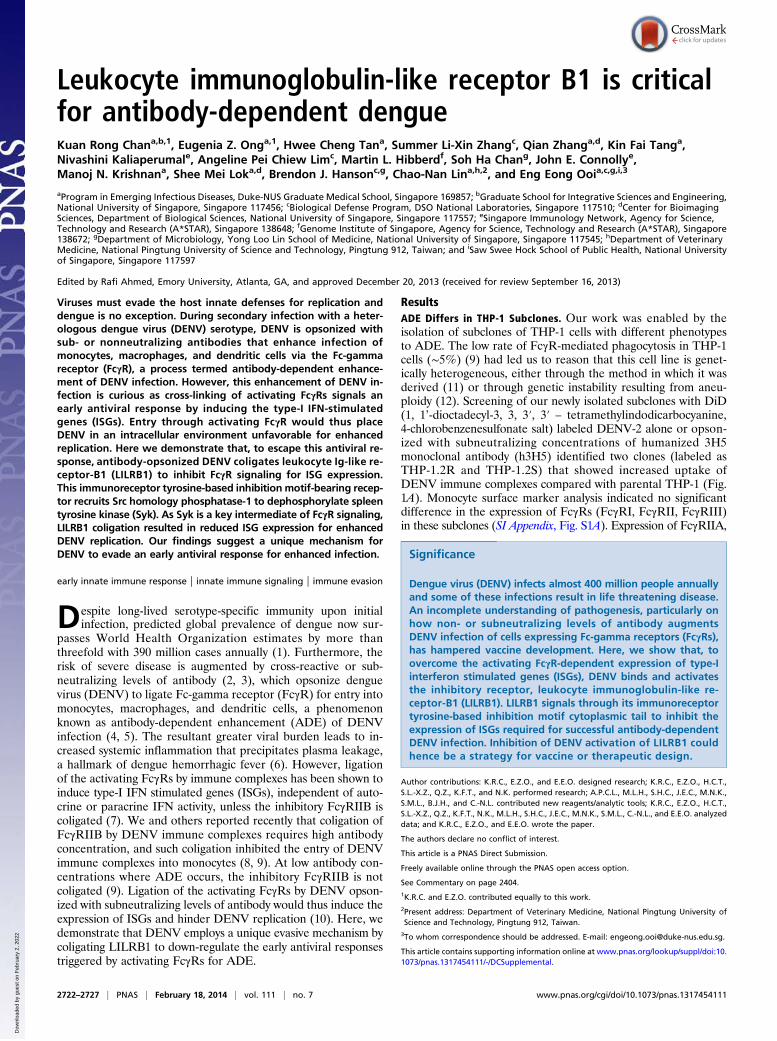
ResultsADE Differs in THP-1 Subclones. Our work was enabled by theisolation of subclones of THP-1 cells with different phenotypesto ADE. The low rate of FcγR-mediated phagocytosis in THP-1cells (∼5%) (9) had led us to reason that this cell line is genet-ically heterogeneous, either through the method in which it wasderived (11) or through genetic instability resulting from aneu-ploidy (12). Screening of our newly isolated subclones with DiD(1, 1’-dioctadecyl-3, 3, 3′, 3′ – tetramethylindodicarbocyanine,4-chlorobenzenesulfonate salt) labeled DENV-2 alone or opson-ized with subneutralizing concentrations of humanized 3H5monoclonal antibody (h3H5) identified two clones (labeled asTHP-1.2R and THP-1.2S) that showed increased uptake ofDENV immune complexes compared with parental THP-1 (Fig.1A). Monocyte surface marker analysis indicated no significantdifference in the expression of FcγRs (FcγRI, FcγRII, FcγRIII)in these subclones (SI Appendix, Fig. S1A). Expression of FcγRIIA,
Significance
Dengue virus (DENV) infects almost 400 million people annuallyand some of these infections result in life threatening disease.An incomplete understanding of pathogenesis, particularly onhow non- or subneutralizing levels of antibody augmentsDENV infection of cells expressing Fc-gamma receptors (FcγRs),has hampered vaccine development. Here, we show that, toovercome the activating FcγR-dependent expression of type-Iinterferon stimulated genes (ISGs), DENV binds and activatesthe inhibitory receptor, leukocyte immunoglobulin-like re-ceptor-B1 (LILRB1). LILRB1 signals through its immunoreceptortyrosine-based inhibition motif cytoplasmic tail to inhibit theexpression of ISGs required for successful antibody-dependentDENV infection. Inhibition of DENV activation of LILRB1 couldhence be a strategy for vaccine or therapeutic design.
Author contributions: K.R.C., E.Z.O., and E.E.O. designed research; K.R.C., E.Z.O., H.C.T.,S.L.-X.Z., Q.Z., K.F.T., and N.K. performed research; A.P.C.L., M.L.H., S.H.C., J.E.C., M.N.K.,S.M.L., B.J.H., and C.-N.L. contributed new reagents/analytic tools; K.R.C., E.Z.O., H.C.T.,S.L.-X.Z., Q.Z., K.F.T., N.K., M.L.H., S.H.C., J.E.C., M.N.K., S.M.L., C.-N.L., and E.E.O. analyzeddata; and K.R.C., E.Z.O., and E.E.O. wrote the paper.
The authors declare no conflict of interest.
This article is a PNAS Direct Submission.
Freely available online through the PNAS open access option.
See Commentary on page 2404.1K.R.C. and E.Z.O. contributed equally to this work.2Present address: Department of Veterinary Medicine, National Pingtung University ofScience and Technology, Pingtung 912, Taiwan.
3To whom correspondence should be addressed. E-mail: [email protected].
This article contains supporting information online at www.pnas.org/lookup/suppl/doi:10.1073/pnas.1317454111/-/DCSupplemental.
2722–2727 | PNAS | February 18, 2014 | vol. 111 | no. 7 www.pnas.org/cgi/doi/10.1073/pnas.1317454111
Dow
nloa
ded
by g
uest
on
Feb
ruar
y 2,
202
2
FcγRIIB, and FcγRIIC were similar in these subclones (SI Ap-pendix, Fig. S1 B and C). Both subclones were also heterozygousfor 131 H/R FcγRIIA polymorphism (SI Appendix, Fig. S1E).Identical HLA haplotyping confirmed that both subclones werederived from THP-1 and not the result of a contamination withanother cell line (SI Appendix, Table S1).Despite no significant differences in uptake and production of
plaque titers when infected with DENV-2 only, infection underADE conditions resulted in significantly different DENV-2 titersin THP-1.2R and THP-1.2S (Fig. 1B). Similar observations werealso made with enhancing titers of convalescent serum (SI Ap-pendix, Fig. S2A) or other DENV serotypes (SI Appendix, Fig.S2B). Furthermore, early DENV RNA replication diverged inthese two subclones, where a significant difference was observedas early as 6 h postinfection (Fig. 1C). Analysis of early geneexpression indicated significant up-regulation of ISGs in THP-1.2R but not THP-1.2S (Fig. 1 D and F–I). These included MX1,MX2, and viperin, which are potent inhibitors of DENV repli-cation (10). The up-regulation of ISGs in THP-1.2R, however,was not due to h3H5 (SI Appendix, Fig. S3) and is independent of
IFN-α, -β, and -γ signaling as both subclones expressed similarIFN transcript levels (Fig. 1E). As expected, addition of antibodiesthat blocked IFNα receptor (IFNαR) signaling (SI Appendix, Fig.S4A) did not reduce this early ISG induction in THP-1.2R fol-lowing infection (SI Appendix, Fig. S4B). The possibility that THP-1.2S had impaired IFNαR-mediated signaling was also excluded,as ISGs were significantly up-regulated in response to exogenousIFN (SI Appendix, Fig. S4C). These subclones thus serve as ex-quisite tools to decipher the signaling requirement to overcomethe early antiviral responses for successful ADE.
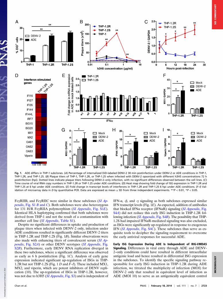
Early ISG Expression During ADE Is Independent of RIG-I/MDA5Signaling. Differences in viral entry through ADE and DENV-2–only conditions could have resulted in different intracellularantigenic load and hence resulted in differential ISG expressionin the subclones. To identify the specific signaling pathway re-sponsible for early ISG induction in THP-1.2R during ADEinfection, we titrated the multiplicity of infection (MOI) forDENV-2 only that resulted in equivalent level of infection asADE (MOI 10) to serve as an antigenically equivalent control
Fig. 1. ADE differs in THP-1 subclones. (A) Percentage of internalized DiD-labeled DENV-2 30 min postinfection under DENV-2 or ADE conditions in THP-1,THP-1.2R, and THP-1.2S. (B) Plaque titers of THP-1, THP-1.2R, or THP-1.2S when infected with DENV-2 opsonized with different h3H5 concentrations 72 hpostinfection (hpi). Dotted lines indicate plaque titers following DENV-2–only infection, with no significant differences observed between the cell lines. (C)Time course of viral RNA copy numbers in THP-1.2R or THP-1.2S under ADE conditions. (D) Heat map showing fold change of ISG expression in THP-1.2R andTHP-1.2S at 6 hpi under ADE conditions. (E) Fold change in transcript levels of interferons in THP-1.2R and THP-1.2S 6 hpi under ADE conditions. (F–I) Val-idation of microarray data in D by quantitative PCR. Data are expressed as mean ± SD from three independent experiments. **P < 0.01, *P < 0.05.
Chan et al. PNAS | February 18, 2014 | vol. 111 | no. 7 | 2723
MICRO
BIOLO
GY
SEECO
MMEN
TARY
Dow
nloa
ded
by g
uest
on
Feb
ruar
y 2,
202
2
(Fig. 2 A and B). Interestingly, lower and higher plaque titerswere observed in THP-1.2R and THP-1.2S, respectively, duringADE relative to DENV-2–only (MOI 60) conditions (Fig. 2C),which corroborates the notion that THP-1.2R has reducedsusceptibility to ADE. Immunofluorescence imaging showednuclear translocation of pSTAT-1 at 3 h post ADE in THP-1.2Rbut not in THP-1.2S or during antigenically equivalent DENV-only infection (Fig. 2D). This early nuclear translocation ofpSTAT-1 is transient as little colocalization could be observed at6 h postinfection.With similar intracellular antigenic load in ADE and DENV-
2–only conditions, we determined whether trafficking of DENVcontaining-phagosomes to cellular compartments enriched withpattern recognition receptors was an explanation for ISG in-duction in THP-1.2R. This was not the case as reduced expres-sion of adaptor molecules [mitochondrial antiviral signalingprotein (MAVS) and IFN regulatory factor 3 (IRF3)] of retinoicacid-inducible gene I (RIG-I)/melanoma differentiation-associ-ated protein 5 (MDA5) resulted in significantly increased early
DENV replication under DENV-2–only but not ADE conditions(Fig. 2E). Reduced TIR-domain containing adapter-inducingIFN β (TRIF) did not result in significant change in DENVreplication under either condition (Fig. 2E). Collectively, theseresults indicate that the early induction of ISG in THP-1.2R isunique to infection under ADE condition and is not mediated byRIG-I/MDA5–dependent type-I IFN expression.
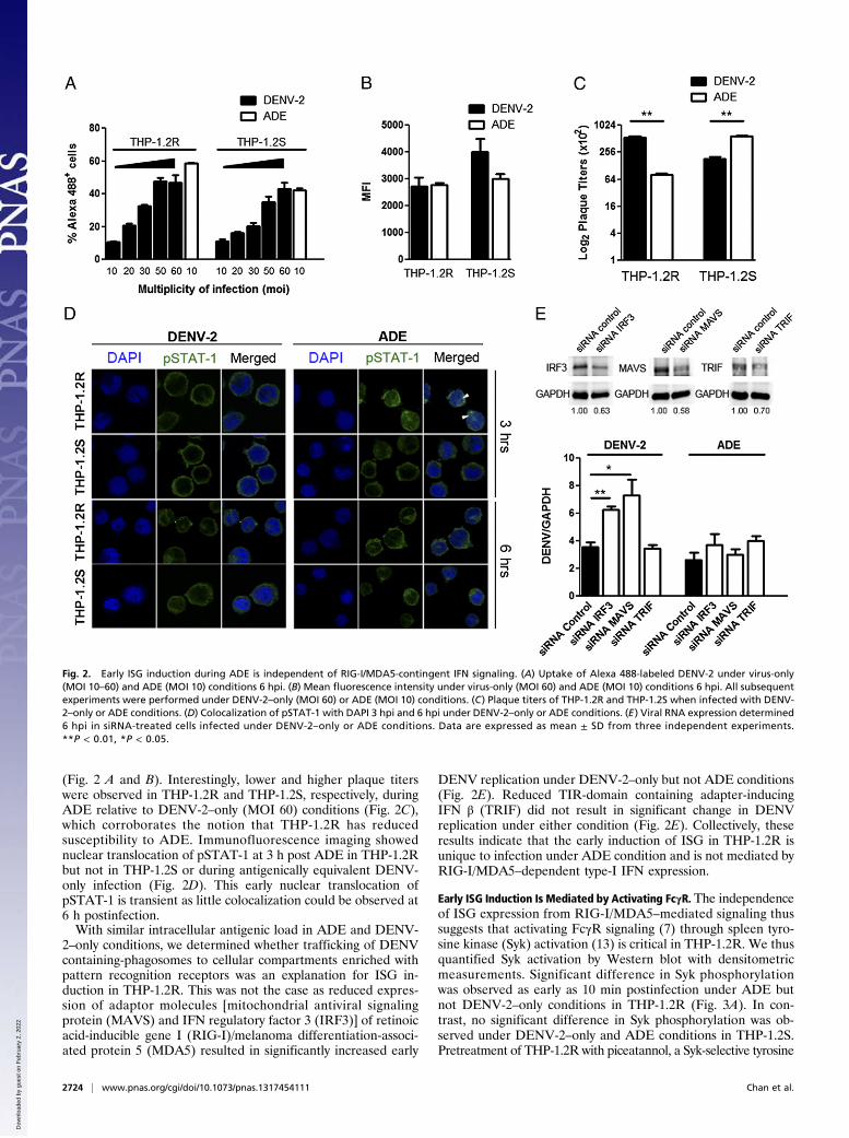
Early ISG Induction Is Mediated by Activating FcγR. The independenceof ISG expression from RIG-I/MDA5–mediated signaling thussuggests that activating FcγR signaling (7) through spleen tyro-sine kinase (Syk) activation (13) is critical in THP-1.2R. We thusquantified Syk activation by Western blot with densitometricmeasurements. Significant difference in Syk phosphorylationwas observed as early as 10 min postinfection under ADE butnot DENV-2–only conditions in THP-1.2R (Fig. 3A). In con-trast, no significant difference in Syk phosphorylation was ob-served under DENV-2–only and ADE conditions in THP-1.2S.Pretreatment of THP-1.2R with piceatannol, a Syk-selective tyrosine
Fig. 2. Early ISG induction during ADE is independent of RIG-I/MDA5-contingent IFN signaling. (A) Uptake of Alexa 488-labeled DENV-2 under virus-only(MOI 10–60) and ADE (MOI 10) conditions 6 hpi. (B) Mean fluorescence intensity under virus-only (MOI 60) and ADE (MOI 10) conditions 6 hpi. All subsequentexperiments were performed under DENV-2–only (MOI 60) or ADE (MOI 10) conditions. (C) Plaque titers of THP-1.2R and THP-1.2S when infected with DENV-2–only or ADE conditions. (D) Colocalization of pSTAT-1 with DAPI 3 hpi and 6 hpi under DENV-2–only or ADE conditions. (E) Viral RNA expression determined6 hpi in siRNA-treated cells infected under DENV-2–only or ADE conditions. Data are expressed as mean ± SD from three independent experiments.**P < 0.01, *P < 0.05.
2724 | www.pnas.org/cgi/doi/10.1073/pnas.1317454111 Chan et al.
Dow
nloa
ded
by g
uest
on
Feb
ruar
y 2,
202
2
kinase inhibitor resulted in greater reduction of ISG expressionunder ADE conditions (Fig. 3B) and a correspondingly greaterincrease in DENV replication (Fig. 3C) compared with DENV-2only. Increase in DENV replication was also greater in THP-1.2Rthan THP-1.2S. These findings suggest that early ISG expressionin THP-1.2R is conditioned upon activating FcγR signaling throughphosphorylated Syk (7).
Coligation of LILRB1 Inhibits ISG Induction. As activating FcγR sig-nals through immunoreceptor tyrosine-based activation motif(ITAM), we postulated that DENV coligates an immunore-ceptor tyrosine-based inhibition motif (ITIM)-bearing receptorto inhibit Syk activation (14) in THP-1.2S. Examination of thegene expression data identified two such possible receptors.LILRB1 (also known as CD85j or Ig-like transcript-2) andLILRB4 were up-regulated preinfection in THP-1.2S relative toTHP-1.2R (SI Appendix, Fig. S5A). Flow cytometry analysis,however, showed that only LILRB1 (Fig. 3D and SI Appendix,Fig. S5B) displayed higher surface expression on THP-1.2S.Because one of the effects of ITIM phosphorylation is the re-cruitment and phosphorylation of SHP-1 (15, 16), we measuredphosphorylated SHP-1 in the two subclones. Higher pSHP-1levels were found in THP-1.2S than THP-1.2R under ADEconditions (Fig. 3 E and F), suggesting that pSHP-1 dephos-phorylated Syk in THP-1.2S.
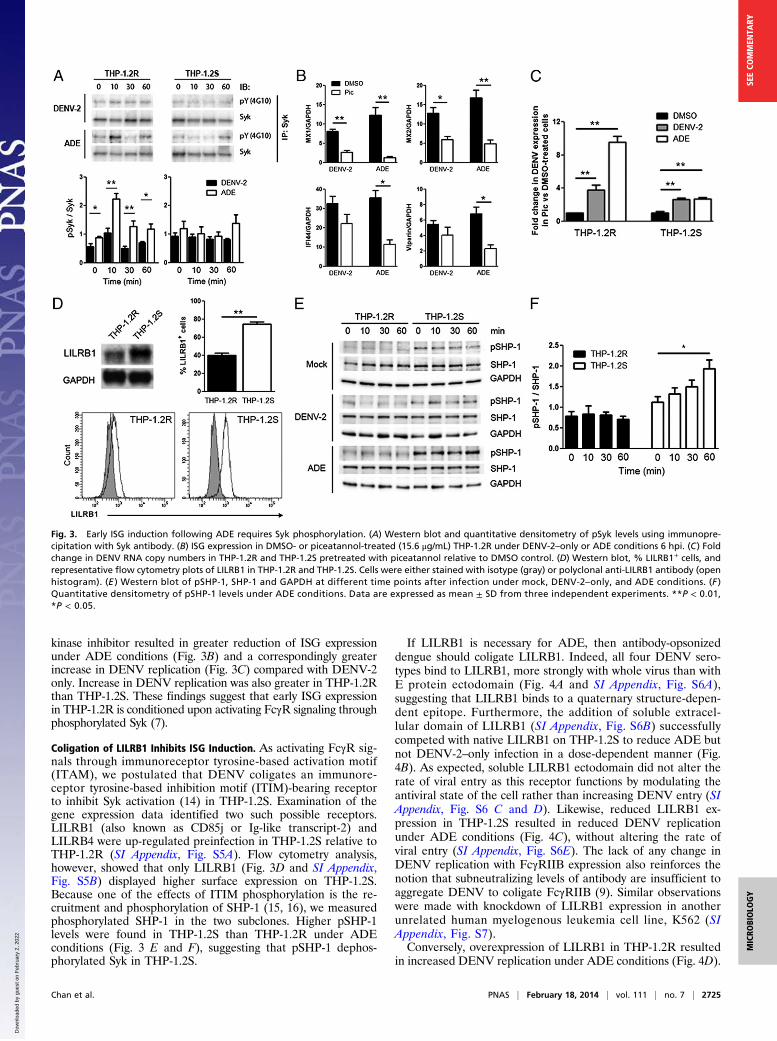
If LILRB1 is necessary for ADE, then antibody-opsonizeddengue should coligate LILRB1. Indeed, all four DENV sero-types bind to LILRB1, more strongly with whole virus than withE protein ectodomain (Fig. 4A and SI Appendix, Fig. S6A),suggesting that LILRB1 binds to a quaternary structure-depen-dent epitope. Furthermore, the addition of soluble extracel-lular domain of LILRB1 (SI Appendix, Fig. S6B) successfullycompeted with native LILRB1 on THP-1.2S to reduce ADE butnot DENV-2–only infection in a dose-dependent manner (Fig.4B). As expected, soluble LILRB1 ectodomain did not alter therate of viral entry as this receptor functions by modulating theantiviral state of the cell rather than increasing DENV entry (SIAppendix, Fig. S6 C and D). Likewise, reduced LILRB1 ex-pression in THP-1.2S resulted in reduced DENV replicationunder ADE conditions (Fig. 4C), without altering the rate ofviral entry (SI Appendix, Fig. S6E). The lack of any change inDENV replication with FcγRIIB expression also reinforces thenotion that subneutralizing levels of antibody are insufficient toaggregate DENV to coligate FcγRIIB (9). Similar observationswere made with knockdown of LILRB1 expression in anotherunrelated human myelogenous leukemia cell line, K562 (SIAppendix, Fig. S7).Conversely, overexpression of LILRB1 in THP-1.2R resulted
in increased DENV replication under ADE conditions (Fig. 4D).
Fig. 3. Early ISG induction following ADE requires Syk phosphorylation. (A) Western blot and quantitative densitometry of pSyk levels using immunopre-cipitation with Syk antibody. (B) ISG expression in DMSO- or piceatannol-treated (15.6 μg/mL) THP-1.2R under DENV-2–only or ADE conditions 6 hpi. (C) Foldchange in DENV RNA copy numbers in THP-1.2R and THP-1.2S pretreated with piceatannol relative to DMSO control. (D) Western blot, % LILRB1+ cells, andrepresentative flow cytometry plots of LILRB1 in THP-1.2R and THP-1.2S. Cells were either stained with isotype (gray) or polyclonal anti-LILRB1 antibody (openhistogram). (E ) Western blot of pSHP-1, SHP-1 and GAPDH at different time points after infection under mock, DENV-2–only, and ADE conditions. (F )Quantitative densitometry of pSHP-1 levels under ADE conditions. Data are expressed as mean ± SD from three independent experiments. **P < 0.01,*P < 0.05.
Chan et al. PNAS | February 18, 2014 | vol. 111 | no. 7 | 2725
MICRO
BIOLO
GY
SEECO
MMEN
TARY
Dow
nloa
ded
by g
uest
on
Feb
ruar
y 2,
202
2
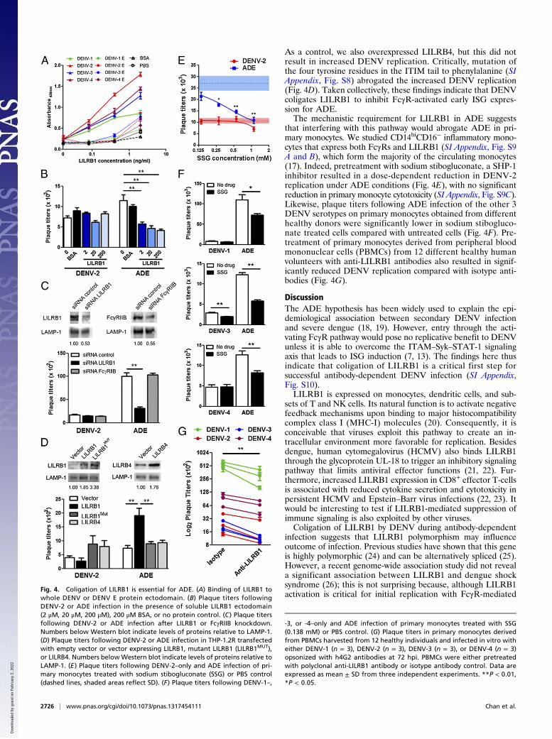
As a control, we also overexpressed LILRB4, but this did notresult in increased DENV replication. Critically, mutation ofthe four tyrosine residues in the ITIM tail to phenylalanine (SIAppendix, Fig. S8) abrogated the increased DENV replication(Fig. 4D). Taken collectively, these findings indicate that DENVcoligates LILRB1 to inhibit FcγR-activated early ISG expres-sion for ADE.The mechanistic requirement for LILRB1 in ADE suggests
that interfering with this pathway would abrogate ADE in pri-mary monocytes. We studied CD14hiCD16− inflammatory mono-cytes that express both FcγRs and LILRB1 (SI Appendix, Fig. S9A and B), which form the majority of the circulating monocytes(17). Indeed, pretreatment with sodium stibogluconate, a SHP-1inhibitor resulted in a dose-dependent reduction in DENV-2replication under ADE conditions (Fig. 4E), with no significantreduction in primary monocyte cytotoxicity (SI Appendix, Fig. S9C).Likewise, plaque titers following ADE infection of the other 3DENV serotypes on primary monocytes obtained from differenthealthy donors were significantly lower in sodium stibogluco-nate treated cells compared with untreated cells (Fig. 4F). Pre-treatment of primary monocytes derived from peripheral bloodmononuclear cells (PBMCs) from 12 different healthy humanvolunteers with anti-LILRB1 antibodies also resulted in signif-icantly reduced DENV replication compared with isotype anti-bodies (Fig. 4G).
DiscussionThe ADE hypothesis has been widely used to explain the epi-demiological association between secondary DENV infectionand severe dengue (18, 19). However, entry through the acti-vating FcγR pathway would pose no replicative benefit to DENVunless it is able to overcome the ITAM–Syk–STAT-1 signalingaxis that leads to ISG induction (7, 13). The findings here thusindicate that coligation of LILRB1 is a critical first step forsuccessful antibody-dependent DENV infection (SI Appendix,Fig. S10).LILRB1 is expressed on monocytes, dendritic cells, and sub-
sets of T and NK cells. Its natural function is to activate negativefeedback mechanisms upon binding to major histocompatibilitycomplex class I (MHC-I) molecules (20). Consequently, it isconceivable that viruses exploit this pathway to create an in-tracellular environment more favorable for replication. Besidesdengue, human cytomegalovirus (HCMV) also binds LILRB1through the glycoprotein UL-18 to trigger an inhibitory signalingpathway that limits antiviral effector functions (21, 22). Fur-thermore, increased LILRB1 expression in CD8+ effector T-cellsis associated with reduced cytokine secretion and cytotoxicity inpersistent HCMV and Epstein–Barr virus infections (22, 23). Itwould be interesting to test if LILRB1-mediated suppression ofimmune signaling is also exploited by other viruses.Coligation of LILRB1 by DENV during antibody-dependent
infection suggests that LILRB1 polymorphism may influenceoutcome of infection. Previous studies have shown that this geneis highly polymorphic (24) and can be alternatively spliced (25).However, a recent genome-wide association study did not reveala significant association between LILRB1 and dengue shocksyndrome (26); this is not surprising because, although LILRB1activation is critical for initial replication with FcγR-mediated
Fig. 4. Coligation of LILRB1 is essential for ADE. (A) Binding of LILRB1 towhole DENV or DENV E protein ectodomain. (B) Plaque titers followingDENV-2 or ADE infection in the presence of soluble LILRB1 ectodomain(2 μM, 20 μM, 200 μM), 200 μM BSA, or no protein control. (C) Plaque titersfollowing DENV-2 or ADE infection after LILRB1 or FcγRIIB knockdown.Numbers below Western blot indicate levels of proteins relative to LAMP-1.(D) Plaque titers following DENV-2 or ADE infection in THP-1.2R transfectedwith empty vector or vector expressing LILRB1, mutant LILRB1 (LILRB1MUT),or LILRB4. Numbers belowWestern blot indicate levels of proteins relative toLAMP-1. (E) Plaque titers following DENV-2–only and ADE infection of pri-mary monocytes treated with sodium stibogluconate (SSG) or PBS control(dashed lines, shaded areas reflect SD). (F) Plaque titers following DENV-1–,
-3, or -4–only and ADE infection of primary monocytes treated with SSG(0.138 mM) or PBS control. (G) Plaque titers in primary monocytes derivedfrom PBMCs harvested from 12 healthy individuals and infected in vitro witheither DENV-1 (n = 3), DENV-2 (n = 3), DENV-3 (n = 3), or DENV-4 (n = 3)opsonized with h4G2 antibodies at 72 hpi. PBMCs were either pretreatedwith polyclonal anti-LILRB1 antibody or isotype antibody control. Data areexpressed as mean ± SD from three independent experiments. **P < 0.01,*P < 0.05.
2726 | www.pnas.org/cgi/doi/10.1073/pnas.1317454111 Chan et al.
Dow
nloa
ded
by g
uest
on
Feb
ruar
y 2,
202
2

entry, multiple other host and viral factors contribute to eventualdisease outcome.Our findings also suggest that generation of antibodies to
quaternary structure-dependent epitopes on DENV that blockLILRB1 interaction can reduce ADE. That heterotypic anti-bodies can enhance dengue infection in FcγR-bearing cells rep-resents a safety concern in the development of a dengue vaccine.Hence, a vaccine that can generate high-titer antibody that bindsthe quaternary structure-dependent epitopes on DENV to preventLILRB1 ligation could reduce the risk of vaccine-induced ADE.Further studies would be needed to clarify this, although caremust be taken in selecting a suitable in vivo model as the LILRB1gene is deleted in laboratory strains of mice (27).In conclusion, DENV coligates LILRB1 to down-regulate the
activating FcγR-mediated early ISG expression for successfulantibody-dependent infection.
Materials and MethodsCells. THP-1.2R and THP-1.2S were subcloned from THP-1 by limiting dilution.Primary monocytes were isolated from healthy donors and cultured as de-scribed (9).
Viruses.DENV-1 (06K2402DK1),DENV-3 (05K863DK1), andDENV-4 (06K2270DK1)are clinical isolates from the EDEN study (28). DENV-2 (ST) is a clinical isolatefrom the Singapore General Hospital.
Virus Infection. Endotoxin-free (LAL Chromogenic Endotoxin Quantitation kit,Pierce) 3H5 and 4G2 chimeric human/mouse IgG1 antibodies were con-structed as described (29). DENV was incubated with media, antibodies, orserum for 1h at 37 °C before adding to cells at indicated MOI. Uptake wasassessed using DiD and Alexa 488-labeled DENV as described (9, 30). For drug
assays, cells were pretreated with piceatannol (Sigma-Aldrich) or sodiumstibogluconate (Santa Cruz Biotechnology) 6 h before infection. Cell viabilitywas assessed using CellTiter 96 AQueous One Solution Cell ProliferationAssay (MTS, Promega) according to the manufacturer’s protocol. Subse-quently, virus replication was assessed using quantitative PCR at indicatedtime points and plaque assay at 72 h postinfection. Protein and proteinphosphorylation levels were assessed using Western blots and analyzedwith ImageJ.
Microarray Analysis. Following RNA extraction, microarray was performed atthe Duke-NUS Genome Biology Core Facility. cRNAs were hybridized toIllumina Human HT-12 v4 Beadchips, according to manufacturer’s instruc-tions. Data analysis was performed using Partek software and normalizedagainst GAPDH.
Competition with Soluble LILRB1 Ectodomain. The extracellular portion ofLILRB1 was cloned into pCMV-XL5 (Origene) and transfected into HEK293Tcells for protein expression. The expressed proteins were then purified andincubated with DENV-2 or h3H5-opsonized DENV-2 for 1 h at 37 °C beforeadding to THP-1.2S.
siRNA Transfection and Overexpression. siRNA transfections and overex-pression were performed as described (9). siRNA targeting FcγRIIB (Qiagen),LILRB1, MAVS, IRF3, and TRIF (SABio) were used, and overexpression studieswere performed with either empty plasmid, plasmid encoding LILRB1 or ty-rosine mutant LILRB1, or LILRB4.
ACKNOWLEDGMENTS. We thank Soman Abraham for his constructive re-view of this work and Mei Fong Chan and Kenneth Goh for their technicalassistance. This work was supported by the Singapore National ResearchFoundation under its Clinician-Scientist Award administered by the NationalMedical Research Council.
1. Bhatt S, et al. (2013) The global distribution and burden of dengue. Nature 496(7446):504–507.
2. Halstead SB, O’Rourke EJ (1977) Dengue viruses and mononuclear phagocytes. I. In-fection enhancement by non-neutralizing antibody. J Exp Med 146(1):201–217.
3. Simmons CP, et al. (2007) Maternal antibody and viral factors in the pathogenesis ofdengue virus in infants. J Infect Dis 196(3):416–424.
4. Halstead SB (1988) Pathogenesis of dengue: Challenges to molecular biology. Science239(4839):476–481.
5. Halstead SB, O’Rourke EJ (1977) Antibody-enhanced dengue virus infection in pri-mate leukocytes. Nature 265(5596):739–741.
6. Libraty DH, et al. (2002) Differing influences of virus burden and immune activationon disease severity in secondary dengue-3 virus infections. J Infect Dis 185(9):1213–1221.
7. Dhodapkar KM, et al. (2007) Selective blockade of the inhibitory Fcgamma receptor(FcgammaRIIB) in human dendritic cells and monocytes induces a type I interferonresponse program. J Exp Med 204(6):1359–1369.
8. Boonnak K, Slike BM, Donofrio GC, Marovich MA (2013) Human FcγRII cytoplasmicdomains differentially influence antibody-mediated dengue virus infection. J Immunol190(11):5659–5665.
9. Chan KR, et al. (2011) Ligation of Fc gamma receptor IIB inhibits antibody-dependentenhancement of dengue virus infection. Proc Natl Acad Sci USA 108(30):12479–12484.
10. Jiang D, et al. (2010) Identification of five interferon-induced cellular proteins thatinhibit west nile virus and dengue virus infections. J Virol 84(16):8332–8341.
11. Tsuchiya S, et al. (1980) Establishment and characterization of a human acutemonocytic leukemia cell line (THP-1). Int J Cancer 26(2):171–176.
12. Sheltzer JM, et al. (2011) Aneuploidy drives genomic instability in yeast. Science333(6045):1026–1030.
13. Tassiulas I, et al. (2004) Amplification of IFN-alpha-induced STAT1 activation and in-flammatory function by Syk and ITAM-containing adaptors. Nat Immunol 5(11):1181–1189.
14. Steevels TA, Meyaard L (2011) Immune inhibitory receptors: Essential regulators ofphagocyte function. Eur J Immunol 41(3):575–587.
15. Fanger NA, et al. (1998) The MHC class I binding proteins LIR-1 and LIR-2 inhibit Fcreceptor-mediated signaling in monocytes. Eur J Immunol 28(11):3423–3434.
16. Scharenberg AM, Kinet JP (1996) The emerging field of receptor-mediated inhibitorysignaling: SHP or SHIP? Cell 87(6):961–964.
17. Passlick B, Flieger D, Ziegler-Heitbrock HW (1989) Identification and characteriza-tion of a novel monocyte subpopulation in human peripheral blood. Blood 74(7):2527–2534.
18. Halstead SB, Mahalingam S, Marovich MA, Ubol S, Mosser DM (2010) Intrinsic anti-body-dependent enhancement of microbial infection in macrophages: Disease reg-ulation by immune complexes. Lancet Infect Dis 10(10):712–722.
19. Simmons CP, Farrar JJ, Nguyen V, Wills B (2012) Dengue. N Engl J Med 366(15):1423–1432.
20. Colonna M, et al. (1997) A common inhibitory receptor for major histocompatibilitycomplex class I molecules on human lymphoid and myelomonocytic cells. J Exp Med186(11):1809–1818.
21. Yang Z, Bjorkman PJ (2008) Structure of UL18, a peptide-binding viral MHC mimic,bound to a host inhibitory receptor. Proc Natl Acad Sci USA 105(29):10095–10100.
22. Cosman D, et al. (1997) A novel immunoglobulin superfamily receptor for cellular andviral MHC class I molecules. Immunity 7(2):273–282.
23. Poon K, Montamat-Sicotte D, Cumberbatch N, McMichael AJ, Callan MF (2005) Ex-pression of leukocyte immunoglobulin-like receptors and natural killer receptors onvirus-specific CD8+ T cells during the evolution of Epstein-Barr virus-specific immuneresponses in vivo. Viral Immunol 18(3):513–522.
24. Kuroki K, et al. (2005) Extensive polymorphisms of LILRB1 (ILT2, LIR1) and their as-sociation with HLA-DRB1 shared epitope negative rheumatoid arthritis. Hum MolGenet 14(16):2469–2480.
25. Jones DC, et al. (2009) Alternative mRNA splicing creates transcripts encoding solubleproteins from most LILR genes. Eur J Immunol 39(11):3195–3206.
26. Khor CC, et al. (2011) Genome-wide association study identifies susceptibility loci fordengue shock syndrome at MICB and PLCE1. Nat Genet 43(11):1139–1141.
27. Kubagawa H, Burrows PD, Cooper MD (1997) A novel pair of immunoglobulin-likereceptors expressed by B cells and myeloid cells. Proc Natl Acad Sci USA 94(10):5261–5266.
28. Low JG, et al. (2006) Early Dengue infection and outcome study (EDEN) - study designand preliminary findings. Ann Acad Med Singapore 35(11):783–789.
29. Hanson BJ, et al. (2006) Passive immunoprophylaxis and therapy with humanizedmonoclonal antibody specific for influenza A H5 hemagglutinin in mice. Respir Res7:126.
30. Zhang SL, Tan HC, Hanson BJ, Ooi EE (2010) A simple method for Alexa Fluor dyelabelling of dengue virus. J Virol Methods 167(2):172–177.
Chan et al. PNAS | February 18, 2014 | vol. 111 | no. 7 | 2727
MICRO
BIOLO
GY
SEECO
MMEN
TARY
Dow
nloa
ded
by g
uest
on
Feb
ruar
y 2,
202
2