lesson 6. metabolism purine and pyrimidine base....
TRANSCRIPT
Biosynthesis of nucleic acids and proteins. Regulation biosyn-
thesis.
33
LESSON 6. METABOLISM PURINE AND PYRIMIDINE BASE. STRUCTURE OF
NUCLEIC ACIDS.
TES T IN WR ITTEN FO R M :
• The structural formula of nitrogenous bases, nucleosides and nucleotides.
• Write the fragments of DNA and RNA.
MA IN TO P IC S :
• Chemical composition of DNA and RNA. Nitrogenus bases of nucleic acids (purine and pyri-
midine). Ribonucleotides and deoxyribonucleotides.The structure and nomenclature.
• Synthesis of the purine nucleotides. Sources of the various atoms of the purine bases. Salvage
of bases. Regulation.
• Synthesis of the pyrimidines. Sources of the various atoms of pyrimidines. Salvage of bases.
Regulation. Orotic aciduria.
• Scheme of the synthesis of deoxyribonucleosides. Regulation.
• Scheme of degradation of nucleic acids. Enzymes, substrates, products.
• Degradation of the purine nucleotides. Hyperuricemia. Gout.
• Degradation of the pyrimidines.
• DNA and RNA. Similarity and differences of composition, primary structure, functions, cells
localization.
• Secondary structure of DNA (model of Watson and Crick). Bonds wich are resposible for sta-
bility of the molecule.
• Specialized forms of RNA. Characterictics of primary, secondary and tertiary structure of
RNA. Structure and biological role of ribosomes.
• Determination of uric acid concentration in blood serum.
STRUCTURE OF NITROGENOUS BASES, NUCLEOSIDES AND NUCLEOTIDES.
Remember the structure of purine and pyrimidine nitrogenous bases, nucleosides, nucleo-
tides (p.151-152)
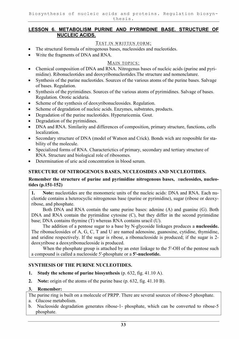
1. Note: nucleotides are the monomeric units of the nucleic acids: DNA and RNA. Each nu-
cleotide contains a heterocyclic nitrogenous base (purine or pyrimidine), sugar (ribose or deoxy-
ribose, and phosphate.
Both DNA and RNA contain the same purine bases: adenine (A) and guanine (G). Both
DNA and RNA contain the pyrimidine cytosine (C), but they differ in the second pyrimidine
base; DNA contains thymine (T) whereas RNA contains uracil (U).
The addition of a pentose sugar to a base by N-glycoside linkages produces a nucleoside.
The ribonucleosides of A, G, C, T and U are named adenosine, guanosine, cytidine, thymidine,
and uridine respectively. If the sugar is ribose, a ribonucleoside is produced; if the sugar is 2-
deoxyribose a deoxyribonucleoside is produced.
When the phosphate group is attached by an ester linkage to the 5'-OH of the pentose such
a compound is called a nucleoside 5'-phosphate or a 5'-nucleotide.
SYNTHESIS OF THE PURINE NUCLEOTIDES.
1. Study the scheme of purine biosynthesis (p. 632, fig. 41.10 A).
2. Note: origin of the atoms of the purine base (p. 632, fig. 41.10 B).
3. Remember:
The purine ring is built on a molecule of PRPP. There are several sources of ribose-5 phosphate.
a. Glucose metabolism.
b. Nucleoside degradation generates ribose-1- phosphate, which can be converted to ribose-5
phosphate.
Biosynthesis of nucleic acids and proteins. Regulation biosyn-
thesis.
34
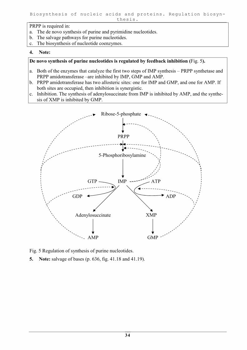
PRPP is required in:
a. The de novo synthesis of purine and pyrimidine nucleotides.
b. The salvage pathways for purine nucleotides.
c. The biosynthesis of nucleotide coenzymes.
4. Note:
De novo synthesis of purine nucleotides is regulated by feedback inhibition (Fig. 5).
a. Both of the enzymes that catalyze the first two steps of IMP synthesis – PRPP synthetase and
PRPP amidotransferase –are inhibited by IMP, GMP and AMP.
b. PRPP amidotransferase has two allosteric sites: one for IMP and GMP, and one for AMP. If
both sites are occupied, then inhibition is synergistic.
c. Inhibition. The synthesis of adenylosuccinate from IMP is inhibited by AMP, and the synthe-
sis of XMP is inhibited by GMP.
Fig. 5 Regulation of synthesis of purine nucleotides.
5. Note: salvage of bases (p. 636, fig. 41.18 and 41.19).
Ribose-5-phosphate
5-Phosphoribosylamine
PRPP
GTP
Adenylosuccinate
GDP
ATP IMP
ADP
XMP
AMP GMP

Biosynthesis of nucleic acids and proteins. Regulation biosyn-
thesis.
35
Fig. 6 Salvage pathways for purine nucleotides.
6. Write the correct letter for the following questions:
The N atoms at the positions 3 and 9 of purine base are derived from theamide nitrogen of:
A. Glutamate.
B. Glutamine.
C. Asparagine.
D. Aspartate.
The carbon atoms at the positions 4 and 5 and the N atom at position 7 of the purine base are sup-
plied from:
A. Valine.
B. Alanine.
C. Glycine.
D. Serine.
SYNTHESIS OF THE PYRIMIDINES.
1. Study the scheme of pyrimidines biosynthesis (p. 633-635, fig. 41.16 A).
2. Note: origin of the atoms of the pyrimidine ring (p. 635, fig. 41.16 B).
3. Remember:
The pyrimidine ring, unlike the purine ring, is not built on a molecule of PRPP. Instead, the pyri-
midine ring is formed, and then it reaacts with PRPP to form the nucleotide.
4. Note:
Regulation of pyrimidine synthesis. The enzyme that catalyzed the synthesis of carbamoyl phos-
phate (carbamoyl phosphate synthetase II) is a single polypeptide called CAD, wich also contains
the enzyme aspartate carbamoyl transferase and dihydroorotase. UTP inhibits the carbamoyl
phosphate synthetase II activity of this multienzyme polypeptide. The enzyme that catalyze the
last two steps of the synthesis of UMP-orotate phosphoribosyltransferase and orotidine 5’- mono-
phosphate (OMP) decarboxylase, also are linked in a single polypeptide. UMP and CMP inhibit
the activity of this multienzyme.
GTP feedback inhibits CTP synthetase.
Hypoxanthine
+
PRPP
Guanine
+
PRPP
HGPRT
APRT
Adenine
+
PRPP
IMP XMP GMP
AMP

Biosynthesis of nucleic acids and proteins. Regulation biosyn-
thesis.
36
5. Remember: The defect of orotate phosphoribosyltransferase leads to orotic aciduria (p. 635, p. 636, fig.41.17).
6. Note: salvage pathways for pyrimidine nucleotides are not significant in mammals as are
those for purine nucleotides.
7. Write the correct letter for the following:
The carbon atoms at the positions 4, 5 and 6 and the N atom at the positions 3 of pyrimidine base
are derived from:
A. Glutamic acid.
B. Aspartic acid.
C. Glycine.
D. Serine.
8. Fill in the following blanks:
1. The synthesis of pyrimidine starts with the formation of carbamoyl phosphate from
, and CO2being catalyzed by carbamoyl phosphate synthetase present
in the cytosol of the cell.
2. Dihydroorotic acid on dehydrogenetion by dihydroorotate dehydrogenase utilizing ________
as coenzyme is converted into .
SYNTHESIS OF DEOXYRIBONUCLEOSIDES.
1. Study the synthesis of deoxyribonucleotides (p. 633, fig. 41.14).
2. Note:
Deoxyribonucleotides are found in DNA. The cellular levels of deoxyribonucleotides are ordinar-
ily low, and they increase at the time of DNA replication.
DEGRADATION OF NUCLEIC ACIDS.
1. Study the scheme of the degradation of nucleic acids. NUCLEIC ACIDS
NUCLEOTIDES
NUCLEOSIDES
PURINE AND PYRIMIDINE BASES
Nucleases
Nucleotidases
H3PO4
(DEOXY)
RIBOSE-1-PHOSPHATE
Nucleoside phosphorylase
Fig. 7 The brief scheme of
intracellular degradation of
nucleic acids in human body.
DEGRADATION OF THE PURINE NUCLEOTIDES.
1. Study the reactions of the degradation of the purine nucleotides (p. 633-634, fig. 41.15).
2. Note:
The final product of purine degradation is uric acid. It is ionized in the body to form urate. Urate
is not very soluble in an aqueous environment. Gout is caused by the precipitation of sodium urate

Biosynthesis of nucleic acids and proteins. Regulation biosyn-
thesis.
37
crystals in the joints and kidneys. Sodium urate crystals precipitate because the serum levels of
urate exceed its solubility limit. Elevated uric acid levels may be due to one of severel disorders.
a. In some patients, PRPP synthetase is abnormal and is not responsive to feedback inhibition by
purine nucleoside diphosphates.
b. A partial deficiency of hypoxanthine-guanine phosphoribosyltransferase (HGPRT) leads to
increased cellular levels of PRPP, which) leads to increased de novo synthesis of purines. This
partial deficiency does not cause any of the neurologic symptoms of Lesch-Nyhan syndrome.
Inflammation and erosion of the joints occur when leukocytes engulf the deposited crystals and
consequently rupture, releasing lysosomal enzymes. Sodium urate crystals in the urinary tract im-
pair renal function.
Allopurinol, which blocks the production of uric acid, is an important drug in the treatment of
gout.
(1) Allopurinol is oxidized by xanthine oxidase to oxypurinol.
(2) Oxypurinol binds tightly to xanthine oxidase, inhibiting it’s ability to oxidize xanthine or hy-
poxanthine. This is an example of suicide inhibition.
(3) HGPRT salvages the hypoxanthine whose levels increased upon inhibition of uric acid forma-
tion. This leads to a decrease in PRPP levels and thus to a decrease in the de novo purine syn-
thesis.
Colchicine is an antiinflammatory drug that may be used to treat an acute gout attack. It inhibits
leukocyte movement by affecting microtubule formation.
3. Note: Lesch-Nyhan syndrome is caused by a defective hypoxanthine-guanine phosphoribo-
syltransferase (p. 637, clinical note).
4. Write the correct letter for the followings: The end product of purine catabolism in other mammals except man:
A. Uric acid.
B. Allantoin.
C. Ammonia.
D. Creatinine.
The healthy adult man typical range of excretion value of total uric acid expressed as milligrams
per 24 hrs is:
A. 100-300.
B. 200-400.
C. 300-500.
D. 400-600.
5. Fill in the blanks of the following questions:
A. Xanthine is oxidized to uric acid by .
B. Xanthine oxidase is inhibited by for which uric acid can not be formed.
C. The chief end product of purine catabolism in man .
D. Blood uric acid level also in hemolytic anemia and thalassemia.
E. Sodium urate crystals are deposited in the soft tissues and these urate deposits are re-
ferred to as .
DEGRADATION OF PYRIMIDINE BASES.
1. Study the degradation of the pyrimidines (p. 635-636).
2. Note:
The products of pyrimidine degradation are excreted in the urine or converted to CO2, H2O and
NH3 (which forms urea). They do not cause any problems for the body, in contrast to urate, which
is produced from the purines and can precipitate, causing gout.

Biosynthesis of nucleic acids and proteins. Regulation biosyn-
thesis.
38
3. Fill in the following blanks:
A. The main site of the catabolism of pyrimidines .
B. The major end products of cytosine, uracil and thymine are and respec-
tively.
PRIMARY STRUCTURE OF DNA AND RNA
Consider the structure of nucleic acids
1. Memorize: DNA and RNA are polymers of nucleotides or nucleoside monophosphates –5-
NMR. Nucleotides are linked together by 3', 5’ - phosphodiester bonds between the 3'-OH
group on the sugar of one nucleotide through a phosphate molecule to the 5'-OH on the sugar
of another nucleotide. The sugar - phosphate linkages form a “backbone” of DNA and RNA.
The terminal nucleotide of one end usually has a free 3- hydroxyl group on its sugar moiety
(3'end) and the other end has a free phosphate group attached to the 5' position of the sugar
(5'end).
2. Study that the sequence of nucleotides in the polynucleotide chains of DNA or RNA is called
the primary structure of the nucleic acids.
N
NN
N
NH2
O
HO
HH
H
CH2
H
PO
O
O-
NH
N
N
O
NH2N
O
H
HH
HHO
P
O
O-
N
NH2
ON
O
HO
HH
H
CH2
H
PO
O
O-
CH2
NH
O
ON
O
HOH
HH
H
CH2
H
PO
O
O-
O
O-
5' end
A
D
3' end
C
B
3. Fig. 8 DNA strand
a) Name the nucleotides that form
the oligonucleotide given in
fig.1.
b) Indicate the 5'- phosphate and
3'-OH ends of the oligonucleo-
tide.
c) Match the following linkages
with the appropriate letters in
fig.1
1. N-glycoside
2. 3′, 5′- phosphodiester
3. 5′-phosphoester
4. 3′- phosphoester
d) What nucleic acid generates this
oligonucleotide on hydrolysis?
4. Write the formula of the dinucleotide A-U and compare the composition of its constituent
nucleotides:
A. UMP 1. Purine base
B. AMP 2. Pyrimidine base
C. Both 3. Ribose
D. None. 4. Pyrophosphate

Biosynthesis of nucleic acids and proteins. Regulation biosyn-
thesis.
39
SECONDARY STRUCTURE OF DNA AND RNA.
Study the secondary structure of DNA (p. 153 - 155)
1. Memorize: double helix is the predominant conformation of DNA and is now referred to as
B-form DNA or secondary structure of DNA. It consists of two polymers, or strands, of
DNA paired to each other and coiled around a common axis in a righthanded manner. Each
strand has an opposite polarity to the other or antiparallel. That is, where one suger-
phosphate backbone has a 5' to 3' direction, the adjacent, paired strand is oriented oppositely
in a 3' to 5' direction. The two DNA strands of the double helix are held together by com-
plementary base pairing: adenine pairs with thymine through two hydrogen bonds (A=T),
and guanine pairs with cytosine through three hydrogen bonds (G=C). The stability of the
double helix owes much to the van der Waals interactions of the bases that lie perpendicular
to the helix axis, which is composed of deoxyribose-phosphate chains. Double helix of DNA
much likes a spiral staircase in which there are 10 base pairs for each complete turn of the
helix.
1. Which one of the following statements is true of the double helix DNA? A. The planes of the bases lie parallel to the helix axis.
B. The chains have a backbone of linked glycosides.
C. The 3' -OH groups of each chain are at opposite ends of the molecule.
D. The duplex structure is stabilized only by hydrogen bonds between bases.
E. The two chains have an identical base sequence.
2. Here is a fragment of DNA chain: .… -A-G-C-T-T-A-G-C-C-.....
A. Write the nucleotide sequence of a complementary DNA chain.
B. Indicate its 5′ and 3′ ends.
LABORATORY MANUAL
DETERMINATION OF URIC ACID CONCENTRATION IN BLOOD SERUM
The method is based on the fact that uric acid reduces the phospho-tungstic reagent (Folin's re-
agent) yielding coloured products whose coloration intensity is presumed to be proportional to the
uric acid concentration.
Procedure:
Reagents Control Standard
solution
Sample
1. Distilled water - - 4 ml
2. Blood serum - - 0,5 ml
Mix
3. Solution of H2SO4 (0,35 M) - - 0,25 ml
4. Solution of tungstic sodium - - 0,25 ml
Mix, centrifuge the mixture at 3000 rpm for 5 min.
5. Centrifugate - - 2 ml
6. Standard solution of uric acid - 2 ml -
7. Distilled water 2 ml - -
8. Solution of sodium carbonate 1 ml 1 ml 1 ml
9. Phospho-tungstic reagent 0,5 ml 0,5 ml 0,5 ml
Mix, wait 30 minutes and measure the absorbance for sample and standard solutions against dis-
tilled water on a photocolorimeter at 510-560 nm (green light filter) using 1cm thick cells.
Calculation is carried out by the equation:
C=Asample/A standard ××××30××××10
where C is the concentration of uric acid in blood serum, mkmol/litre; A sample is the absorbance
measured for sample solution; A standard is the absorbance measured for standard solution; 30
mkmol - is the mass of uric acid contained in standard solution, used for reaction; 10 - is the scal-
ing factor for conversion to per litre blood serum.

Biosynthesis of nucleic acids and proteins. Regulation biosyn-
thesis.
40
Norm for male – 240-500 mkmol/l; for female – 160 –400 mkmol/l.
Presentation of results. Calculate the concentration of uric acid in blood serum by making use of
the measured absorbance values and record the result in a report sheet. Compare the found uric
acid concentration to the norm. Suggest an explanation for the difference (if any) between these
two indices from the standpoint of disturbed purine metabolism.
Homework:
Study lesson 7.

Biosynthesis of nucleic acids and proteins. Regulation biosyn-
thesis.
41
LESSON 7. STRUCTURE OF DNA AND RNA. NUCLEOPROTEINS. SYNTHESIS OF
DNA (REPLICATION). REPARATION OF DNA.
• The peculiarity of formation of nucleoproteins. DNA-binding proteins: helix-spire (round)-
helix; lucines zip-fasteners; Zn-fingers; histones.
• Role of histones in the formation of tertiary structure of DNA. Chromatin. Chromosomes.
Covalent modifications of histones and their role in regulation structure and activity of chro-
matin.
• Replication of DNA. Definition of the process. Principles of replication ( complementary,
antiparallel, semiconservatism, unipolarity, bidirectionality, coordination of replication and
cell cycle). Stages of replication.
• Replication in prokaryotes. Initiation. Enzymes and proteins which participate in formation of
replication fork: helicase, topoisomerase, single-strand binding proteins, primase.
• Elongation and termination. Kinds and functions of DNA polymerases. Leading and lagging
strands, Okazaki fragments. Function of DNA ligase.
• Replication in eukaryotes. Points of origin for replication. Eukaryotic DNA polymerases.
• Replication of the ends of chromosomes. Telomeres. Action of telomerase.
• Mechanism replication of various viruses. Retrovirus. Reversal transcriptase.
• Damage and repair of DNA. Types of damage. Molecular mutations.
Mechanisms of repair. Enzymes wich take part in this process. Defects in DNA repair sys-
tems and hereditary diseases.
• Hydrolysis of deoxyribonucleoproteins and definition of DNP components in hydrolysate.
TERTIARY STRUCTURE OF DNA AND RNA.
Study the secondary and tertiary structure of DNA (p. 153 - 160)
1. Note, that eukaryotic DNA interacts with an equal weight of small, basic proteins known as
histones which contain a large amount of arginine and lysine. It allows to package DNA into
a more compacted tertiary structure. During mitosis, DNA is packaged in structures called
chromosomes, but during interphase, when DNA needs to be accessible to the transcription
and replication enzymes, it is packaged less densely in a structure known as chromatin.
When chromatin is extracted from cells, it has the appearance of beads on a string. These
beads are known as nucleosomes: two molecules of each of four histone classes (H2A, H2B,
H3 and H4) form a core around which approximately 140 base pairs of double-stranded DNA
are wound.
2. All of the following statements regarding nucleosomes are true EXCEPT A. The bead-like structures are made of core histones around which DNA is wrapped.
B. The bead-like structures are linked by a DNA chain.
C. Each bead-like structure contains two molecules of histones: HI, H2a, H2b, H3, H4.
D. Approximately 140 base pairs of DNA are wound around each nucleosome.
E. The primary structure of the histones is similar in all cell types and in all eucaryotes.
Study the secondary and tertiary structure of RNA.
I.Mark: The three major types of RNA are messenger RNA (mRNA), ribosomal RNA
(rRNA), and transfer RNA (tRNA) (p.160-164).
All RNA is single-stranded. RNA strands may loop back on themselves, and bases in the por-
tions of the strand that run in opposite direction can pair, guanine with cytosine and adenine with
uracil forming secondary structure (fig. 11.33, 11.35A).
Some RNAs serve structural roles, while some interact with specific proteins and few have cata-
lytic functions requiring that they be able to form some very complex tertiary structures (fig.
11.31, n.35B).
Remember the functions of mRNA, tRNA and rRNA in the process of protein synthesis.
3. All of the following statements about RNA are true EXCEPT

Biosynthesis of nucleic acids and proteins. Regulation biosyn-
thesis.
42
A. The sugar group of ribonucleotides has an OH group at the 2′ position.
B. In RNA helices, A can base pair with U through two hydrogen bonds.
C. In RNA helices, G can base pair with C through three hydrogen bonds.
D. The mole fraction of A equals the mole fraction of U.
E. Ribonucleotides are linked by phosphodiester bonds between the 3′-OH on the sugar of
one ribonucleotide through a phosphate to the 5'-OH on the sugar of another ribonucleo-
tide.
4. Compare the secondary structures of DNA and RNA.
A. DNA 1. Hydrogen bonds are formed between nitrogen bases.
B. RNA 2. Hydrogen bonds are formed between C and T and between A and G
C. Both 3. Its molecule consists of one polynucleotide chain
D. None 4. The chains in its molecule are antiparallel.
5. Match the figures with the letters. The nucleic acids in the cell may be:
A. DNA 1. Adaptors of amino acids to mRNA codons.
B. mRNA 2. Structural components of ribosomes.
C. tRNA 3. Templates for protein synthesis.
D. rRNA 4. Carriers of genetic information.
5. Templates for RNA synthesis.
6. Complete the sentences using the corresponding nucleic acids:
A. Histones take part in the arrangement of ..... in the cell. 1. DNA
B. The molecules of ..... contain modified nucleotides and an anticodon. 2. mRNA
C. The 18S ... molds the 40S subunit of ribosome. 3. tRNA
D. The "cap" at the 5'-end of ... plays a role in initiation of translation. 4. rRNA
E. …contain the CCA sequence whereby they bind with amino acids.
SYNTHESIS OF DNA (REPLICATION, REPAIR).
Study the synthesis of DNA in eukaryotes (p. 171-173)
1. Memorize: DNA replication occurs only in S phase of the cell cycle and provides the infor-
mation inherited by daughter cells. During replication, each of the two parental strands of
DNA serves a template for the synthesis of a complementary strand. In the cause of the M
phase each daughter cell receives one intact parental strand and one newly synthesized
strand, replication is semiconservative.
2. Remember that DNA polymerases α and δ replicate chromosomal DNA, DNA polymerase β
and έ repair DNA, and DNA polymerase γ replicates mitochondrial DNA. Look at fig. 12.8,
12.9 and note that DNA replication occurs at many chromosomal origins. Synthesis is bi-
directional from each point of origin, DNA polymerase α synthesizes the lagging strand,
via Okazaki fragments, and DNA polymerase β synthesizes the leading strand, RNA pri-
mers are synthesized by DNA polymerases α which carries a primase subunit. Look at fig.
12.5 and 12.6 and learn the function of DNA ligase.
3. Answer the question. Which of the following statements concerning Okazaki fragments is
true? A. They are produced by restricting nucleases.
B. They are synthesized on the leading strand during replication.
C. They are regions of DNA that do not code for the amino acids in a protein.
D. They are relatively short polydeoxyribonucleotides with a few ribonucleotide residues at
the 5’ end.
E. They are the products of the action of DNA ligase.
Study DNA repair (p.173-176).

Biosynthesis of nucleic acids and proteins. Regulation biosyn-
thesis.
43
1. Note that errors that occur during replication could lead to mutations. Many of them are cor-
rected by enzymes associated with the complex at the replication fork. Repair mechanisms
correct DNA damage, usually by removing and replacing the damaged region (fig. 12.12,
12.13, p.l75). Remember enzymes involved in DNA repair.
2. Arrange the following enzymes in accordance with the processes they catalyze.
A. Glycosidase 1. Replication
B. Exonucleases 2. Repair
C. Helicase 3. Both
D. Topoisomerase 4. None
E. DNA polymerase α
F. DNA polymerase β
G. DNA ligase
3. Choose the enzymes written above with the correct description of its action.
A. Topoisomerase 1. Can relieves the supercoiling produced by
unwinding of the parental duplex
B. DNA ligase 2. Can joins Okazaki fragments
C. Special RNAses (f.e. RNAse H) 3. Can removes the RNA primers
D. DNA polymerase α 4. Can produces RNA primers
E. DNA polymerase β 5. Can elongate leading chain
F. Helicase 6. Can unwise double helix of DNA
4. Match the figures with the letters:
A. dNTPs incorporation into the polynucleotide chain. 1. Replication
B. Elimination of DNA errors 2. Repair
C. Synthesis of new DNA molecules 3. Both
D. Synthesis of templates for translation. 4. None
EFFECTS OF MUTATIONS: POINT, INSERTION, DELETION.
EXAMPLES OF HEREDITARY DISEASES.
Study the types of mutations (table 14.2, p. 203 )
1. Estimate the possible changes in the protein structure, protein polymorphism in some exam-
ples of hereditary diseases (p. 203-204).Read the clinical cases of Michael Sichel, Annie
Myck, Jay Sakz ( p. 200,203-205,212 ).
Mind that mutations are chemical or physical changes in the genetic material of a cell or or-
ganism, which lead to a change in the genetic information.
2. Select the appropriate characteristics for different kinds of mutations.
A. Missens-mutation 1. Synthesized protein has one amino acid alteration
B. Nonsens-mutation 2. Protein coded by the mutated gene is shorter than nor-
mal for some amino acids
C. Insertion with frame-shift 3. Synthesized protein has accidental amino acid se-
quence in the part that codes after the point mutation.
D. Deletion without frame-shift
E. Silence mutation
3. Choose the correct answers. Causes of protein polymorphisms are
A. Changes in the base sequence of DNA
B. Gene amplification
C. Genetic variation
D. Recombinations
E. Increase in the activity of DNA polymerase α and β

Biosynthesis of nucleic acids and proteins. Regulation biosyn-
thesis.
44
4. Choose the correct answers. HbA and HbS:
A. Are the products of the allel genes
B. Have similar spatial structure
C. Interact with the same ligand
D. Have many differences in amino acid composition
E. Are the result of missens mutation
LABORATORY MANUAL.
Hydrolysis of deoxyribonucleoproteins and definition of DNP com-
ponents in hydrolysate.
Yeast cells contain a lot of nucleoproteins. Peptides, nitrogenous bases, pentoses and phos-
phatic acid are formed by acid catalyzed hydrolysis of yeast. The end products of hydrolysis may
be detected by qualitative reactions.
Practical procedures:
Expose the dry yeast (0,5 g) to a wide tube with reverse condenser. Add 10 ml of 10% sulfate
acid. Place the tube into boiling water for 1 hour then cool the tube and determine compounds of
DNP.
1. Peptides Biuret test.
Place test sample (0,5 ml) into the tube, add 10 drops of 10% solution sodium hydroxide and
2 drops of 1% solution of copper sulfate. Write down the results and conclusion.
2.”Silver” test on purine nitrogenous bases.
Place test sample (1ml) into the tube, add 10 drops of solution ammonium (for alkaline me-
dia) and 20 drops of 2% solution silver in ammonia. Incubate them for 5 min at room temperature
to let the color develop. Write down the results and conclusion.
3. Qualitative pentose reaction (reaction of Trommer).
Place tested sample (1ml) into the tube, add 10 drops of 10% solution sodium hydroxide and
5 drops of 7% solution copper sulfate. Heat the upper part of mixture till boiling (carefully), boil
for 1 min. Write your results and conclusion.
4. “Molybdenum” test on phosphatic acid.
Place tested sample (10 drops) into the tube, add 20 drops of molybdic reagent and boil to
the appearance of yellow color. Cool the tube. The yellow precipitate must appear on the bottom
of the tube. Write your results and conclusion.
Write the scheme of DNP hydrolysis down into pad.
Essay for lesson 7.
1. International programe “Human genome project”.
2. Gene Therapy.
3. Polymerase chain reaction – method of studing of genome and laboratory diagnostic.
Homework:
Study lesson 8.

Biosynthesis of nucleic acids and proteins. Regulation biosyn-
thesis.
45
LESSON 8. SYNTHESIS OF RNA (TRANSCRIPTION). PROTEIN SYNTHESIS. INHIBITORS OF THE DNA, RNA AND PROTEIN SYNTHESIS.
MA IN TO P IC S :
• Gene as functional unit of DNA.The human genome.Features of organization genome in eu-
karyotes (enhancers, silencers, cis-elements). Structure of gene in eukaryotes. Mosaic of struc-
tural genes in eukaryotes ( introns and exons). 3 kinds of eukaryotic genes. Features of organi-
zation genome in prokaryotes. Structure of operon.
• Transcription. Definition. Principles of transcription (complementarity, antiparallelity, semi-
conservatism, unipolarity, RNA polymerases can initiate the synthesis of new strands, asym-
metricity). Stages of transcription. Transcription in prokaryotes. Сharacteristic оf transcrip-
tional apparatus: substrats, template (coding strand and template strand), expense of energy,
enzymes. Structure of RNA polymerase: role of subunits (α2 ββ'σ). Structure of promoter
(Pribnov box). Initiation of this process.
• Elongation and termination of transcription (ρ – dependent and ρ- independent termination)
• Transcription in eukaryotes: 3 types of RNA polymerases (I, II, III). Structure of promoter.
Ваsal factors of transcription. Role of TFIID and TFIIH factors in initiation of transcription.
Structure of regulatory DNA binding proteins ( gene- specific transcription factors).
• Processing of the primary transcript RNA. Capping of 5' – end, addition of a poly (A) tail,
splicing of pre-mRNA. Spliceosome. Role of snRNA in the removal of introns and splicing
splicing of exons.
• Synthesis of protein. Translation. The genetic code, features in eukaryotes. Basic components
of complex, wich takes part in synthesis of proteins: amino acids, tRNA, ribosomes, source
of energy, factors, enzymes.
• Structure and functions of ribosomes. Binding sites of ribosomes: peptidyl (P) and aminoacyl
(A) sites.
• tRNA as adaptor molecule. Aminoacyl-tRNA biosynthesis. Aminoacyl-tRNA synthetases.
The high specificity of aminoacyl-tRNA synthetases to substrate.
• Synthesis of polypeptide on ribosome. Formation of initiation complex in prokaryotes. Shine-
Dalgarno sequence. Elongation: formation of peptide bond. Peptidyltransferase activity of
rRNA. Translocation. Termination of translation. Role of protein factors on each stage..
• Peculiarity of protein synthesis in eukaryotes.
• Posttranslational processing of proteins: limited proteolysis, formation of disulfide bridges,
addition of prosthetic groups, covalent modification (glycosylation, methylation, phosphoryla-
tion, acetylation).
• The formation of spatial conformation (folding)of proteins. Enzymes of folding (disulfide
isomerase and peptidilprolil isomerase). The participation of heat shock proteins in this proc-
ess. Classification of chaperones. Folding of proteins with participation of chaperonine sys-
tem. Prionic proteins. Deseases as results of folding disordes.
• The process of translation on the ribosome. Elongation and termination. The peculiarities of
synthesis and processing of secretory proteins (collagen and insulin).
• Turnover of proteins. The ubiquitin-proteasome pathway.
• Inhibitors of DNA and RNA synthesis. Antibiotics in chemotherapy, antiviral drugs.
• Inhibitors of protein synthesis. The influence of antibiotics and toxins on protein synthesis.
Interferons.
GENETIC CODE, FEATURES.
Study the features of the genetic code (p.200 - 202).

Biosynthesis of nucleic acids and proteins. Regulation biosyn-
thesis.
46
1. Memorize: The genetic message encoded in DNA is first transcribed into mRNA, and the
nucleotide sequence of the mRNA then determines the amino acid sequence of the protein in
the form of a genetic code. The portion of mRNA that specifies the amino acid sequence of
the protein is read in codons, which are sets of three nucleotides. The triplet codons are read
in 5' to 3'direction. The code is read sequentially, without spacer bases, from a fixed starting
point. Because mRNA is composed of four bases and each codon is three bases, there are 4 or
64 possible codon sequences. 61 codons specify 20 amino acids. The remaining three co-
dons - UAA, UAG and UGA - are called stop codons. These codons facilitate the termina-
tion of translation. Pay attention to the main features of genetic code: unambiguous, degen-
eracy, colinearity, nonoverlapping and universality.
2. Match the following statements and properties of the genetic code:
A. Degeneracy 1. A given codon designates only one amino acid
B. Unambiguous 2. The genetic code is the same for all examined species
C. Universality of plants, animals and people
D. Colinearity 3. More than 1 codon can specify the same amino acid.
E. Nonoverlapping
3. Choose the correct answer. Degeneracy of the genetic code means that:
A. A given base triplet can code for more than one amino acid
B. There is no punctuation in the code sequences
C. The third base in a codon is not important in coding
D. Each amino acid can be coded for by more than one base triplet
E. Codons are not ambiguous
4. Estimate the validity of the following statement: The product of a gene is the peptide specified by the base sequence of the exon regions of
the, gene because introns sequences are transcribed and then spliced out.
5. What property of the genetic code has been written in statement 4?
A. Degeneracy
B. Unambiguous
C. Universality
D. Colinearity
E. Nonoverlapping
RNA SYNTHESIS AND PROCESSING.
Study RNA synthesis (transcription) and processing (p. 181-187).
1. Note: RNA polymerases copy a DNA template in the 3' to 5' direction and synthesize an
RNA strand in the 5' to 3' direction (fig. 13.1). RNA polymerases can initiate the synthesis of
new strands. Look at table 13.1, p. 183 and remember that eukariotic RNA is transcribed in
the nucleus by three different RNA polymerases produced precursors of mRNA, rRNA and
tRNA.
2. The following statements about RNA polymerases are true EXCEPT A. Initiate the synthesis of new RNA chains.
B. Copy DNA template in the 3' to 5'direction.
C. Perform the splicing of the precursors of mRNA, rRNA and tRNA
D. Associate with promoters of genes.
E. All human RNAs are synthesized by a single RNA polymerase.
Study post-transcriptional modification of RNA (188-193).
1. Memorize that RNA primary transcripts are modified and trimmed to produce the mature
RNAs, which travel to the cytoplasm to participate in translation. The processing occurs pri-

Biosynthesis of nucleic acids and proteins. Regulation biosyn-
thesis.
47
marily within the nucleus. The processing includes capping (fig. 13.11), nucleolyc and liga-
tion reactions (fig. 13.13, 13.15, 13.22), terminal additions of nucleotides (fig. 13.12) and
nucleoside modification (p. 193).
2. Choose the most complete answer: during the processing:
A. The "cap" is bonded with the 5'-end of mRNA precursors.
B. RNA precursors turn into mature molecules that are involved in protein synthesis
C. Intrones are excised
D. Some nitrogen bases undergo methylation
E. The poly (A) fragment is added to the 3′-end of mRNA.
3. Fill in the table:
№ Process Replication Repair Transcription
1 Template
2 Substrate
3 Energy sources
4 Direction of elongation of newly synthesized chain
5 Characteristics of the reaction product:
A. Complementary and identical to the template
B. Complementary but not identical to the template
FORMATION OF AMINOACYL-tRNA, AMINOACYL-tRNA SYNTHETASES
Study the formation of aminoacyl-tRNA (fig. 14.5, p. 204 - 205).
1. Note: In a ribosome, the message is carried by mRNA and is read or translated to produce a
protein. The linkage between the message with protein is made by tRNAs which are known
as adaptor molecules. Each tRNA has an attachment site for a specific aminoacid and a
three-base nucleotide sequence - anticodon - that recognizes a specific codon on them RNA
(Fig. 9).
Fig. 9 Function of the tRNA as adaptor molecule.
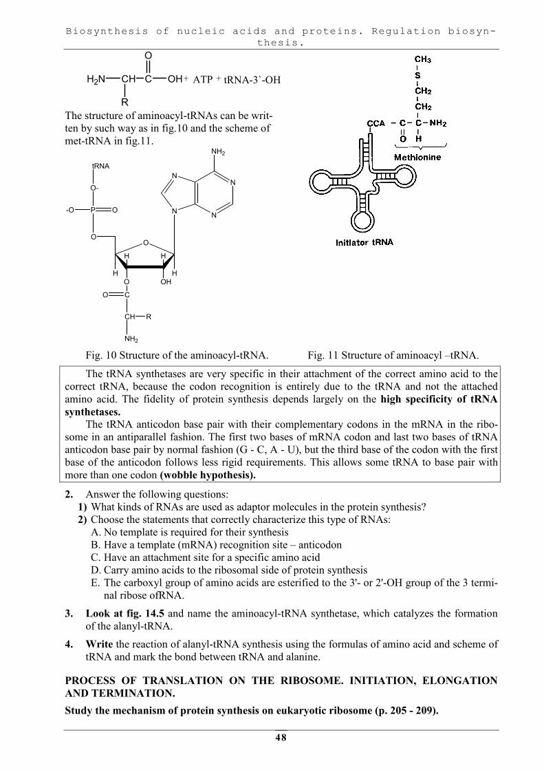
Aminoacyl- tRNA synthetases are enzymes that activate amino acids and attach them to their
appropriate tRNA: a-carboxyl group of amino acids are joined to the 2' or 3' hydroxyl group of the
3' terminal adenosine of tRNA. The sum reaction of this step is expressed as:
Biosynthesis of nucleic acids and proteins. Regulation biosyn-
thesis.
48
H2N CH C
R
OH
O
ATP+ + tRNA-3`-OH
The structure of aminoacyl-tRNAs can be writ-
ten by such way as in fig.10 and the scheme of
met-tRNA in fig.11.
N
NN
N
NH2
O
OHO
HH
HH
P O
O-
O
-O
tRNA
NH2
CH
C
R
O
Fig. 10 Structure of the aminoacyl-tRNA. Fig. 11 Structure of aminoacyl –tRNA.
The tRNA synthetases are very specific in their attachment of the correct amino acid to the
correct tRNA, because the codon recognition is entirely due to the tRNA and not the attached
amino acid. The fidelity of protein synthesis depends largely on the high specificity of tRNA
synthetases. The tRNA anticodon base pair with their complementary codons in the mRNA in the ribo-
some in an antiparallel fashion. The first two bases of mRNA codon and last two bases of tRNA
anticodon base pair by normal fashion (G - C, A - U), but the third base of the codon with the first
base of the anticodon follows less rigid requirements. This allows some tRNA to base pair with
more than one codon (wobble hypothesis).
2. Answer the following questions:
1) What kinds of RNAs are used as adaptor molecules in the protein synthesis?
2) Choose the statements that correctly characterize this type of RNAs:
A. No template is required for their synthesis
B. Have a template (mRNA) recognition site – anticodon
C. Have an attachment site for a specific amino acid
D. Carry amino acids to the ribosomal side of protein synthesis
E. The carboxyl group of amino acids are esterified to the 3'- or 2'-OH group of the 3 termi-
nal ribose ofRNA.
3. Look at fig. 14.5 and name the aminoacyl-tRNA synthetase, which catalyzes the formation
of the alanyl-tRNA.
4. Write the reaction of alanyl-tRNA synthesis using the formulas of amino acid and scheme of
tRNA and mark the bond between tRNA and alanine.
PROCESS OF TRANSLATION ON THE RIBOSOME. INITIATION, ELONGATION
AND TERMINATION.
Study the mechanism of protein synthesis on eukaryotic ribosome (p. 205 - 209).

Biosynthesis of nucleic acids and proteins. Regulation biosyn-
thesis.
49
1. Mind that translation of the mRNA commences near its 5' terminal with the formation of the
corresponding amino terminal of the protein molecule. The message is read from 5' to 3',
concluding with the formation of the carboxyl terminal of the protein. Protein synthesis oc-
curs in three stages: initiation, elongation, and termination.
a) Initiation of translation (fig. 14.8, p. 206) begins with the formation of a 40S initiation com-
plex, which contains the following: a strand of mRNA, at least 10 eukaryotic initiation factors
(elFs), 40S ribosomal subunit, initiator Met-tRNAiMet
, GTP and ATP. The 40S ribosomal
subunit binds near the 5' end of the mRNA and scans the mRNA in the 3' direction (ATP hy-
drolysis may be essential for this scanning process) until it finds the first AUG codon (initia-
tion codon). Other elFs bind, GTP is hydrolyzed, the large ribosomal subunit (60 S) binds
and initiation factors are released. The rapid association of the 40S and 60S subunit forms
the 80S ribosome. Two binding sites for tRNA, known as P (peptidyl) and A (aminoacyi)
sites, are present on the ribosome. At this stage Met-tRNAi Met
binds at the P site of the ri-
bosome, ready for elongation cycle to commence.
b) Elongation (fig. 14.10, p.208), a cyclic process, involves several steps:
• Binding of aminoacyl-tRNA to the A site with the use of GTP and elongation factors
eEF lα and eEF 1βγ (eEF= eukaryotic elongation factor).
• Peptide bond formation is catalyzed by peptidyltransferase (fig.3.5), which is not pro-
tein but 28S rRNA of the large ribosomal (60S) subunit. The tRNA in the A site now
contains the growing polypeptide chain, and the tRNA in the P site is uncharged.
• Translocation moves the mRNA and its base-paired tRNA with respect to the ribosome
toward the 3'-end. As the result the uncharged tRNA quickly releases from the P site. The
newly formed peptidyl-tRNA at the A site moves into the empty P site and the next co-
don of the mRNA occupies the A site. This process involves another elongation factor
EF2 and GTP which is hydrolyzed to GDP during translocation.
c) Termination of translation proceeds when terminating codon of mRNA (UUA, UAG, UGA)
appears in the A site. Releasing factors (eRF) are capable of recognizing that a termination
signal resides in the A site, they in conjunction with GTP and the peptidyltransferase promote
to hydrolyze the bond between the peptide chain and tRNA, releasing the protein and tRNA,
and to dissociate 80 S ribosome into its 40S and 60S subunits.
Fig. 12 Formation of a peptide bond.

Biosynthesis of nucleic acids and proteins. Regulation biosyn-
thesis.
50
2. Choose the correct statements. In protein biosynthesis:
A. Each amino acid recognizes its own codon by a direct interaction with the mRNA tem-
plate.
B. The formation of peptide bonds by ribosomal messenger RNA complex continues until a
stop codon on mRNA is reached.
C. Peptide bond formation ceases when the ribosome reaches the 5' end of the mRNA.
D. A given codon and its anticodon must have identical base sequences in order for them to
be proper base.
E. Each amino acid is added in its proper place to a growing peptide chain through the
"adaptor" function of tRNA.
3. Add the components, which are required for receiving complete cell-free protein synthesiz-
ing system:
1) ATP and GTP;
2) Amino acids (20);
3) Ribosomes;
4) 4Mg2+.
5) Factors of initiation; elongation and termination;
6) >20 tRNA;
7) ?;
8) ?
4. Match the function of 7 and 8 components in protein synthesis:
A. Enzyme catalyzes the formation of peptide bonds
B. The sources of energy
C. Substrates for mRNA synthesis
D. Enzymes activate amino acids and attach them to tRNA
E. Template of protein synthesis.
5. The figure below shows the structure of the non-transcribed strand of the DNA:
5'.............-CCACTATAAAG........GCCATGGGGGTA.........3'
A. Write down the sequence of the complementary DNA strand.
B. Underline the TATA box.
C. Write the sequence of the nucleotides of the mRNA coding the underlining part of DNA.
D. Underline the start codon for the protein.
E. Write the amino acids sequence of this fragment of protein.
F. Using fig. 14.1 draw scheme of incorporating amino acid coded by GUA triplet in the
growing polypeptide chain, in your copy-book.
POSTTRANSLATIONAL PROCESSING OF PROTEINS
Study Posttranslational processing of proteins.
1. Study the unit "Posttranslational processing of protein on p. 209-210 and answer the ques-
tion:
2. What proteins bind to a nascent polypeptide and mediate the folding process?
3. Choose appropriate alteration of amino acids residues in the nascent polypeptide chain and
fill in table:
Posttranslational alterations of polypeptide chains
Amino acid radicals Posttranslational alteration
N-terminal met
Cys

Biosynthesis of nucleic acids and proteins. Regulation biosyn-
thesis.
51
Pro
Lys
Ser, Thr, Tyr
INHIBITORS OF DNA, RNA AND PROTEIN SYNTHESIS
Study some inhibitors of DNA, RNA and protein synthesis.
1. Note that the cessation of DNA, RNA or protein synthesis causes the death of cells.
Inhibitors of the template synthesis are used as drugs in the treatment of various neoplasms or
bacterial infections. For these purposes antibiotics are often used.
a) Antibiotics are drugs in chemotherapy. Successful treatment of tumors with drugs de-
pends upon the greater sensitivity of neoplastic cells to the treatment than that of normal cells.
Proliferating cells, such as neoplastic cells, are generally more sensitive to these agents than are
quiescent cells, because they are more penetrative and have usually high activity of DNA and
RNA synthesis. In this case cells that normally proliferate rapidly: hair follicle cells, cells of the
hematopoietic system and cells, that line the gut, are also damaged by such drugs.
b) Antibiotics are antibacterial drugs. On the other hand many antibiotics selectively in-
hibit protein synthesis in bacteria. The bacterial ribosome is smaller (70S with 50S and 30S sub-
units) than 80S mammalian ribosome and has a different complement of RNA and protein mole-
cules. Many antibiotics interact with the proteins of prokaryotic ribosomes and thus inhibit protein
synthesis. This results in growth arrest or death of the bacterium. Such antibiotics do not interact
with components of eukaryotic ribosome and thus are not toxic to eukaryotes.
Some antibiotics (puromycin for example) inhibit protein synthesis on all ribosomes but the
others only on eukaryotic cells (cycloheximide). They are not clinically useful but important in
scientific purposes.
c) Poisons, toxins and interferons are inhibitors of the template biosynthesis For exam-
ple: αααα-amanitin, a compound driven from the mushroom Amanita phalloides, inhibits eukaryotic
RNA polymerases; diphtheria toxin, an exotoxin of Corynebacterium diphtheriae infected with
specific lysogenic phage, catalyzes the ADP-ribosylation of eEF-2 in mammalian cells. This mod-
ification inactivates eEF-2 and inhibits mammalian protein synthesis. In the human organism in-
terferons possess a high antiviral effect. They inhibit synthesis of the proteins in cells that have
been infected by a virus. They:
- inhibit synthesis of the proteins required for viral replication;
- stimulate synthesis of an enzyme that produces an oligonucleotide (2'-5'-oligo(A))that acti-
vates a ribonuclease. This RNase degrades mRNA and in such a way inhibits protein synthesis.
2. Look at table 2 and choose the correct couples. Drug used for:
A. - antibacterial treatment;
B. - chemotherapy.
Drugs as inhibitors of template biosynthesis
№ Drugs Mode of action
1 Doxorubicin binds to DNA by intercalation (insertion between adjacent base pairs)
2 Tetracycline Binds to the 30S ribosomal subunit and inhibits binding of aminoacyl-tRNA
3 Bleomycin Causes chromosomal breaks, fragmentation
4 Vinblastine Produces metaphase arrest during mitosis
5 Erythromycin Binds to the 50S ribosomal subunit and prevents translocation
3. Which of the following statements about inhibitors of protein synthesis is correct A. Erythromycin inhibits DNA synthesis causing chromosomal breaks.
B. Diphtheria toxin inactivates the eukaryotic elongation factor eEF-2.
C. α-Amanitin inhibits eukaryotic RNA polymerases.
D. Doxorubicin binds to the 30S subunit and distorts its structure.
E. Bleomycin inhibits DNA synthesis causing fragmentation of molecule.

Biosynthesis of nucleic acids and proteins. Regulation biosyn-
thesis.
52
Essey for lesson 8:
The inhibitors of protein synthesis. An effect of antibiotics and toxins on this process.
Homework:
Study lesson 9.

Biosynthesis of nucleic acids and proteins. Regulation biosyn-
thesis.
53
LESSON 9. REGULATION OF GENE EXPRESSION. POLYMORPHISM OF THE
PROTEINS. USE OF RECOMBINANT DNA TECHNIQUES IN MEDICINE.
MA IN TO P IC S :
• Regulation of gene expression in prokaryotes. Hypothesis of operon. Lac-inducible operon.
His-repressible operon.
• Regulation of gene expression in prokaryotes by stimulation of RNA polymerase binding, by
sigma factors, by attenuation of transcription.
• Regulation of gene expression in eukaryotes at multiple levels.
• Mechanism of action of hormones wich act through secondary messenger cAMP.
• Mechanism of action of steroid hormones on genetic apparatus.
• Regulation biosynthesis of proteins at the level of translation and the stability of mRNA. Al-
ternative splicing and RNA editing.
• Degradation of proteins in eukaryotic cells. The role of ubiquetine, lysosomes, autho-
phagosomes, endosomes, secondary lysosomes, peroxisomes.
• Hereditary diseases as a result of genetic desturbances (biochemical aspects).
• International programme “Human genome project”.
• Gene therapy.
• Polymerase chain reaction – method of studing genome and laboratory diagnostics.
• Use of recombinant DNA techniques in medicine.
HYPOTHESIS OF OPERON: LAC- INDUCIBLE AND TRP REPRESSIBLE
OPERONS.
Study the regulation of gene expression in prokaryotes (p. 215 - 222).
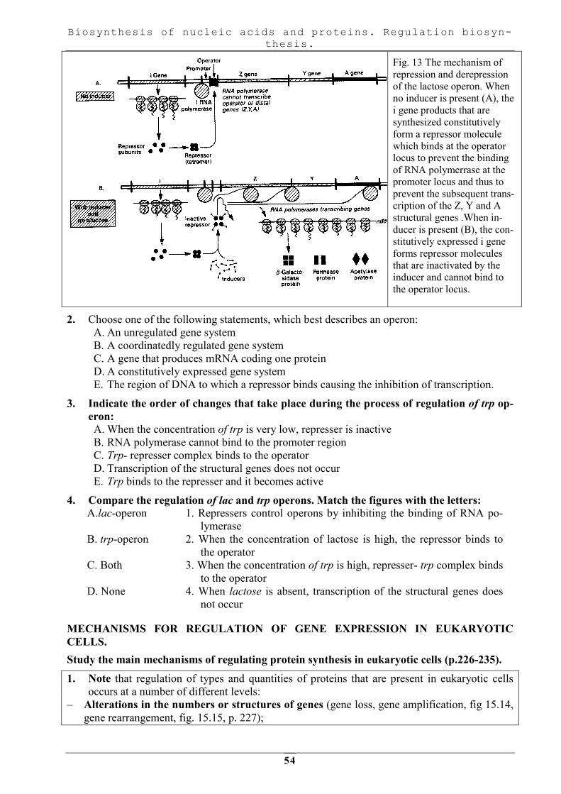
1. Memorize. Prokaryotes regulate expression of genes mainly at the transcriptional level
through genetic units known as operons. An operon consists of a set of genes that produce a
series of proteins under the control of a single promoter. Regulatory proteins bind to the pro-
moter and facilitate or inhibit the binding of RNA polymerase. There are the following kinds
of genes:
- Inducible genes. Some proteins are called to be inducible because they are produced in sig-
nificant amounts when a specific inducing substance (inducer) is present. For example, the
production of the enzymes, which use lactose, is induced by the presence of its substrate, lac-
tose, in the medium (Fig. 3.6, 15.4, p.218 and 15.6, p.219).
- Repressible genes. Operons regulated by repression are expressed until small molecules
known as corepressors enter the cell. The corepressor binds to the repressor protein. The re-
pressor - corepressor complex then binds to the operator and prevents binding of RNA-
polymerase. The structural genes no longer produce proteins (Fig. 15.7, p.218).
Constitutive genes refer to prokaryotic genes whose expression is not regulated.
The products of these genes are produced at a constant, often low rate and their expression is
said to be constitutive.
Biosynthesis of nucleic acids and proteins. Regulation biosyn-
thesis.
54
Fig. 13 The mechanism of
repression and derepression
of the lactose operon. When
no inducer is present (A), the
i gene products that are
synthesized constitutively
form a repressor molecule
which binds at the operator
locus to prevent the binding
of RNA polymerrase at the
promoter locus and thus to
prevent the subsequent trans-
cription of the Z, Y and A
structural genes .When in-
ducer is present (B), the con-
stitutively expressed i gene
forms repressor molecules
that are inactivated by the
inducer and cannot bind to
the operator locus.
2. Choose one of the following statements, which best describes an operon:
A. An unregulated gene system
B. A coordinatedly regulated gene system
C. A gene that produces mRNA coding one protein
D. A constitutively expressed gene system
E. The region of DNA to which a repressor binds causing the inhibition of transcription.
3. Indicate the order of changes that take place during the process of regulation of trp op-
eron: A. When the concentration of trp is very low, represser is inactive
B. RNA polymerase cannot bind to the promoter region
C. Trp- represser complex binds to the operator
D. Transcription of the structural genes does not occur
E. Trp binds to the represser and it becomes active
4. Compare the regulation of lac and trp operons. Match the figures with the letters:
A.lac-operon 1. Repressers control operons by inhibiting the binding of RNA po-
lymerase
B. trp-operon 2. When the concentration of lactose is high, the repressor binds to
the operator
C. Both 3. When the concentration of trp is high, represser- trp complex binds
to the operator
D. None 4. When lactose is absent, transcription of the structural genes does
not occur
MECHANISMS FOR REGULATION OF GENE EXPRESSION IN EUKARYOTIC
CELLS.
Study the main mechanisms of regulating protein synthesis in eukaryotic cells (p.226-235).
1. Note that regulation of types and quantities of proteins that are present in eukaryotic cells
occurs at a number of different levels:
– Alterations in the numbers or structures of genes (gene loss, gene amplification, fig 15.14,
gene rearrangement, fig. 15.15, p. 227);
Biosynthesis of nucleic acids and proteins. Regulation biosyn-
thesis.
55
– At the level of transcription (condensation of chromatin, fig. 15.16, activation of specific
genes, fig. 15.17, p.229);
– At the level of posttranscriptional modification (during processing of the primary transcript,
fig. 15.20, p. 231, transport of mRNA from the nucleus to the cytoplasm);
– At the level of translation (with the help of the initiation factors for translation, fig. 15.24,
p.235 or regulatory proteins, which bind to the mRNA, fig. 15.23, p.234);
– At the level of posttranslational maturation by proteolytic degradation.
2. Answer the following question: Some genes that normally exist in a low copy number are able to undergo selective gene amplifi-
cation. For example, metallothionein is a low molecular weight protein that binds heavy metals
such as copper, mercury, zinc, and cadmium.
The binding of heavy metals to metallothionein protects cells from heavy metal toxicity. In re-
sponse to increasing amounts of heavy metals, cells amplify their metallothionein genes.
What kind of template synthesis will be increased in this case?
3. Regulation at the level of transcription. A typical nucleus contains chromatin that is condensed (heterochromatin) and chromatin that is
diffused (euchromatin). The genes in heterochromatin are inactive, while those in euchromatin
produce mRNA. Specific genes are activated by inducers. The inducers, such as steroid hormones,
enter the cells and bind to the receptor proteins (Fig. 15.17.). The hormone-receptor complex
binds to a response element on DNA: enhancers or silencers and activates (if it binds to enhan-
cer) or, in some cases, inactivates (if it binds to silencer) gene transcription. Look at fig. 15.18.
and answer the following question:
What differs the activation of sets of genes by single inducer in eukaryotes from the process that
regulates bacterial operons?
Study fig. 13.8, p. 186 and remember the regions of DNA molecule that are recognized. by RNA
polymerase a and proteins that regulate the frequency of transcription..
Note that enhancers and silencers differ from promoters in that their sequences are dissimilar and
they may be located thousands of base pairs from the startpoint of transcription. They are tissue-
or species – specific.
3. Choose the correct statements Enhancer:
A. Is a segment of DNA
B. Is required for the transcription of gene at a basal level
C. Activates transcription of gene irrespective of their relative position in DNA
D. Has no polarity
E. Is nonspecific for tissue or species.
5. Regulation at the level of posttranscriptional modification. Study fig. 15.20, p.231 and fig.
15.21, p.233 and pay attention that the use of alternative splicing and polyadenylation sites
causes different proteins to be produced from the same gene.
6. The regulation of gene expression in eukaryotic cells may be accomplished by: A. The binding of trans-elements to the response -elements within the DNA sequence
B. Alternative splicing
C. Gene amplification
D. Condensation of chromatin
E. Activation of sets of genes by a single inducer.
USE OF RECOMBINANT DNA TECHNIQUES IN MEDICINE.
Study recombinant DNA technology, which increases our knowledge of gene expression and
cause of many diseases.

Biosynthesis of nucleic acids and proteins. Regulation biosyn-
thesis.
56
1. Note that recombinant DNA technology leads to new approaches for the diagnosis and
treatment of many diseases. Read chapter 16 ( p. 239-258 ) and remember that DNA must be
isolated from the appropriate source and adequate amounts must be available for study to
recognize normal or pathological genetic variations. These include:
• The use of restriction endonucleases that permit the dissection of huge DNA molecules into
defined fragments (p.241,242).
• The development of cloning technigues, polymerase chain reaction (PCR) provides the me-
chanisms for amplification of specific nucleotide sequences.
Gel electrophoresis, blotting into nitrocellulose paper and the preparation of labeled probes, which hybridize to the appropriate target DNA sequences have allowed the identification and ma-
nipulation of nucleotide sequences of interest. These experimental approaches have permitted the
identification of nucleotide sequences in DNA that are responsible for certain genetic diseases
such as: growth hormone deficiency, sickle cell anemia, a- and p- thalassemia, phenylketonuria
and some others. A profoundly simple concept has been established: mutant genes cause the pro-
duction of defective or deficient proteins resulting in impaired function and ultimately clinical
disease.
2. Choose the one best answer. The components of PCR are a DNA template, dNTP and heat-stable DNA polymerase. These
components are mixed and placed into a thermal cycler, which will cycle repeatedly at the tem-
perature of 55C°. 72C° and 92C°for various periods of incubation.
What is the major function of the 92C° temperature?
A. It is the optimal temperature for DNA polymerization
B. To increase the specificity of the binding of the primers
C. To denature the DNA strands
D. To drive the polymerase reaction to completion
E. To reanneal newly synthesized DNA strands.
Essey for lesson 9:
1. The translocation V, D, J - segments of gene immunoglobulins (Ig) is source of variety speci-
ficity genes.
2. Use of recombinant DNA techniques in medicine.
Homework: prepare to colloquium.
Textbook: “Basic Medical Biochemistry", D.B. Marks et al. Lecture.

Biosynthesis of nucleic acids and proteins. Regulation biosyn-
thesis.
57
LESSON 10. COLLOQUIUM: BIOSYNTHESIS OF NUCLEIC ACIDS AND PROTEINS. REGULATION OF BIOSYNTHESIS.
MA IN TH E O R E TIC A L TO P IC S FO R R EV IS IO N :
1. Chemical composition of DNA and RNA. Nitrogenus bases of nucleic acids (purine
and pyrimidine). Ribonucleotides and deoxyribonucleotides.The structure and no-
menclature.
2. Synthesis of the purine nucleotides. Sources of the various atoms of the purine bases.
Salvage of bases. Regulation.
3. Synthesis of the pyrimidines. Sources of the various atoms of pyrimidines. Salvage of
bases. Regulation. Orotic aciduria.
4. Scheme of the synthesis of deoxyribonucleosides. Regulation.
5. Scheme of degradation of nucleic acids. Enzymes, substrates, products.
6. Degradation of the purine nucleotides. Hyperuricemia. Gout.
7. Degradation of the pyrimidines.
8. DNA and RNA. Similarity and differences of composition, primary structure, func-
tions, cells localization.
9. Secondary structure of DNA (model of Watson and Crick). Bonds wich are resposible
for stability of the molecule.
10. Specialized forms of RNA. Characterictics of primary, secondary and tertiary struc-
ture of RNA. Structure and biological role of ribosomes.
11. The peculiarity of formation of nucleoproteins. DNA-binding proteins: helix-spire
(round)-helix; lucines zip-fasteners; Zn-fingers; histones.
12. Role of histones in the formation of tertiary structure of DNA. Chromatin. Chromo-
somes. Covalent modifications of histones and their role in regulation structure and
activity of chromatin.
13. Replication of DNA. Definition of the process. Principles of replication ( complemen-
tary, antiparallel, semiconservatism, unipolarity, bidirectionality, coordination of rep-
lication and cell cycle). Stages of replication.
14. Replication in prokaryotes. Initiation. Enzymes and proteins which participate in for-
mation of replication fork: helicase, topoisomerase, single-strand binding proteins,
primase.
15. Elongation and termination. Kinds and functions of DNA polymerases. Leading and
lagging strands, Okazaki fragments. Function of DNA ligase.
16. Replication in eukaryotes. Points of origin for replication. Eukaryotic DNA poly-
merases.
17. Replication of the ends of chromosomes. Telomeres. Action of telomerase.
18. Mechanism replication of various viruses. Retrovirus. Reversal transcriptase.
19. Damage and repair of DNA. Types of damage. Molecular mutations.
20. Mechanisms of repair. Enzymes wich take part in this process. Defects in DNA repair
systems and hereditary diseases.
21. Gene as functional unit of DNA.The human genome.Features of organization genome
in eukaryotes (enhancers, silencers, cis-elements). Structure of gene in eukaryotes.
Mosaic of structural genes in eukaryotes ( introns and exons). 3 kinds of eukaryotic
genes. Features of organization genome in prokaryotes. Structure of operon.
22. Transcription. Definition. Principles of transcription (complementarity, antiparallelity,
semiconservatism, unipolarity, RNA polymerases can initiate the synthesis of new
strands, asymmetricity). Stages of transcription. Transcription in prokaryotes. Сharac-
teristic оf transcriptional apparatus: substrats, template (coding strand and template
strand), expense of energy, enzymes. Structure of RNA polymerase: role of subunits
(α2 ββ'σ). Structure of promoter (Pribnov box). Initiation of this process.
Biosynthesis of nucleic acids and proteins. Regulation biosyn-
thesis.
58
23. Elongation and termination of transcription (ρ – dependent and ρ- independent termi-
nation)
24. Transcription in eukaryotes: 3 types of RNA polymerases (I, II, III). Structure of
promoter. Ваsal factors of transcription. Role of TFIID and TFIIH factors in initia-
tion of transcription. Structure of regulatory DNA binding proteins ( gene- specific
transcription factors).
25. Processing of the primary transcript RNA. Capping of 5' – end, addition of a poly (A)
tail, splicing of pre-mRNA. Spliceosome. Role of snRNA in the removal of introns
and splicing splicing of exons.
26. Synthesis of protein. Translation. The genetic code, features in eukaryotes. Basic
components of complex, wich takes part in synthesis of proteins: amino acids,
tRNA, ribosomes, source of energy, factors, enzymes.
27. Structure and functions of ribosomes. Binding sites of ribosomes: peptidyl (P) and
aminoacyl (A) sites.
28. tRNA as adaptor molecule. Aminoacyl-tRNA biosynthesis. Aminoacyl-tRNA syn-
thetases. The high specificity of aminoacyl-tRNA synthetases to substrate.
29. Synthesis of polypeptide on ribosome. Formation of initiation complex in prokaryo-
tes. Shine-Dalgarno sequence. Elongation: formation of peptide bond. Peptidyltrans-
ferase activity of rRNA. Translocation. Termination of translation. Role of protein
factors on each stage..
30. Peculiarity of protein synthesis in eukaryotes.
31. Posttranslational processing of proteins: limited proteolysis, formation of disulfide
bridges, addition of prosthetic groups, covalent modification (glycosylation, methyla-
tion, phosphorylation, acetylation).
32. The formation of spatial conformation (folding)of proteins. Enzymes of folding (di-
sulfide isomerase and peptidilprolil isomerase). The participation of heat shock pro-
teins in this process. Classification of chaperones. Folding of proteins with participa-
tion of chaperonine system. Prionic proteins. Deseases as results of folding disordes.
33. The process of translation on the ribosome. Elongation and termination. The peculi-
arities of synthesis and processing of secretory proteins (collagen and insulin).
34. Turnover of proteins. The ubiquitin-proteasome pathway.
35. Inhibitors of DNA and RNA synthesis. Antibiotics in chemotherapy, antiviral drugs.
36. Inhibitors of protein synthesis. The influence of antibiotics and toxins on protein syn-
thesis. Interferons.
37. Regulation of gene expression in prokaryotes. Hypothesis of operon. Lac-inducible
operon.
38. His-repressible operon.
39. Regulation of gene expression in prokaryotes by stimulation of RNA polymerase
binding, by sigma factors, by attenuation of transcription.
40. Regulation of gene expression in eukaryotes at multiple levels.
41. Mechanism of action of hormones wich act through secondary messenger cAMP.
42. Mechanism of action of steroid hormones on genetic apparatus.
43. Regulation biosynthesis of proteins at the level of translation and the stability of
mRNA. Alternative splicing and RNA editing.
44. Degradation of proteins in eukaryotic cells. The role of ubiquetine, lysosomes, autho-
phagosomes, endosomes, secondary lysosomes, peroxisomes.
45. Hereditary diseases as a result of genetic desturbances (biochemical aspects).
46. International programme “Human genome project”.
47. Gene therapy.
48. Polymerase chain reaction – method of studing genome and laboratory diagnostics.
49. Use of recombinant DNA techniques in medicine.
Biosynthesis of nucleic acids and proteins. Regulation biosyn-
thesis.
59
Home work:
Study lesson 11.