1 metabolism of purine and pyrimidine nucleotides dna replication department of biochemistry 2013...
TRANSCRIPT
1
Metabolism of purine and pyrimidine nucleotides
DNA replication
Department of Biochemistry 2013 (E.T.)
2
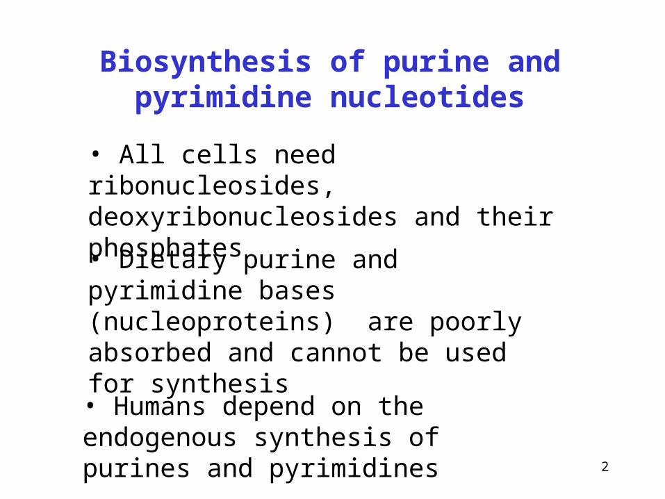
Biosynthesis of purine and pyrimidine nucleotides
• Dietary purine and pyrimidine bases (nucleoproteins) are poorly absorbed and cannot be used for synthesis
• Humans depend on the endogenous synthesis of purines and pyrimidines
• All cells need ribonucleosides, deoxyribonucleosides and their phosphates
3
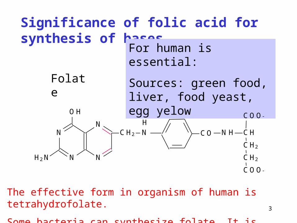
Significance of folic acid for synthesis of bases
Folate
For human is essential:
Sources: green food, liver, food yeast, egg yelow
N
N
O H
N
NC H 2 N
H 2 N
C O N H C H
C H2
C H2
C O O -
C O O -
H
The effective form in organism of human is tetrahydrofolate.
Some bacteria can synthesize folate. It is growth factor for them

4
(dihydro)folate reductase
(catalyzes the both reactions in animals and some microorganisms)
Folate
N
N
OH
NH2N
CO NH CHCH2
CH2
COO-
COO-H
CH2 NN
H
H
tetrahydrofolate
dihydrofolate
NADPH + H+
NADP
NADP
NADPH + H+
N
N
OH
NH2N
CO NH CHCH2
CH2
COO-
COO-H
CH2 NNH
Formation of tetrahydrofolate
+
+
5
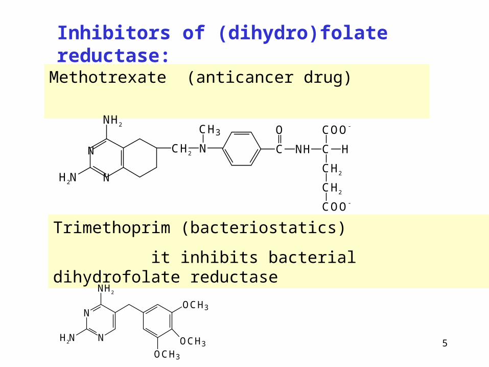
Methotrexate (anticancer drug)
Inhibitors of (dihydro)folate reductase:
NH2
H2N
CH2 N
CH3
C NH
O
C
COO-
H
CH2
CH2
COO-
N
N
Trimethoprim (bacteriostatics)
it inhibits bacterial dihydrofolate reductase
N
N
NH2
H2N
OCH3
OCH3
OCH3

6
• Sulfonamides (e.g. Sulfamethoxazol) are structural analogs of p-aminobenzoic acid.
• p-aminobenzoic acid is necessary for bacterial synthesis of folic acid
• Folic acid is a growth factor for bacterias.
Sulfonamides act as competitve inhibitors of the synthesis.
Sulfonamides stop growth of bacterias dependent on folic acid (streptococcus, haemophilus etc.)
O
OH
NH2
S
O
O
NH2
NH2
Inhibitors of folate synthesis
c c
p-aminobenzoic acid (PABA) sulfanilamide

7
N
N
OH
NH2N
CO NH CHCH2
CH2
COO-
COO-
CH2 NN
H
CH2
N-5,N-10- methylen H4F – synthesis of thymine
N
N
OH
NH2N
CO NH CHCH2
CH2
COO-
COO-
CH2 NN
H
CHOH
N-10-formyl H4F – synthesis of purine
510
1
3
Using of tetrahydrofolate in synthesis of purines and pyrimidines
8
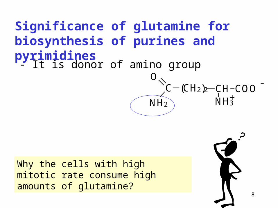
Significance of glutamine for biosynthesis of purines and pyrimidines
CH COONH3
(CH2)2C -
+
O
NH2
- It is donor of amino group
Why the cells with high mitotic rate consume high amounts of glutamine?
9
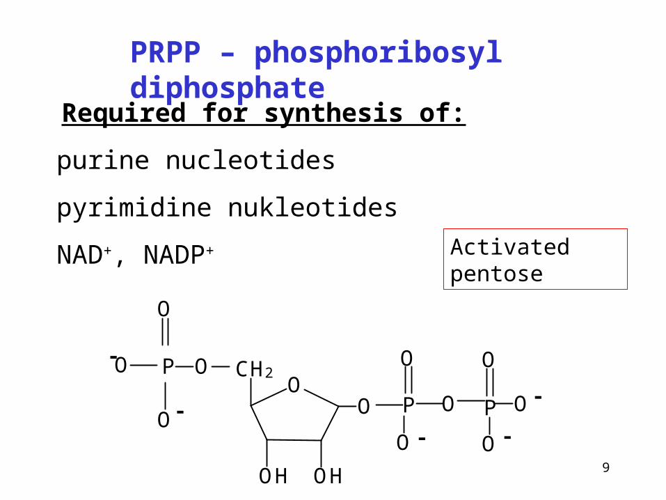
Required for synthesis of:
purine nucleotides
pyrimidine nukleotides
NAD+, NADP+
PRPP – phosphoribosyl diphosphate
OO
CH2OP
O
O
O-
-
OH OH
P P
O
OO
O
-- O
O
-
Activated pentose

10
Ribose-5P + ATP PRPP + AMP
PRPP-synthase
Synthesis of phosphoribosyl diphosphate (PRPP)
OO
CH2OP
O
O
O-
-
OH OH
P P
O
OO
O
-- O
O
-
(kinase)

11
Differences in purine and pyrimidine synthesis
Purines
Synthesis starts with PRPP, purine ring is built step-by-step with C-1 of PRPP as a primer
Pyrimidines
The pyrimidine ring is synthetized before ribose is added
OO
CH2OP
O
O
O-
-
OH OH
P P
O
OO
O
-- O
O
-
N
NO
O
COO -
H
12
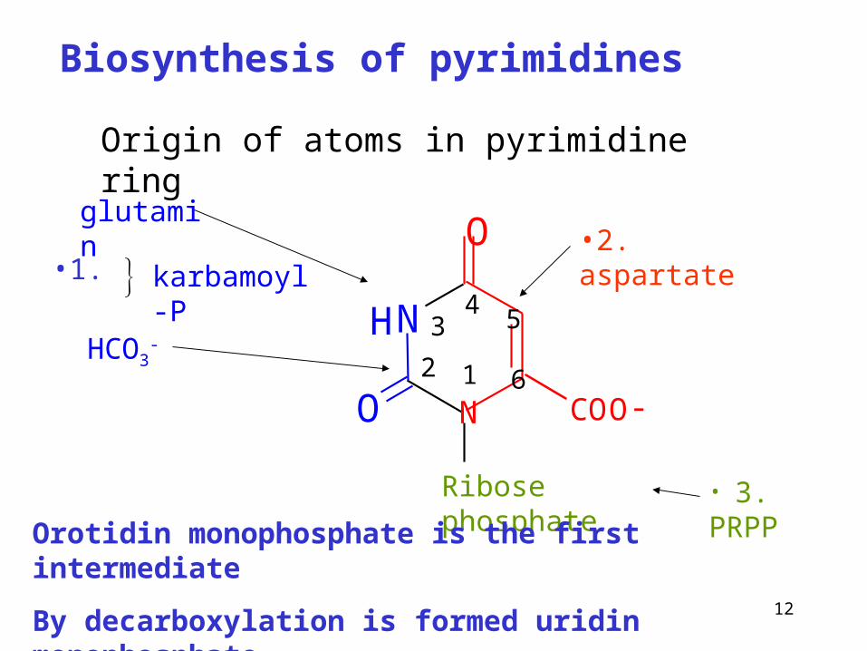
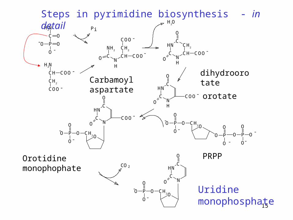
Biosynthesis of pyrimidines
N
O
H
O12
34 5
6N
Ribose phosphate
glutamin
HCO3-
•2. aspartate
• 3. PRPP
COO-
Orotidin monophosphate is the first intermediate
By decarboxylation is formed uridin monophosphate
Origin of atoms in pyrimidine ring
karbamoyl-P•1.
13
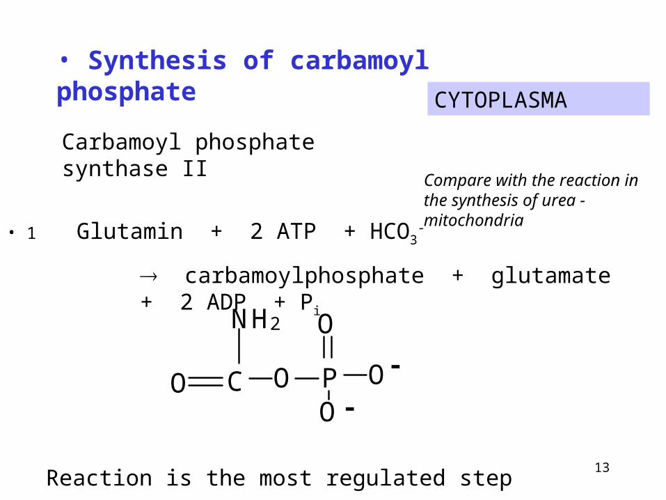
CYTOPLASMA
• 1 Glutamin + 2 ATP + HCO3-
carbamoylphosphate + glutamate + 2 ADP + Pi
• Synthesis of carbamoyl phosphate
CO
NH2
O PO
O
O-
-
Compare with the reaction in the synthesis of urea - mitochondria
Carbamoyl phosphate synthase II
Reaction is the most regulated step

14
Comparision of carbamoyl phosphate synthetases
Enzyme typ Carbamoyl phosphate synthetase I
Carbamoyl phosphate synthetase II
Localization in the cell
mitochondria cytoplasma
Metabolic pathway
synthesis of urea synthesis of pyrimidine
Source of nitrogen
ammonia glutamin
Regulation activation: N-acetylglutamate inhibition: UTP
activation: ATP
15
H2N
CH
CH2
COO
COO
-
-
H2N
C O
P OO
O
-
-
Pi
CN
CH
CH2
COOO
COO
NH2
H
-
H2O
-
HN
CN
CH
CH2
C
O
COOO
-
H
HN
CN
C
O
COOO
-
H
O
O P
O
O
CH2
OP
O
O
O
O P
O
O
O
-
--
-
--
-
HN
CN
C
O
COOO
-
O
O
O
P O CH2O
-
-
HN
CN
C
O
O
O
O
O
P O CH2O
CO2
Steps in pyrimidine biosynthesis - in detail
Carbamoyl aspartatedihydroorotate
orotate
Orotidine monophophate PRPP
Uridine monophosphate
16
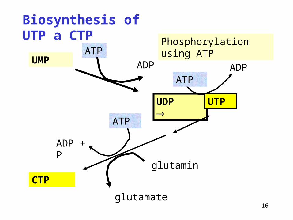
ATP
Biosynthesis of UTP a CTP
UMP
UDP
ADP
ATP
ADP + P
glutamin
glutamate
CTP
ATPADP
UTP
Phosphorylation using ATP

17
N
NH
OO
O
CH3
OH
OH
dUMP dTMP
P
N
NH
OO
O
CH3
OH
OHP
CH2- FH4 FH2
H4F is required for methylation
Formation of dTMP (methylation)
18
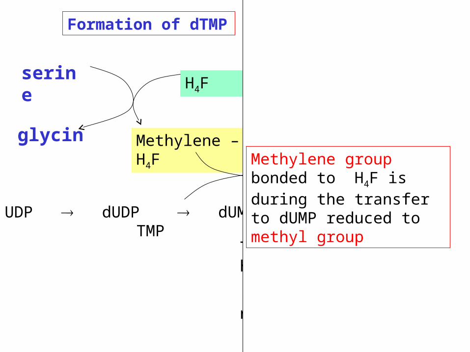
UDP dUDP dUMP TMP
Methylene –H4F DHF
serine
Thymidylátsynthasa
(enzym závislý na folátu)
H4F
Dihydrofolátreduktasa
glycin
Formation of dTMP
NADPH
NADP
Methylene group bonded to H4F is during the transfer to dUMP reduced to methyl group

19
UDP dUDP dUMP TMP
Thymidylate synthase
(folate dependent enzyme)
Dihydrofolate reductase
Formation of TMP
Methylene –H4F H2F
serinH4F
glycin
NADPH
NADP
After release of methylene H4F becomes oxidized to H2F
N
N
O
O
CH3
CH2
OH
O
H

20
N
N
OH
NH2N
CO NH CHCH2
CH2
COO-
COO-
CH2 NN
H
CH2
N
N
OH
NH2N
CO NH CHCH2
CH2
COO-
COO-
CH2 NHN
N
N
OH
NH2N
CO NH CHCH2
CH2
COO-
COO-H
CH2 NN
H
H
NADPH + H+
NADP+
CH2
CH2-H4F
H2F
H4F
H
HH
H
Dihydrofolate reductase

21
(Dihydro)folate reductase
reduces dihydrofolate (H2F) back to tetrahydrofolate (H4F)
Why metotrexate (amethopterine) functions as antineoplastic agent?
NH2
H2N
CH2 N
CH3
C NH
O
C
COO-
H
CH2
CH2
COO-
22
Many antineoplastic drugs inhibit nucleotide metabolism
• The development of drugs with selective toxicity for cancer cells is difficult because cancer cells are too similar to normal cells
• Therefore, agents that are toxic for cancer cells are toxic also for normal cells
• Cancer cells do, however, have a higher mitotic rate than normal cells
• Therefore they have a higher requirement for DNA synthesis
• Most antineoplastic drugs act as antagonists of nucleotide synthesis

23
Dihydrofolate reductase - target of anti-tumour therapy. Aminopterin (4-amino-dihydrofolate) and methotrexate (amethopterin, 4-amino-10-methyl-dihydrofolate) are anti-folate drugs - potent competitive inhibitors of dihydrofolate reductase.
They bind the enzyme 1000x more tightly than folate, they function as competitive inhibitors.

24
Also thymidylate synthase can be inhibited
Cytostatic effect – cell division is stopped
Fluorouracil is converted in vivo into fluorodeoxyuridylate
It irreversibly inhibits thymidylate synthase (suicide inhibition)
All antineoplastic drugs are toxic not only for cancer cells but for all rapidly dividing cells, including those in bone marrow, intstinal mucosa and hair bulbs. Therefore, bone marrow depression, diarrhea, and hair loss are common side effects of cancer chemotherapy.
25
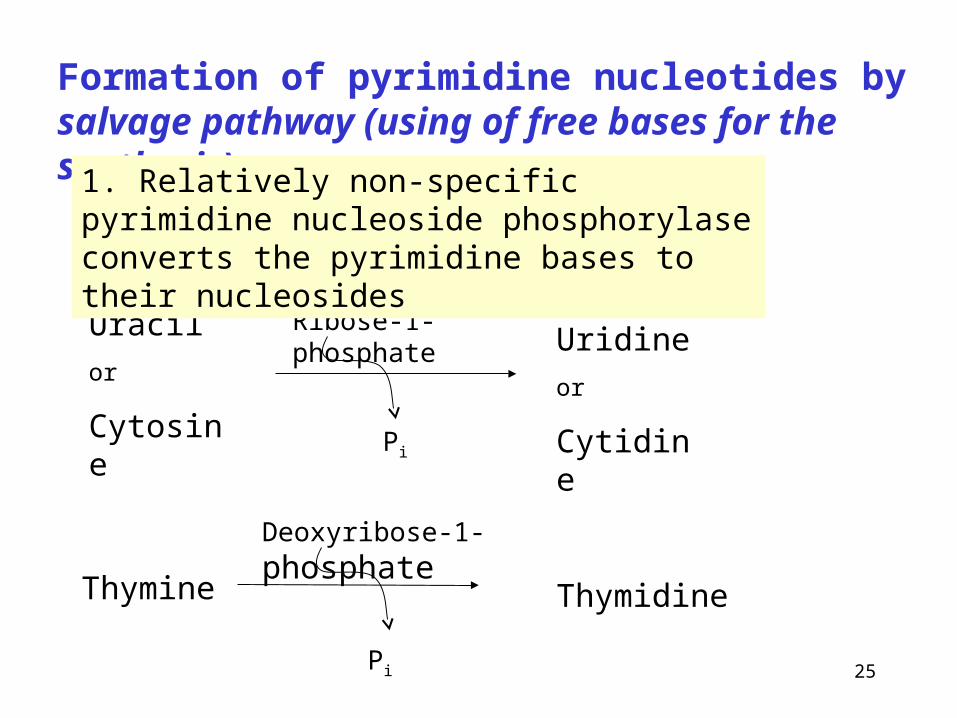
Formation of pyrimidine nucleotides by salvage pathway (using of free bases for the synthesis)
Uracil
or
Cytosine
Ribose-1-phosphate
Pi
Uridine
or
Cytidine
Thymine
Deoxyribose-1-phosphate
Pi
Thymidine
1. Relatively non-specific pyrimidine nucleoside phosphorylase converts the pyrimidine bases to their nucleosides

26
•thymidine + ATP TMP + ADP
•cytidine + ATP CMP + ADP
•deoxycytidine + ATP dCMP + ADP
•uridine + ATP UMP + ADP
2. Formation of nucleotides from nucleosides by action of kinases
27
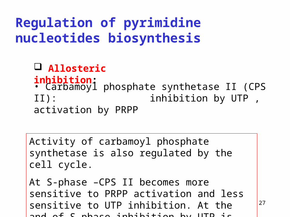
Regulation of pyrimidine nucleotides biosynthesis
• Carbamoyl phosphate synthetase II (CPS II): inhibition by UTP , activation by PRPP
Allosteric inhibition:
Activity of carbamoyl phosphate synthetase is also regulated by the cell cycle.
At S-phase –CPS II becomes more sensitive to PRPP activation and less sensitive to UTP inhibition. At the and of S-phase inhibition by UTP is more pronounced and activation by PRPP is reduced
28
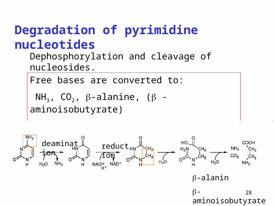
Degradation of pyrimidine nucleotides
Free bases are converted to:
NH3, CO2, -alanine, ( -aminoisobutyrate)
Soluble metabolites – excretion in urine
deamination reduction
-alanin
-aminoisobutyrate from thymine
Dephosphorylation and cleavage of nucleosides.
29
Ribose-5-phosphate
3 glycinHCO3
-
aspartate
formyl-H4F
glutamine
1 PRPP
formyl-H4F
2
4
67
8
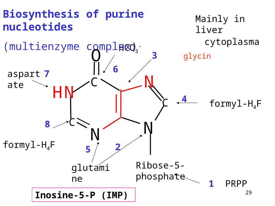
Biosynthesis of purine nucleotides
(multienzyme complex)
Inosine-5-P (IMP)
5
cytoplasma
Mainly in liver
N
HN
O
N
NC
C
C
30
O
O P
O
O
CH2
OP
O
O
O
O P
O
O
O
-
--
-
- OCH
2OP
O
O
O NH2
-
-
OCH
2OP
O
O
O NH
C O
CH2
NH3
-
-
OCH
2OP
O
O
O NH
C O
CH2
NH CHO
-
-OCH
2OP
O
O
O NH
C NH
CH2
NH CHO
-
-
N
N
O
O
O
P O CH2O
H2N-
-
N
N
O
O
O
P O CH2O
H2N
OOC-
-
N
N
O
O
O
P O CH2 O
H2N
COH2N
-
N
N
O
O
O
P O CH2 O
NH
COH2N
OHC
-
O
O
O
P O CH2 O
-
N
N
O
N
N
H
H
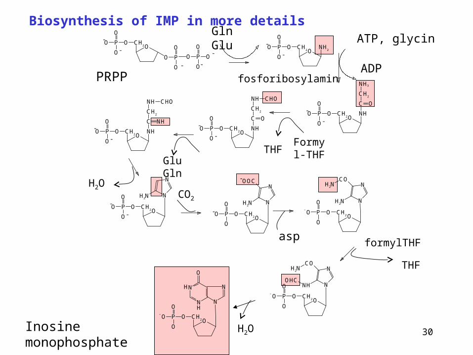
Biosynthesis of IMP in more detailsGln Glu
Inosine monophosphate
ATP, glycin
ADP
Formyl-THFTHF
Glu Gln
H2OCO2
aspformylTHF
THF
H2O
fosforibosylaminPRPP

31
Inosine-5-P (IMP)
Serves as the branchpoint from which adenine and guanine nucleotides can be produced
aspartate, GTP
amination
AMP
oxidation amination GMP
Glutamine, ATP
XMPmycofenolic acid
N
N
O
N
NH
ribose-5-P

32
Mycophenolic acid
• Potent, reversible, uncompetitive inhibitor of IMP dehydrogenase
Used in preventing graft rejection
It blocks de novo formation of GMP supress the proliferation of T and B cells
O
O
O
OH
O
OH
CH3
CH3
CH3

33
Synthesis of AMP and GMP
(more detailed)
IMP
Asp
GTPGDP + Pi fumarate
AMPNAD+
NADH + H+ ATP AMP + PPi
XMP GMP
Gln Glu
H2O
N
N
O
N
N
Ribose
H
P
N
N
N
CH CH2COO
COO
N
N
Ribose P
--
N
N
NH2
N
N
Ribose P
N
N
O
ON
N
RiboseH P
N
N
O
H2N N
N
RiboseH P
HH
34
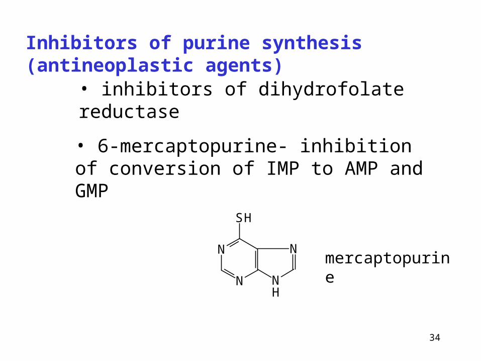
Inhibitors of purine synthesis (antineoplastic agents)
• inhibitors of dihydrofolate reductase
• 6-mercaptopurine- inhibition of conversion of IMP to AMP and GMP
N
N
SH
N
NH
mercaptopurine

35
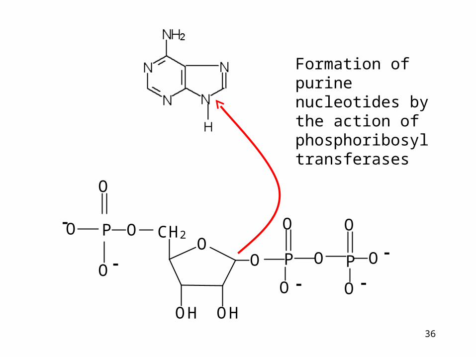
Synthesis of purine nucleotides by salvage pathway
Extrahepatal tissues
Recyclation of free bases
Purine + PRPP purine nucleotide monphosphate + PP
Phosphoribosyltransferases:
Adenine phosphoribosyltransferase Hypoxantine phosphoribosyltransferase
Recyclation of purine bases by phosphoribosyltransferase.
Purine nucleotides are sythesized preferentially by salvage pathway, so long as the free bases are available.
36
OO
CH2OP
O
O
O-
-
OH OH
P P
O
OO
O
-- O
O
-
Formation of purine nucleotides by the action of phosphoribosyl transferases

37
Deficiency of phosphoribosyl transferase results in Lesch-Nyhan syndrom
• X-linked hereditary disease
• purine bases cannot be salvaged
• accumulation of PRPP
• overproduction of purine bases that are degraded to uric acid
• accumulation of uric acid – gout
• neurologic problems : mental retardation, self-mutilation
38
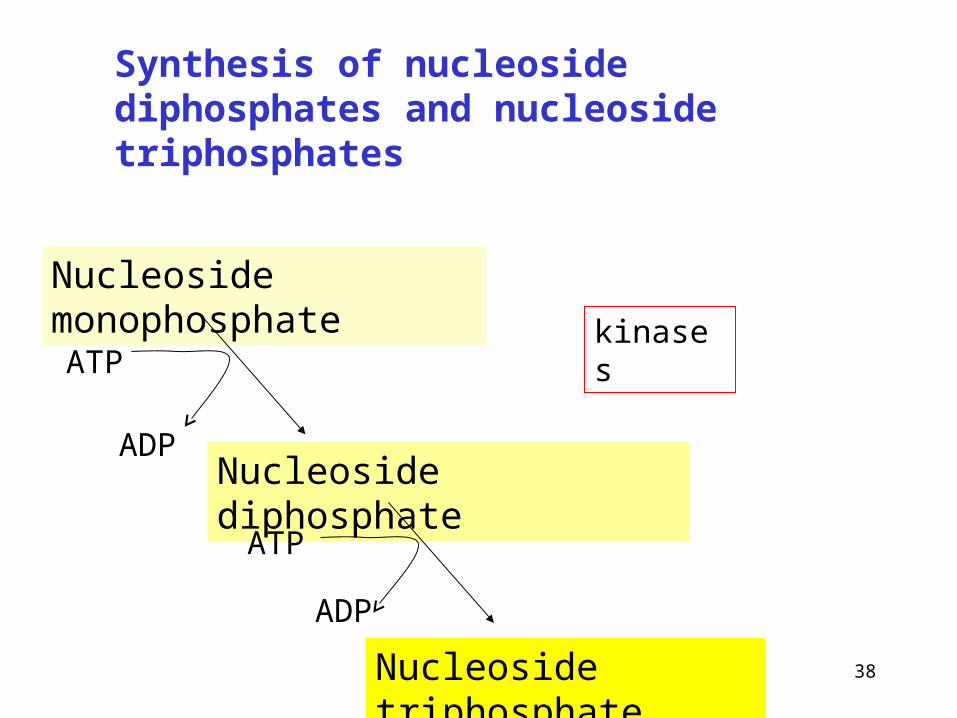
Synthesis of nucleoside diphosphates and nucleoside triphosphates
Nucleoside monophosphate
Nucleoside diphosphate
Nucleoside triphosphate
ATP
ADP
ATP
ADP
kinases

39
Regulation of purine nucleotide biosynthesis
• inhibition of PRPP-glutamylamidotransferase by AMP, GMP, IMP (end-products), activation by PRPP
IMP
AMP
GMP
GMP
AMP
1.
2.
The main factor is availability of PRPP

40
Formation od 2-deoxyribonucleotides
(purine and pyrimidine)
OCH2O
OH OH
basePP
Nucleoside diphosphate 2-deoxynucleoside diphosphate
reduction Thioredoxin, thioredoxin reductase and NADPH are required
H
Thioredoxin reductase is selenoenzyme deoxygenation
41
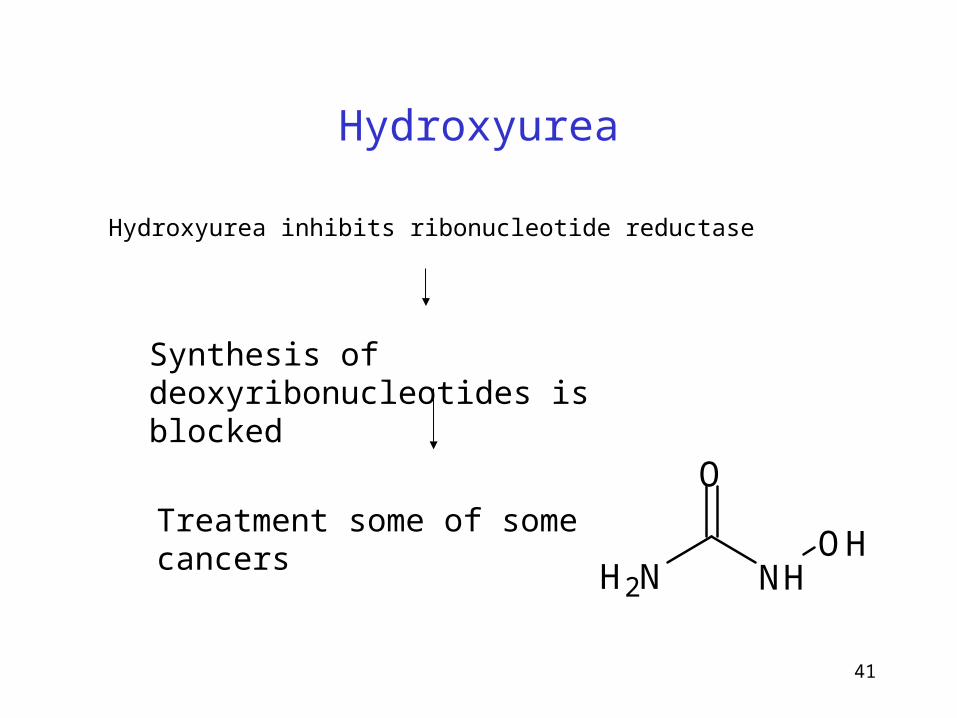
Hydroxyurea
Hydroxyurea inhibits ribonucleotide reductase
Synthesis of deoxyribonucleotides is blocked
Treatment some of some cancers
O
NHNH2
OH
42
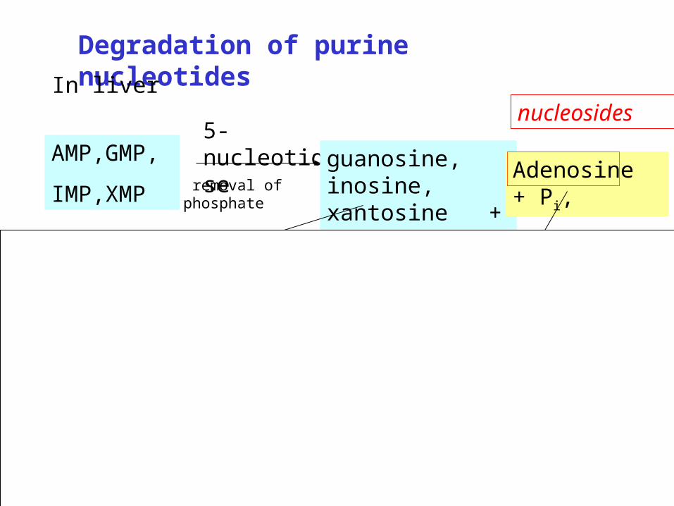
Degradation of purine nucleotides
AMP,GMP,
IMP,XMP
5-nucleotidaseguanosine, inosine, xantosine + Pi
nukleosidfosforylasa
Adenosine + Pi,
guanin,
hypoxantin,
xantin
+ riboso-1-P
adenosindeaminasa
inosinnukleosidfosforylasa
removal of phosphate
In livernucleosides
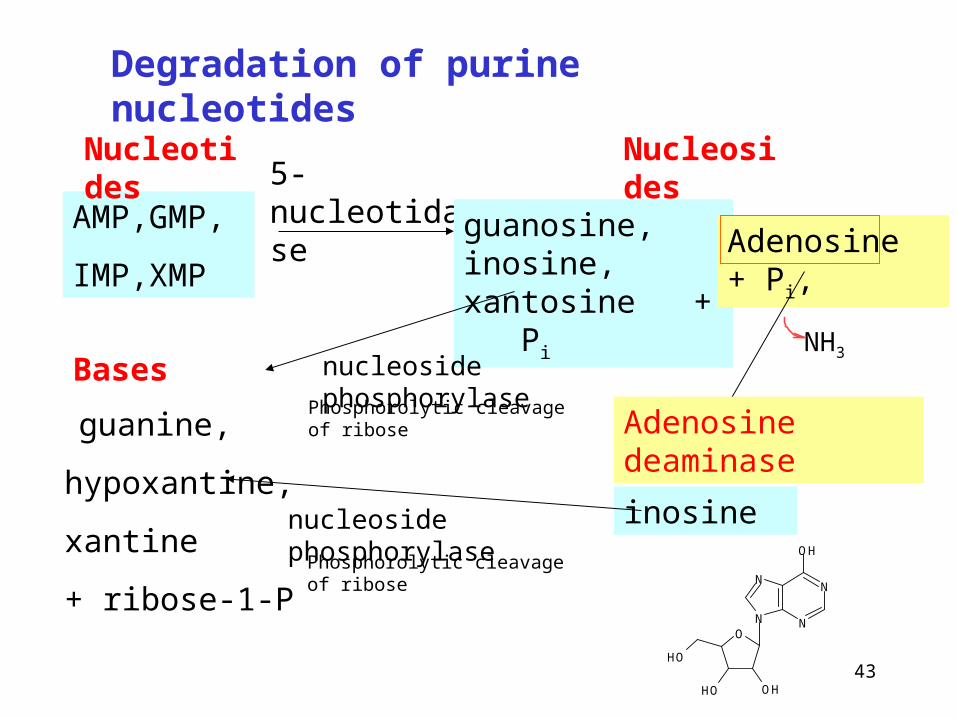
43
AMP,GMP,
IMP,XMP
5-nucleotidase
guanosine, inosine, xantosine + Pi
nucleoside phosphorylase
Adenosine + Pi,
guanine,
hypoxantine,
xantine
+ ribose-1-P
inosinenucleoside phosphorylase
Degradation of purine nucleotides
Nucleotides Nucleosides
BasesNH3
Adenosine deaminase
N
N
N
N
OH
O
OHOH
OH
Phosphorolytic cleavage of ribose
Phosphorolytic cleavage of ribose

44
Adenosine deaminase deficiency
Enzyme deficiency acummulation of adenosine in cells (esp. lymphocytes) conversion to AMP,dAMP, ADP by cellular kinases.
Inhibition of ribonucleotide reductase
Synthesis of other deoxynucleotides drops
Cells cannot make DNA and devide.
One of the causes severe combined immunodeficiency disease (SCID).
Treatment by gene therapy
Findings of deoxyadenosine in urine

45
hypoxantine xantine Uric acid
Xantine oxidase
guaninguanase
Final product of human purine degradation (400-600 mg /day)
Inhibition by oxypurinol
Degradation of purine bases
46
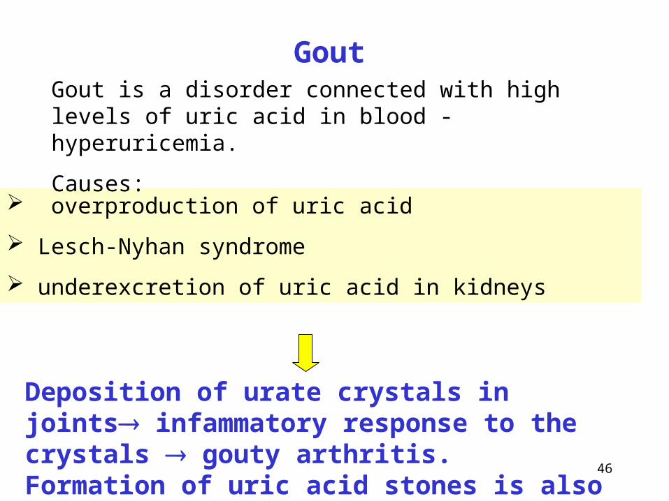
overproduction of uric acid
Lesch-Nyhan syndrome
underexcretion of uric acid in kidneys
Deposition of urate crystals in joints infammatory response to the crystals gouty arthritis. Formation of uric acid stones is also possible.
GoutGout is a disorder connected with high levels of uric acid in blood - hyperuricemia.
Causes:
47
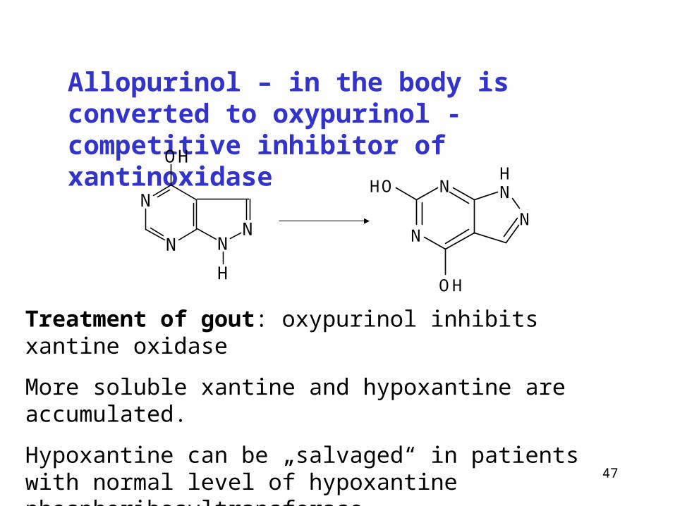
Allopurinol – in the body is converted to oxypurinol - competitive inhibitor of xantinoxidase
Treatment of gout: oxypurinol inhibits xantine oxidase
More soluble xantine and hypoxantine are accumulated.
Hypoxantine can be „salvaged“ in patients with normal level of hypoxantine phosphoribosyltransferase.
N
N
OH
NN
H
N NH
NN
OH
OH

48
Replication of DNA

49
Replication of DNA
Each of the two parental strands serves as a template for the synthesis of complementary strand
Bases in the new strand are attached on the principle of complementarity to the bases in the template strand
Location: nucleus
50
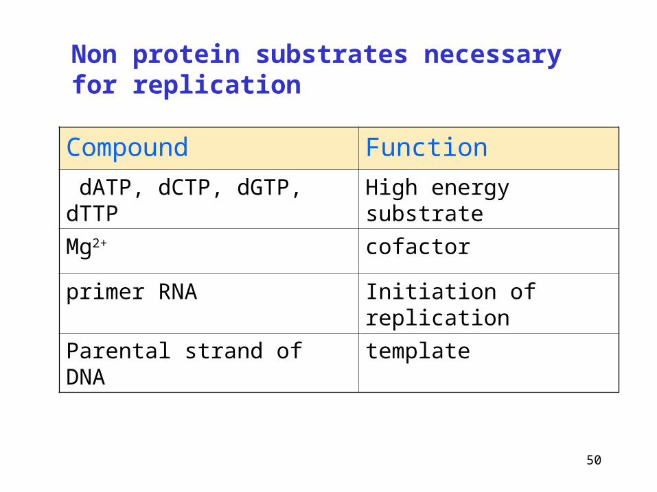
Non protein substrates necessary for replication
Compound Function
dATP, dCTP, dGTP, dTTP High energy substrate
Mg2+ cofactor
primer RNA Initiation of replication
Parental strand of DNA template
51
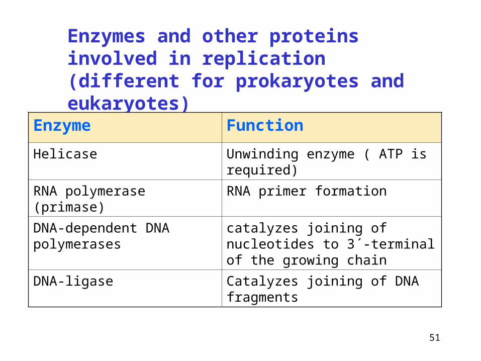
Enzymes and other proteins involved in replication (different for prokaryotes and eukaryotes)
Enzyme Function
Helicase Unwinding enzyme ( ATP is required)
RNA polymerase (primase) RNA primer formation
DNA-dependent DNA polymerases
catalyzes joining of nucleotides to 3´-terminal of the growing chain
DNA-ligase Catalyzes joining of DNA fragments
52
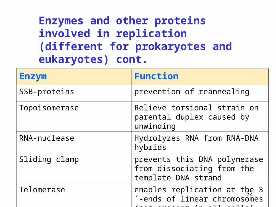
Enzym Function
SSB-proteins prevention of reannealing
Topoisomerase Relieve torsional strain on parental duplex caused by unwinding
RNA-nuclease Hydrolyzes RNA from RNA-DNA hybrids
Sliding clamp prevents this DNA polymerase from dissociating from the template DNA strand
Telomerase enables replication at the 3´-ends of linear chromosomes (not present in all cells)
Enzymes and other proteins involved in replication (different for prokaryotes and eukaryotes) cont.

53
The all DNA polymerases attach nucleotides on 3´-end of a growing chain
(new DNA is formed in the direction 5´3´)
Chemical reaction of DNA synthesis
Synthetic process is catalyzed by DNA-polymerases
Already formed strand (DNA or RNA) reacts with deoxyribonucleoside triphosphate (dNTP)
Diphosphate is released and dNMP is attached by ester bond
(DNA)n + dNTP (DNA)n+1 + PPi

54
O
O
CH2
O
P O
O
O-
base 1
H
H
O
O
CH2
O
OP
Obase 2OP
O
OOP
OO
O
-
Mg
Formation of a bond between new deoxynucleotide and the chain of DNA during elongation
dNTP-2 reacts with 3´-terminal of the growing chain
3´-terminal of the growing chain
DNA-polymerase

55
-
base 1O
O
CH2
O
O
O P O
H
O
O
CH2
O
OP base 2O
Chain elongation
dNTP3
+ PPi
Ester bond is formed between the 3´-OH group of growing chain and 5´-phosphate of entering nucleotide
5´
3´
Diphosphate is released (complexed with Mg2+ ions).
56
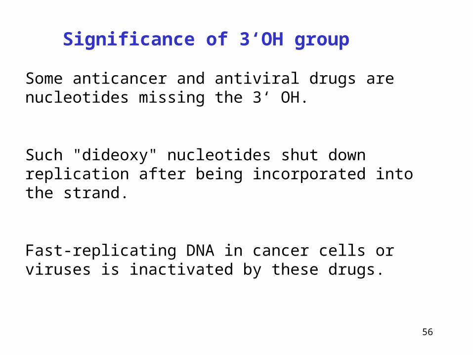
Some anticancer and antiviral drugs are nucleotides missing the 3‘ OH.
Such "dideoxy" nucleotides shut down replication after being incorporated into the strand.
Fast-replicating DNA in cancer cells or viruses is inactivated by these drugs.
Significance of 3‘OH group

57
Replication proceeds on both strands
• double helix must be unwinded – enzyme helicase
• formation of replication fork
• reannealing of strands is prevented by ssb-proteins (single strain binding proteins)
• each newly synthesized strand of DNA base-pairs with its complementary parental template strand

58
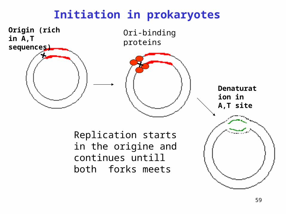
• replication in prokaryotes and eukaryotes starts at the given point origin
• it occurs in both directions from the origin, two replication forks are formed that move away from the origin bidirectionally (in both direction at the same time)
• replication bubbles are formed - replicons
5´
3´
3´
5´
Initiation of replication
origin
Differences between prokaryotes and eukaryotes
Beginning with one parental double helix two newly synthesized stretches of nucleotide chains must grow in opposite direction – one in 53 direction toward the replication fork and the other 53 direction away from replication fork
59
Ori-binding proteins
Origin (rich in A,T sequences)
Denaturation in A,T site
Replication starts in the origine and continues untill both forks meets
Initiation in prokaryotes
60
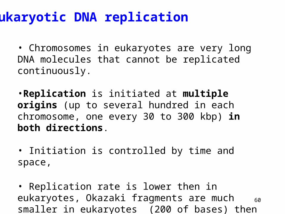
• Chromosomes in eukaryotes are very long DNA molecules that cannot be replicated continuously.
•Replication is initiated at multiple origins (up to several hundred in each chromosome, one every 30 to 300 kbp) in both directions.
• Initiation is controlled by time and space,
• Replication rate is lower then in eukaryotes, Okazaki fragments are much smaller in eukaryotes (200 of bases) then prokaryotes (1000-2000 of bases)
•Occurs in S-phase
Eukaryotic DNA replication
61
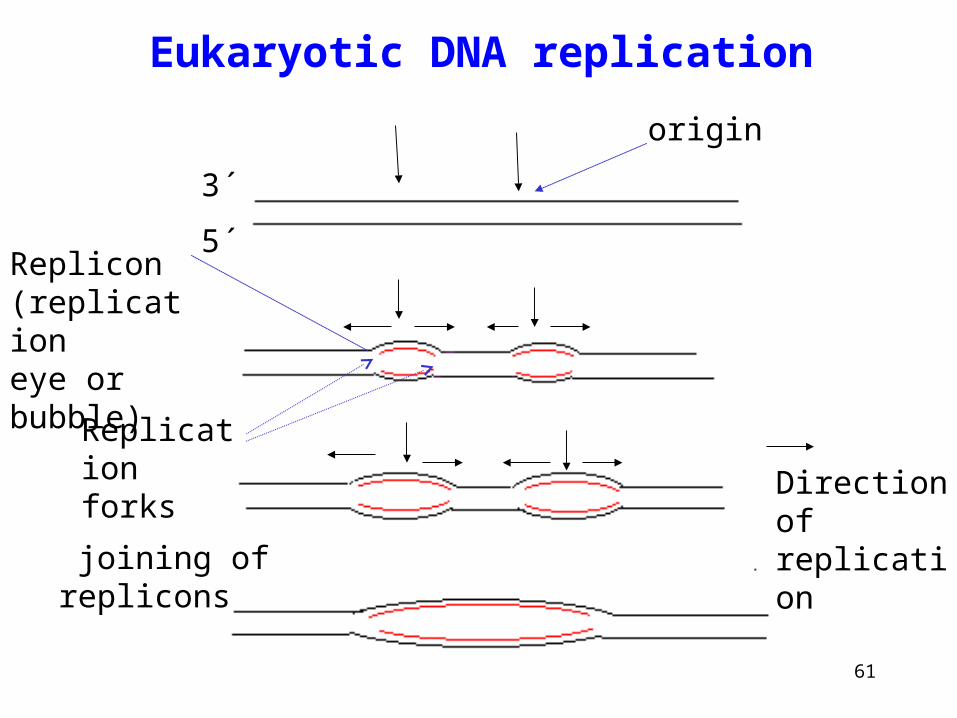
Eukaryotic DNA replication
3´
5´
Direction of replication
origin
Replicon (replicationeye or bubble)
joining of replicons
Replication forks

62
Nuclear DNA is replicated only in the S phase of the cell cycle,mitosis takes place after the replication of all DNA sequences has been completed. Two gaps in time separate the two processes.
Eukaryotic DNA replication
synthetic phase- replication of DNA
(6 – 8 h)
preparation for mitosis - G2
(2 – 6 h)
G1 (~ 10 h) or G0 (variable quiescence phase)
M
mitosis andcell division
(1 h)
S

63
Unwinding protein (ATP-dependent) (helicase)
ATP
ADP
Proteins stabilizing single strand structure
(ssb-proteins single strain binding)
Proteins that are involved in unwinding and prevention of reannealing3´
5´

64
RNA primer is necessary for DNA synthesis
•DNA polymerase cannot initiate de novo synthesis of the chain, it requires free 3´-OH group for linking a new nucleotide.
• This primer is RNA oligonucleotide (10-20 bases)
•RNA primer is synthesized in direction 5´3´ by the action of RNA polymerase (primase)
•Primer is coded according to template sequence
3´RNA-DNA hybride
5´
3´
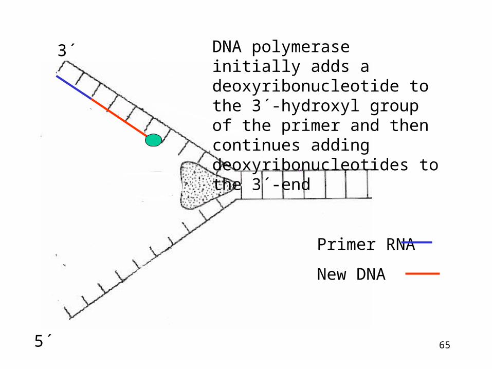
65
DNA polymerase initially adds a deoxyribonucleotide to the 3´-hydroxyl group of the primer and then continues adding deoxyribonucleotides to the 3´-end
3´
5´
Primer RNA
New DNA

66
RNA primer is subsequently removed by the action of exonuclease and the gap is filled by by DNA polymerase
Degraded RNA
3´
5´

67
Synthesis of DNA proceeds always in 5´ 3´ direction
The synthesis of new DNA along the A parental strand occurs without problems
3´Parental chain A (leading strand)
Parental chain B (lagging strand)
How will occur the synthesis among the B chain?
5´
5´
68
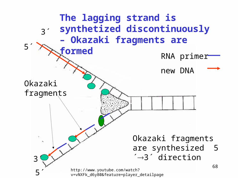
The lagging strand is synthetized discontinuously – Okazaki fragments are formed
Okazaki fragments
Okazaki fragments are synthesized 5´3´ direction
3´
5´
5´
3´
RNA primer
new DNA
http://www.youtube.com/watch?v=vNXFk_d6y80&feature=player_detailpage

69
Okazaki fragments
3´
5´
5´
3´
As replication progress, the RNA primers are removed from Okazaki fragments. DNA polymerase fills the gaps produced by removal of the primers.DNA ligase will join the fragments of DNA
Lagging strand – replication occurs discontinuously in direction away from the fork
70
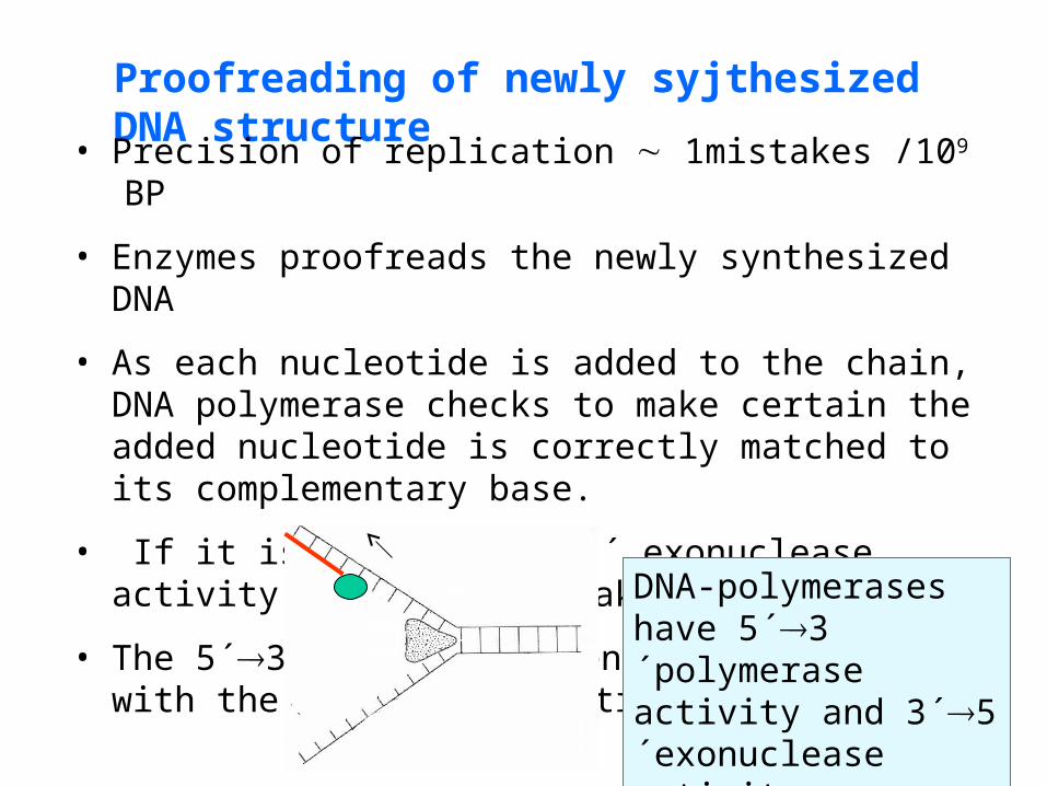
Proofreading of newly syjthesized DNA structure
• Precision of replication 1mistakes /109 BP
• Enzymes proofreads the newly synthesized DNA
• As each nucleotide is added to the chain, DNA polymerase checks to make certain the added nucleotide is correctly matched to its complementary base.
• If it is not, the 3´5´ exonuclease activity edits the mistake.
• The 5´3´polymerase then replaces it with the correct nucleotide.
DNA-polymerases have 5´3´polymerase activity and 3´5´exonuclease activity

71
Polymerase Polymerase activity(for all enzymes 5´ → 3´)
Exonuclease activity
DNA polymerase I Filling if gap after removal RNA primer, DNA repair, removal of RNA primers
5´→3´ and 3´→5´
DNA polymerase II DNA repair 3´→5´
DNA polymerase III* Replication, proofreading and editing
3´→5´
*The main enzyme of replication
Prokaryotic DNA-polymerases

72
Polymerase* Polymerase activity(for all enzymes 5´ → 3´)
Exonuclease activity
DNA polymerase replication, DNA repair
no
DNA polymerase DNA repair no
DNA polymerase replication in mitochondria
3´→5´
DNA polymerase ** replication,
DNA repair
3´→5´
DNA polymerase replication 3´→5´
* At least 9 polymerases is known **major replicative enzyme
Eukaryotic DNA-polymerases

73
Topoisomerase
(Topology od DNA = tridimensional structure of DNA)
Topoisomerase regulates the formation of superhelices
These enzymes catalyze the concerted breakage and rejoining of DNA strands, producing a DNA that is more or less superhelical than the original
The precise regulation of the cellular level of DNA superhelicity is important to facilitate protein interaction with DNA
DNA topoisomerases have many functions (at replication, transcription, repairs, etc.)
http://www.youtube.com/watch?v=EYGrElVyHnU
positive negative supercoiling

74
Supercoiling at unwinding double helix DNA
Positive supercoilingNegative supercoiling
DNA topoisomerases have many functions (at replication, transcripti, repair, …)

75
Make a transient single-strand break in negatively supercoiled DNA double helix. Passage of the unbroken strand through the gap eliminates one supercoil from DNA.
Energy is not required.
Present in prokaryotes and eukaryotes.
Topoisomerase I
Topoisomerase II It binds to double helix DNA and cleave both strands. It can relax supercoiled DNA or introduce supercoil into DNA.
Present in prokaryotes and eukaryotes.
Requires ATP cleavage energy..

76
Action of topoisomerase I
Interuption of phosphodiester bond followed by rotation around the second strand and closing the break by ligation

77
Inhibitors of human topoisomerase prevent replication
Antineoplastic drugs
Examples of topoisomerase inhibitors
camptothecine – plant product
antracyclines (daunorubicine) -bacterial products
podophyllotoxines-plant product
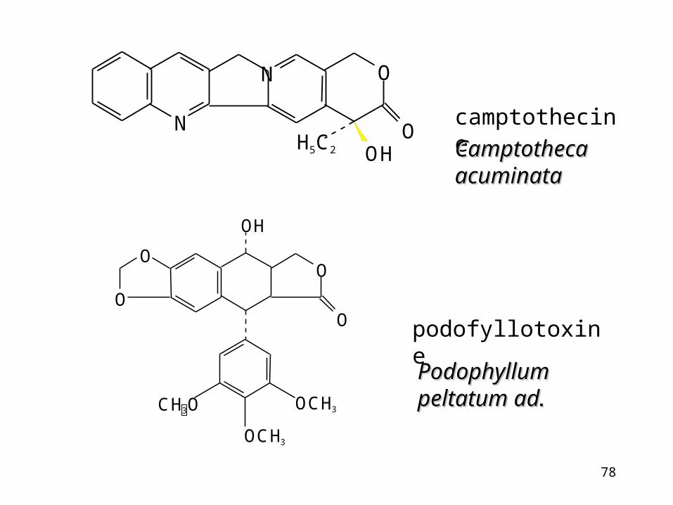
78
podofyllotoxine
camptothecine
Camptotheca Camptotheca acuminataacuminata
PodophyllumPodophyllumpeltatum ad.peltatum ad.
N
N O
OOH
H5C2
OH
O
O
CH3O OCH3
OCH3
O
O

79
Eukaryotic chromosomes are linear. A solution must be found to two problems:
• First, the ends of the chromosomes must be protected from degradation.
• Secondly, there must be some mechanism to ensure replication of a complete chromosome
Telomeres
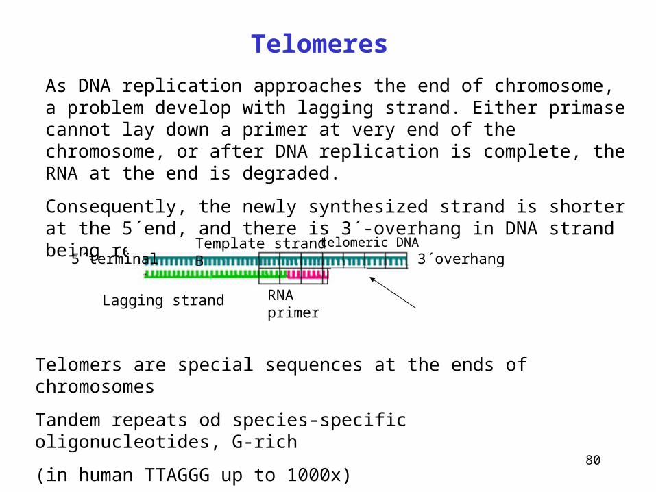
80
Telomeres
As DNA replication approaches the end of chromosome, a problem develop with lagging strand. Either primase cannot lay down a primer at very end of the chromosome, or after DNA replication is complete, the RNA at the end is degraded.
Consequently, the newly synthesized strand is shorter at the 5´end, and there is 3´-overhang in DNA strand being replicated.
Lagging strand RNA primer
3´overhang5´terminalTemplate strand B
Telomers are special sequences at the ends of chromosomes
Tandem repeats od species-specific oligonucleotides, G-rich
(in human TTAGGG up to 1000x)
They protect the ends of chromosomes against nuclease activities.
telomeric DNA

81
Telomerase
• completition of DNA synthesis
• adds newly synthesized hexanucleotide to 3´-end template strand
• it is reverse transcriptase – it carries its own RNA template (CA), this is added to 3´-end of DNA template and new DNA is synthesized that lengthens the 3´-end of DNA strand.
•Then the telomerase moves down the DNA toward the new 3´end and repeats the process a number of times.
http://faculty.plattsburgh.edu/donald.slish/Telomerase.html
RNA template is a component of telomerase
Template strand B is lengthened on the base of reversal transcription from RNA template
TTAGGG
Direction of telomerase movement
telomerase adds tandem repeats to the telomere's 3´-end

82
Telomerase actionReplicating leading strand is not included in the scheme
RNA template
5´-
3´-
pol
RNA-primer Telomerase actionLagging strand
5´-ending strand is extended by normal lagging strand synthesis
replicated strand complementary to the 3´-end of the chromosome
83
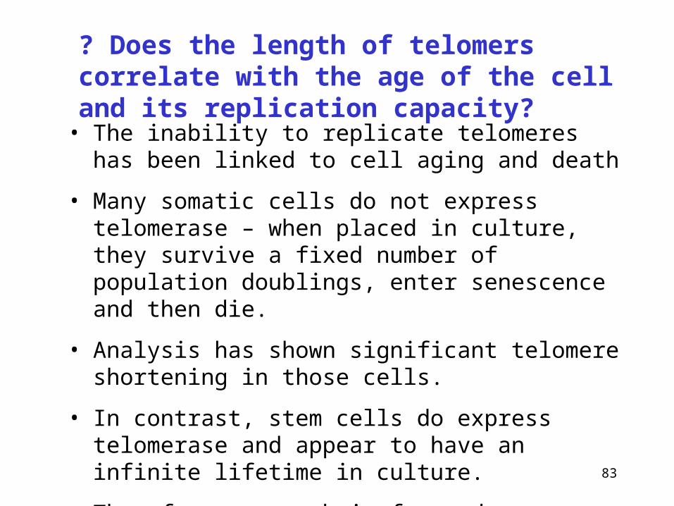
? Does the length of telomers correlate with the age of the cell and its replication capacity?
• The inability to replicate telomeres has been linked to cell aging and death
• Many somatic cells do not express telomerase – when placed in culture, they survive a fixed number of population doublings, enter senescence and then die.
• Analysis has shown significant telomere shortening in those cells.
• In contrast, stem cells do express telomerase and appear to have an infinite lifetime in culture.
• Therefore research is focused on understanding of the role of telomeres in aging, growth and cancer

84
It is estimated that the number of damaging interventions into the DNA structure in the human cell is about:
104-106/day
In addult human (1012 cells) it results in 1016-1018 repair processes per day.
DNA damage and repair

85
DNA damage and repair
Type of damage Cause of damage
Missing base Depurination (104purines/day)
Altered base Ionizing radiation, alkylating agents
Non-correct base Spontaneous deamination
Deletion-insertion Intercalating drugs (acridines)
Formation of dimers UV radiation
Strand breaks Ionizing radiation, chemicals (bleomycine)
Cross-linkages Chemicals (derivateves of psoralene, mitomycine C)
Tautomer formation Spontaneous and temporary
86
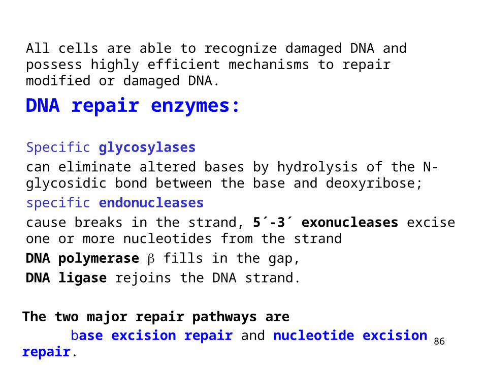
All cells are able to recognize damaged DNA and possess highly efficient mechanisms to repair modified or damaged DNA.
DNA repair enzymes:
Specific glycosylases
can eliminate altered bases by hydrolysis of the N-glycosidic bond between the base and deoxyribose;
specific endonucleases
cause breaks in the strand, 5´-3´ exonucleases excise one or more nucleotides from the strand
DNA polymerase fills in the gap,
DNA ligase rejoins the DNA strand.
The two major repair pathways are
base excision repair and nucleotide excision repair.