left-sided pancreatic incidentalomas treated with
TRANSCRIPT

RESEARCH Open Access
Left-sided pancreatic incidentalomastreated with laparoscopic approach: areport of 20 casesMarco Chiarelli1, Martino Gerosa4, Fulvio Tagliabue1, Luca Fumagalli1, Angelo Guttadauro2, Francesco Gabrielli2,Alessandro Marando3, Matilde De Simone5 and Ugo Cioffi5*
Abstract
Background: The diffusion of cross-sectional imaging has recently permitted the detection of an increasingnumber of incidentalomas localized in the distal pancreas.Currently, there are no studies in the literature exploring the laparoscopic approach as treatment for left-sidedpancreatic incidentalomas.
Methods and results: We report a series of 20 incidentalomas localized in the body and tail of the pancreastreated with laparoscopic surgery over the period 2010–2014. The incidental masses of our series included a greatvariety of histotypes and a relevant proportion of malignant lesions. In two cases, the laparoscopic procedures wereconverted to open surgery. No postoperative death was observed. The postoperative pancreatic fistula rate was20 %, and the new-onset diabetes rate was 25 %.
Conclusions: Left-sided pancreatic incidentalomas in patients with minor comorbidities can be safely treated withlaparoscopic approach. Only clinical trials will confirm whether laparoscopic surgery is an effective treatment formalignant lesions.
Keywords: Pancreatic incidentaloma, Distal pancreatectomy, Pancreas, Laparoscopic surgery
BackgroundPancreatic incidentalomas (PIs) are asymptomatic massesaccidentally diagnosed by radiological, endoscopic, or la-boratory exams performed for symptoms not suggestingpancreatic diseases [1]. Masses confined to the body andtail of the gland are frequently asymptomatic, but recently,an increasing number of these lesions has been detected,due to the large use of cross-sectional imaging [2]. To date,only few series of PIs have been reported in the literature,and consequently, some aspects of their management arestill debated [3–7].Laparoscopic distal pancreatectomy (LDP) is actually
considered an effective and safe treatment for benignand premalignant left-sided pancreatic tumors [8–11].Minimally invasive surgery could be a good choice forthe treatment of incidental masses of the distal pancreas,
but currently, there are no studies in the literature con-firming this hypothesis.We report a series of 20 incidentalomas localized in
the body and tail of the pancreas treated with laparo-scopic approach.
MethodsAfter obtaining local ethics committee approval, weretrospectively reviewed the medical records of all thepatients who underwent distal pancreatectomy at theGeneral Surgery Division of Alessandro ManzoniHospital, from January 2010 to December 2015. Ourinstitution is categorized as a medium-volume hospitalfor pancreatic surgery [12]. Only patients with asymp-tomatic incidentally identified lesions treated withlaparoscopic approach were included into the study. Thedata collected were as follows: preoperative data—age,gender, ethnicity, body mass index (BMI), AmericanSociety of Anesthesiologists (ASA) physical status
* Correspondence: [email protected] of Surgery, University of Milan, Milan, ItalyFull list of author information is available at the end of the article
© 2016 The Author(s). Open Access This article is distributed under the terms of the Creative Commons Attribution 4.0International License (http://creativecommons.org/licenses/by/4.0/), which permits unrestricted use, distribution, andreproduction in any medium, provided you give appropriate credit to the original author(s) and the source, provide a link tothe Creative Commons license, and indicate if changes were made. The Creative Commons Public Domain Dedication waiver(http://creativecommons.org/publicdomain/zero/1.0/) applies to the data made available in this article, unless otherwise stated.
Chiarelli et al. World Journal of Surgical Oncology (2016) 14:204 DOI 10.1186/s12957-016-0949-7
classification, indication and type of imaging exams, andtumor size and location; intraoperative data—type ofresection and operative time; pathological diagnosis andstaging (according to the 7th edition of American JointCommittee on Cancer TNM staging system); and post-operative outcomes—perioperative mortality, length ofhospital stay, readmission, postoperative pancreatic fis-tulas (POPF) [13], post-pancreatectomy diabetes (PPD)[14], and generic complications.LDP was performed according to the standard tech-
nique described in the literature [15]. The pancreas wastransected with a linear cutting stapler (Endopath® ETSLinear Cutter—Ethicon Endo-Surgery Inc., CincinnatiOH, USA); no extra suture was performed routinely. Onesuction drain was left close to the transected pancreas.In patients with malignant neoplasm, the radiological
follow-up consisted in CT scan every 6 months. Informedconsent for publishing personal data was obtained fromeach patient included in the study.
ResultsWe retrospectively collected 34 cases of tumors of the dis-tal pancreas that underwent surgery during the period2010–2015: 22 patients (64.7 %) were asymptomatic and20 (58.8 %) were treated with laparoscopic approach. Themean age was 63.4 years (range 26–78). Fourteen patientswere female and 6 male. All patients were Caucasian. Themedian BMI was 24.75—interquartile (IQ) range 23.7–26.2. Four patients were classified ASA 1 (20 %), 12 pa-tients ASA 2 (60 %), and 4 patients ASA 3 (20 %). Themain radiological, pathological, and surgical characteristicsof the series are summarized in Tables 1 and 2.In 12 patients, diagnosis was made during diagnostic
work-up in the emergency department (ED). In three pa-tients, the suspected diagnosis was pulmonary embolism;in two, colonic diverticulitis; and in one, pericarditis. Inthree patients presenting with hematuria and three withrenal stones, PI was an incidental finding.In six cases, imaging was performed for follow-up
(two for pelvic cyst, two for uterine carcinoma, and onefor prostate carcinoma) or for staging (one for oral neo-plasm). In two patients, PI was detected due to an in-crease of serum amylases.Radiological diagnosis was made by abdominal ultrason-
ography (US) in 5 patients and computed tomography(CT) scan in 15 patients. In nine cases, CT was performedin ED.In two cases, the laparoscopic procedures were con-
verted to open surgery for the large size of PI. The medianoperative time was 203.5 min (IQ range 193–218.5). Themedian postoperative hospital stay was 7.5 days (IQ range6–10). Four patients (20 %) developed POPF: 2 were gradeA and 2 grade B fistulas. No emergent reintervention wasrequired and conservative management was adopted in all
cases. Two patients with grade B fistulas were treated withenteral nutrition and antibiotic therapy; the drainage wasmaintained in place until leakage resolution. In one case,the patient was discharged with drain in situ and reevalu-ated in an outpatient setting. In our series, the medianpersistency of POPF was 16 days. In 4 cases (20 %), post-operative pleural effusion was observed: in 2 patients, itwas associated with grade B pancreatic fistulas, and in 2cases, it was secondary to pneumonia; one patient re-quired thoracentesis. Five patients (25 %) developed PPDduring the postoperative course. In our series, no case ofpostoperative death or readmission was observed at 90-day follow-up.In 8 patients (40 %), the PI was located in the body of
the gland; in 9 patients (45 %), in the tail; and in 3 patients(15 %), between the body and tail.In all the patients, the resection margins were nega-
tive for tumor involvement. Histology revealed ductaladenocarcinoma (DAC) in six patients—associated withundifferentiated carcinoma and intraductal papillarymucinous neoplasm (IPMN) in two patients; neuroen-docrine tumor in five patients (two presented the cysticvariant); and two cases of acinar cell carcinoma (ACC).Serous cystic neoplasm was detected in two patients,mucinous cystic neoplasm in two patients, solid pseu-dopapillary neoplasm in two patients, and one patientshowed an isolated IPMN.The mean follow-up of the cohort was 31 months. All
the patients with cystic neoplasms or neuroendocrine tu-mors were alive without disease recurrence in December2015. In 8 patients with DAC and ACC, the median num-ber of lymph nodes removed was 15 (IQ range 13.5–17).In this specific sub-group, pathological staging was stage IB in one patient, stage II A in four patients, and stage II Bin three patients. The median follow-up time of this sub-group was 17 months: six of eight patients died of tumorrecurrence, while two patients are alive in December 2015(one with disease recurrence).
DiscussionThe preoperative work-up of a pancreatic lesion shoulddetermine its nature in order to plan the most accuratetreatment. Nevertheless, in some cases PIs are not easyto characterize preoperatively [2]. Incidental masses ofthe pancreas include a great variety of lesions andunusual histotypes are frequently counted in the seriespresent in the literature [4, 5, 7]. Differently from DAC,uncommon histological types with a lower biologicalaggressiveness, such as mucinous cystic neoplasms orneuroendocrine tumors, are preferentially located in thedistal pancreas [16–18]: in our series, we found twoACC, two cystic neuroendocrine tumors, and two solidpseudopapillary neoplasms (Figs. 1, 2, and 3). Further-more, in about 7 % of patients with a pancreatic mass is
Chiarelli et al. World Journal of Surgical Oncology (2016) 14:204 Page 2 of 7
not possible to establish a definitive diagnosis before sur-gery despite a complete preoperative imaging [19].The diagnostic work-up of our series was character-
ized by the exiguity of preoperative exams. Solid masseslocalized in the distal pancreas are very frequentlysuitable for surgical resection, and consequently, CTfindings are sufficient to plan the more appropriate man-agement of a left-sided incidentaloma in the majority ofthe cases [7, 20].All patients underwent a contrast-enhanced multide-
tector CT scan with a biphasic protocol (arterial andvenous phases). Currently, intravenous contrast-enhanced CT scan is considered the pivotal radiologicaltechnique for the detection, definition, and staging ofpancreatic masses [21–23]. In the last decade, the useof CT imaging increased by threefold in emergency-treated patients [24]: in our series, eight cases of PIwere discovered by a CT scan performed in patientsevaluated in the ED.A relevant proportion of PIs were detected by transab-
dominal US. US is considered the first-level imaging inhepato-biliary disease, but the deep location of the pan-creas and the operator dependency make the US an
exam with low accuracy for the correct assessment of apancreatic tumor [21].Magnetic resonance imaging (MRI) and endoscopic
ultrasound (EUS) were performed in selected cases. Acomplete pancreatic MRI study including pre- andpost-gadolinium T1-weighted sequences, T2-weightedsequences, and magnetic resonance cholangiopancrea-tography was performed for the definition of the localextension of poorly defined solid masses and the dif-ferentiation of cystic neoplasms [22, 23, 25].EUS with fine-needle aspiration (FNA) provides high-
resolution images and relevant information aboutcytology and tumor markers of solid and cystic lesions,but the high operator dependency is the limit of theexam [21, 22]. EUS presents a high accuracy for smallsolid tumors, and EUS-FNA may be useful in the differ-ential diagnosis of pancreatic cystic masses [25–27].Over recent years, we have employed EUS more fre-quently in the diagnostic work-up of PIs.Surgical resection is considered the standard treatment
for asymptomatic pancreatic solid neoplasms [1, 6].Conversely, the management of cystic PIs is generallyconservative due to the benign nature of the majority of
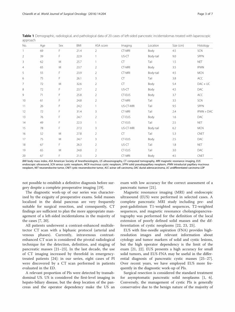
Table 1 Demographic, radiological, and pathological data of 20 cases of left-sided pancreatic incidentalomas treated with laparoscopicapproach
No. Age Sex BMI ASA score Imaging Location Size (cm) Histology
1 69 F 21.4 2 CT-MRI Body 4.5 SCN
2 39 F 22.9 1 US-CT Body-tail 9.0 SPPN
3 62 M 25.7 1 CT Tail 1.5 NET
4 65 M 23.7 2 CT-MRI Body 3.5 IPMN
5 55 F 23.9 2 CT-MRI Body-tail 4.5 MCN
6 75 F 26.1 3 CT Tail 3.8 ACC
7 72 M 32.6 2 CT Body 5.4 DAC + UC
8 72 F 23.7 2 US-CT Body 4.5 DAC
9 71 F 25.8 2 CT-EUS Body 3.7 ACC
10 61 F 24.8 2 CT-MRI Tail 3.5 SCN
11 26 F 24.2 1 US-CT-MRI Tail 9.5 SPPN
12 72 F 31.4 3 CT-MRI Tail 2.4 IPMN + DAC
13 76 F 24.7 2 CT-EUS Body 1.6 DAC
14 49 F 22.3 1 CT-EUS Tail 2.5 NET
15 78 F 27.3 3 US-CT-MRI Body-tail 6.2 MCN
16 52 M 27.8 2 CT Tail 5.3 CNET
17 75 M 24.7 3 CT-EUS Body 2.5 DAC
18 67 F 26.3 2 US-CT Tail 1.8 NET
19 65 M 24.8 2 CT-EUS Tail 3.0 DAC
20 67 F 21.5 2 CT-MRI Body 4.5 CNET
BMI body mass index, ASA American Society of Anesthesiologists, US ultrasonography, CT computed tomography, MRI magnetic resonance imaging, EUSendoscopic ultrasound, SCN serous cystic neoplasm, MCN mucinous cystic neoplasm, SPPN solid pseudopapillary neoplasm, IPMN intraductal papillary mucinousneoplasm, NET neuroendocrine tumor, CNET cystic neuroendocrine tumor, ACC acinar cell carcinoma, DAC ductal adenocarcinoma, UC undifferentiated carcinoma LDP
Chiarelli et al. World Journal of Surgical Oncology (2016) 14:204 Page 3 of 7

these lesions [2]. Serous cystic neoplasm (SNC) is consid-ered a benign lesion, and surgery should be consideredonly in case of large tumors (size >4 cm) or when pre-operative exams are not conclusive [28]. Mucinous cysticneoplasms (MCN) and IPMN are frequently incidental[25]. Surgical resection is mandatory for MCN and mainduct-type IPMN [29]. Branch duct-type IPMN should beconsidered for surgery only in case of a lesion greater than3 cm associated with main duct dilatation >10 mm or anenhanced solid component [29].A relevant proportion of PIs are malignant or prema-
lignant lesions [6, 7]. Malignancies detected inciden-tally are diagnosed in earlier stages, and long-termsurvivals seem to be more favorable than symptomaticlesions [7]. In our series, in six cases the histologicdiagnosis of left-sided PI was DAC, and in twopatients, it was ACC. Despite a complete preoperativediagnostic work-up, the final pathological staging dem-onstrated a relevant proportion of tumors with infiltra-tion of surrounding tissues or microscopic lymph nodeinvolvement: four patients were classified as stage II Aand three patients as stage II B. Consequently, when apancreatic mass is detected, surgical treatment shouldbe always considered [20].The distal pancreatectomy consists in the resection of
body and tail of the pancreas including the spleen and
regional lymph nodes: it is considered the standardtreatment of left-sided malignancies. Minimally inva-sive surgery was introduced for DAC staging, and sub-sequently, it was employed for pancreatic resections[8, 15]. LDP is currently considered an effective treat-ment for benign and low-grade malignant lesions ofthe distal pancreas, but it is still debating if this tech-nique is an appropriate treatment for DAC [8–11]. Inthe series presented in the literature, LDP was per-formed for non-malignant lesions in the majority ofcases, but there are not clinical trials comparing long-term survivals of patients with DAC treated with lap-aroscopic or open approach [11]. In the meta-analysisof the literature, the tumor free margin rate and num-ber of lymph nodes dissected are comparable in bothtechniques, but no definitive conclusions can bedrawn about outcomes of laparoscopic resection formalignancy [9, 10]. However, LDP presents lowerblood loss, shorter time to oral intake, and reducedlength of hospital stay as compared to open surgery,while the rate of postoperative pancreatic fistulas issimilar for the two surgical techniques [8–10].We consider the presence of lymph node involve-
ment and infiltration of surrounding tissues at pre-operative work-up as contraindications to laparoscopicapproach. In our series, conversion to open surgery
Table 2 Surgical and follow-up data of 20 cases of left-sided pancreatic incidentalomas treated with laparoscopic approach
No. Surgery Operative time (min) Postop stay Complications Follow-up
1 LDP 204 7 – AD
2 LDP 215 10 P, PE AD
3 LSPDP 228 5 – AD
4 LDP 220 12 POPF AD
5 LDP 217 10 P, PE AD
6 LDP 210 5 – D
7 LDP-CO 235 8 PPD D
8 LDP 191 11 POPF, PE D
9 LDP 188 9 PPD D
10 LDPDP 198 5 – AD
11 LDP 162 6 – AD
12 LDP 238 8 PPD PD
13 LDP 172 14 POPF D
14 LDPDP 197 5 – AD
15 LDP-CO 212 7 PPD AD
16 LDPDP 230 6 – AD
17 LDP 203 22 POPF, PE D
18 LDP 185 6 – AD
19 LDP 195 7 – AD
20 LDP 200 9 PPD AD
LDP laparoscopic distal pancreatectomy, LSPDP laparoscopic spleen-preserving distal pancreatectomy, CO, converted to open, PE pleural effusion, POPF postoperativepancreatic fistula, P pneumonia, PPD post-pancreatectomy diabetes, AD absence of disease, PD progression of disease, D died
Chiarelli et al. World Journal of Surgical Oncology (2016) 14:204 Page 4 of 7

was determined by the intraoperative detection ofconsiderable-size masses causing the failure to progress.Laparoscopic spleen-preserving distal pancreatectomy wasperformed in two patients for small benign lesions local-ized in the tail. In patients with diagnosis of DAC, thenumber of lymph nodes removed was adequate for a cor-rect oncologic resection.The absence of postoperative deaths could be related
to the epidemiologic features of our cohort: the series
was composed by relatively young patients without con-siderable comorbidities.In our series, the postoperative fistula rate was slightly
high, but it was still in the range reported in the litera-ture [10, 30]. No grade C fistula was observed, and intwo cases, the pancreatic leak was only biochemicalwithout any clinical relevance. Our high POPF rate maybe attributed to lack of oversewing of the pancreaticstump, but any conclusions about the correlation
Fig. 2 Case 2. a Surgical specimen of distal pancreatectomy containing a 9.5-cm-large encapsulated pancreatic tail mass with areas of cysticdegeneration. The histological diagnosis was solid pseudopapillary neoplasm. b The microscopic pattern of the neoplasm is solid and pseudopapillary withpoorly cohesive monomorphic cells, admixed with thin-walled blood vessels. At the center of the image, there are characteristic cholesterolcrystals, surrounded by foreign-body giant cells (hematoxylin-eosin; magnification ×200)
Fig. 1 Case 16. a CT scan demonstrating a 3 × 4 cm pancreatic tail cystic lesion. b Surgical specimen of spleen-preserving distal pancreatectomywith a cystic lesion of the tail. A well-demarcated, solitary, and cystic mass of the pancreatic tail is a rare macroscopic presentation of a neuroendocrinetumor. c The characteristic trabecular and gyriform pattern of a pancreatic neuroendocrine tumor with relatively uniform cells (hematoxylin-eosin;magnification ×40). d The immunohistochemical staining shows strong and diffuse expression of chromogranin A (magnification ×200)
Chiarelli et al. World Journal of Surgical Oncology (2016) 14:204 Page 5 of 7

between the type of surgical closure of the remnantgland and POPF cannot be drawn due to the limitednumber of the series.Pleural effusion, a rare complication [8], was observed in
four patients. In two cases, pleural effusion was associatedwith grade B pancreatic fistulas; in two cases was secondaryto pneumonia, and it was self-limiting. Pleural and pulmon-ary complications are considered infrequent complicationsin pancreatic surgery, but a large observational study showshow LDP is associated with a pleuro-pulmonary morbidityrate of 26 % [11].The incidence of PPD was consistent with the data of
the literature [31].
ConclusionsIn conclusion, circumscribed incidentalomas of the distalpancreas in patients with minor comorbidities could besafely treated with laparoscopic approach. Left-sided PIsare frequently uncommon pancreatic neoplasms, but in asignificant share of patients, DAC in early stages arefound. In case of large-sized tumors or lymph nodeinvolvement, open surgery should be considered. Onlyclinical trials will confirm whether laparoscopic surgery isan effective treatment for malignant lesions [11].
AbbreviationsACC, acinar cell carcinoma; ASA, American society of anesthesiologists; BMI,body mass index; CT, computed tomography; DAC, ductal adenocarcinoma; ED,emergency department; EUS, endoscopic ultrasound; FNA, fine-needle aspiration;IPMN, intraductal papillary mucinous neoplasm; LDP, laparoscopic distalpancreatectomy; MCN, mucinous cystic neoplasm; MRI, magnetic resonanceimaging; PIs, pancreatic incidentalomas; POPF, postoperative pancreatic fistulas;PPD, post-pancreatectomy diabetes; SCN, serous cystic neoplasm; US,ultrasonography
AcknowledgementsWe thank Dr. Gerardo Cioffi, a native speaker, for reviewing the English language.
FundingNo source of funding has a role in the study’s design, conduct, andreporting.
Authors’ contributionsMC carried out the study, drafted the manuscript, and revised it. MG collectedthe information on the patients and wrote the contents of the methods and
results of the manuscript. FT collected the information on the patients andrevised the contents of the methods and results of the manuscript. LF helpedin drafting the manuscript and revised the contents of the discussion of themanuscript. AG and FG collected the information on the patients and revisedthe contents of discussion of the manuscript. AM checked the histopathologyand wrote the contents of histopatologic legends of the manuscript. MDS andUC carried out the concept and the design of the study and revised themanuscript. All authors read and approved the final manuscript.
Competing interestThe authors declare that they have no competing interests.
Consent for publicationWritten informed consent was obtained from the patients of this study forthe publication of personal data and images.
Ethics approval and consent to participateThe clinical study was approved by the Comitato Etico Interaziendale delleProvince di Lecco, Como e Sondrio (no. 0018832/16U), and written informedconsent to participate was obtained from the patients.
Data sharing statementWe provide as attachments the database containing all the informationcollected to perform the study and all documents related to it (ethicscommittee approval, informed consent, consent for the processing ofpersonal data, study protocol, and synopses).
Author details1Department of Surgery, Ospedale Alessandro Manzoni, Lecco, Italy.2Department of Surgery, University of Milan-Bicocca, Istituti Clinici Zucchi, ViaZucchi, Monza, MB, Italy. 3Department of Pathology, Ospedale AlessandroManzoni, Lecco, Italy. 4Department of Surgery, Istituto Clinico HumanitasMater Domini, Castellanza, VA, Italy. 5Department of Surgery, University ofMilan, Milan, Italy.
Received: 6 April 2016 Accepted: 13 July 2016
References1. Zarate X, Williams N, Herrera MF. Pancreatic incidentalomas. Best Pract Res
Clinical Endocrinol Metab. 2012;26:97–103.2. Berland LL, Silverman SG, Gore RM, et al. Managing incidental findings on
abdominal CT: white paper of the ACR incidental findings committee.J Am Coll Radiol. 2010;7:754–73.
3. Fitzgerald TL, Smith AJ, Ryan M, et al. Surgical treatment of incidentallyidentified pancreatic masses. Can J Surg. 2003;46:413–8.
4. Winter JM, Cameron JL, Lillemoe KD, et al. Periampullary and pancreaticincidentaloma. A single institution’s experience with an increasinglycommon diagnosis. Ann Surg. 2006;243:673–80.
5. Bruzoni M, Johnston E, Sasson AR. Pancreatic incidentalomas: clinical andpathologic spectrum. Am J Surg. 2008;195:329–32.
6. Sachs T, Pratt WB, Callery MP, et al. The incidental asymptomatic pancreaticlesion: nuisance or threat? J Gastrointest Surg. 2009;13:405–15.
Fig. 3 Case 6. a Histological picture of an acinar cell carcinoma: the neoplasm composed by cells arranged in small acinar units (hematoxylin-eosin;magnification ×200). b The immunohistochemical staining proves the expression of pancreatic exocrine enzymes (trypsin; magnification ×200)
Chiarelli et al. World Journal of Surgical Oncology (2016) 14:204 Page 6 of 7
7. Lahat G, Ben Haim M, Nachmany I, et al. Pancreatic incidentalomas: highrate of potentially malignant tumors. J Am Coll Surg. 2009;209:313–9.
8. Borja-Cacho D, Al-Refaie WB, Vickers SM, et al. Laparoscopic distalpancreatectomy. J Am Coll Surg. 2009;209:758–65.
9. Jin T, Altaf K, Xiong JJ, et al. A systematic review and meta-analysis of studiescomparing laparoscopic and open distal pancreatectomy. HPB. 2012;14:711–24.
10. Venkat R, Edil BH, Schulick RD, et al. Laparoscopic distal pancreatectomy isassociated with significantly less overall morbidity compared to the opentechnique: a systematic review and meta-analysis. Ann Surg. 2012;255:1048–59.
11. Sulpice L, Farges O, Goutte N, et al. Laparoscopic distal pancreatectomy forpancreatic ductal adenocarcinoma: time for a randomized controlled trial? Resultsof an all-inclusive national observational study. Ann Surg. 2015;262:868–74.
12. Balzano G, Zerbi A, Capretti G, et al. Effect of hospital volume on outcomeof pancreaticoduodenectomy in Italy. Br J Surg. 2008;95:357–62.
13. Bassi C, Dervenis C, Butturini G, et al. Postoperative pancreatic fistula: aninternational study group (ISGPF) group. Surgery. 2005;138:8–13.
14. American Diabetes Association. Diagnosis and classification of diabetesmellitus. Diabetes Care. 2012;35:S64–71.
15. Patterson EJ, Gagner M, Salky B, et al. Laparoscopic pancreatic resection:single-institution experience of 19 patients. J Am Coll Surg. 2001;193:281–7.
16. Sener SF, Fremgen A, Menk HR, et al. Pancreatic cancer: a report oftreatment and survival trends for 100,313 patients diagnosed from 1985–1995, using the National Cancer Database. J Am Coll Surg. 1999;189:1–7.
17. Crippa S, Fernandez-Del Castillo C, Salvia R, et al. Mucin-producingneoplasms of the pancreas: an analysis of distinguishing clinical andepidemiologic characteristics. Clin Gastroenterol Hepatol. 2010;8:213–9.
18. Zerbi A, Falconi M, Rindi G, et al. Clinicopathological features of pancreaticendocrine tumors: a prospective multicenter study in Italy of 297 sporadiccases. Am J Gastroenterol. 2010;105:1421–9.
19. De la Fuente SG, Ceppa EP, Reddy SK, et al. Incidence of benign disease inpatients that underwent resection for presumed pancreatic cancerdiagnosed by endoscopic ultrasonography (EUS) and fine-needle aspiration(FNA). J Gastrointest Surg. 2010;14:1139–42.
20. Sheehan M, Latona C, Araha G, et al. The increasing problem of unusualpancreatic tumors. Arch Surg. 2000;135:644–8.
21. Clarke DL, Thomson SR, Madiba TE, et al. Preoperative imaging ofpancreatic cancer: a management-oriented approach. J Am Coll Surg.2003;196:119–29.
22. Low G, Panu A, Millo N, et al. Multimodality imaging of neoplastic andnonneoplastic solid lesions of the pancreas. Radiographics. 2011;31:993–1015.
23. Buerke B, Domagk D, Heindel W, et al. Diagnostic and radiologicalmanagement of cystic pancreatic lesions: important features for radiologists.Clin Radiol. 2012;67:727–37.
24. Kocher KE, Meurer WJ, Fazel R, et al. National trends in use of computedtomography in the emergency department. Ann Emerg Med. 2011;58:452–62.
25. Italian Association of Hospital Gastroenterologists and Endoscopists;Italian Association for the Study of the Pancreas, Buscarini E, Pezzilli R,Cannizzaro R, et al. Italian consensus guidelines for the diagnosticwork-up and follow-up of cystic pancreatic neoplasms. Dig Liver Dis.2014;46:479–93.
26. Petrone MC, Arcidiacono PG. Role of endoscopic ultrasound in thediagnosis of cystic tumours of the pancreas. Dig Liver Dis. 2008;40:847–53.
27. Khalid A, Brugge W. ACG practice guidelines for the diagnosis andmanagement of neoplastic pancreatic cysts. Am J Gastroenetrol. 2007;102:2339–49.
28. Sakorafas GH, Smyrniotis V, Reid-Lombardo KM, et al. Primary pancreaticcystic neoplasms revisited. Part I: serous cystic neoplasms. Surg Oncol.2011;20:e84–92.
29. Tanaka M, Fernandez-del Castillo C, Adsay V, et al. International consensusguidelines 2012 for the management of IPMN and MCN of the pancreas.Pancreatology. 2012;12:183–97.
30. Natan H, Cameron JL, Goodwin CR, et al. Risk factors for pancreatic leakafter distal pancreatectomy. Ann Surg. 2009;250:277–81.
31. Shirakawa S, Matsumoto I, Toyama H, et al. Pancreatic volumetricassessment as a predictor of new-onset diabetes following distalpancreatectomy. J Gastrointest Surg. 2012;16:2212–9.
• We accept pre-submission inquiries
• Our selector tool helps you to find the most relevant journal
• We provide round the clock customer support
• Convenient online submission
• Thorough peer review
• Inclusion in PubMed and all major indexing services
• Maximum visibility for your research
Submit your manuscript atwww.biomedcentral.com/submit
Submit your next manuscript to BioMed Central and we will help you at every step:
Chiarelli et al. World Journal of Surgical Oncology (2016) 14:204 Page 7 of 7