lecture slides prepared by meg flemming austin …napavalley.edu/people/briddell/documents/bio...
TRANSCRIPT

© 2013 Pearson Education, Inc.
PowerPoint® Lecture Slides
prepared by
Meg Flemming
Austin Community College
C H A P T E R 11
The
Cardiovascular
System: Blood

© 2013 Pearson Education, Inc.
Chapter 11 Learning Outcomes
• 11-1
• Describe the components and major functions of blood, and list the
physical characteristics of blood.
• 11-2
• Describe the composition and functions of plasma.
• 11-3
• List the characteristics and functions of red blood cells, describe the
structure and function of hemoglobin, indicate how red blood cell
components are recycled, and explain erythropoiesis.
• 11-4
• Discuss the factors that determine a person's blood type, and
explain why blood typing is important.
© 2013 Pearson Education, Inc.
Chapter 11 Learning Outcomes
• 11-5
• Categorize the various white blood cells on the basis of their
structures and functions, and discuss the factors that regulate their
production.
• 11-6
• Describe the structure, function, and production of platelets.
• 11-7
• Describe the mechanisms that control blood loss after an injury.

© 2013 Pearson Education, Inc.
Introduction to the Cardiovascular System
(Introduction)
• Includes heart, blood vessels, and blood
• Major transportation system for:
• Substances we need from the external environment
• Substances we need to eliminate through wastes
• Substances we synthesize that need delivery to other
organs
© 2013 Pearson Education, Inc.
Functions of Blood (11-1)
1. Transports dissolved gases, nutrients, hormones,
and metabolic wastes
2. Regulates pH and ion makeup of interstitial fluids
3. Restricts fluid loss at injury sites
4. Defends against toxins and pathogens
5. Stabilizes body temperature

© 2013 Pearson Education, Inc.
Composition of Blood (11-1)
• A liquid connective tissue made of plasma and
formed elements
• Temperature is 38oC, a little above body temperature
• Blood is five times more viscous than water
• Viscosity refers to thickness, stickiness
• Caused by plasma proteins, formed elements
• pH is slightly alkaline in a range of 7.35–7.45
© 2013 Pearson Education, Inc.
Blood Collection and Analysis (11-1)
• Whole blood is usually collected from veins
• Called venipuncture
• Common site is median cubital vein
• Can also be collected from peripheral capillary
• A drop from fingertip or earlobe
• Occasionally collected from arterial puncture
• To evaluate gas exchange efficiency in lung function

© 2013 Pearson Education, Inc.
Checkpoint (11-1)
1. List five major functions of blood.
2. What two components make up whole blood?
3. Why is venipuncture a common technique for
obtaining a blood sample?

© 2013 Pearson Education, Inc.
Plasma (11-2)
• Along with interstitial fluid, makes up most of ECF
• Contains:
• Plasma proteins
• Hormones
• Nutrients
• Gases
• Water
© 2013 Pearson Education, Inc.
Three Major Types of Plasma Proteins (11-2)
1. Albumins
• Most abundant
• Maintains osmotic pressure of plasma
2. Globulins
• Act as transport proteins and antibodies
3. Fibrinogen
• Functions in blood clotting, converting to fibrin

© 2013 Pearson Education, Inc.
Plasma Proteins (11-2)
• Plasma, minus the clotting proteins like fibrinogen,
is called serum
• 90 percent of plasma proteins are synthesized by
liver
• Liver disorders can result in altered blood
composition and function

© 2013 Pearson Education, Inc.
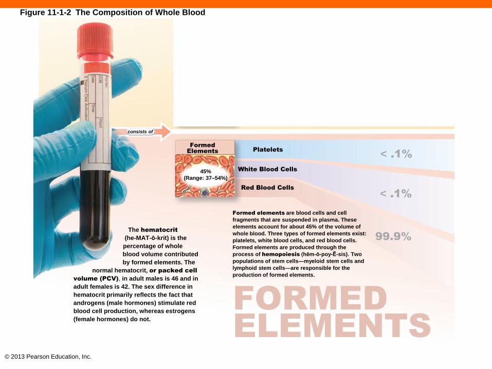
Figure 11-1 The Composition of Whole Blood
A Fluid Connective Tissue
Blood is a fluid connective tissue with a unique composition.
Plasma, the matrix of blood, makes up about 55% of the volume of whole blood.
Plasma
55% (Range: 46–
63%)
Plasma Proteins
Other Solutes
Water
7%
1%
92%
< .1%
< .1%
Platelets
White Blood Cells
Red Blood Cells
Formed
Elements
45% (Range: 37–54%)
consists of
PLASMA
99.9%
Formed elements are blood cells and cell fragments that are suspended in plasma. The hematocrit
(he-MAT- -krit) is the percentage of whole blood volume contributed by formed elements.
FORMED
ELEMENTS
Plasma Proteins
Plasma proteins are in solution rather than forming insoluble fibers like those in other connective tissues, such as loose connective tissue or cartilage.
Albumins
(al-BŪ-minz) constitute
roughly 60% of the plasma
proteins. As the most
abundant plasma proteins,
they are major contributors
to the osmotic pressure
of plasma.
Fibrinogen
(fī-BRIN-ō-jen) functions
in clotting, and normally
accounts for roughly 4% of
plasma proteins. Under certain
conditions, fibrinogen molecules
interact, forming large, insoluble
strands of fibrin (FĪ-brin) that
form the basic framework
for a blood clot.
Globulins
(GLOB-ū-linz) account for
approximately 35% of the
proteins in plasma. Important
plasma globulins include antibodies
and transport globulins. Antibodies,
also called immunoglobulins
(i-mū-nō-GLOB-ū-linz), attack foreign
proteins and pathogens. Transport
globulins bind small ions,
hormones, and other
compounds.
Plasma also contains
enzymes and hormones
whose concentrations
vary widely.
Other Solutes
Other solutes are generally present in concentrations similar to those in the interstitial fluids. However, because blood is a transport medium there may be differences in nutrient and waste product concentrations between arterial blood and venous blood.
Organic
Nutrients: Organic
nutrients are used for ATP
production, growth, and
maintenance of cells. This
category includes lipids (fatty
acids, cholesterol, glycerides),
carbohydrates (primarily
glucose), amino acids,
and vitamins.
Electrolytes:
Normal extracellular ion
composition is essential for
vital cellular activities. The
major plasma electrolytes are
Na+, K+, Ca2+, Mg2+, Cl–,
HCO3–, HPO4
–, and
SO42–.
Organic
Wastes: Waste
products are carried to sites
of breakdown or excretion.
Examples of organic wastes
include urea, uric acid,
creatinine, bilirubin,
and ammonium
ions.
Platelets
Platelets are small, membrane- bound cell fragments that contain enzymes and other substances important to clotting.
White Blood Cells
White blood cells (WBCs), or leukocytes (LOO-k -sīts; leukos, white + -cyte, cell), participate in the body’s defense mechanisms. There are five classes of leukocytes.
Neutrophils
Eosinophils
Basophils Lymphocytes
Monocytes
Red Blood Cells
Red blood cells (RBCs), or erythrocytes (e-RITH-rō-sits; erythros, red + -cyte, cell), are the most abundant blood cells.
SPOTLIGHT FIGURE 11-1
The Composition of Whole Blood

© 2013 Pearson Education, Inc.
SPOTLIGHT FIGURE 11-1
The Composition of Whole Blood
Blood is a fluid connective tissue with a
unique composition. It consists of a matrix
called plasma (PLAZ-muh) and formed
elements (cells and cell fragments). The term
whole blood refers to the combination of
plasma and the formed elements together.
The cardiovascular system of an adult male
contains 5–6 liters (5.3–6.4 quarts) of whole
blood; that of an adult female contains 4–5
liters (4.2–5.3 quarts). The sex differences in
blood volume primarily
reflect differences in
average body size.
Plasma, the matrix of blood, makes up about 55% of the volume of whole blood. In many respects, the composition of plasma resembles that of interstitial fluid. This similarity exists because water, ions, and small solutes are continuously exchanged between plasma and interstitial fluids across the walls of capillaries. The primary differences between plasma and interstitial fluid involve (1) the levels of respiratory gases (oxygen and carbon dioxide, due to the respiratory activities of tissue cells), and (2) the concentrations and types of dissolved proteins (because plasma proteins cannot cross capillary walls).
PLASMA
A Fluid Connective Tissue
55%
(Range: 46–63%)
Plasma
consists of
1%
92%
Plasma Proteins
Other Solutes
Water
< .1%
99.9%
< .1%
The hematocrit
(he-MAT-ō-krit) is the
percentage of whole
blood volume contributed
by formed elements. The
normal hematocrit, or packed cell
volume (PCV), in adult males is 46 and in
adult females is 42. The sex difference in
hematocrit primarily reflects the fact that
androgens (male hormones) stimulate red
blood cell production, whereas estrogens
(female hormones) do not.
Formed elements are blood cells and cell
fragments that are suspended in plasma. These elements account for about 45% of the volume of whole blood. Three types of formed elements exist: platelets, white blood cells, and red blood cells. Formed elements are produced through the process of hemopoiesis (hēm-ō-poy-Ē-sis). Two
populations of stem cells—myeloid stem cells and lymphoid stem cells—are responsible for the production of formed elements.
FORMED
ELEMENTS
Platelets
White Blood Cells
Red Blood Cells
Formed Elements
45% (Range: 37–54%)
7%
Figure 11-1a The Composition of Whole Blood

© 2013 Pearson Education, Inc.
PLASMA Plasma, the matrix of blood, makes up about 55% of the
volume of whole blood. In many respects, the
composition of plasma resembles that of interstitial
fluid. This similarity exists because water, ions, and
small solutes are continuously exchanged between
plasma and interstitial fluids across the walls of
capillaries. The primary differences between plasma and
interstitial fluid involve (1) the levels of respiratory gases
(oxygen and carbon dioxide, due to the respiratory
activities of tissue cells), and (2) the concentrations and
types of dissolved proteins (because plasma proteins
cannot cross capillary walls).
Blood is a fluid connective tissue with a unique composition. It consists of a matrix called plasma (PLAZ-muh) and formed elements (cells and cell fragments). The term whole blood refers to the combination of plasma and the formed elements together. The cardiovascular system of an adult male contains 5–6 liters (5.3–6.4 quarts) of whole blood; that of an adult female contains 4–5 liters (4.2–5.3 quarts). The sex differences in blood volume primarily reflect differences in average body size. 7%
1%
92%
Plasma Proteins
Other Solutes
Water
55%
(Range: 46–63%)
Plasma
A Fluid Connective Tissue
consists of
Figure 11-1-1 The Composition of Whole Blood
© 2013 Pearson Education, Inc.
Platelets
White Blood Cells
Red Blood Cells
Formed
Elements
45%
(Range: 37–54%)
consists of
< .1%
99.9%
< .1%
Formed elements are blood cells and cell
fragments that are suspended in plasma. These
elements account for about 45% of the volume of
whole blood. Three types of formed elements exist:
platelets, white blood cells, and red blood cells.
Formed elements are produced through the
process of hemopoiesis (hēm-ō-poy-Ē-sis). Two
populations of stem cells—myeloid stem cells and
lymphoid stem cells—are responsible for the
production of formed elements.
The hematocrit
(he-MAT-ō-krit) is the
percentage of whole
blood volume contributed
by formed elements. The
normal hematocrit, or packed cell
volume (PCV), in adult males is 46 and in
adult females is 42. The sex difference in
hematocrit primarily reflects the fact that
androgens (male hormones) stimulate red
blood cell production, whereas estrogens
(female hormones) do not.
FORMED
ELEMENTS
Figure 11-1-2 The Composition of Whole Blood

© 2013 Pearson Education, Inc.
Figure 11-1 The Composition of Whole Blood (3–4)
Albumins
(al-BŪ-minz) consti
tute roughly 60% of the
plasma proteins. As the
most abundant plasma
proteins, they are major
contributors to the
osmotic pressure
of plasma.
Fibrinogen
(fī-BRIN-ō-jen) functions
in clotting, and normally
accounts for roughly 4% of
plasma proteins. Under certain
conditions, fibrinogen molecules
interact, forming large, insoluble
strands of fibrin (FĪ-brin) that
form the basic framework
for a blood clot.
Globulins
(GLOB-ū-linz) account for
approximately 35% of the
proteins in plasma. Important
plasma globulins include antibodies
and transport globulins. Antibodies,
also called immunoglobulins
(i-mū-nō-GLOB-ū-linz), attack foreign
proteins and pathogens. Trans-
port globulins bind small ions,
hormones, and other
compounds.
Plasma also contains enzymes and hormones whose concentrations vary widely.
Plasma proteins are in solution rather
than forming insoluble fibers like those
in other connective tissues, such as
loose connective tissue or cartilage.
On average, each 100 mL of plasma
contains 7.6 g of protein, almost five
times the concentration in interstitial
fluid.The large size and globular
shapes of most blood proteins prevent
them from crossing capillary walls, so
they remain trapped within the
bloodstream. The liver synthesizes and
releases more than 90% of the plasma
proteins, including all albumins and
fibrinogen, most globulins, and various
prohormones.
Plasma Proteins
Other Solutes
Other solutes are generally present in
concentrations similar to those in the
interstitial fluids. However, because blood
is a transport medium there may be
differences in nutrient and waste product
concentrations between arterial blood and
venous blood.
Organic
Nutrients: Organic
nutrients are used for ATP
production, growth, and
maintenance of cells. This
category includes lipids (fatty
acids, cholesterol, glycerides),
carbohydrates (primarily
glucose), amino acids,
and vitamins.
Electrolytes:
Normal extracellular ion
composition is essential for
vital cellular activities. The
major plasma electrolytes are
Na+, K+, Ca2+, Mg2+, Cl–,
HCO3–, HPO4
–, and
SO42–.
Organic
Wastes: Waste prod-
ucts are carried to sites of
breakdown or excretion.
Examples of organic wastes
include urea, uric acid,
creatinine, bilirubin,
and ammonium
ions.

© 2013 Pearson Education, Inc.
Figure 11-1 The Composition of Whole Blood (5–7)
Platelets
Platelets are small, membrane-bound cell
fragments that contain enzymes and other
substances important to clotting.
White Blood Cells
White blood cells (WBCs), or leuko-
cytes (LOO-kō-sīts; leukos, white + -cyte,
cell), participate in the body’s defense
mechanisms. There are five classes of
leukocytes, each with slightly different
functions that will be explored later in the
chapter.
Neutrophils
Eosinophils
Basophils Lymphocytes
Monocytes
Red Blood Cells
Red blood cells (RBCs), or erythrocytes
(e-RITH-rō-sits; erythros, red + -cyte, cell),
are the most abundant blood cells. These
specialized cells are essential for the
transport of oxygen in the blood.

© 2013 Pearson Education, Inc.
Checkpoint (11-2)
4. List the three major types of plasma proteins.
5. What would be the effects of a decrease in the
amount of plasma proteins?
© 2013 Pearson Education, Inc.
Erythrocytes or Red Blood Cells (11-3)
• RBCs
• Make up 99.9 percent of formed elements
• Measured in red blood cell count, cells/µL
• Men have 5.4 million/µL
• Women have 4.8 million/µL
• Measured as a percentage of whole blood
• Hematocrit in men is 46 percent
• In women, it's 42 percent
• Contain pigment molecule hemoglobin
• Transports oxygen and carbon dioxide
© 2013 Pearson Education, Inc.
Structure of RBCs (11-3)
• Unique biconcave shape provides advantages
• Increased surface area increases rate of diffusion
• Increased flexibility to squeeze through narrow
capillaries
• During RBC formation organelles are lost
• Cannot go through cell division
• Can only rely on glucose from plasma for energy
© 2013 Pearson Education, Inc.
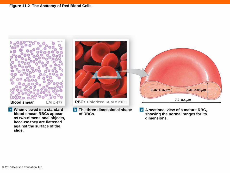
Figure 11-2 The Anatomy of Red Blood Cells.
When viewed in a standard blood smear, RBCs appear as two-dimensional objects, because they are flattened against the surface of the slide.
The three-dimensional shape of RBCs.
A sectional view of a mature RBC, showing the normal ranges for its dimensions.
0.45–1.16 µm 2.31–2.85 µm
7.2–8.4 µm Colorized SEM x 2100 RBCs LM x 477 Blood smear
© 2013 Pearson Education, Inc.
Hemoglobin Structure (11-3)
• Hb structure
• 95 percent of all RBC intracellular proteins
• Transports oxygen and carbon dioxide
• Composed of two pairs of globular proteins, called
subunits
• Each subunit contains heme, with an iron atom
• Oxygen binds to heme, carbon dioxide binds to the
globular subunits

© 2013 Pearson Education, Inc.
Hemoglobin Function (11-3)
• O2–heme bond is fairly weak
• High plasma O2
• Causes hemoglobin to gain O2 until saturated
• Occurs as blood circulates through lung capillaries
• Low plasma O2 and high CO2
• Causes hemoglobin to release O2
• Occurs as blood circulates through systemic capillaries
© 2013 Pearson Education, Inc.
Anemia (11-3)
• A reduction in oxygen-carrying capacity
• Caused by:
• Low hematocrit
• Low hemoglobin content in RBCs
• Symptoms include:
• Muscle fatigue and weakness
• Lack of energy in general
© 2013 Pearson Education, Inc.
RBC Life Span and Circulation (11-3)
• RBCs are exposed to stresses of friction and wear
and tear
• Move through small capillaries
• Bounce against walls of blood vessels
• Life span is about 120 days
• About 1 percent of all RBCs are replaced each day
• About 3 million new RBCs enter circulation per second

© 2013 Pearson Education, Inc.
Hemoglobin Recycling (11-3)
• If RBCs hemolyze in bloodstream, Hb breaks
down in blood
• Kidneys filter out Hb
• If a lot of RBCs rupture at once it causes
hemoglobinuria, indicated by reddish-brown urine
• Most RBCs are phagocytized in liver, spleen, and
bone marrow
• Hb components are recycled

© 2013 Pearson Education, Inc.
Three Steps of Hemoglobin Recycling (11-3)
1. Globular proteins are broken into amino acids
2. Heme is stripped of iron, converted to biliverdin
• Biliverdin is converted to bilirubin, orange-yellow
• Liver absorbs bilirubin, it becomes part of bile
• If not put into bile, tissues become yellow, jaundiced
3. Iron can be stored or released into blood to bind
with transferrin

© 2013 Pearson Education, Inc.
Figure 11-4 Recycling of Hemoglobin.
Events Occurring in the
Red Bone Marrow
Events Occurring in
Macrophages
Macrophages in liver,
spleen, and bone marrow
RBC
formation
Fe2+ transported in the bloodstream by transferrin
Amino acids Heme
Biliverdin
Bilirubin
Old and
damaged
RBCs
90%
10% In the bloodstream,
the rupture of RBCs is called hemolysis.
New RBCs released into circulation
Bilirubin bound
to albumin in
bloodstream Hemoglobin that is not
phagocytized breaks down,
and the polypeptide subunits
are eliminated in urine.
Liver
Bilirubin
Average life span of
RBC is 120 days
Excreted
in bile
Absorbed into the bloodstream
Kidney
Urobilins
Eliminated
in urine Urobilins, stercobilins
Eliminated
in feces
Bilirubin
Events Occurring in
the Kidney Events Occurring in
the Large Intestine
Events Occurring
in the Liver

© 2013 Pearson Education, Inc.
Gender and Iron Reserves (11-3)
• Men have about 3.5 g of ionic Fe2+, 2.5 g of that is
in Hb, providing a reserve of 1 g
• Women have 2.4 g of Fe2+ and 1.9 g in Hb,
providing a reserve of only 0.5 g
• Women often require dietary supplements
• If low, iron deficiency anemia may appear

© 2013 Pearson Education, Inc.
Stages of Erythropoiesis (11-3)
• Also called RBC formation
• Embryonic cells differentiate into multipotent stem cells,
called hemocytoblasts
• Erythropoiesis occurs in red bone marrow, or myeloid
tissue
• Hemocytoblasts produce myeloid stem cells
• Erythroblasts are immature and are synthesizing Hb
• When nucleus is shed they becomes reticulocytes
• Reticulocytes enter bloodstream to mature into RBCs

© 2013 Pearson Education, Inc.
Red bone marrow
Hemocytoblasts
Multi-CSF
Lymphoid Stem
Cells Myeloid Stem Cells
Progenitor Cells
GM-CSF EPO
G-CSF
Blast Cells
Proerythroblast Myeloblast Monoblast Lymphoblast
Myelocytes
Band Cells
Promonocyte Prolymphocyte
Monocyte Lymphocyte
Agranulocytes Granulocytes
Basophil Eosinophil Neutrophil Platelets
Megakaryocyte
Red Blood Cells
(RBCs)
Reticulocyte
Erythrocyte
Ejection of nucleus
Erythroblast stages
White Blood Cells (WBCs)
EPO M-CSF
Figure 11-5 The Origins and Differentiation of RBCs, Platelets, and WBCs.
© 2013 Pearson Education, Inc.
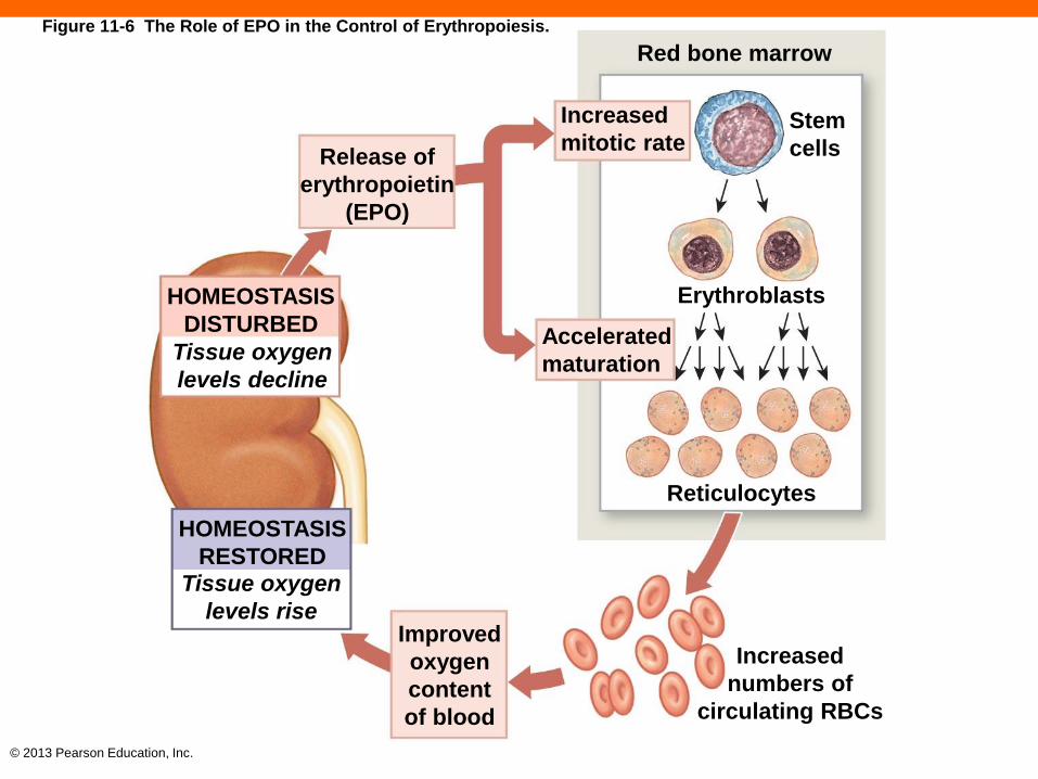
Regulation of Erythropoiesis (11-3)
• Requires amino acids, iron, and B vitamins
• Stimulated by low tissue oxygen, called hypoxia
• Kidney hypoxia triggers release of erythropoietin
• When blood flow to kidney decreases
• When anemia occurs
• When oxygen content of air declines
• When damage to respiratory membrane occurs
© 2013 Pearson Education, Inc.
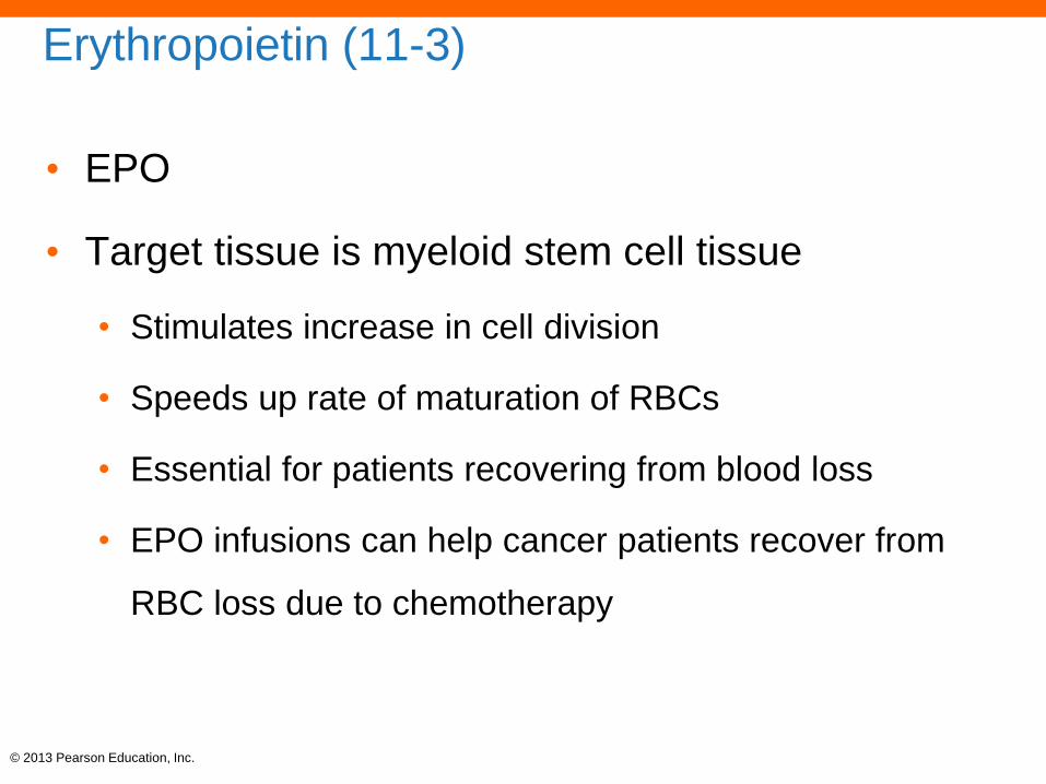
Erythropoietin (11-3)
• EPO
• Target tissue is myeloid stem cell tissue
• Stimulates increase in cell division
• Speeds up rate of maturation of RBCs
• Essential for patients recovering from blood loss
• EPO infusions can help cancer patients recover from
RBC loss due to chemotherapy
© 2013 Pearson Education, Inc.
Red bone marrow
Increased
mitotic rate Stem
cells
Erythroblasts
Accelerated
maturation
Release of
erythropoietin
(EPO)
HOMEOSTASIS
DISTURBED
Tissue oxygen
levels decline
Reticulocytes
HOMEOSTASIS
RESTORED Tissue oxygen
levels rise Improved
oxygen
content
of blood
Increased
numbers of
circulating RBCs
Figure 11-6 The Role of EPO in the Control of Erythropoiesis.

© 2013 Pearson Education, Inc.
Checkpoint (11-3)
6. Describe hemoglobin.
7. What effect does dehydration have on an
individual's hematocrit?
8. In what way would a disease that causes liver
damage affect the level of bilirubin in the blood?
9. What effect does a reduction in oxygen supply to
the kidneys have on levels of erythropoietin in
the blood?

© 2013 Pearson Education, Inc.
ABO Blood Types and Rh System (11-4)
• Based on antigen–antibody responses
• Antigens, or agglutinogens, are substances that can
trigger an immune response
• Your surface antigens are considered normal, not
foreign, and will not trigger an immune response
• Presence or absence of antigens on membrane of RBC
determines blood type
• Three major antigens are A, B, and Rh (or D)

© 2013 Pearson Education, Inc.
Blood Types (11-4)
• Type A blood has antigen A only
• Type B blood has antigen B only
• Type AB blood had both A and B
• Type O blood has neither A nor B
• Rh positive notation indicates the presence of the
Rh antigen; Rh negative, the absence of it

© 2013 Pearson Education, Inc.
Table 11-1 The Distribution of Blood Types in Selected Populations

© 2013 Pearson Education, Inc.
Antibodies (11-4)
• Also called agglutinins
• Found in plasma, will not attack your own antigens on your
RBCs
• Will attack foreign antigens of different blood type
• Type A blood contains anti-B antibodies
• Type B blood contains anti-A antibodies
• Type AB blood contains neither antibodies
• Type O blood contains both antibodies

© 2013 Pearson Education, Inc.
Cross-Reactions in Transfusions (11-4)
• Occur when antibodies in recipient react with their
specific antigen on donor's RBCs
• Cause agglutination or clumping of RBCs
• Referred to as cross-reactions or transfusion
reactions
• Checking blood types before transfusions ensures
compatibility

© 2013 Pearson Education, Inc.
The Difference between ABO and Rh (11-4)
• Anti-A or anti-B antibodies
• Spontaneously develop during first six months of life
• No exposure to foreign antigens needed
• Anti-Rh antibodies in Rh negative person
• Do not develop unless individual is exposed to Rh
positive blood
• Exposure can occur accidentally, during a transfusion or
during childbirth

© 2013 Pearson Education, Inc.
Figure 11-7a Blood Types and Cross-Reactions.
Type A Type B Type AB Type O
Type A blood has RBCs with
surface antigen A only.
Type B blood has RBCs with
surface antigen B only.
Type AB blood has RBCs
with both A and B surface
antigens.
Type O blood has RBCs
lacking both A and B surface
antigens.
Surface
antigen A
If you have Type A blood, your
plasma contains anti-B
antibodies, which will attack
Type B surface antigens.
If you have Type B blood,
your plasma contains anti-A
antibodies, which will attack
Type A surface antigens.
If you have Type AB blood,
your plasma has neither
anti-A nor anti-B antibodies.
If you have Type O blood,
your plasma contains both
anti-A and anti-B antibodies.
Blood type depends on the presence of surface antigens (agglutinogens) on RBC surfaces.
The plasma contains antibodies (agglutinins) that will react with foreign surface antigens.
Surface
antigen B

© 2013 Pearson Education, Inc.
Figure 11-7b Blood Types and Cross-Reactions.
RBC
Surface antigens Opposing antibodies Agglutination (clumping) Hemolysis
In a cross-reaction, antibodies react with their target antigens causing agglutination and hemolysis of
the affected RBCs.

© 2013 Pearson Education, Inc.
Figure 11-8 Blood Type Testing.
Anti-A Anti-B Anti-Rh Blood
type
A+
B+
AB+
O-
© 2013 Pearson Education, Inc.
Checkpoint (11-4)
10. Which blood type(s) can be safely transfused
into a person with Type AB blood?
11. Why can't a person with Type A blood safely
receive blood from a person with Type B blood?
© 2013 Pearson Education, Inc.
Leukocytes or White Blood Cells (11-5)
• WBCs
• Larger than RBCs, involved in immune responses
• Contain nucleus and other organelles and lack
hemoglobin
• Granulocytes
• Neutrophils, eosinophils, basophils
• Agranulocytes
• Lymphocytes and monocytes

© 2013 Pearson Education, Inc.
WBC Circulation and Movement (11-5)
• Four characteristics of WBCs
1. All are capable of amoeboid movement
2. All can migrate outside of bloodstream through
diapedesis
3. All are attracted to specific chemical stimuli, referred to
as positive chemotaxis, guiding them to pathogens
4. Neutrophils, eosinophils, and monocytes are
phagocytes
© 2013 Pearson Education, Inc.
Types of WBCs (11-5)
• Neutrophils, eosinophils, basophils, and
monocytes
• Respond to any threat
• Are part of the nonspecific immune response
• Lymphocytes
• Respond to specific, individual pathogens
• Are responsible for specific immune response

© 2013 Pearson Education, Inc.
Neutrophils (11-5)
• Make up 50–70 percent of circulating WBCs
• Have a dense, contorted multilobular nucleus
• Usually first WBC to arrive at injury
• Phagocytic, attacking and digesting bacteria
• Numbers increase during acute bacterial infections
© 2013 Pearson Education, Inc.
Eosinophils (11-5)
• Make up 2–4 percent of circulating WBCs
• Similar in size to neutrophils
• Have deep red granules and a two-lobed nucleus
• Are phagocytic, but also attack through exocytosis
of toxic compounds
• Numbers increase during parasitic infection or
allergic reactions
© 2013 Pearson Education, Inc.
Basophils (11-5)
• Somewhat smaller than neutrophils and
eosinophils
• Rare, less than 1 percent of circulating WBCs
• Granules contain:
• An anticoagulant, heparin
• Inflammatory compound, histamine
© 2013 Pearson Education, Inc.
Monocytes (11-5)
• About twice the size of a RBC with a large, kidney
bean–shaped nucleus
• Usually 2–8 percent of circulating WBCs
• Migrate into tissues and become macrophages
• Aggressive phagocytes

© 2013 Pearson Education, Inc.
Lymphocytes (11-5)
• Slightly larger than typical RBC with nucleus
taking up most of cell
• About 20–40 percent of circulating WBCs
• Large numbers are migrating in and out of tissues
and lymphatics
• Some attack foreign cells, others secrete
antibodies into circulation
© 2013 Pearson Education, Inc.
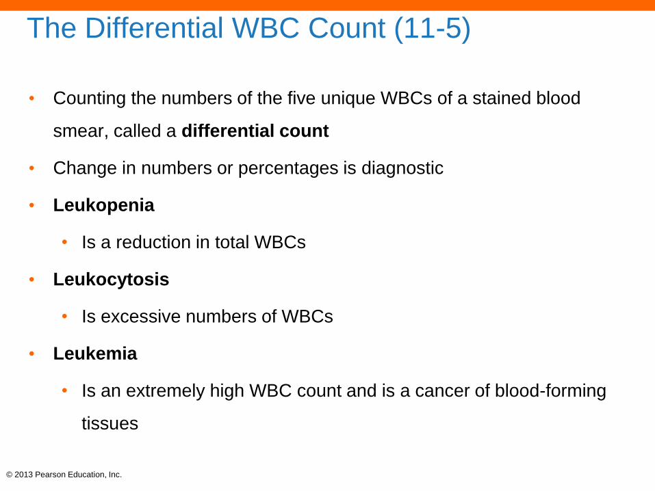
The Differential WBC Count (11-5)
• Counting the numbers of the five unique WBCs of a stained blood
smear, called a differential count
• Change in numbers or percentages is diagnostic
• Leukopenia
• Is a reduction in total WBCs
• Leukocytosis
• Is excessive numbers of WBCs
• Leukemia
• Is an extremely high WBC count and is a cancer of blood-forming
tissues
© 2013 Pearson Education, Inc.
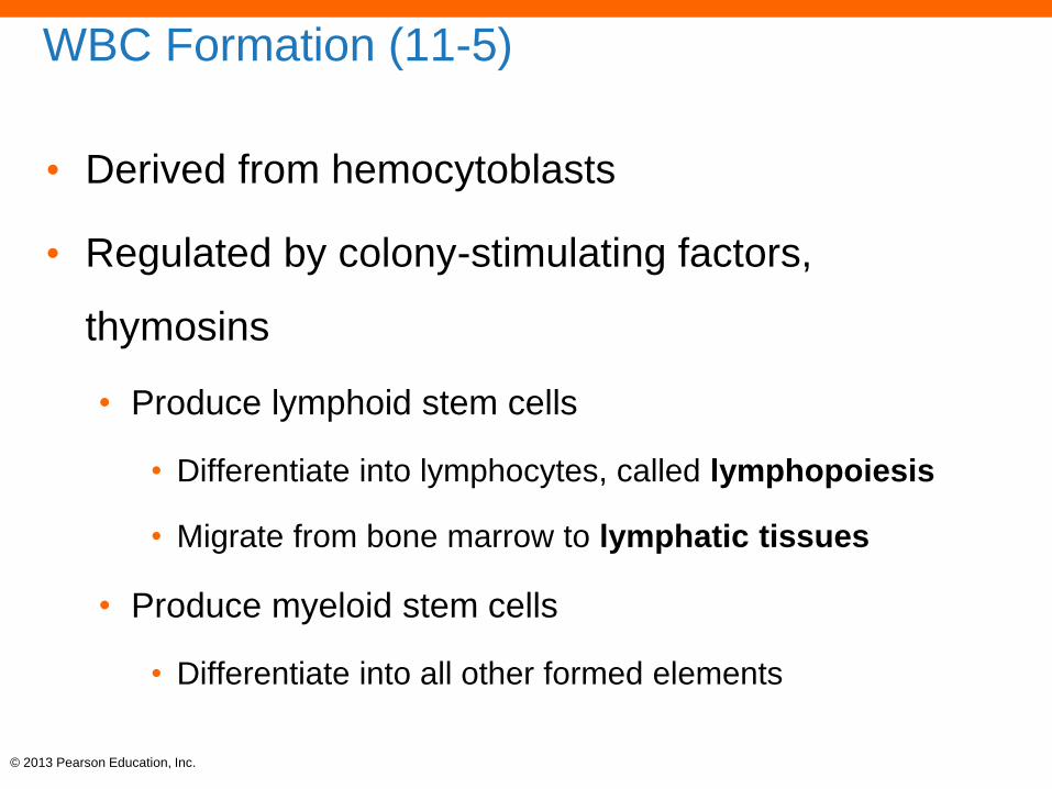
WBC Formation (11-5)
• Derived from hemocytoblasts
• Regulated by colony-stimulating factors,
thymosins
• Produce lymphoid stem cells
• Differentiate into lymphocytes, called lymphopoiesis
• Migrate from bone marrow to lymphatic tissues
• Produce myeloid stem cells
• Differentiate into all other formed elements

© 2013 Pearson Education, Inc.
Checkpoint (11-5)
12. Identify the five types of white blood cells.
13. Which type of white blood cell would you expect
to find in the greatest numbers in an infected cut?
14. Which type of cell would you find in elevated
numbers in a person producing large amounts of
circulating antibodies to combat a virus?
15. How do basophils respond during inflammation?
© 2013 Pearson Education, Inc.
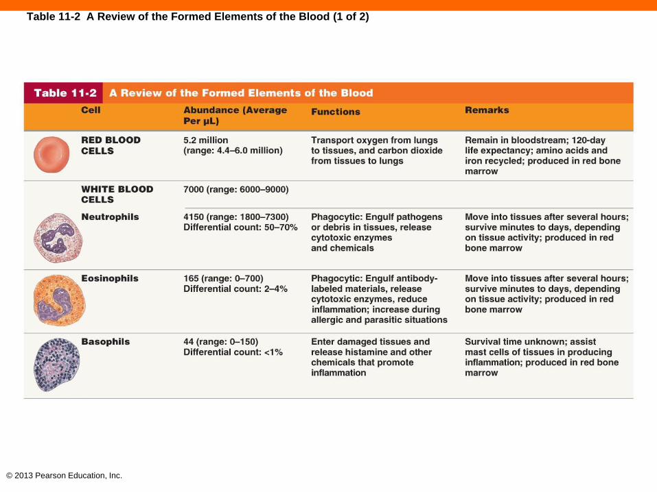
Table 11-2 A Review of the Formed Elements of the Blood (1 of 2)
© 2013 Pearson Education, Inc.
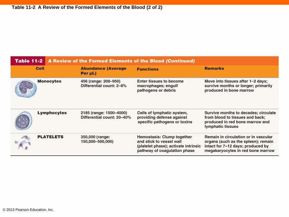
Table 11-2 A Review of the Formed Elements of the Blood (2 of 2)

© 2013 Pearson Education, Inc.
Platelets (11-6)
• Cell fragments involved in prevention of blood loss
• Hemocytoblasts differentiate into megakaryocytes
• Contain granules of chemicals
• Initiate clotting process and aid in closing tears in blood
vessels
• Normal count is 150,000–500,000/µL
• Low count is called thrombocytopenia
© 2013 Pearson Education, Inc.
Checkpoint (11-6)
16. Explain the difference between platelets and
thrombocytes.
17. List the primary functions of platelets.
© 2013 Pearson Education, Inc.
Three Phases of Hemostasis (11-7)
• Halts bleeding and prevents blood loss
1. Vascular phase
2. Platelet phase
3. Coagulation phase
© 2013 Pearson Education, Inc.
The Vascular Phase (11-7)
• Blood vessels contain smooth muscle lined with
endothelium
• Damage causes decrease in vessel diameter
• Endothelial cells become sticky
• A vascular spasm of smooth muscle occurs

© 2013 Pearson Education, Inc.
The Platelet Phase (11-7)
• Platelets attach to sticky endothelium and exposed
collagen
• More platelets arrive and stick to each other
forming a platelet plug
• May be enough to close a small break

© 2013 Pearson Education, Inc.
The Coagulation Phase (11-7)
• Also called blood clotting
• A chemical cascade of reactions that leads to fibrinogen
being converted to fibrin
• Fibrin mesh grows, trapping cells and more platelets
forming a blood clot
© 2013 Pearson Education, Inc.
The Clotting Process (11-7)
• Requires clotting factors
• Calcium ions, vitamin K and 11 different plasma
proteins
• Proteins are converted from inactive proenzymes to
active enzymes involved in reactions
• Cascade event
• Step-by-step
• Product of first reaction is enzyme that activates
second reaction, etc.

© 2013 Pearson Education, Inc.
The Extrinsic Pathway of Blood Clotting (11-7)
• Begins with damaged tissue releasing tissue
factor
• Combines with calcium and other clotting proteins
• Leads to formation of enzyme that can activate
Factor X
© 2013 Pearson Education, Inc.
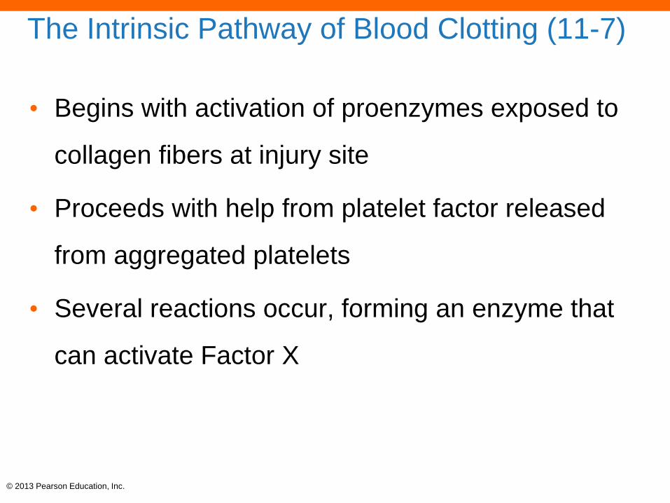
The Intrinsic Pathway of Blood Clotting (11-7)
• Begins with activation of proenzymes exposed to
collagen fibers at injury site
• Proceeds with help from platelet factor released
from aggregated platelets
• Several reactions occur, forming an enzyme that
can activate Factor X
© 2013 Pearson Education, Inc.
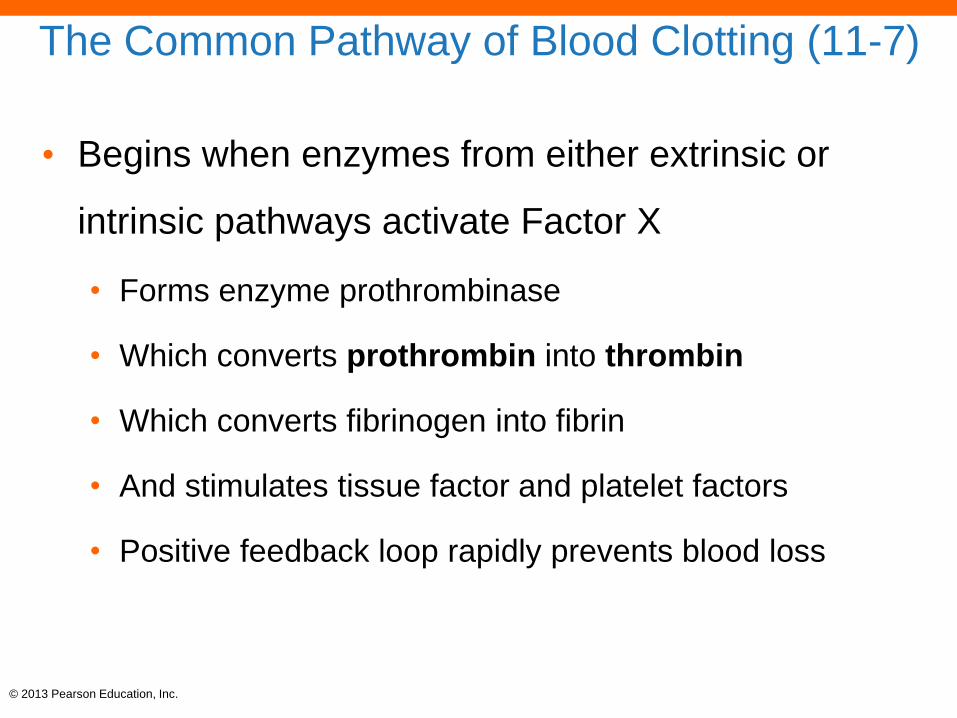
The Common Pathway of Blood Clotting (11-7)
• Begins when enzymes from either extrinsic or
intrinsic pathways activate Factor X
• Forms enzyme prothrombinase
• Which converts prothrombin into thrombin
• Which converts fibrinogen into fibrin
• And stimulates tissue factor and platelet factors
• Positive feedback loop rapidly prevents blood loss

© 2013 Pearson Education, Inc.
Figure 11-10 The Structure of a Blood Clot.
Trapped
RBC
Fibrin
network
Platelets
Blood clot containing trapped RBCs SEM x 2060
© 2013 Pearson Education, Inc.
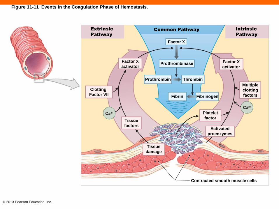
Figure 11-11 Events in the Coagulation Phase of Hemostasis.
Extrinsic
Pathway
Common Pathway Intrinsic
Pathway
Factor X
Prothrombinase
Prothrombin Thrombin
Factor X
activator
Clotting
Factor VII
Factor X
activator
Multiple
clotting
factors Fibrin Fibrinogen
Platelet
factor
Activated
proenzymes
Tissue
damage
Tissue
factors
Contracted smooth muscle cells
© 2013 Pearson Education, Inc.
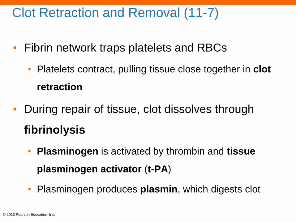
Clot Retraction and Removal (11-7)
• Fibrin network traps platelets and RBCs
• Platelets contract, pulling tissue close together in clot
retraction
• During repair of tissue, clot dissolves through
fibrinolysis
• Plasminogen is activated by thrombin and tissue
plasminogen activator (t-PA)
• Plasminogen produces plasmin, which digests clot
© 2013 Pearson Education, Inc.
Checkpoint (11-7)
18. If a sample of red bone marrow has fewer than
normal numbers of megakaryocytes, what body
process would you expect to be impaired as a
result?
19. Two alternate pathways of interacting clotting
proteins lead to coagulation, or blood clotting.
How is each pathway initiated?
20. What are the effects of a vitamin K deficiency on
blood clotting (coagulation)?