lecture 25 viruses. outline epigenetics cont. histone modification rnai viruses
TRANSCRIPT

Lecture 25Viruses

Outline Epigenetics Cont. Histone Modification RNAi
Viruses

Epigenetics describes phenomena in which genetically identical cells or organisms express their genomes differently, causing phenotypic differences.
Genetically identical cells or individuals
Different epigenetic modifications leading to
different expression patterns

Epigenetic modifications
Epigenetic modifications include:Cytosine methylation of DNAHistone modifications Collectively, these
changes contribute to the distribution of DNA into silent, heterochromatin and active euchromatin
heterochromatineuchromatin
Deal, R.B., Topp, C.N., McKinney, E.C., and Meagher, R.B. (2007) Repression of flowering in Arabidopsis requires activation of FLOWERING LOCUS C expression by the histone variant H2A.Z. Plant Cell 19: 74-83.

DNA methylation
cytosine 5-methylcytosine
Methyltransfe
rase
TTCGCCGACTAA
Methyl-cytosine
DNA can be covalently modified by cytosine methylation.

Histone proteins can be modified to affect chromatin structure
DNA
Histone octamer
The amino terminal regions of the histone monomers extend beyond the nucleosome and are accessible for modification.
NUCLEOSOME

The Histone Code Histones can be modified by Acetylation (Ac) Ubiquitination (Ub) Methylation (Me) Phosphorylation (P) Sumoylation (Su)
Depending on their position, these can contribute to transcriptional activation or inactivation.

Histone modification affects chromatin structure
Closed configuration
Me Me P
K9 K27 S28H3
Open configuration
Me P Ac
K4 S10 K14H3
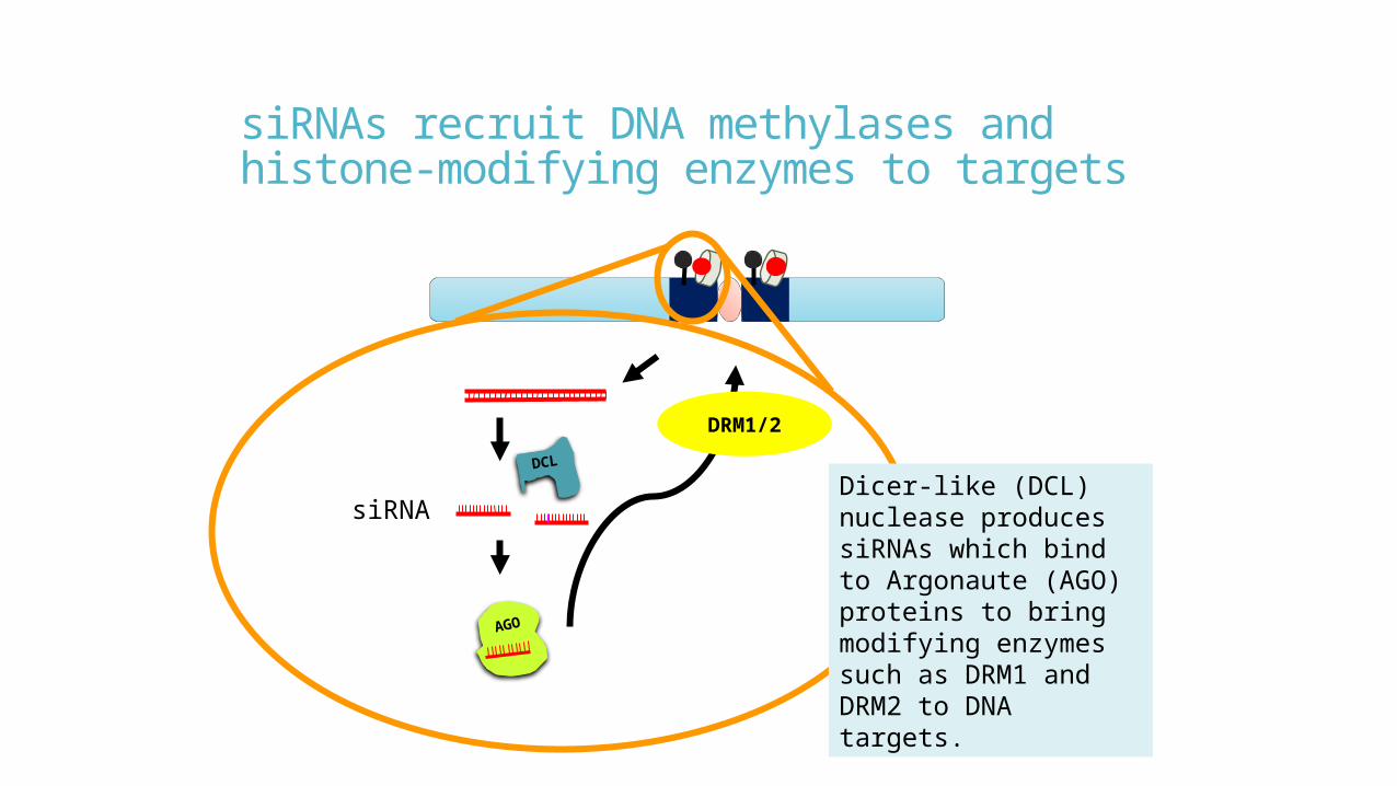
siRNAs recruit DNA methylases and histone-modifying enzymes to targets
siRNA
DCL
DRM1/2
AGO
Dicer-like (DCL) nuclease produces siRNAs which bind to Argonaute (AGO) proteins to bring modifying enzymes such as DRM1 and DRM2 to DNA targets.
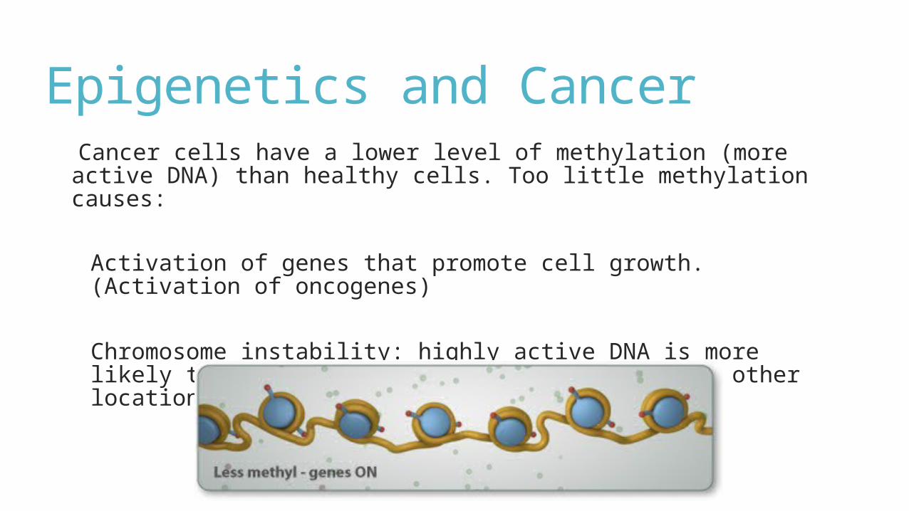
Epigenetics and Cancer Cancer cells have a lower level of methylation (more active DNA) than healthy cells. Too little methylation causes:
Activation of genes that promote cell growth. (Activation of oncogenes)
Chromosome instability: highly active DNA is more likely to be duplicated, deleted, and moved to other locations.
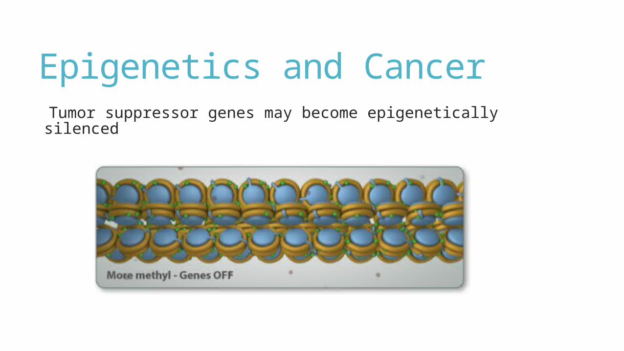
Epigenetics and Cancer Tumor suppressor genes may become epigenetically silenced
Epigenetics and Cancer therapy Target epigenetic changes in cells Therapeutic HDAC inhibitors – effective in treating
Lymphoproliferative disorders. 5 Azacytidine and 5-aza-2-deoxycytidine restore normal
methylation patterns in vitro and invivo in several genes – Inhibitors of DNA methylation
Clinical trial show some promise.
Use epigenetics to silence oncogenes siRNAs can be made to target and silence specific genes that are
driving cell division.
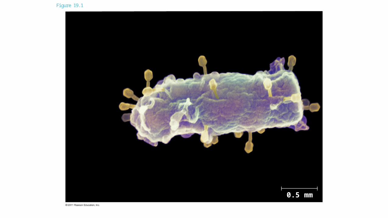
Figure 19.1
0.5 mm

The Discovery of Viruses Tobacco mosaic disease stunts growth of tobacco plants and gives their leaves a mosaic coloration
In the late 1800s, researchers hypothesized that a particle smaller than bacteria caused the disease
In 1935, Wendell Stanley confirmed this hypothesis by crystallizing the infectious particle, now known as tobacco mosaic virus (TMV)


Figure 19.2
Extracted sapfrom tobaccoplant withtobacco mosaicdisease
RESULTS
Passed sapthrough aporcelain filterknown to trapbacteria
Healthy plantsbecame infected
Rubbed filteredsap on healthytobacco plants
1 2 3
4

Structure of Viruses Viruses are not cells
A virus is a very small infectious particle consisting of nucleic acid enclosed in a protein coat and, in some cases, a membranous envelope

Viral Genomes Viral genomes may consist of either
Double- or single-stranded DNA, or Double- or single-stranded RNA
Depending on its type of nucleic acid, a virus is called a DNA virus or an RNA virus

Capsids and Envelopes A capsid is the protein shell that encloses the viral genome
Capsids are built from protein subunits called capsomeres
A capsid can have various structures

Figure 19.3
Capsomereof capsid
RNA CapsomereDNA
Glycoprotein Glycoproteins
Membranousenvelope RNA
CapsidHead
DNA
Tailsheath
Tailfiber
18 250 nm 80 225 nm70–90 nm (diameter) 80–200 nm (diameter)
20 nm 50 nm 50 nm 50 nm(a)Tobacco
mosaic virus(b) Adenoviruses (c) Influenza viruses (d) Bacteriophage T4

Viral Envelopes Some viruses have membranous envelopes that help them infect hosts
These viral envelopes surround the capsids of influenza viruses and many other viruses found in animals
Viral envelopes, which are derived from the host cell’s membrane, contain a combination of viral and host cell molecules

Bacteriophage Capsids Bacteriophages, also called phages, are viruses that infect bacteria
They have the most complex capsids found among viruses
Phages have an elongated capsid head that encloses their DNA
A protein tail piece attaches the phage to the host and injects the phage DNA inside

Viruses only replicate in host cells Viruses are obligate intracellular parasites, which means they can replicate only within a host cell
Each virus has a host range, a limited number of host cells that it can infect

Viral Replicative Cycles Once a viral genome has entered a cell, the cell begins to manufacture viral proteins
The virus makes use of host enzymes, ribosomes, tRNAs, amino acids, ATP, and other molecules
Viral nucleic acid molecules and capsomeres spontaneously self-assemble into new viruses

VIRUS
2
1
3
4
Entry anduncoating
Replication
Transcriptionand manufacture ofcapsid proteins
Self-assembly ofnew virus particlesand their exit fromthe cell
DNA
Capsid
HOSTCELL
Viral DNA
ViralDNA
mRNA
Capsidproteins
Figure 19.4

Replicative Cycle of Phages Phages are the best understood of all viruses
Phages have two reproductive mechanisms: the lytic cycle and the lysogenic cycle

The Lytic Cycle The lytic cycle is a phage replicative cycle that culminates in the death of the host cell
The lytic cycle produces new phages and lyses (breaks open) the host’s cell wall, releasing the progeny viruses
A phage that reproduces only by the lytic cycle is called a virulent phage
Bacteria have defenses against phages, including restriction enzymes that recognize and cut up certain phage DNA

Figure 19.5-1
Attachment1

Figure 19.5-2
Attachment
2
1
Entry of phageDNA anddegradation of host DNA
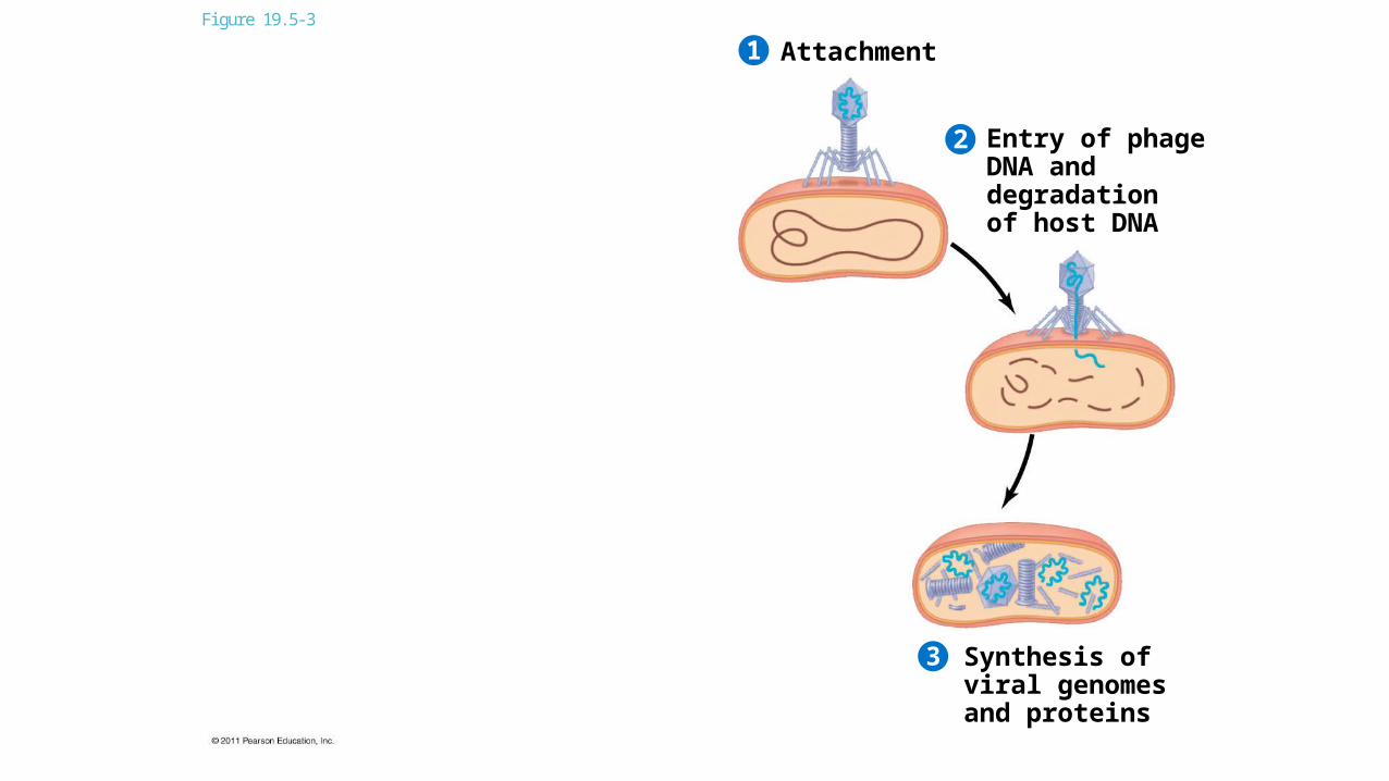
Figure 19.5-3
Attachment
2
1
3
Entry of phageDNA anddegradation of host DNA
Synthesis ofviral genomesand proteins

Figure 19.5-4
Attachment
2
1
43
Entry of phageDNA anddegradation of host DNA
Synthesis ofviral genomesand proteins
Assembly
Phage assembly
Head Tail Tailfibers

Figure 19.5-5
Attachment
2
1
5
43
Entry of phageDNA anddegradation of host DNA
Release
Synthesis ofviral genomesand proteins
Assembly
Phage assembly
Head Tail Tailfibers

The Lysogenic Cycle The lysogenic cycle replicates the phage genome without destroying the host
The viral DNA molecule is incorporated into the host cell’s chromosome
This integrated viral DNA is known as a prophage
Every time the host divides, it copies the phage DNA and passes the copies to daughter cells

An environmental signal can trigger the virus genome to exit the bacterial chromosome and switch to the lytic mode
Phages that use both the lytic and lysogenic cycles are called temperate phages
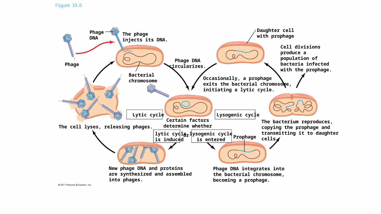
Figure 19.6
New phage DNA and proteinsare synthesized and assembledinto phages.
The cell lyses, releasing phages.
Phage
PhageDNA
The phageinjects its DNA.
Bacterialchromosome
Lytic cycle
lytic cycleis induced
or
Phage DNAcircularizes.
Certain factorsdetermine whether
lysogenic cycleis entered
Lysogenic cycle
Prophage
Daughter cellwith prophage
Occasionally, a prophageexits the bacterial chromosome,initiating a lytic cycle.
Cell divisionsproduce apopulation ofbacteria infectedwith the prophage.
The bacterium reproduces,copying the prophage andtransmitting it to daughtercells.
Phage DNA integrates intothe bacterial chromosome,becoming a prophage.

Replicative Cycles of Animal Viruses There are two key variables used to classify viruses that infect animals
DNA or RNA? Single-stranded or double-stranded?

Table 19.1a

Table 19.1b

Viral Envelopes Many viruses that infect animals have a membranous envelope
Viral glycoproteins on the envelope bind to specific receptor molecules on the surface of a host cell
Some viral envelopes are formed from the host cell’s plasma membrane as the viral capsids exit
Other viral membranes form from the host’s nuclear envelope and are then replaced by an envelope made from Golgi apparatus membrane

Figure 19.7
Capsid
RNA
Envelope (withglycoproteins)
Capsid and viral genomeenter the cell
HOST CELL
Viral genome(RNA)Template
mRNA
ERCapsidproteins
Copy ofgenome(RNA)
New virus
Glyco-proteins

RNA as Viral Genomic Material The broadest variety of RNA genomes is found in viruses that infect animals
Retroviruses use reverse transcriptase to copy their RNA genome into DNA
HIV (human immunodeficiency virus) is the retrovirus that causes AIDS (acquired immunodeficiency syndrome)

Glycoprotein
Reversetranscriptase HIV
Viral envelope
Capsid
RNA (twoidenticalstrands)
HOSTCELL
Viral RNAReversetranscriptase
RNA-DNAhybrid
DNA
NUCLEUSProvirus
ChromosomalDNA
RNA genomefor thenext viralgeneration
mRNA
New virus
HIV
Membraneof whiteblood cell
0.25 m
HIV entering a cell
New HIV leaving a cell
Figure 19.8

The viral DNA that is integrated into the host genome is called a provirus
Unlike a prophage, a provirus remains a permanent resident of the host cell
The host’s RNA polymerase transcribes the proviral DNA into RNA molecules
The RNA molecules function both as mRNA for synthesis of viral proteins and as genomes for new Avirus particles released from the cell

Viruses and Disease Diseases caused by viral infections affect humans, agricultural crops, and livestock worldwide
Smaller, less complex entities called viroids and prions also cause disease in plants and animals, respectively

Viral Diseases in Animals Viruses may damage or kill cells by causing the release of hydrolytic enzymes from lysosomes
Some viruses cause infected cells to produce toxins that lead to disease symptoms
Others have molecular components such as envelope proteins that are toxic

Combating Viruses Vaccines are harmless derivatives of pathogenic microbes that stimulate the immune system to mount defenses against the harmful pathogen
Vaccines can prevent certain viral illnesses
Viral infections cannot be treated by antibiotics
Antiviral drugs can help to treat, though not cure, viral infections

Emerging Viruses Emerging viruses are those that suddenly become apparent
Recently, a general outbreak (epidemic) of a flu-like illness appeared in Mexico and the United States, caused by an influenza virus named H1N1
Flu epidemics are caused by new strains of influenza virus to which people have little immunity
Viral diseases in a small isolated population can emerge and become global
New viral diseases can emerge when viruses spread from animals to humans
Viral strains that jump species can exchange genetic information with other viruses to which humans have no immunity

Pandemics These strains can cause pandemics, global epidemics
The 2009 flu pandemic was likely passed to humans from pigs; for this reason it was originally called the “swine flu”

Figure 19.9
(c) 1918 flu pandemic
2009 pandemicscreening
(b)2009 pandemic H1N1influenza A virus
(a)
1 m