lecture 12: mucosal immunity - … · lecture 12: mucosal immunity gut structure - small intestine...
TRANSCRIPT
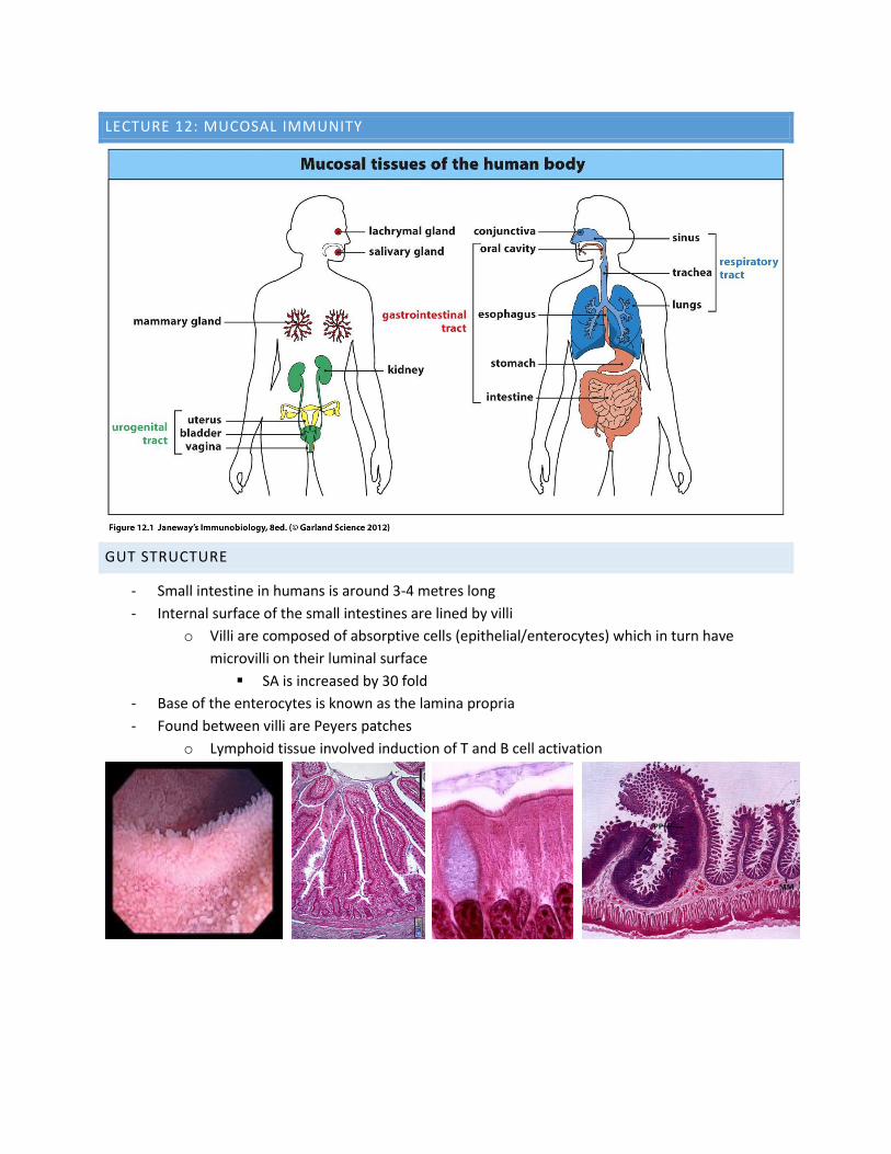
LECTURE 12: MUCOSAL IMMUNITY
GUT STRUCTURE
- Small intestine in humans is around 3-4 metres long
- Internal surface of the small intestines are lined by villi
o Villi are composed of absorptive cells (epithelial/enterocytes) which in turn have
microvilli on their luminal surface
SA is increased by 30 fold
- Base of the enterocytes is known as the lamina propria
- Found between villi are Peyers patches
o Lymphoid tissue involved induction of T and B cell activation
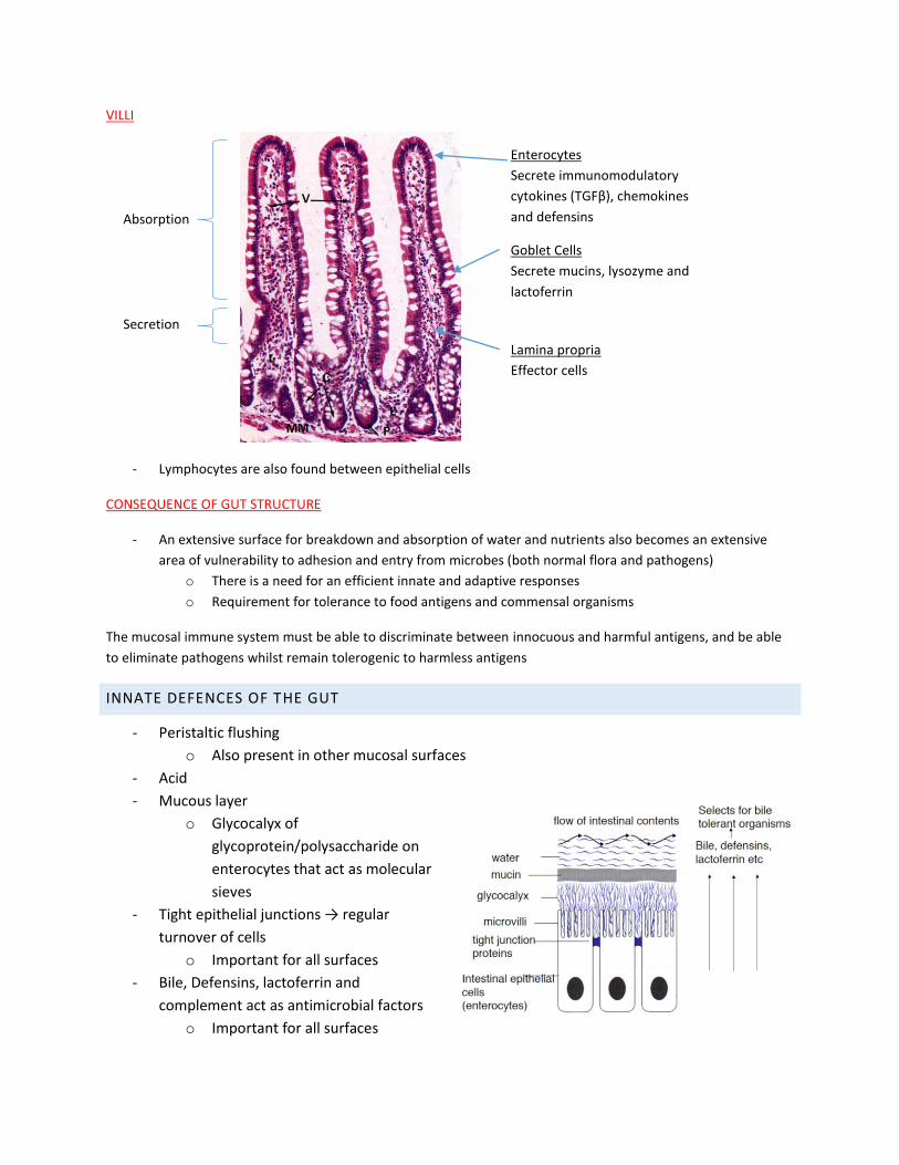
VILLI
- Lymphocytes are also found between epithelial cells
CONSEQUENCE OF GUT STRUCTURE
- An extensive surface for breakdown and absorption of water and nutrients also becomes an extensive
area of vulnerability to adhesion and entry from microbes (both normal flora and pathogens)
o There is a need for an efficient innate and adaptive responses
o Requirement for tolerance to food antigens and commensal organisms
The mucosal immune system must be able to discriminate between innocuous and harmful antigens, and be able
to eliminate pathogens whilst remain tolerogenic to harmless antigens
INNATE DEFENCES OF THE GUT
- Peristaltic flushing
o Also present in other mucosal surfaces
- Acid
- Mucous layer
o Glycocalyx of
glycoprotein/polysaccharide on
enterocytes that act as molecular
sieves
- Tight epithelial junctions → regular
turnover of cells
o Important for all surfaces
- Bile, Defensins, lactoferrin and
complement act as antimicrobial factors
o Important for all surfaces
Absorption
Secretion
Enterocytes
Secrete immunomodulatory
cytokines (TGFβ), chemokines
and defensins
Goblet Cells
Secrete mucins, lysozyme and
lactoferrin
Lamina propria
Effector cells
- Lymphocytes, macrophages and dendritic cells
o Important for all surfaces (several other types will exist depending on the site)
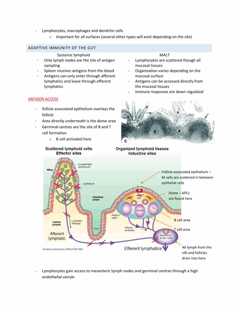
ADAPTIVE IMMUNITY OF THE GUT
Systemic lymphoid MALT - Only lymph nodes are the site of antigen
sampling - Spleen receives antigens from the blood - Antigens can only enter through afferent
lymphatics and leave through efferent lymphatics
- Lymphocytes are scattered though all mucosal tissues
- Organisation varies depending on the mucosal surface
- Antigens can be accessed directly from the mucosal tissues
- Immune responses are down regulated
ANTIGEN ACCESS
- follicle associated epithelium overlays the
follicle
- Area directly underneath is the dome area
- Germinal centres are the site of B and T
cell formation
o B cell activated here
- Lymphocytes gain access to mesenteric lymph nodes and germinal centres through a high
endothelial venule
Follicle associated epithelium –
M cells are scattered in between
epithelial cells
Dome – APCs
are found here
All lymph from the
villi and follicles
drain into here
B cell area
T cell area
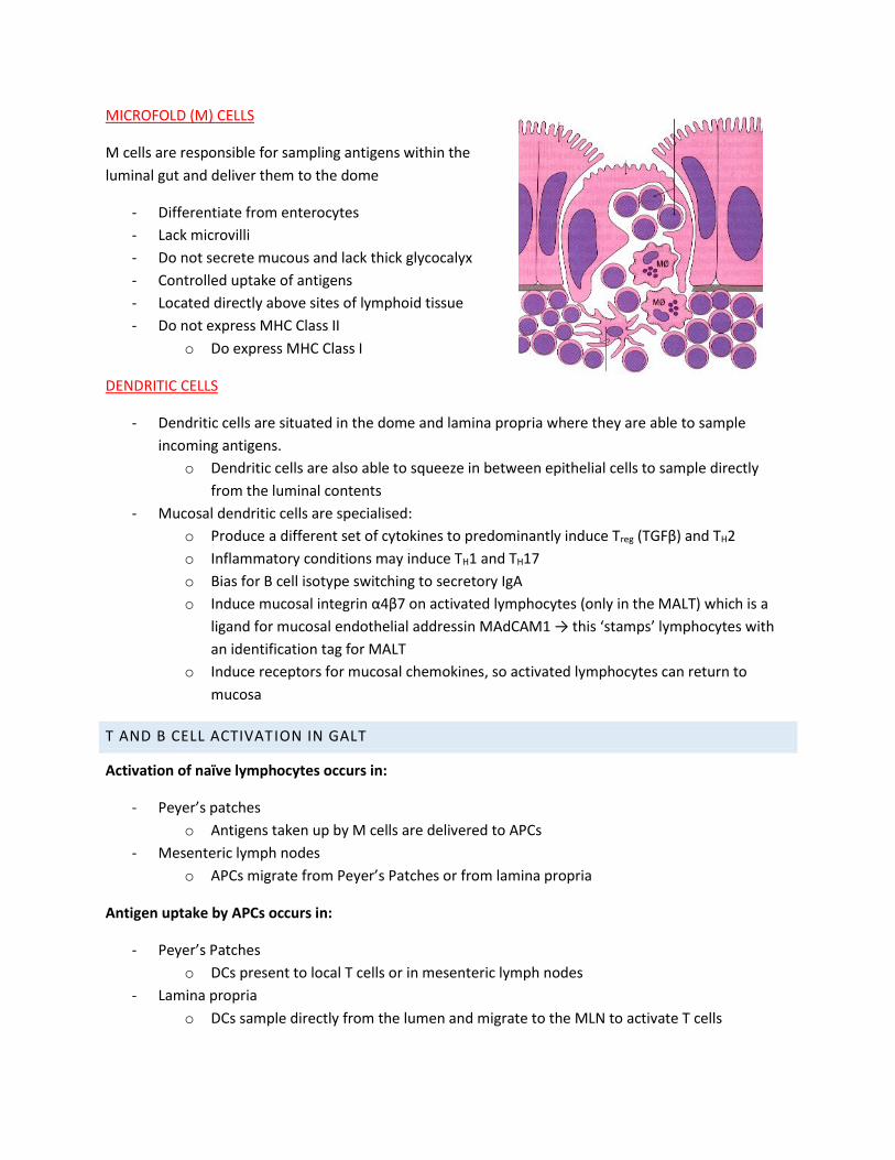
MICROFOLD (M) CELLS
M cells are responsible for sampling antigens within the
luminal gut and deliver them to the dome
- Differentiate from enterocytes
- Lack microvilli
- Do not secrete mucous and lack thick glycocalyx
- Controlled uptake of antigens
- Located directly above sites of lymphoid tissue
- Do not express MHC Class II
o Do express MHC Class I
DENDRITIC CELLS
- Dendritic cells are situated in the dome and lamina propria where they are able to sample
incoming antigens.
o Dendritic cells are also able to squeeze in between epithelial cells to sample directly
from the luminal contents
- Mucosal dendritic cells are specialised:
o Produce a different set of cytokines to predominantly induce Treg (TGFβ) and TH2
o Inflammatory conditions may induce TH1 and TH17
o Bias for B cell isotype switching to secretory IgA
o Induce mucosal integrin α4β7 on activated lymphocytes (only in the MALT) which is a
ligand for mucosal endothelial addressin MAdCAM1 → this ‘stamps’ lymphocytes with
an identification tag for MALT
o Induce receptors for mucosal chemokines, so activated lymphocytes can return to
mucosa
T AND B CELL ACTIVATION IN GALT
Activation of naïve lymphocytes occurs in:
- Peyer’s patches
o Antigens taken up by M cells are delivered to APCs
- Mesenteric lymph nodes
o APCs migrate from Peyer’s Patches or from lamina propria
Antigen uptake by APCs occurs in:
- Peyer’s Patches
o DCs present to local T cells or in mesenteric lymph nodes
- Lamina propria
o DCs sample directly from the lumen and migrate to the MLN to activate T cells
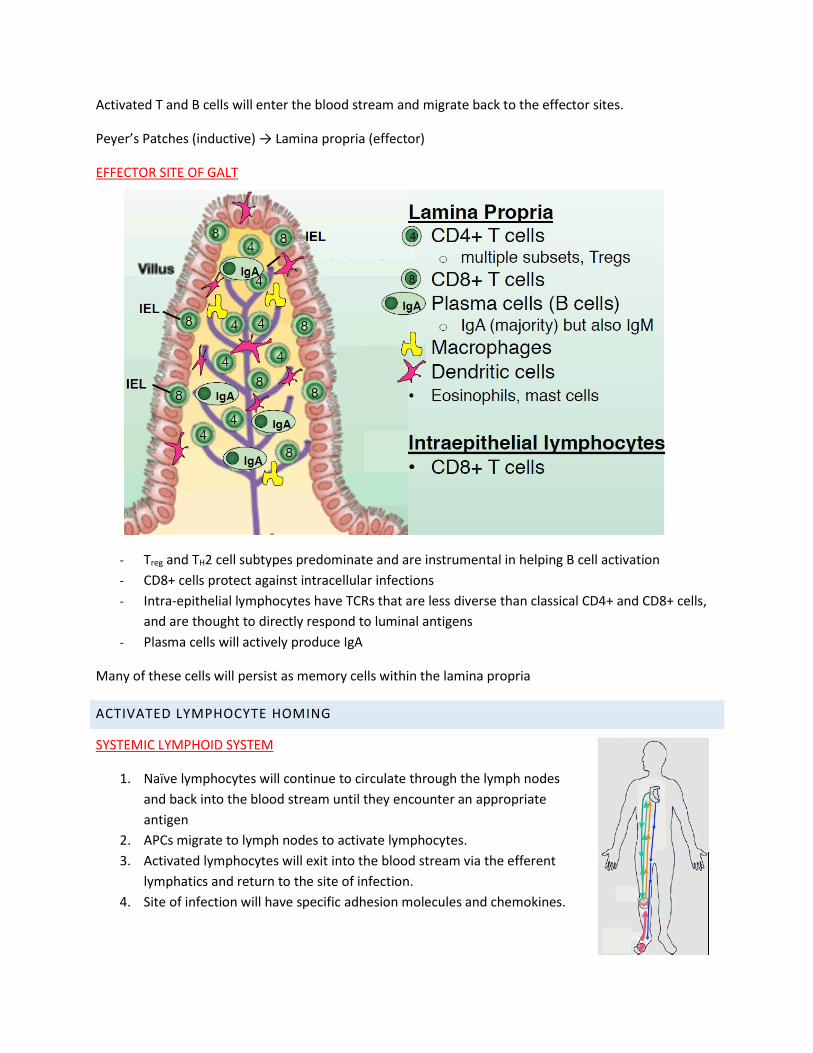
Activated T and B cells will enter the blood stream and migrate back to the effector sites.
Peyer’s Patches (inductive) → Lamina propria (effector)
EFFECTOR SITE OF GALT
- Treg and TH2 cell subtypes predominate and are instrumental in helping B cell activation
- CD8+ cells protect against intracellular infections
- Intra-epithelial lymphocytes have TCRs that are less diverse than classical CD4+ and CD8+ cells,
and are thought to directly respond to luminal antigens
- Plasma cells will actively produce IgA
Many of these cells will persist as memory cells within the lamina propria
ACTIVATED LYMPHOCYTE HOMING
SYSTEMIC LYMPHOID SYSTEM
1. Naïve lymphocytes will continue to circulate through the lymph nodes
and back into the blood stream until they encounter an appropriate
antigen
2. APCs migrate to lymph nodes to activate lymphocytes.
3. Activated lymphocytes will exit into the blood stream via the efferent
lymphatics and return to the site of infection.
4. Site of infection will have specific adhesion molecules and chemokines.
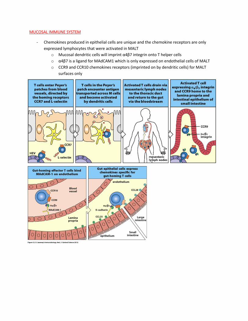
MUCOSAL IMMUNE SYSTEM
- Chemokines produced in epithelial cells are unique and the chemokine receptors are only
expressed lymphocytes that were activated in MALT
o Mucosal dendritic cells will imprint α4β7 integrin onto T helper cells
o α4β7 is a ligand for MAdCAM1 which is only expressed on endothelial cells of MALT
o CCR9 and CCR10 chemokines receptors (imprinted on by dendritic cells) for MALT
surfaces only
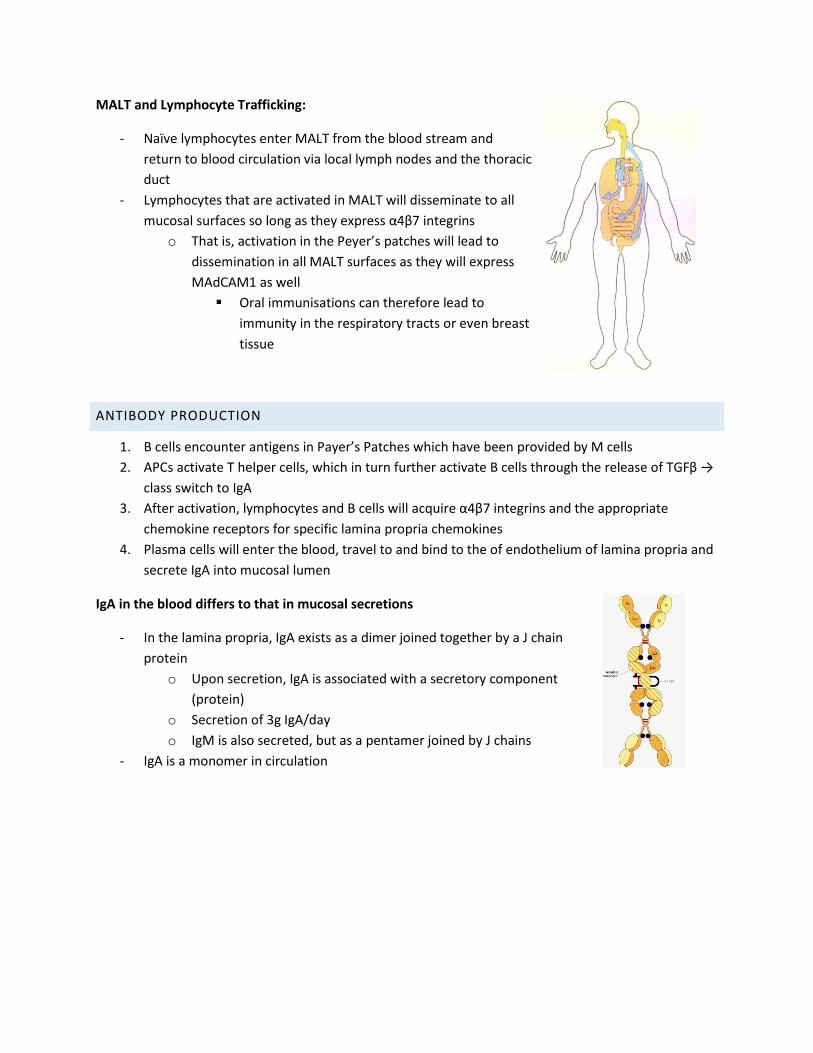
MALT and Lymphocyte Trafficking:
- Naïve lymphocytes enter MALT from the blood stream and
return to blood circulation via local lymph nodes and the thoracic
duct
- Lymphocytes that are activated in MALT will disseminate to all
mucosal surfaces so long as they express α4β7 integrins
o That is, activation in the Peyer’s patches will lead to
dissemination in all MALT surfaces as they will express
MAdCAM1 as well
Oral immunisations can therefore lead to
immunity in the respiratory tracts or even breast
tissue
ANTIBODY PRODUCTION
1. B cells encounter antigens in Payer’s Patches which have been provided by M cells
2. APCs activate T helper cells, which in turn further activate B cells through the release of TGFβ →
class switch to IgA
3. After activation, lymphocytes and B cells will acquire α4β7 integrins and the appropriate
chemokine receptors for specific lamina propria chemokines
4. Plasma cells will enter the blood, travel to and bind to the of endothelium of lamina propria and
secrete IgA into mucosal lumen
IgA in the blood differs to that in mucosal secretions
- In the lamina propria, IgA exists as a dimer joined together by a J chain
protein
o Upon secretion, IgA is associated with a secretory component
(protein)
o Secretion of 3g IgA/day
o IgM is also secreted, but as a pentamer joined by J chains
- IgA is a monomer in circulation
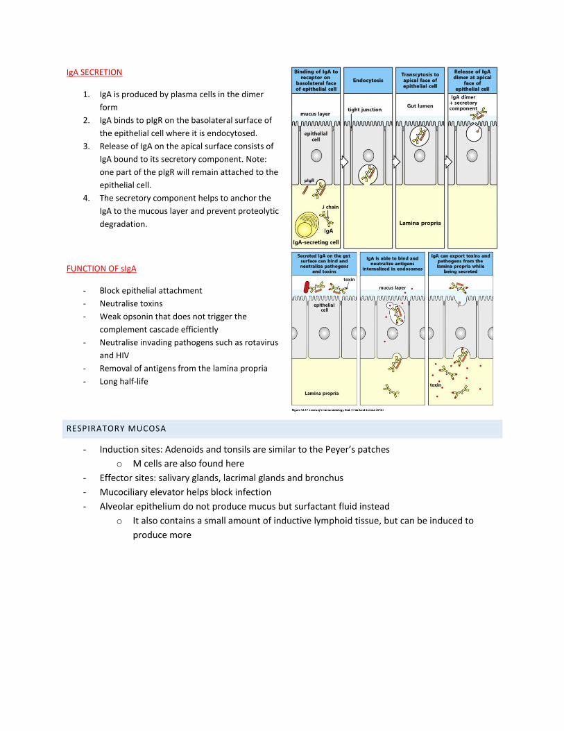
IgA SECRETION
1. IgA is produced by plasma cells in the dimer
form
2. IgA binds to pIgR on the basolateral surface of
the epithelial cell where it is endocytosed.
3. Release of IgA on the apical surface consists of
IgA bound to its secretory component. Note:
one part of the pIgR will remain attached to the
epithelial cell.
4. The secretory component helps to anchor the
IgA to the mucous layer and prevent proteolytic
degradation.
FUNCTION OF sIgA
- Block epithelial attachment
- Neutralise toxins
- Weak opsonin that does not trigger the
complement cascade efficiently
- Neutralise invading pathogens such as rotavirus
and HIV
- Removal of antigens from the lamina propria
- Long half-life
RESPIRATORY MUCOSA
- Induction sites: Adenoids and tonsils are similar to the Peyer’s patches
o M cells are also found here
- Effector sites: salivary glands, lacrimal glands and bronchus
- Mucociliary elevator helps block infection
- Alveolar epithelium do not produce mucus but surfactant fluid instead
o It also contains a small amount of inductive lymphoid tissue, but can be induced to
produce more