le allergie alimentari non ige-mediate - altervista · iride dello iacono unità operativa di...
TRANSCRIPT

Iride Dello IaconoUnità Operativa di Pediatria
ed AllergologiaOspedale Fatebenefratelli
Benevento
LE ALLERGIE
ALIMENTARI NON
IgE-MEDIATE
24 Maggio 2014

FOOD ALLERGY
IgE-mediated reactionsNon IgE-mediated reactions or cell-mediated reactionsCombined IgE and cell-mediated reactions

FOOD ALLERGYIgE-mediated reactions
IgE-mediated reactions are characterized by an acute onset of symptoms generally within 2 hours after ingestion of or exposureto the trigger food. IgE-mediated reactions to foods typically involve the skin, gastrointestinal tract, and respiratory tract.Allergic sensitization occurs when food-specific IgE (sIgE) antibodies are produced by plasma cells that have differentiated from allergen-specific B lymphocytes.

FOOD ALLERGYIgE-mediated reactions
The sIgE antibodies bind to the surface of tissue mast cells and blood basophils, and on reexposure to the food, antigenic proteins in the food bind to and cross-link these cell surface–bound sIgE antibodies, which triggers the release of symptom-causing mediators, such as histamine and leukotrienes.

FOOD ALLERGYIgE-mediated reactions
Subjects can have allergic sensitization (production of sIgE) to food allergens without having clinical symptoms of an allergic reaction on exposure. Thus sensitization alone is not sufficient to define food allergy. An sIgE-mediated food allergy requires both the presence of sensitization and the development of specific signs and symptoms on exposure to that food.

Sensibilizzazione allergica
non equivale ad
ALLERGIA

Specific food-induced allergic conditios

FOOD ALLERGY
Non–IgE-mediated immunologic reactions (eg, cell mediated) include food protein–induced enterocolitis, proctocolitis, and enteropathy
syndromes.
These conditions primarily affect infants or young children who present with abdominal complaints, such as vomiting, abdominal
cramps, diarrhea, and occasionally blood in the stool and failure to thrive or poor weight gain.
Non IgE-mediated reactions

Specific food-induced allergic conditios

Specific food-induced allergic conditios


FOOD PROTEIN INDUCED ENTEROCOLITIS SYNDROME (FPIES)
• Enterocolite allergica o food protein-induced enterocolitis syndrome: un pò di storia……………
• Nel 1940, Rubin descrisse un bambino con severa diarrea ematica responsiva alla dieta priva di LV .
Rubin M. Allergic intestinal bleeding in the newborn. Am J Med Sci 1940;200: 385-7.
• Nel 1967 Grybosky descrisse 21 bambini in cui challenges orali dimostrarono sintomi gastrointestinali associati ad ingestione di LV .Gryboski J. Gastrointestinal milk allergy in infancy. Pediatrics 1967;40:354-62.

FOOD PROTEIN-INDUCED ENTEROCOLITIS SYNDROME (FPIES)
Powell GK.
Milk-and soy-induced enterocolitis in infancy.
J Pediatr 1978;93:553-60
• Powell propose i seguenti criteri per definire laenterocolite allergica infantile:
1. inizio dei sintomi prima dei due mesi di vita
2. una risposta positiva al challenge eseguito durante i primi nove mesi di vita
3. la cessazione della diarrea con l’eliminazione della proteina sospetta e
4. la ricorrenza dei sintomi dopo ingestione della proteina offending.

FOOD PROTEIN-INDUCED ENTEROCOLITIS SYNDROME
Sicherer SH, Eigenmann PA, Sampson HA.
Clinical features of food protein-induced enterocolitis syndrome.
J Pediatr 1998:133:214-9.
Nel 1998 Sicherer puntualizzò le caratteristiche cliniche della enterocolite allergica in una review di 16 pazienti e definì la
sindrome Food protein induced enterocolitis syndrome (FPIES),
ampliando i criteri iniziali proposti da Powell e includendo pazienti che avevano un’età superiore ai 2 mesi al momento della diagnosi.

FOOD PROTEIN-INDUCED ENTEROCOLITIS SYNDROME
Segni tipici di FPIES secondo Sicherer:
• età inferiore ai 9 mesi alla diagnosi iniziale;
• la ripetuta esposizione all’alimento incriminato suscitava diarrea e/o vomito ripetuto entro 24 ore senza nessuna altra causa dei sintomi;
• non vi erano sintomi, oltre a quelli gastrointestinali, provocati dall’alimento incriminato;
• la rimozione delle proteine offending dalla dieta comportava la risoluzione dei sintomi;
• un challenge orale standardizzato provocava diarrea e/o vomito entro 24 ore dalla somministrazione dell’alimento;
• se monitorato durante un challenge, un aumento della conta assoluta di neutrofili al di sopra di 3550/mm3 a 5 ed 8 ore dal challenge costituiva un segno evidente addizionale di una risposta positiva.

FOOD PROTEIN-INDUCED ENTEROCOLITIS SYNDROME
Tipicamente i soggetti descritti da Sicherer erano negativi alla ricerca delle IgEs nei confronti degli alimenti offending (principalmente
LV e soia).
Sei ulteriori soggetti, che soddisfacevano i criteri clinici di FPIES, ma che avevano più di 9 mesi di età alla diagnosi o che presentavano anticorpi IgE verso l’alimento incriminato erano considerati avere una FPIES “atipica”.

FOOD PROTEIN-INDUCED ENTEROCOLITIS SYNDROME
Sono stati segnalati reports di FPIES che rispondevano ai criteri clinici,
in cui i bambini mostravano IgE nei confronti delle proteine causali, sia alla presentazione che durante il follow-up.
Questi bambini avevano un decorso più prolungato dell’allergia, non potendosi escludere la progressione verso reazioni tipiche che riflettono la sensibilità IgE-mediata.
Sicherer ritiene, pertanto, prudente che, nel seguire il decorso della FPIES, si includa lo screening delle IgE per gli alimenti sospetti.
Scott H, Sicherer. Food protein-induced enterocolitis syndrome: case presentations and management lessons. J Allergy Clin Immunol 2005;115:149-56.

FOOD PROTEIN-INDUCED ENTEROCOLITIS SYNDROME
PATOGENESI
Recenti studi hanno evidenziato il ruolo delle cellule T e
l’importanza del tumor necrosis factor (TNF)-α nella patogenesi
dell’affezione.
Heyman et al. hanno dimostrato che l’α-interferon, secreto da
cellule T proteinospecifiche nei confronti del LV, è in grado di
incrementare la permeabilità intestinale, contribuendo, in tal
modo, al passaggio di antigeni nella sottomucosa, con successiva
attivazione di linfociti antigene-specifici.
Heyman M, Darmon N, Dupont C, Dugas B, Hirribaren A, Blaton MA, et al.
Mononuclear cells from infants allergic to cow’s milk secrete tumor necrosis factor alpha,
altering intestinal function. Gastroenterology 1994;106:1514-23.

FOOD PROTEIN-INDUCED ENTEROCOLITIS SYNDROME
PATOGENESI
Chung HL, Hwang JB, Park JJ, Kim SG. Expression of transforming growth factor beta1,
transforming growth factor type i and ii receptors, and tnf-alfa in the mucosa of the small
intestine in infants with foodprotein-induced enterocolitis syndrome. J Allergy Clin Immunol
2002;109;150-4.
Si è, infine, ipotizzato, un deficit di risposta di β-
transforming growth factor (TGF),
fattore citochino-regolatore, che agisce nella protezione
della barriera epiteliale dell’intestino dalla penetrazione
di antigeni estranei, accanto ad una eccitata risposta α-
TNF, nella patogenesi immunitaria della malattia.

Curr Opin Allergy Clin Immunol 2014, 14:222–228

2010
DIAGNOSISThe NIAID Food Allergy Guidelines recommend using the medical history and OFC to
establish a diagnosis of FPIES . However, when history indicates that infants or children
have experienced hypotensive episodes or multiple reactions to the same food, a
diagnosis may be based on a convincing history and absence of symptoms when the
causative food is eliminated from the diet.
The original diagnostic criteria as proposed by Powell
were as follows:
- exposure to the incriminating food elicits repetitive vomiting and/or diarrhea within
4 h, without any other cause for the symptoms;
- symptoms are limited to the gastrointestinal tract;
-avoidance of the offending protein from the diet results in resolution of symptoms;
- a standardized OFC or isolated reexposure elicits the typical symptoms

In a review published in 2013, Miceli Sopo et al. proposed criteria to aid the
clinician in diagnosis, which include the following:
- less than 2 years of age at first presentation (not mandatory);
-exposure to trigger food elicits repetitive vomiting, pallor, and lethargy within 2–4 h, and
usually last less than 6 h;
- absence of symptoms that suggest an IgE-mediated reaction;
- avoidance of offending protein from the diet results in resolution of symptoms;
- recurrence of typical symptoms within 2–4h of reexposure.

DIAGNOSI DI FPIES
La FPIES infantile è una diagnosi clinica
• Non sono, infatti, riportati studi in cui biopsie intestinali siano state eseguiteunicamente per questa diagnosi.
• Tuttavia, in alcuni case reports di soggetti che rispondevano ai criteri clinici di unaFPIES, biopsie del colon effettuate in pazienti sintomatici, hanno rivelato ascessicriptici ed un diffuso infiltrato cellulare infiammatorio con predominantiplasmacellule; biopsie intestinali del piccolo intestino, hanno mostrato edema,infiammazione acuta e lieve insulto sui villi.
• In alcuni casi,sono state descritte una gastrite erosiva focale ed una esofagite conuna predominante eosinofilia.
Goldman H, et al. Allergic proctitis and gastroenteritis in children. Am J Surg Pathol 1986;10:75-86
DIAGNOSI DI FPIES
La FPIES infantile è una diagnosi clinica
Murray e Christie hanno riportato sei bambini che si sono presentati con acidosi, metaemoglobinemia e con cianosi evidente clinicamente su circa 17 FPIES.
La metaemoglobinemia si ipotizzava fosse il risultato dell’incremento della eme-ossidazione, causata da una elevazione di nitriti nell’intestino per la ridotta attività catalasica durante l’infi ammazione
Murray K, Christie D. Dietary protein intolerance in infants with transient methemoglobinemia and diarrhea. J Pediatr 1993;122:90-2.

ORAL FOOD CHALLENGE IN FOOD PROTEIN-INDUCED ENTEROCOLITIS
SYNDROME
The OFC remains the gold standard for an initial diagnosis of FPIES as well as for
monitoring the resolution of FPIES.

ORAL FOOD CHALLENGE IN FOOD PROTEIN-INDUCED ENTEROCOLITIS
SYNDROME
The OFC remains the gold standard for an initial diagnosis of FPIES as well as for
monitoring the resolution of FPIES.

ORAL FOOD CHALLENGE IN FOOD PROTEIN-INDUCED ENTEROCOLITIS
SYNDROME
The OFC remains the gold standard for an initial diagnosis of FPIES as well as for
monitoring the resolution of FPIES.

For infants, exclusively breastfeeding can be protective.
If this is not possible or the infant is exclusively formula-fed, casein hydrolysate-
based formulais recommended due to frequent concomitant cow’s milk and soy
FPIES
Management of FPIES consists of removing the offending food from the diet
Rarely, amino acid formula or, in severe cases, intravenous fluids are need


La storia di Giulia
Giulia, 4 mesi, viene condotta in PS per la comparsa di stato letargico ed ipotonia generalizzata insorti dopo vomito a getto, profuso.
La piccola, secondogenita, nata a termine da PS, con PN di Kg 3.200, si è regolarmente accresciuta, con esclusivo allattamento al seno.
Da qualche giorno assume integrazione con LVA, che non sembra gradire molto

Quali ipotesi diagnostiche possiamo fare
di fronte ad una storia come questa?
La storia di Giulia

La storia di Giulia
Poco prima del ricovero Giulia ha presentato 3 episodi di vomito a getto, seguiti da sonnolenza, insorti dopo circa tre ore dal pasto abituale di LM integrato con LVA (60 ml) e, in pochi minuti, è diventata sempre meno reattiva; per tale
motivo è stata condotta in PS.

La storia di Giulia
L’esame clinico mostra una bambina compromessa nello stato generale: colorito cutaneo grigiastro, estremità fredde, lieve cianosi periorale, tempo di Refil > 2 secondi, FC = 160’,
PA = 50/20; iporeattività, ipotonia generalizzata.
Il nostro orientamento è per un quadro settico.
Si incannula una vena periferica e vengono eseguite indagini ematologiche e colturali in urgenza.
Frattanto si inizia ad infondere soluzione fisiologica alla dose di 20 ml/Kg PC in ½ ora + Flebocortid: 10 mg/kg PC e.v. per
combattere lo shock.

La storia di Giulia
Perviene immediatamente l’EAB che evidenzia un quadro di acidosi metabolica: pH = 7,25, BE= -8 mmol/l, glicemia= 60
mg/dl; Na+= 124 mmol/l; K+= 6 mmol/l.
L’Emocromo rivela una leucocitosi neutrofila: G.B. = 22.000 con 70% di neutrofili, PCR = 1,2 mg/dl.
Il sospetto di una sepsi sembra confermato ed iniziamo terapia con Ceftriaxone e.v.

La storia di Giulia
Dopo circa un’ora dall’arrivo in PS, già solo con l’infusione di fluidi ed il trattamento cortisonico, Giulia si mostra più reattiva, anche se persiste pianto sofferente. La PA è
70/50, il colorito cutaneo appare meno grigiastro; frattanto, tuttavia, inizia a mostrare una distensione addominale con ipertimpanismo.
Esplorazione rettale negativa, con fuoriuscita solo di feci liquide e maleodoranti.Nelle 3 ore successive lo stato generale della piccola migliora e, gradatamente,
ritorna alla normale reattività, sebbene persista il pianto sofferente ed un addome disteso.
L’EAB a tre ore: pH = 7,32 con BE = -6 mmol/L; Na+=132 mmol/l; glicemia = 80 mg/dl.Dopo sei ore dall’ingresso Giulia presenta una evacuazione abbondante, acquosa con
emissione di una discreta quantità di sangue, gelatina di ribes.

La storia di Giulia
Perviene immediatamente l’EAB che evidenzia un quadro di acidosi metabolica: pH = 7,25, BE= -8 mmol/l, glicemia= 60
mg/dl; Na+= 124 mmol/l; K+= 6 mmol/l.
L’Emocromo rivela una leucocitosi neutrofila: G.B. = 22.000 con 70% di neutrofili, PCR = 1,2 mg/dl.
Il sospetto di una sepsi sembra confermato ed iniziamo terapia con Ceftriaxone e.v.

La storia di Giulia
Dopo circa un’ora dall’arrivo in PS, già solo con l’infusione di fluidi ed il trattamento cortisonico, Giulia si mostra più reattiva, anche se persiste pianto sofferente. La PA è
70/50, il colorito cutaneo appare meno grigiastro; frattanto, tuttavia, inizia a mostrare una distensione addominale con ipertimpanismo.
Esplorazione rettale negativa, con fuoriuscita solo di feci liquide e maleodoranti.Nelle 3 ore successive lo stato generale della piccola migliora e, gradatamente,
ritorna alla normale reattività, sebbene persista il pianto sofferente ed un addome disteso.
L’EAB a tre ore: pH = 7,32 con BE = -6 mmol/L; Na+=132 mmol/l; glicemia = 80 mg/dl.Dopo sei ore dall’ingresso Giulia presenta una evacuazione abbondante, acquosa con
emissione di una discreta quantità di sangue, gelatina di ribes.

Cosa avreste fatto a questo punto?
La storia di Giulia

La storia di Giulia
.In urgenza, nel sospetto di invaginazione intestinale, si esegue
ecografia addome: negativa.Dopo 12 ore deciso miglioramento delle condizioni generali, colorito cutaneo roseo, normale reattività, EAB: pH = 7,35; BE = -2 mmol/l ;
Na+= 135 mmol/l.Giulia, nelle successive 12 ore, presenta ancora tre evacuazioni
liquide, la prima con muco striato di sangue, le altre due senza sangue evidente.
Si effettua esame citologico del muco fecale che evidenzia granulociti eosinofili e neutrofili.

Quali ulteriori esami avreste eseguito?
La storia di Giulia
La storia di Giulia
.
SPT per lattoalbumina, beta-lattoglobulina e caseina sono negativi.
Prick by Prick LV: negativo
IgEs per LV < 0,35 KUA/L.
Si conferma l’orientamento di una sepsi a partenza gastrointestinale.

Voi avreste ipotizzato altre patologie con questa storia
clinica e con le indagini laboratoristiche e strumentali
in nostro possesso?
La storia di Giulia

.Dopo 24 ore Giulia riprende un’alimentazione con Nutramigen (per prudenza) e presenta pieno recupero delle condizioni generali.
Dopo qualche giorno dimettiamo la piccola con il consiglio di effettuare, dopo 6 settimane, TPO con latte formulato, ma ci scontriamo con il rifiuto della madre, la quale è, comunque, convinta che tutta la sintomatologia sia stata una conseguenza dell’alimentazione con LVA ; lei ritiene, infatti, che sua figlia, questo alimento, non lo abbia mai ben tollerato.
La storia di Giulia

Considerate giusta la scelta di dimettere la bambina con
idrolisato di proteine del LVpur non avendo posto
diagnosi di APLV?
La storia di Giulia

.
Giulia, pertanto, si dimette con Nutramigen e non presenta più alcuna manifestazione clinicamente importante.
Viene svezzata secondo le indicazioni del pediatra curante senza problemi, inserendo nella dieta tutti i cibi solidi e continuando solo alimentazione priva di latte e derivati.
La storia di Giulia

Avreste effettuato un TPO a Giulia, dopo sei settimane
di dieta?
La storia di Giulia

.
A 15 mesi effettua TPO presso la nostra Unità Operativa volto a valutare una eventuale acquisita tolleranza.
Si somministra LV alla dose di 0,6 g/Kg PC, in tre dosi frazionate, in 60 minuti.
Dopo 2 ore dall’ultima somministrazione Giulia presenta vomito a getto, sonnolenza ed ipotensione. Viene trattata con fluidi e.v e cortisonici. Dopo circa sei ore diarrea muco-ematica con presenza di eosinofili nel muco fecale. Resterà a ricovero per due giorni.
La storia di Giulia

La storia di Giulia
Food Protein-Induced Enterocolitis Syndrome
o Enterocolite Allergica.
IL CASO DI AKRAM
9 mesi, accesso in DEA per la comparsa di
VOMITI ripetuti (9 episodi)
DIARREA, numerose scariche
insorti dopo circa 3 ore dal pasto (LM + LA)

IL CASO DI AKRAM
anamnesi familiare: positiva per atopia
anamnesi personale:
PS a 39 W, PN 3.770 g, Apgar 9/10
LM esclusivo dalla nascita, divezzamento a 6 mesi
età di 9 mesi: integrazione LA per riduzione LM
3 ore dopo il primo pasto con LM + LA (50 ml): vomiti ripetuti
dopo 5 ore: persistono i vomiti, compare la diarrea

IL CASO DI AKRAM
in DEA:
lieve iporeattività
cute rosea, non segni di disidratazione
T 36°7 C
FC 137’, satO2 in AA: 99%
terapia infusiva EV (SF 40 ml/h)
esami ematochimici urgenti

EGA: modesta acidosi metabolica
(PH 7.34, BE -5.7 mmol/l, HCO3- 19.1 mmol/l)
Na+ 130 mmol/l glicemia 74 mg/dl
EMOCROMO: leucocitosi neutrofila, piastrinosi
(WBC 24510, PMN 63.8%; PLTs 577000)
PCR: nella norma
graduale ripresa della reattività dopo infusione EV
si ricovera in osservazione
IL CASO DI AKRAM

in OB:
rapida normalizzazione condizioni generali
ancora 3 evacuazioni diarroiche
non più vomiti (a 2 ore dall’arrivo in DEA)
IL CASO DI AKRAM
proseguita infusione
EGA e Na+ normalizzatisi dopo 24 ore
diarrea cessata (dopo 48 ore dall’arrivo in DEA)
pasti regolari con LM

IL CASO DI AKRAM
dopo 48 ore dal ricovero in OB:
DIMISSIONE (diagnosi: GASTROENTERITE)
raccomandazioni:
LM esclusivo per alcuni giorni
graduale integrazione con LA; fermenti lattici

DA FEBBRAIO 2009 A LUGLIO 2009
ALTRI 10 EPISODI ANALOGHI
SEMPRE 3-4 ORE DOPO
L’ASSUNZIONE DI LATTE VACCINO O DERIVATI
SEMPRE CON ACCESSO IN DEA
E DIAGNOSI DI GASTROENTERITE
IL CASO DI AKRAM

IL CASO DI AKRAM
finché…
IL CASO DI AKRAM: LA DIAGNOSI
età 14 mesi:
ultimo accesso in DEA
raccolta anamnesi accurata
richiesto ricovero per sospetta APLV
in reparto:
ripresa dell’alimentazione (LM + Nutramigen)
SPT e PbP per proteine del latte: NEGATIVI
APT per latte: POSITIVI +++

si decide per TPO con latte vaccino
dose: 0.6 g/Kg, in tre dosi frazionate, in 60 minuti
a 3 ore dall’ultima dose:
DIARREA ( 1 scarica, feci lievemente sfatte )
nelle ore successive il bambino sta bene, beve il LM
IL CASO DI AKRAM
dopo aver bevuto 75 ml di LV
RIFIUTO A PROSEGUIRE l’assunzione

il giorno successivo riprende il TPO
a 3 ore dall’ultima dose:
VOMITI A GETTO, profusi ( 6 episodi )
IL CASO DI AKRAM
dopo aver bevuto 10 ml di LV
RIFIUTO A PROSEGUIRE l’assunzione

a 5 ore dall’ultima dose:
DIARREA ( 2 episodi )
IL CASO DI AKRAM
EGA: modesta acidosi metabolica
(PH 7.33, BE -4 mmol/l, HCO3- 20.6 mmol/l)
Na+ 137 mmol/l
EMOCROMO: leucocitosi neutrofila
(WBC 15340, PMN 68.4%; PLTs 437000)

terapia infusiva EV
SF 20 ml/Kg in 30’
idrocortisone 10 mg/Kg
pronta risposta alla terapia
EGA e Na+ normalizzatisi dopo 4 ore
pasti regolari con LM

DIAGNOSI:
Food Protein- Induced Enterocolitis Syndrome
alias
Enterocolite Allergica
da allergia alle proteine del latte vaccino
IL CASO DI AKRAM

J Allergy Clin Immunol 2011;127:647-53


Whereas earlier studies focused on chronic FPIES , the majority of published series now describe the acute phenotype.
Chronic FPIES may have become a less common entity, because of improved and earlier recognition by pediatricians that cow milk/soy can induce allergic gastrointestinal reactions in newborns and the availability of hydrolyzed formulas for treatment of such presentations.
Based on cases presenting to pediatric allergy outpatients, the yearly prevalence of FPIES in two series was approximately 1% .
Katz et al. were the first to perform a population-based case study.
The prevalence of cow milk FPIES over a 2-year period in this Israeli population was 0.34%.
2014

2014

FOOD PROTEIN-INDUCED ENTEROCOLITIS SYNDROME
Diagnosi Differenziale (Sampson et al. 2000)
Sampson HA, Anderson JA. Summary and recommendations: classification of
gastrointestinal manifestations due to immunologic reactions to foods in infants and young
children. J Pediatr Gastroenterol Nutr 2000;30(Suppl):S87-94.
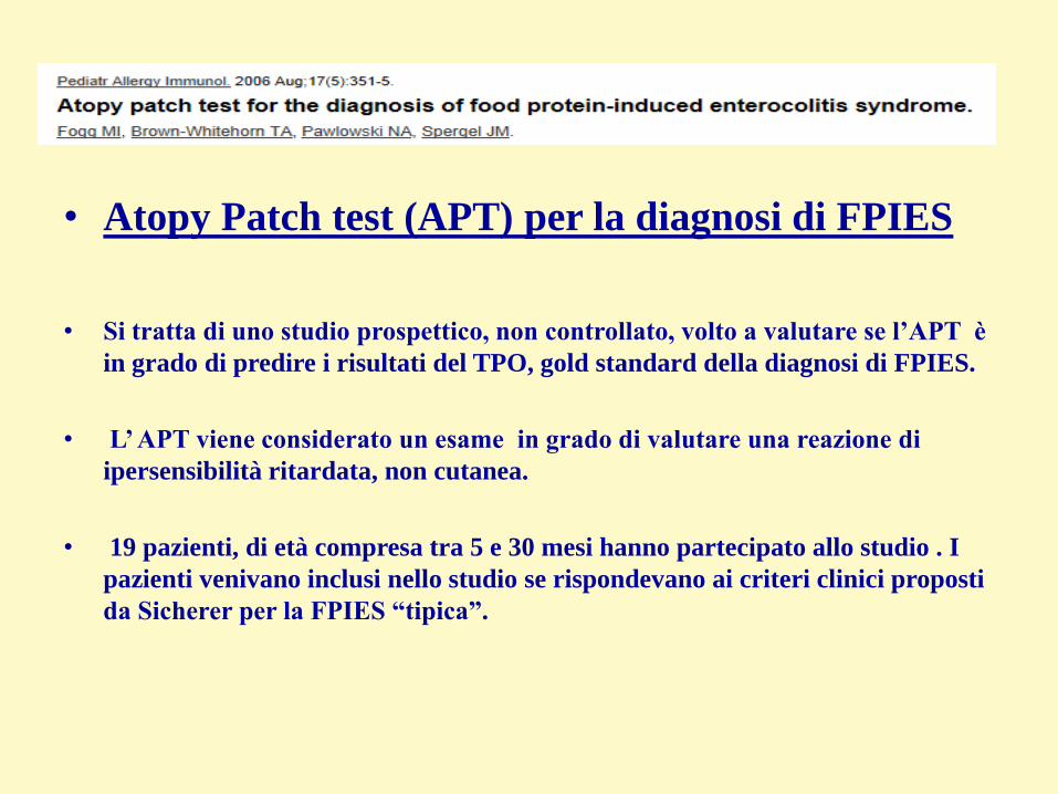
• Atopy Patch test (APT) per la diagnosi di FPIES
• Si tratta di uno studio prospettico, non controllato, volto a valutare se l‟APT è
in grado di predire i risultati del TPO, gold standard della diagnosi di FPIES.
• L‟ APT viene considerato un esame in grado di valutare una reazione di
ipersensibilità ritardata, non cutanea.
• 19 pazienti, di età compresa tra 5 e 30 mesi hanno partecipato allo studio . I
pazienti venivano inclusi nello studio se rispondevano ai criteri clinici proposti
da Sicherer per la FPIES “tipica”.

• Atopy Patch test (APT) per la diagnosi di FPIES
• Entro 2 mesi dall‟APT veniva effettuato un challenge orale, non cieco, per
accertare se i pazienti avessero FPIES. Durante questi due mesi i pazienti
erano istruiti ad evitare tutti gli alimenti che erano positivi all‟APT.
• I risultati dell‟APT e del TPO venivano comparati ed usati per calcolare
sensibilità e specificità dell‟APT

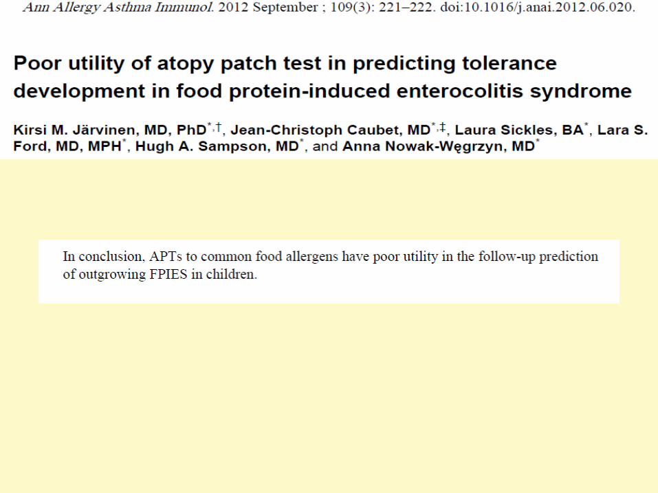
L„analisi statistica dei risultati di questi 19 pazienti , fece
concludere per:
- un valore predittivo positivo del 75%,
-valore predittivo negativo del 100%
-sensibilità del 100%
-specificità del 71%
Qualora in studi ulteriori, standardizzando
l’esecuzione del test, tali risultati dovessero
essere riconfermati, ’APT sembrerebbe essere
un esame diagnostico promettente per la
diagnosi di FPIES.

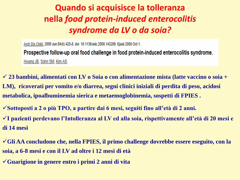
Quando si acquisisce la tolleranzanella food protein-induced enterocolitis
syndrome da LV o da soia?
Sottoposti a 2 o più TPO, a partire dai 6 mesi, seguiti fino all‟età di 2 anni.
23 bambini, alimentati con LV o Soia o con alimentazione mista (latte vaccino o soia +
LM), ricoverati per vomito e/o diarrea, segni clinici iniziali di perdita di peso, acidosi
metabolica, ipoalbuminemia sierica e metaemoglobinemia, sospetti di FPIES .
I pazienti perdevano l‟Intolleranza al LV ed alla soia, rispettivamente all‟età di 20 mesi e
di 14 mesi
Gli AA concludono che, nella FPIES, il primo challenge dovrebbe essere eseguito, con la
soia, a 6-8 mesi e con il LV ad oltre i 12 mesi di età
Guarigione in genere entro i primi 2 anni di vita

LA FPIES E’ INDOTTA SOLO DA LV E DA SOIA?
-Riso (n=14)-Soia (n=12)-Latte Vaccino (n=7)-Vegetali e frutta (in totale n=3, di cui 1 reagì alla banana)-Carni (n=2)-Avena (n=2)-Pesce (n=1).


An 8-month-old girl was initially breastfed and supplemented with a cow’s milk–
based formula. From birth she had issues with increased gas and appeared
uncomfortable. She also had recurrent vomiting, which typically would occur
soon after eating, and intermittent bloody streaks in her stool, which were green
in color and often very loose.
Her symptoms did not improve when a short trial of soy formula was given at 2
weeks of age. At 3 weeks of age she was prescribed ranitidine and formula was
changed to a casein hydrolysate–based formula with resolution of vomiting and
improvement in her symptoms.

At 4 months of age she was given soy formula and within 1 hour developed
repetitive vomiting, became lethargic and pale, and her parents described her as
nonresponsive. Emergency services were called, and she was brought to the
emergency department, where she was treated with oral rehydration. Her brother
had diarrhea around the same time, and it was thought that the infant’s symptoms
could be secondary to viral gastroenteritis.

She was seen by an allergist, and food allergy testing was performed.
The results of skin testing to milk, soy, and egg were negative at the time, and
specific IgE was undetectable to milk, soy, egg, wheat, and peanut. Because of
negative test results, an attempt was made to slowly reintroduce soy into her diet
at home. She was given a bottle with 1 oz of soy formula and 7 oz of casein
hydrolysate–based formula and did not complete the bottle.
Within 2 hours, she developed repetitive vomiting but was not pale or lethargic
and she recovered within 1 hour. She did not have any shortness of breath, rash,
or angioedema with any of these reactions.

Her diet currently includes hydrolyzed casein formula, grains (rice,
wheat and oat), several fruits and vegetables, and chicken.
She has dry patches of skin on her legs at times but has never been diagnosed
as having eczema. She has no history of asthma or wheezing. There is a strong
family of history of allergy, including cousins with food allergy and eczema, as
well as her father with a history of allergic rhinitis.

This infant’s history is consistent with milk and soy food protein– induced
enterocolitis.
A plan was made to continue with a casein hydrolysate–based formula and avoid
soy and milk. A follow-up in 1 year was recommended to consider performing a
physician-supervised food challenge to milk or soy at approximately 2 years of
age.
This challenge would be done with intravenous access in place and rapid
hydration available.

Quando si acquisisce la tolleranzanella food protein-induced enterocolitis
syndrome da LV o da soia?
Sottoposti a 2 o più TPO, a partire dai 6 mesi, seguiti fino all‟età di 2 anni.
23 bambini, alimentati con LV o Soia o con alimentazione mista (latte vaccino o soia +
LM), ricoverati per vomito e/o diarrea, segni clinici iniziali di perdita di peso, acidosi
metabolica, ipoalbuminemia sierica e metaemoglobinemia, sospetti di FPIES .
I pazienti perdevano l‟Intolleranza al LV ed alla soia, rispettivamente all‟età di 20 mesi e
di 14 mesi
Gli AA concludono che, nella FPIES, il primo challenge dovrebbe essere eseguito, con la
soia, a 6-8 mesi e con il LV ad oltre i 12 mesi di età
Guarigione in genere entro i primi 2 anni di vita


FOOD PROTEIN INDUCED ENTEROCOLITIS SYNDROME
CASO CLINICO
• S. è un secondogenito con anamnesi familiare e personale negativa per malattie allergiche.
• A 40 giorni di vita, per una ipogalattia materna, assume integrazione con formula presentando, la prima volta, vomito a getto dopo due ore dall’ingestione di circa 60 ml, senza compromissione dello stato generale e, la seconda volta, vomito profuso ed “abbattimento” dopo circa tre ore dall’introduzione.

FOOD PROTEIN INDUCED ENTEROCOLITIS SYNDROME
• La Pediatra curante esegue: skin prick test (SPT) con lattalbumina, beta-lattoglobulina e caseina: negativi; prick by prick latte formulato: negativo. Si orienta per una allergia alle proteine del latte vaccino (APLV) non (Ig)E-mediata e pone il piccolo a dieta con latte di soia.
• Dopo sei settimane S. giunge alla nostra osservazione. Si conferma la negatività dei test cutanei e delle IgEs per latte vaccino (LV) e si effettua test di provocazione orale (TPO): 0,3 g di proteine di LV/kg PC in un’ora. Dopo circa 20 minuti dalla dose bolo, S. presenta vomito profuso e ipotensione lieve, risolti rapidamente con infusione di soluzione fi siologica.
• Nei giorni successivi presenterà diarrea striata di sangue.

FOOD PROTEIN INDUCED ENTEROCOLITIS SYNDROME
• Confermiamo il sospetto diagnostico di enterocolite allergica da latte vaccino e prescriviamo prosecuzione della dieta di esclusione dell’alimento.
• S. ritorna alla nostra osservazione dopo 18 mesi per ripetere TPO.
• SPT con lattalbumina e caseina risultano negativi; SPT con beta-lattoglobulina è positivo: 5 mm diametro medio; prick by prick LV fresco positivo: 7 mm; IgEs LV: 10 KUA/l.

FOOD PROTEIN INDUCED ENTEROCOLITIS SYNDROME
• Effettua secondo TPO, seguendo lo schema della graduale somministrazione poiché risulta, allo stato attuale, IgE positivo.
• Nessuna manifestazione clinica fino alla dose bolo di 100 ml che evoca, in successione:
- vomito profuso
- edema della glottide
- stato letargico e
- shock (pressione arteriosa 50/20). Si somministra adrenalina i.m., cortisonico ed idratazione massiva e.v.

FOOD PROTEIN-INDUCED ENTEROCOLITIS SYNDROME
Prima descrizione in età adulta

FOOD PROTEIN-INDUCED ENTEROCOLITIS SYNDROME
FPIES TRASMESSA ATTRAVERSO
LATTE MATERNO


Curr Opin Allergy Clin Immunol 2014, 14:240–245

CLINICAL CASE NUMBER 1• When Gabriele was 1 year old, he received a diagnosis of egg-
FPIES; he had already experienced three severe acute episodes. Rigorous avoidance of dietary egg was prescribed, and the parents were given an action plan of instructions to be followed in case of accidental egg ingestion; this could be shown to the medical personnel in case of emergency.
• Some months later, Gabriele accidentally ate a small piece of omelet at nursery school, without anyone noticing.
Curr Opin Allergy Clin Immunol 2014, 14:240–245

CLINICAL CASE NUMBER 1• Two hours later his fourth acute FPIES episod occurred. The
child presented with repetitive and profuse vomiting, pallor, and severe lethargy.
• He was immediately taken to the emergency department, where his parents produced the action plan; intravenous fluids and steroids were immediately administered.
• Six hours after the onset of symptoms, he had recovered fully.
Curr Opin Allergy Clin Immunol 2014, 14:240–245

Curr Opin Allergy Clin Immunol 2014, 14:240–245

Curr Opin Allergy Clin Immunol 2014, 14:240–245
CLINICAL CASE NUMBER 2• Carmen suffers from atopic eczema; she has been exclusively breast-
fed. From her second month of life she presented frequent episodes of vomiting and diarrhea, sometimes watery; her growth was normal.
• When she was 5 months, she drank 70ml of cow’s milk formula; 2 h later she presented with repetitive and profuse vomiting, pallor, lethargy, and watery stools.
• Two hours later the symptoms resolved, and this episode was diagnosed as a viral gastroenteritis.
• After 2 weeks, she again drank 90ml of cow’s milk formula, and 2 h later suffered a similar acute episode. After the symptoms had improved, she still had episodes of regurgitation, colic, and occasional diarrhea.
Curr Opin Allergy Clin Immunol 2014, 14:240–245
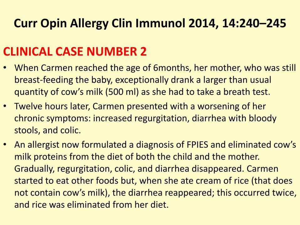
CLINICAL CASE NUMBER 2• When Carmen reached the age of 6months, her mother, who was still
breast-feeding the baby, exceptionally drank a larger than usual quantity of cow’s milk (500 ml) as she had to take a breath test.
• Twelve hours later, Carmen presented with a worsening of her chronic symptoms: increased regurgitation, diarrhea with bloody stools, and colic.
• An allergist now formulated a diagnosis of FPIES and eliminated cow’s milk proteins from the diet of both the child and the mother. Gradually, regurgitation, colic, and diarrhea disappeared. Carmen started to eat other foods but, when she ate cream of rice (that does not contain cow’s milk), the diarrhea reappeared; this occurred twice, and rice was eliminated from her diet.

Curr Opin Allergy Clin Immunol 2014, 14:240–245
LONG-TERM DIETARY MANAGEMENT AND INTRODUCTION OF AT-RISK FOODS
• Carmen’s story clearly exemplifies the chronic FPIES phenotype, which is rare. This case can be considered even rarer, because the culprit food affected her after passing through the breast milk. Although chronic FPIES is less frequent than acute FPIES, its long-term management is very similar: both require the culprit food to be eliminated from the patient’s diet, a point on which all experts agree.

Curr Opin Allergy Clin Immunol 2014, 14:240–245
LONG-TERM DIETARY MANAGEMENT AND INTRODUCTION OF AT-RISK FOODS
• Whereas in the case of IgE-mediated food allergies management is moving toward decreasing dietary restrictions, albeit with great caution (e.g. administration of baked milk or egg, ora immunotherapy), and although the current standard of care continues to entail strict avoidance of the food allergen for food allergy patients, in the case of FPIES patients, avoidance of culprit foods is compulsory, and surrounded by little or no doubt.

Curr Opin Allergy Clin Immunol 2014, 14:240–245
LONG-TERM DIETARY MANAGEMENT AND INTRODUCTION OF AT-RISK FOODS
This certainty is due to at least three reasons:
• first, most FPIES patients achieve tolerance spontaneously before their fifth year of life, an important reason making oral immunotherapy less attractive;
• second, for the individual patient, it is difficult to know the minimal dose of culprit food that may cause an adverse reaction; it may even be infinitesimal. This is due to the latency of symptoms; the child can finish the meal before any symptoms appear. It is thus very difficult to formulate an oral immunotherapy program with an initial dose that will almost certainly be tolerated;

Curr Opin Allergy Clin Immunol 2014, 14:240–245
LONG-TERM DIETARY MANAGEMENT AND INTRODUCTION OF AT-RISK FOODS
• third, although the immunopathogenic mechanisms are not as yet fully clear, the most convincing hypothesis assigns an important role to the T lymphocytes and their inflammatory cytokines; this would mean that pathogenic chemical bonds involve sequential peptides that are so small that any heating process, as for example in baking food, will be irrelevant in terms of the immune system’s recognition of epitopes.

Curr Opin Allergy Clin Immunol 2014, 14:240–245
LONG-TERM DIETARY MANAGEMENT AND INTRODUCTION OF AT-RISK FOODS
• In the case of cow’s milk-FPIES, if breast milk is lacking, the use of an extensively hydrolyzed casein formula is usually recommended, whereas soy milk should be avoided.
• These recommendations were developed on the basis of studies observing some clinical cohorts some years ago in the USA; the studies showed that 50% of children affected by cow’s milk-FPIES had adverse reactions to soy.
• More recently, Ruffner et al. confirmed these data in a study likewise conducted on U.S. children. However, Israeli, Italian, and Australian reports have not found the same association between cow’s milk-FPIES and soy-FPIES.
• .

Curr Opin Allergy Clin Immunol 2014, 14:240–245
LONG-TERM DIETARY MANAGEMENT AND INTRODUCTION OF AT-RISK FOODS
• More recently, Ruffner et al. confirmed these data in a study likewise conducted on U.S. children. However, Israeli, Italian, and Australian reports have not found the same association between cow’s milk-FPIES and soy-FPIES.
• Soy milk may thus be considered as a cow’s milk substitute, but only after having performed an OFC, to ensure that no adverse events will occur. In rare cases, it may be necessary to use an amino-acid formula.

Two hypothetical liberalization in the diet• It has been reported that culprit foods (in particular, cow’s
milk and hen’s egg) may be tolerated if baked; this runs counter to the theory of an exclusively cell-mediated pathogenesis of FPIES, and provides support for a role played by specific IgEs, as has been hypothesized.
• However, very few studies have addressed this point, so that in expectation of further research, the current recommendation is still to avoid culprit food even if baked.
• In this case, too, possible tolerance of baked culprit food should be verified by means of an OFC.
Curr Opin Allergy Clin Immunol 2014, 14:240–245

Two hypothetical liberalization in the diet• In the case of fish-FPIES, which is the commonest form of
solid-food-induced FPIES in Italy, it may not be necessary to eliminate all kinds of fish from the diet.
• Sopo et al. reported that three of eight children who reacted to one or more types of fish tolerated other fish (e.g. tuna, swordfish, salmon).
• It is known that cross-reactivity between species of fish is not absolute in IgE-mediated fish allergy; this may also be true for fish-FPIES.
• Be that as it may, tolerance to other types of fish must
• in all cases be verified via an OFC.
Curr Opin Allergy Clin Immunol 2014, 14:240–245

• Should a restrictive diet be prescribed to breast-feeding mothers?
• Considering the rarity of FPIES passing through breastmilk, the more tolerant approach is currently preferable
Curr Opin Allergy Clin Immunol 2014, 14:240–245

CONCLUSION
• In 2005, Sicherer *1+ wrote: ‘. . . but much more research is needed to determine the best course of dietary management, develop laboratory tests to avoid the need for oral food challenges, address prevention, and determine specific treatment modalities.
• These goals will most likely be reached through more intensive laboratory investigation of the immunopathologic basis of the disorder. More work also needs to be done to determine whether disorders with similar symptoms are pathophysiologically distinct from FPIES or part of a spectrum with a similar cause whose clinical expression varies with environmental influences.’ In 2014, we still agree with those conclusions.
Curr Opin Allergy Clin Immunol 2014, 14:240–245
MESSAGES
• La FPIES rappresenta una sindrome non ancora
perfettamente conosciuta e ciò spiega il numero elevato di
indagini che, abitualmente, vengono eseguite in PS e di
diagnosi errate effettuate prima che sia posta la diagnosi
corretta.
• Si tratta di una diagnosi prevalentemente clinica. I criteri
di Sicherer, se soddisfatti, sono in grado di far formulare
correttamente la diagnosi.

CONCLUSIONI• Un TPO diagnostico è superfluo nei casi “tipici”, che
soddisfino tutti i criteri clinici.
• Il TPO, allorquando se ne valuti l‟opportunità di
esecuzione, va effettuato in ambiente attrezzato e con
personale esperto a fronteggiare eventuali gravi reazioni.
• L‟acquisizione della tolleranza totale verso l‟alimento
responsabile si verifica nella stragrande maggioranza dei
casi

BENEVENTOVIII GIORNATA DI ALLERGOLOGIA ED IMMUNOLOGIA
PEDIATRICA 20-22 Novembre 2014