layers of the human skull - web.lemoyne.edu
TRANSCRIPT
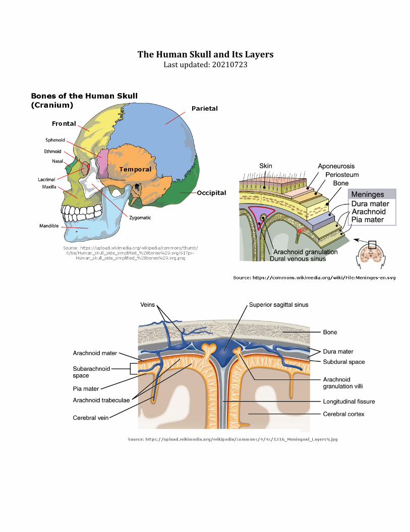
TheHumanSkullandItsLayersLastupdated:20210723
Aponeurosis=“Theepicranialaponeurosis,orgaleaaponeurotica,isatoughlayerofdensefibroustissuewhichrunsfromthefrontalismuscleanteriorlytotheoccipitalisposteriorly…[The]primaryfunction[oftheaponeurosis]istojoinmusclesandthebodypartstheyactupon,whetheritbeboneorothermuscles”{Wikipedia}Periosteum(akapericranium)=“thefibrousmembranethatformsthecoveringofbonesexceptattheirarticularsurfaces.Itconsistsofadenseexternallayercontainingnumerousbloodvesselsandaninnerlayerofconnectivetissuecellsthatfunctionasosteoblasts[=cellsthatformnewbones]whentheboneisinjuredandalsoparticipateinnewboneformation.Theperiosteumservesasasupportingstructureforbloodvesselsnourishingboneandforattachmentoftendonsandligaments”(Venes,2009,p.1752).SkullBone([Neuro-]Cranium)=Thehumanskullisgenerallyconsideredtoconsistoftwenty-twobones—eightcranialbonesandfourteenfacialskeletonbones.Intheneurocraniumthesearetheoccipitalbone,twotemporalbones,twoparietalbones,thesphenoid,ethmoidandfrontalbones.{Wikipedia].Notethatthefourlobesofthecerebralcortexarenamedaftertheboneswhichcoverthem.Thesectionoftheskullcoveringthecerebrumisalsoknownasthecalvarium.[Latin:calvaria=“skull’]Aswithbonesthroughoutthebody,theinteriorofthecranialbonescontainsmarrow.Recentresearchpointstothecranialbonemarrowasasourceofcellstocombatinflectionorotherformsofinjurytothebrain(Cugarraetal.,2021;Herissonetal.,2018)Meninges=belowtheneurocranium(skullbones)arethreelayersofmembraneswhichcoverthebrain.
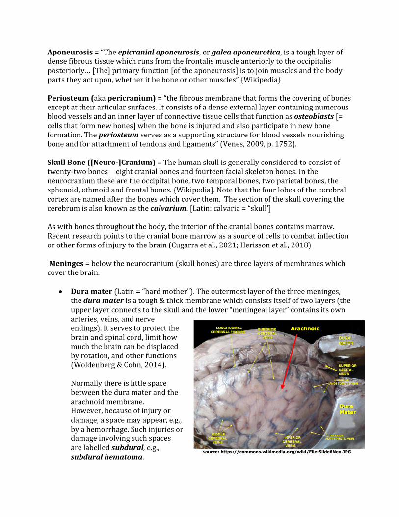
• Duramater(Latin=“hardmother”).Theoutermostlayerofthethreemeninges,theduramaterisatough&thickmembranewhichconsistsitselfoftwolayers(theupperlayerconnectstotheskullandthelower“meningeallayer”containsitsownarteries,veins,andnerveendings).Itservestoprotectthebrainandspinalcord,limithowmuchthebraincanbedisplacedbyrotation,andotherfunctions(Woldenberg&Cohn,2014).Normallythereislittlespacebetweentheduramaterandthearachnoidmembrane.However,becauseofinjuryordamage,aspacemayappear,e.g.,byahemorrhage.Suchinjuriesordamageinvolvingsuchspacesarelabelledsubdural,e.g.,subduralhematoma.

• Arachnoidmater(Greek:arachne=“spider”,Latin:“arachnoīdes”=“likeacobweborspiderweb”).Subarachnoidspace.Belowthearachnoidmembraneandabovethepiamateristhesubarachnoidspace.Thisareacontainscerebralspinalfluid(CSF)aswellasmultiplebranchesofthebrain’sarteriesandveins.Overthegyriofthecortex,thisspacetendstobequitethin.However,inthesulciofthecortex,thespacebetweenthearachnoidmembraneandpiamatercanbecomequitelargeandformswhatareknownassubarachnoidorbasalcisterns(Latin=“boxes”).Tumorsgrowinginthemeningesarecalledmeningiomas.TheygenerallyariseinthearachnoidandpressdownuponthePiamater.
• Piamater(Latin=“tendermother”)Thepiamaterisathinmembranewhich
coverstheentireCNSincludingtheinsidesofthesulci.Functionallyitappearstoprovideacoveringofthecorticaltissuethatallowswaterandsomesmallsolublemoleculestopassthroughtothebrain’sinterstitialfluid(thefluidwhichsurroundstheneuronsandglialcellsofthebrain.)ThePiamaterandArachnoidtogetheraresometimescalledtheleptomeninges.
• Notethatmeningitisisausuallyseenasaninflammationoftheleptomeningeswiththeduramaterlessfrequentlyinvolveds
References
Cugarra,A.,Mamuladze,T.,…Kipnis,J.(2021).Skullandveretebralbonemarroware
myeloidcellrevervoirsforthemeningesandCNSparenchyma.Science,373,eabf7844.https://doi.org/10.1126/science.abf7844
Herisson,F.,Frodermann,V.,Courties,G.,Rohde,D.,Sun,Y.,Vandoorne,K.,…Nahrendorf,
M.(2018).Directvascularchannelsconnectskullbonemarrowandthebrainsurfaceenablingmyeloidcellmigration.NatureNeuroscience,21,1209-1217.https://dx.doi.org/10.1038/s41593-018-0213-2
Venes,D.(Ed.).(2009).Taber’scyclopedicmedicaldictionary(21sted.).Philadelphia,PA:F.
A.DavisCompanyWoldenberg,R.F.,&Kohn,S.A.(2014).Duramater.Encyclopediaoftheneurological
sciences,1039–1042.https://dx.doi.org/10.1016/b978-0-12-385157-4.01143-x