laboratory x-ray nano-ct system - nondestructive · pdf filelaboratory x-ray nano-ct system...
TRANSCRIPT

Laboratory X-Ray Nano-CT System
Alexander SASOV, SkyScan, Aartselaar, Belgium
Abstract. Using advanced X-ray technologies and X-ray scattering enhancement in signal detection, a compact laboratory X-ray scanner for 3D non-invasive imaging with 150-200 nanometers 3D detail detectability has been created. This spatial resolution in the volume terms is equal or better than can be achieved in synchrotron tomography, 5 orders better than in existing laboratory instruments and 10-12 orders better in comparison to clinical CT. The instrument is built using X-ray source with LaB6 cathode and two electromagnetic lenses. Small-angle scattering enhances the object details up to 150-200nm. An object manipulator allows precision positioning and rotation with accuracy better than 100nm. The X-ray detector is based on an intensified CCD with single photon sensitivity. The typical acquisition cycle for 3D reconstruction of the full object volume takes from 15 to 100 minutes with collection of several hundreds angular views. Subsequent volumetric reconstruction produces results as a set of virtual slices with isotropic voxel size up to 150 x 150 x 150nm or as a 3D-model, which can be virtually manipulated and measured. The object stays in normal environmental conditions without any coating, vacuum treatment or other preparation. Unique spatial resolution in non-invasive 3D-investigation allows obtaining heretofore unachievable 3D images in the wide range of application areas, such as composite materials, carbon-based materials, fuel cells, paper and wood microstructure, biomedical applications, etc.
Introduction Using modern technologies in x-ray sources, precision mechanics and detection systems, a compact laboratory x-ray scanner has been created for non-invasive imaging of internal microstructure of objects with 150-200 nanometers isotropic three-dimensional spatial resolution. The system allows the imaging of previously unattainable details of internal three-dimensional micro-architecture in a wide range of applications. 1. Nano-CT Scanner The X-Ray nano-CT instrument is based on the same general principles as clinical CT-scanners and other micro-CT systems [1]. The object mechanically rotates inside the x-ray beam and an x-ray camera acquires hundreds or thousands angular projections for subsequent reconstruction of the object’s three-dimensional internal 3D microstructure in the computer memory using back-projection algorithms. The differences in the new nano-CT instrument compared to previous micro-CT instruments, which allow us to reach submicron resolution, are based on (a) the use of different contrast formation physics and
ECNDT 2006 - We.1.5.4
1
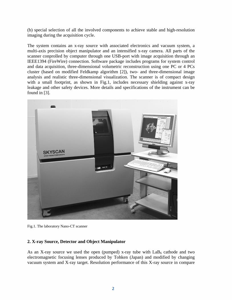
(b) special selection of all the involved components to achieve stable and high-resolution imaging during the acquisition cycle. The system contains an x-ray source with associated electronics and vacuum system, a multi-axis precision object manipulator and an intensified x-ray camera. All parts of the scanner conprolled by computer through one USB-port with image acquisition through an IEEE1394 (FireWire) connection. Software package includes programs for system control and data acquisition, three-dimensional volumetric reconstruction using one PC or 4 PCs cluster (based on modified Feldkamp algorithm [2]), two- and three-dimensional image analysis and realistic three-dimensional visualization. The scanner is of compact design with a small footprint, as shown in Fig.1, includes necessary shielding against x-ray leakage and other safety devices. More details and specifications of the instrument can be found in [3].
Fig.1. The laboratory Nano-CT scanner 2. X-ray Source, Detector and Object Manipulator As an X-ray source we used the open (pumped) x-ray tube with LaB6 cathode and two electromagnetic focusing lenses produced by Tohken (Japan) and modified by changing vacuum system and X-ray target. Resolution performance of this X-ray source in compare
2

to “standard” microfocus X-ray source is shown in Fig.2 by comparison of images from resolution test pattern produced by XRadia company [4].
Fig.2. X-ray sources resolution comparison: microfocus X-ray source (left) and X-ray source used in Nano-CT (right) Submicron spot size in the X-ray source creates big difference in the image formation during interaction of the X-ray beam with an object. The best known phenomenon in the interaction of an x-ray beam with material is x-ray absorption, shown in the left part of Fig.3. It is used in all clinical x-ray imaging systems and CT-scanners as well as in industrial CT and micro-CT instruments. In the case of source spot-size around and under one micron another fortuitous physical phenomenon exists in contrast formation, shown on the right side of Fig.3 and called “small angle scattering” or “small angle reflection”. Any external surface or internal border between different densities inside the object creates small angle scattering (reflection) of a polychromatic x-ray beam, which (in the case of small spot-size of the source) enhances the image of the object’s external and internal edges. This image enhancement is defined by the spot size of the source and the object-detector distance. The correct choice of the object-detector distance for the high-resolution mode allows selection of the necessary balance of absorption contrast and scattering enhancement. If an averaging kernel in the reconstruction program is adjusted for exact compensation of the small angle scattering in the projection image, signal-to-noise ratio will be improved without reducing spatial resolution.
3

Fig.3. Absorption contrast (left) and small angle scattering (right). The X-ray detector contains an intensified 1280x1024 pixels / 12-bit digital CCD camera with direct connection to PC (IEEE-1394). The intensified CCD with Be-window includes 18 mm format MCP-based image intensifiers, fiber-optically coupled to a Sony ICX285 CCD-sensor: the gain is 5000 cd/sq.m/lx, 62 lp/mm resolution. The camera is mounted on the translation stage with 25mm travel / 1µm repeatability for alignment. The object manipulator can move the specimen stage between the camera and the detector to change image magnification. The object position closest to the detector corresponds to the minimum magnification with a field of view (maximum object diameter) near 10mm. An object position close to the source corresponds to the maximum magnification with the voxel size of the image (at the object location) close to 100nm. One of the most important parts of the Nano-CT system is the object X-Y-Z positioning and rotation system. The necessity to acquire several hundred angular views during 10-60 minutes from a well-defined object location requires very high accuracy and stability. Therefore for object rotation in the Nano-CT instrument we use an air-bearing with integrated stepping motor and optical encoder and with the following specifications: flatness <100nm, eccentricity <100nm, wobble <2.5microrad, repeatability 0.001deg, speed up to 360 deg/sec, load up to 5 kg. The rotatable part of this stage floats in an airflow layer without physical contact to the static part. The top of the rotation stage holds an X-Y-Z manipulator for high-precision specimen positioning: Z axis -10mm travel with 400nm resolution, XY axis – piezo drives with 1.2mm travel with 1um steps All electronics for X-Y-Z drives in the top of the rotation stage are powered through a non-contact slotted transformer and controlled by wireless infrared data link. A special microprocessor
4

controller in the top of the rotation stage controls X-Y-Z drives and continuously measures local temperature in the top of the stage. Temperature measurement allows compensation for thermal extension of the metal parts in the object support with overall accuracy around 150nm. 3. Application Examples To confirm the overall spatial resolution of three-dimensional reconstruction at the submicron level, we performed a scan of a platinum (Pt) wire with 600nm nominal diameter as a test object. The diameter of the Pt wire was verified by SEM imaging. Fig.4a shows the Pt wire on top of a mesh with 5x5µm openings. Measurements show that the Pt wire has a diameter of 600-800nm. Fig.4b shows two-dimensional X-ray shadow image of the same Pt wire object in the Nano-CT system. Subsequent acquisition and reconstruction were done for a smaller part of the volume, containing only Pt-wire and keeping the thick Ag-rod outside the field of view. One of the reconstructed cross sections with 170nm isotropic voxels is shown in Fig.6c. The cross section through the thin Pt wire can be seen as an ellipse since the reconstruction plane crosses the wire at a significant angle.
Fig.4. SEM-image of the Pt-wire (a), a Nano-CT x-ray shadow image (b) and a reconstructed Nano-CT cross section with 170nm voxel size (c). Another application example is a reconstruction of the internal microstructure inside a paper sample. Fig.5 shows the reconstruction results in the form of three orthogonal virtual slices through the sample. The samples scanned with 360nm isotropic voxel size. Scanning time is 40 minutes for entire volume.
5

Fig.5. A three orthogonal virtual slices reconstructed through a paper sample with 360nm isotropic voxel size. Conclusions The compact Nano-CT scanner described here allows non-invasive investigation of the three-dimensional internal microstructure of a wide range of objects and materials, with isotropic spatial resolution in the range of hundreds nanometers. References [1] A.Sasov, Desk-top X-ray Micro-CT Instruments. In: Developments in X-Ray Tomography III, Editor U.Bonse, SPIE vol.4503, pp 282-290,(2001). [2] L.A.Feldkamp, L.C.Davis and J.W.Kress, Practical Cone-beam Algorithm. J.Opt.Soc.Am. A, vol.1, No.6, June 1984. [3] www.skyscan.be [4] www.xradia.com
6