laboratory - ncsu
TRANSCRIPT

127
lab
ora
tory
8
8 Laboratory R
ober
t Jo
hnso
n/S
hutt
erst
ock
.com
LEARNING OBJECTIVES
Students will….
• identify associations between plants and pollinators by analyzing flower structures
and determining potential pollination strategies for each plant.
• practice communication and presentation skills by presenting their findings on
flower structure and pollination strategies to the class.
• practice microscopy skills and extend their knowledge of symbiotic relationships
by making slides of live specimens of microscopic organisms that aid in digestive
processes.
• analyze endo- and ectoparasite groups by observing live, preserved, and micro-
scopic specimens and using online materials; and they will present their findings
to the class.
• apply their knowledge of parasitic relationships by locating and identifying para-
sites on the surface and internal organs of a fish specimen.
• demonstrate their knowledge of fish anatomy by pointing out morphological and
anatomical structures.
• extend their knowledge of symbiotic relationships by answering and discussing
specific questions at the end of lab.
INTRODUCTIONAssociations between organisms of different species are known as interspecific inter-
actions. The organisms involved may benefit from, be harmed by, or not be affected
by the interaction. By this definition, we can use the term symbiosis to represent any
Species Relationships: Symbiosis
NOT FOR DISTRIBUTION - FOR INSTRUCTORS USE ONLY

128
Laboratory 8 Species Relationships: Symbiosis
association between organisms, excluding interactions between members of the
same species, or intraspecific interactions. Symbiotic relationships exist between all
types of organisms including bacteria, protozoans, fungi, plants, and animals.
Various types of symbioses, whether beneficial or harmful, are described by the terms
mutualism, commensalism, and parasitism. In these relationships, we refer to the
symbiont as the organism that lives inside (endoparasites) or on (ectoparasites)
another organism, the host. In symbioses where the organisms interact with each
other, either living inside or on the other, both organisms are termed symbionts.
In mutualistic relationships, both partners benefit equally. Most commonly, organ-
isms enable the acquisition of nutrients for one another. An example of a mutualistic
relationship is the one between Aiptasia pallida, a small sea anemone found in the
Caribbean and along the east coast of the United States, and a dinoflagellate algae.
The dinoflagellate is called an endosymbiont, because it lives inside the sea anemone,
its host. The sea anemone receives oxygen and photosynthetic products from the
dinoflagellate, whereas the dinoflagellate receives protection and molecules for pho-
tosynthesis from the sea anemone.
The protozoans found in the stomach of herbivores, which help the animals digest
food, receiving nutrients in the process, represent another mutualistic relationship.
Protozoans also inhabit the gut of termites, helping them digest wood material. You
will have the opportunity to observe these protozoans when you complete the nutri-
tion lab. Mutualistic relationships also occur between plants and fungi. Fungi called
Mycorrhizae form an association with the roots of a plant, in which they help plants
to extract nutrients from nutrient-poor soils and in exchange receive organic com-
pounds from the plant’s photosynthetic processes. Another important symbiotic
relationship involves a fungi and a photosynthetic organism like an algae, which un-
dergoes a remarkable change during their association, resulting in a new entity called
a lichen.
An association in which the symbiont benefits while the host organism is neither
harmed nor benefited is called commensalism. The relationship between the tube
worm Chaetopterus and pea crabs is an example of a commensalistic relationship.
In this relationship, the worm shares its tube-like dwelling with a crab. The crab gets
protection from the tube and receives food and oxygen from the water that passes
into the tube. The shark and sucker fish Echeneis is another example of commensal-
ism. The sucker fish rides along with the shark by sticking to its underside with a
modified dorsal fin shaped like a suction disc. Close proximity to the shark allows the
sucker fish to scavenge bits of food left over from the shark’s meal.
Parasitism is a symbiosis in which the symbiont benefits at the expense of the host.
As in most symbiotic relationships, the driving force behind parasitic associations
is usually food/nutrients, since the parasite obtains its food from the host. Parasitic
relationships affect the host to varying degrees. Some parasites are so patheno-
genic (disease-causing) that they cause symptoms in the host almost immediately
after infection. In these cases, the host may die, but most parasites do not kill their
host until they have reproduced and completed their life cycles (see life cycle Figures
8-2 through 8-8). Some parasites need more than one host. Hosts are then either
NOT FOR DISTRIBUTION - FOR INSTRUCTORS USE ONLY
129
Laboratory 8 Species Relationships: Symbiosis
intermediate or final. You will have a chance to observe some of these parasites in the
laboratory. Hosts can also serve as reservoirs for a parasite (a breeding ground and
source of infection for another host) without showing signs of infection themselves,
or they can be vectors—carriers of the parasite to a final host. Plants can also be
parasitic. There are thousands of parasitic plant species, ranging from trees to small,
herbaceous plants. The best-known group of parasitic plants is mistletoe. There are
about 800 species, most occurring in the tropics and subtropics (Paracer and Ah-
madjian 2000). Mistletoe parasitize tree branches, but the giant mistletoe, Nuylsia,
forms a tree that grows as high as 10 meters and parasitizes roots of nearby grasses
and plants.
Symbioses between plants and their pollinators (Figure 8-1) are considered a prime
example of coevolution of these two groups of organisms over the past 200 million
years. So remarkable is the “fit” between pollinator and flower that a fairly novice
observer can predict the type of pollinator for which a flower is adapted by examining
the flower’s color, shape, scent, and other characteristics. Similarly, specialized struc-
tures on a pollinator, like the shape and length of the proboscis (the tubular feeding
organ) closely match the flower’s anatomy. Pollinators, usually insects or some other
animal, carry pollen from the anther of one plant where it is produced to the stigma of
another plant, while plants provide the animal with a food source in the form of nectar
or pollen (see Figure 8-1). Nearly 70% of flowering plants rely on insects for pollination
and 30% of our food comes from bee-pollinated crops (Kearns and Inouye 1997).
Symbiotic relationships between animals and microorganisms are also important in
the process of nutrient acquisition (you will learn more about specific nutritional
adaptations in the nutrition lab). Ruminants and other animals rely on certain spe-
cies of bacteria and protists within their digestive tracts for digesting tough, cellulose
material. The most advanced fiber processing digestive tract, which is found in graz-
ing types of mammals, is the ruminant system. Cows, sheep, and deer, among many
others, are ruminants. These animals are often described as having four stomachs,
because the stomach is partitioned into four chambers that each have a specific func-
tion for digesting plant material before reaching the small intestine. In order, the
chambers are: rumen, reticulum, omasum, and abomasum. Symbiotic bacteria and
protists in the first two processing chambers use enzymes to degrade the plant ma-
terial and yield large quantities of a waste product called volatile fatty acids through
fermentation reactions. These fatty acids are absorbed into the blood and are trans-
ported to the liver where they are converted to sugars that are used in metabolism.
The fluid from these chambers is often referred to as “rumen fluid,” since the rumen
is the largest chamber that contains microorganisms. The third chamber of the ru-
minant stomach acts as a particle sieve keeping the larger, less degraded particles in
the first two chambers. It also reabsorbs water. The final chamber acts as the “true
stomach,” with acids and enzymes that break down materials just as they do in the
stomach of an animal with a monogastric system.
The alimentary canals of animals also possess symbiotic bacteria housed in spe-
cialized intestinal structures, such as the cecum. In herbivores, the cecum can be a
large fermentation chamber. For example, the koala has a long, tubular cecum with
abundant symbiotic bacteria. Bacteria in the cecum use enzymes to break down the
NOT FOR DISTRIBUTION - FOR INSTRUCTORS USE ONLY

130
Laboratory 8 Species Relationships: Symbiosis
fibrous eucalyptus leaves, which are the sole food source of the koala. On the other
hand, carnivores have a small cecum, since plant material is not common in their
diets. Regardless of the anatomical structures present, most animals possess gut mi-
croorganisms collectively known as the “gut flora.” In addition to helping with the
digestion of dietary fiber, these microorganisms perform other important roles. For
instance, in mammals, beneficial intestinal Escherichia coli synthesize vitamin K. A
healthy “gut flora” is also essential for maintaining a healthy gastrointestinal system
and plays an important role in the immune system.
In the invertebrates, termites are a classic example of a type of organism that has a
coevolutionary relationship with gut microorganisms. Termites rely on bacteria and
protists to digest cellulose from the wood they consume. Termites and their diverse
community of microorganisms form obligate symbiotic relationships in which one
cannot live without the other. In this lab, you will have the opportunity to analyze the
content of a termite gut and find some of these microorganisms. The most common
organism you will find is a protist called, Trychonympha spp. You will also observe
rumen fluid from a cow to view the gut flora.
Activity One: Plants and Pollinators
PROCEDURE
1. Read Table 8-1. It describes various pollinator and flower characteristics (you
may also refer to pictures of flowers seen in Appendix F). You will also watch a
series of video clips on pollination.
2. Work in groups at your laboratory bench during this activity to try to predict the
type of pollinator for the flowers you observe in the laboratory. Fill out Table 8-2
as you make your observations. Each group should present their findings to the
class.
NOT FOR DISTRIBUTION - FOR INSTRUCTORS USE ONLY

131
Laboratory 8 Species Relationships: Symbiosis
Table 8-1. Flower attractants commonly associated with pollinators
BIRDS BUTTERFLIES MOTHS BATS FLIES BEES WIND
Color Bright;
scarlet red,
orange,
yellow
Bright; red,
orange, yel-
low, or pink
Pale yellow
or bright
white
Dull greenish,
yellowish, or
purple; often
creamy white
Light colors Bright; most
commonly
yellow or blue,
cannot see red
Very pale;
drab
Scent None Weak to
moderate
Strong, sweet
like perfume
Strong,
musky
Mild; often
fungal
Sweet None
Flower
shape
Tubular and
usually long,
to house the
bird’s bill
Showy flow-
ers; often
tubular
Very con-
cealed
nectar; often
tubular
Big and wide;
sometimes a
brush-type
or bowl-
shaped
Flat or
concave;
reward
exposed;
shallow
Complex;
reward often
concealed
No petals;
mainly
anthers
and
carpels
Pollina-
tion time
Day Day Dusk and
night
Night Day Day None
Flower
position
Lack
landing
platforms,
stigma/
anthers pro-
trude well
beyond the
petals; flow-
ers often
hanging
Have land-
ing platform
of clustered
flowers,
stigma/
anthers
protrude
beyond
petals
Often hang-
ing; erect or
horizontal
Exposed on
sturdy stems
or tips of
branches
Mostly
erect
Have landing
platforms for
pollen attach-
ment; many
positions; of-
ten hanging
Erect
Reward Nectar in
the tube
(large quan-
tity)
Nectar in
the tube
Nectar (more
than in bee-
pollinated
flowers)
Highest nec-
tar quantity,
pollen
Pollen,
nectar
Nectar, pollen None
Other Birds hover
while pol-
linating
Butterfly-
pollinated
flowers
resemble
bird-
pollinated
flowers but
are smaller
and contain
less nectar.
Moths are
nocturnal
and usually
find females
by following
a pheromone
or other
chemical
trail; good
sense of
smell
Carrion
flies are at-
tracted to
mottled
purple or
dark blue
flowers that
smell like
rotting meat
and are of-
ten near the
ground
Flowers are
not red be-
cause bees
cannot see
red light well;
often show
patterns (nec-
tar guides)
visible only
in ultraviolet
light that bees
can perceive
NOT FOR DISTRIBUTION - FOR INSTRUCTORS USE ONLY

132
Laboratory 8 Species Relationships: Symbiosis
Table 8-2. Record of observed pollinators and flowers
FLOWER NAME OR TYPE
PREDICTED POLLINATOR
JUSTIFICATION
©Hayden-McNeil, LLC
Anther sheddingpollen onto bee
Flower nectaries
Petal
Bee proboscis
Stigma receivingplatform for pollen
Figure 8-1. Flower–Pollinator Interaction
NOT FOR DISTRIBUTION - FOR INSTRUCTORS USE ONLY

133
Laboratory 8 Species Relationships: Symbiosis
Activity Two: Symbiosis in Termites
Termites are invertebrates that possess a complete digestive tract. They consume
wood, which is primarily composed of cellulose; however, they cannot manufacture
a key enzyme necessary for its breakdown. Instead they rely on a symbiotic relation-
ship with bacteria and protozoa to digest the cellulose and absorb the waste products
(volatile fatty acids) produced for use as food.
1. Using forceps, gently remove a termite from the stock culture.
2. Place the termite on a glass microscope slide.
3. Using a dissecting needle, remove the head of the termite and discard it.
4. Place one drop of Insect Ringer’s solution on the termite abdomen and then place
a coverslip over the abdomen and gently press down with your dissecting needle
so that the material in the abdomen is dispersed on the slide.
5. View the symbionts with a compound microscope. (Primarily what you will see
is protozoan, but you can see bacteria on a much higher power with stain.)
6. Draw your observations below.
NOT FOR DISTRIBUTION - FOR INSTRUCTORS USE ONLY

134
Laboratory 8 Species Relationships: Symbiosis
Activity Three: Bacteria and Protists in the Rumen
Like the termites, ruminants and other herbivores rely on microorganisms to process
the fibrous cellulose present in plant leaves. Within the rumen chamber, bacteria and
protozoa are suspended in the fiber particle-filled rumen fluid. The microorganisms
attach themselves to the fiber particles and hydrolyze the cellulose. As with the ter-
mites, the waste products of their metabolism are absorbed by the host animal and
used for food. One major benefit to having the rumen before the true stomach of the
animal is that, as bacteria die and flow through the digestive tract, they can be used
as a source of dietary protein.
1. Take a microscope slide and place 2–3 drops of rumen fluid on it, using the plas-
tic pipette.
2. View this slide, with a coverslip, under 40# power. You should be able to see large
protists moving around on the slide.
3. Bacteria are usually seen in the forms of rods, cocci, and spirilla, but you may not
be able to see them without stain.
4. Draw two of the different endosymbionts that you can see on your slide.
NOT FOR DISTRIBUTION - FOR INSTRUCTORS USE ONLY

135
Laboratory 8 Species Relationships: Symbiosis
Activity Four: Introduction to Parasites
PROCEDURE
1. Work in groups to familiarize yourself with various endo- and ectoparasites of
humans and other animals. Each student group will be assigned one group of
endoparasites and at least one ectoparasite to examine. Your group will then be-
come the experts at identifying these types of parasites, and you will use your
expertise when you move to Activity Five: Examining Fish Parasites. Refer to
Table 8-3 to view your assignment.
Table 8-3. Parasite groups
STUDENT GROUP I
STUDENT GROUP II
STUDENT GROUP III
STUDENT GROUP IV
STUDENT GROUP V
STUDENT GROUP VI
Endoparasites Amoebas Ciliates Flagellates Apicom-plexans
Platyhel-minthes
(flat worms: flukes and
tapeworms)
Nematoda or Nema-
thelminthes (round-worms)
Ectoparasites Tooth amoebas
(Entamoeba gingivalis)
AND Ticks
Bedbugs Fleas Lice Mites Leeches
NOT FOR DISTRIBUTION - FOR INSTRUCTORS USE ONLY

136
Laboratory 8 Species Relationships: Symbiosis
2. Use the available prepared slides, biomounts, preserved specimens, Web site in-
formation, laboratory charts, and lab manual to gather as much information as
you need for your selected parasites (see life cycle Figures 8-2 through 8-8).
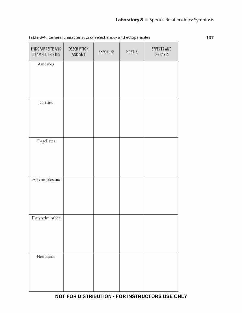
3. You will have to find the following information to enter into Table 8-4:
a. Example species: Examine at least three species in your endoparasitic group
as well as your given ectoparasite(s). List the scientific names of each parasite
species in the first column of Table 8-4.
b. Description and size: Provide some general background information for
your parasite group, including key characteristics and interesting facts. Give
the average size of your parasites. If looking at slides, note total magnification
and field of view to estimate size. Also, note the life cycle stage of the parasite
you are viewing. For example, size may differ between a cyst (or egg) and
the adult form of your parasite. Your description should have enough detail
(think back to Organism X from lab 1 and how you described it) for someone
else to get a good overview of your parasite. For example, “big and brown” is
not an acceptable description.
c. Exposure: Briefly describe how humans and other animals are exposed to
your particular parasite, for example, contaminated food or drink. If relevant,
include the geographic location of the most frequent exposure.
d. Host(s): List the hosts your parasite uses. If it has multiple hosts, then specify
which one is intermediate and which one is final.
e. Effects/Diseases: List and briefly describe the effects and/or diseases caused
by your parasites. Include symptoms and treatments when possible. Since we
are looking for parasites during the fish dissection (Activity Five), you may
want to note if your parasite affects fish.
4. As a class, you will fill out the entire Table 8-4. Be prepared to describe your
parasites to the class. You will need to familiarize yourself with each group of
parasites in order to find parasites during the fish dissection in Activity Five.
5. Make a drawing or find a picture of a representative endoparasite and ectopara-
site for your given parasitic organisms to share with the class.
NOT FOR DISTRIBUTION - FOR INSTRUCTORS USE ONLY
137
Laboratory 8 Species Relationships: Symbiosis
Table 8-4. General characteristics of select endo- and ectoparasites
ENDOPARASITE AND EXAMPLE SPECIES
DESCRIPTION AND SIZE
EXPOSURE HOST(S)EFFECTS AND
DISEASES
Amoebas
Ciliates
Flagellates
Apicomplexans
Platyhelminthes
Nematoda
NOT FOR DISTRIBUTION - FOR INSTRUCTORS USE ONLY

138
Laboratory 8 Species Relationships: Symbiosis
ECTOPARASITEDESCRIPTION
AND SIZEEXPOSURE HOST(S)
EFFECTS AND DISEASES
Bedbugs
Fleas
Mites
Ticks
Lice
Leeches
Tooth amoebas
NOT FOR DISTRIBUTION - FOR INSTRUCTORS USE ONLY

139
Laboratory 8 Species Relationships: Symbiosis
Life Cycles of Common Endoparasitic Protozoans
2
1
Cyst
Trophozoites
Multiplication
Exitshost
Trophozoites arealso passed in
stool but they donot survive in the
environment
Contaminationof water, food, or
hands withinfective cysts
©Hayden-McNeil, LLC
Trophant
Trophozoites in the host’s skin
Mature trophantwith hundreds ofmaturing tomites
Releaseof tomites
Tomite
©Hayden-McNeil, LLC
Figure 8-2. Giardiasis Figure 8-3. Ichthyophthirius multifiliis
3
1
2
4
Excystation
Cysts
Trophozoites
45
Trophozoites
Multiplication
Exitshost
Mature cystsingested
Cysts andtophozoites
passedin feces ©Hayden-McNeil, LLC
Sporozoites
Liver stageparasites
Merozoites
Gametocytes
Ring
Trophozoite
Schizont(cyst)
Rupturingschizont
©Hayden-McNeil, LLC
Figure 8-4. Entamoeba histolytica Figure 8-5. Plasmodium sp.
NOT FOR DISTRIBUTION - FOR INSTRUCTORS USE ONLY

140
Laboratory 8 Species Relationships: Symbiosis
Life Cycles of Common Endoparasitic Worms
Metacercariae hatch(excyst) in duodenum
(the first part of the smallintestine), pass into the
abdominal cavity, andenter liver.
©Hayden-McNeil, LLC
7
Metacarceriae on aquatic plants canbe ingested by human, sheep, or cattle
6
Adults live in liver andhepatic biliary ducts and
lay thousands of eggseach day.
8
Eggs are passed in feces.
1
Eggs enter water2
Eggs develop and hatch.The larvae are called miracidia.
3
Miracidia penetrate snail anddevelop into free-swimming larvae.
Sporocysts
4
Cercariae form cysts called metacercariaeon aquatic plants. (This is called “encysting.”)
5
4aRediae
4b
Free-swimming larvaecalled cercariae develop.
4c
Figure 8-6. Fasciola hepatica
Encysted larvae are ingested byhumans and develop into adulttapeworm.
©Hayden-McNeil, LLC
Scolex of adulttapewormattaches tointestinal wallof human host
1
2
4
Eggs are encased in a tapeworm segment called a proglottid, which is passed out in feces. The proglottid is termed “gravid” when filled with eggs. Eggs are relased into the environment.
Cattle and pigs become infected by ingesting vegetation contaminated by eggs or gravid proglottids.
Larval cysts called cysticeri develop in muscle tissue.
3
Small intestinal larvae called oncospores hatch out of eggs, penetrate intestinal wall, and circulate to musculature
5
Egg
Proglottid
Adulttapeworm
Scolex
Figure 8-7. Taenia saginata
NOT FOR DISTRIBUTION - FOR INSTRUCTORS USE ONLY

141
Laboratory 8 Species Relationships: Symbiosis
©Hayden-McNeil, LLC
5
Metacarceriae infish can be ingestedby humans
4
Adults live in hepaticbiliary ducts and gallbladder and laythousands of eggseach day.
6
Eggs are passed in feces.
1
Eggs are ingestedby snail and develop
into free-swimming larvae.
Sporocysts
2
Cercariae penetrate the muscletissue of fish and form cystscalled metacercariae.3
Miracidia
2a
Rediae
2b
Free-swimming larvaecalled cercariae develop.
2c
2dMetacercariae hatch
(excyst) in duodenum(the first part of the small
intestine), pass into theabdominal cavity, and
enter liver.
Figure 8-8. Clonorchis sinensis
NOT FOR DISTRIBUTION - FOR INSTRUCTORS USE ONLY

142
Laboratory 8 Species Relationships: Symbiosis
Activity Five: Examining Fish Parasites
PROCEDURE
Work in groups during this activity. Obtain your fish, measure it, and document its
length and type in Table 8-5. Refer to Figures 8-9 and 8-10, and enter all of your data
in Table 8-5 as you complete the following steps. NOTE: As you go through the pro-
cedure, try to identify any parasites you find. Keep in mind that parasites need close
contact with their host and access to host nutrients, be it digested food or nutrient-
rich blood supply. You can use the small sample jars to store and label your specimens
for further observation. Share any interesting findings with your laboratory instruc-
tor and peers. Before you begin, take a few moments to familiarize yourself with the
external anatomy and anatomical planes of your fish.
ANTERIOR POSTERIOR
Nostril
Anterior dorsal fin
OperculumPosterior dorsal fin
Caudal fin
Anal finPectoral fin
Pelvic fin
Lateral line
©Hayden-McNeil, LLC
VENTRAL
DORSAL
Figure 8-9. External Anatomy of a Fish
PART I: EXTERIOR EXAMINATION1. Look for parasites of the skin. Under the pectoral fin is a good place to look,
but make sure to examine your fish thoroughly. Using a scalpel, scrape along
the scales of the fish and collect the scrapings on a microscope slide. Gather the
scrapings on the center of the slide and prepare a wet mount.
2. Remove the operculum (flap of skin that covers the gills on either side) with scis-
sors to expose the gills for attached parasites. If you find an attached parasite,
view it under both the dissecting and compound microscopes.
3. Carefully remove the gills that you have exposed. Try removing them as one unit
and place in a watch glass to observe under a dissecting scope. If upon closer ex-
amination you find anything that may be a parasite, extract it, make a wet mount
slide, and view it on your compound microscope.
NOT FOR DISTRIBUTION - FOR INSTRUCTORS USE ONLY

143
Laboratory 8 Species Relationships: Symbiosis
4. Study the inside of the mouth for visible parasites. Scrape the roof of the mouth
and smear the scrapings on the center of a slide. This is called a “tissue smear.”
Put a tiny drop of methylene blue stain on it and let it dry. View it under the
compound light microscope.
PART II: DISSECTION1. Begin the dissection by inserting the tip of a pair of scissors into the anus (also
called the vent) of the fish and making a starting incision. Follow the incision
ventrally (along the fish’s belly) toward the head and stop short of the operculum.
You want to make sure that the incision is above the pelvic (ventral) fins. Now cut
out a rectangle of skin from behind the operculum, ventral to the backbone and
anterior to the anus. Carefully pull apart the skin to expose the internal organs.
2. Look for parasites around the digestive system and liver (esophagus to the anus).
Make sure to look at the stomach and the pyloric ceca (finger-like pouches be-
tween the stomach and small intestine). Examine the wall of the coelom (the
peritoneum), as well.
©Hayden-McNeil, LLC
Swim bladder
Gill filaments
Heart Liver
Intestine
Gonad
Kidney
Vent
Stomach
Pyloric caeca
Figure 8-10. Internal Anatomy of a Fish
3. Cut out the liver and place it in a watch glass to view under the dissecting scope.
Look for parasites and make a slide of any tissue that you want to further examine
under the compound light microscope.
4. Cut out the small and large intestines and use a disposable pipette to flush out
the inside to remove any parasites. Do this over a watch glass. View under the
dissecting scope to locate parasites and make wet mounts as needed to locate
parasites.
5. Once you are done with the digestive system, remove it to expose other organs
such as the swim bladder (also known as the air bladder, it controls the fish’s
buoyancy), gonads, and kidneys. Also look for parasites around these organs.
Use the techniques discussed above to examine your specimen.
NOT FOR DISTRIBUTION - FOR INSTRUCTORS USE ONLY

144
Laboratory 8 Species Relationships: Symbiosis
6. Examine any other organ in your fish and make slides as needed to locate para-
sites.
7. Dispose of the fish remains where directed and wash all equipment and your
laboratory bench with cleaning solution.
8. Discuss your findings with your class.
Table 8-5. Fish parasite data
Fish type:
Fish length:
Parasites
NAME LOCATION (ORGAN) QUANTITYDESCRIPTION/ OBSERVATIONS
REFERENCESDabhill, E. 1995. Access excellence national health museum: Studying living organ-
isms. http://www.accessexcellence.org/AE/AEPC/WWC/1995/parasite.html
Kearns, C.A., Inouye, D.W. 1997. Pollinators, flowering plants and conservation biol-
ogy. Bioscience, 47: 297–307.
Noble, E. R., Noble, G.A. 1982. Parasitology: The Biology of Animal Parasites. 5th Ed.
London: Lea and Febiger Publishing.
Paracer, S., Ahmadjian, V. 2000. Symbiosis: An Introduction to Biological Associa-
tions. Oxford University Press.
Rollinson, D., Anderson, R. M. 1985. The Ecology and Genetics of Host-Parasite
Interactions. Orlando, FL: Academic Press.
Smith, J. D. 1994. Introduction to Animal Parasitology. Cambridge University Press.
NOT FOR DISTRIBUTION - FOR INSTRUCTORS USE ONLY

[Questions]145
Laboratory 8 Species Relationships: Symbiosis
Name Lab/Section
Partner’s Name (if applicable) Date (of Lab Meeting)
1. What was the most interesting flower that you looked at in lab? What did you
predict for its most probable pollinator? Why did you think this?
2. Botanically, a fruit is a structure that is derived from a fertilized flower. Many of
the vegetables that we eat are in fact fruits. Given that most fruits contain seeds,
name three “vegetables” that are actually fruits.
Which pollinators do you think are most important to agriculture? What might
you expect most “vegetable flowers” to look like?
3. Both termites and cows (ruminants) possess a complete digestive system, but on
their own cannot digest their own food. Explain how these animals actually get
their nutrients.
NOT FOR DISTRIBUTION - FOR INSTRUCTORS USE ONLY

146
Laboratory 8 Species Relationships: Symbiosis
4. What is the advantage of a parasitic life cycle? The disadvantages?
5. Name one of the endoparasites you examined at your table. What hosts does this
parasite infect? How are the hosts exposed? What effect can this endoparasite
have on their host?
6. Name one of the ectoparasites you examined at your table. What hosts does this
parasite infect? How are the hosts exposed? What effect can this ectoparasite
have on their hosts?
7. How is the endoparasite from question 5 and the ectoparasite from question 6
similar? How are they different?
8. Compare and contrast the protozoan and worm endoparasitic life cycles.
NOT FOR DISTRIBUTION - FOR INSTRUCTORS USE ONLY

[Questions]147
Laboratory 8 Species Relationships: Symbiosis
Name Lab/Section
Partner’s Name (if applicable) Date (of Lab Meeting)
9. Give an example of an endoparasite that has only one host and another endo-
parasite that uses multiple hosts to carry out its life cycle.
One-host parasite:
Two- or more-host parasites:
What advantage might there be in having just one host OR requiring multiple
hosts to complete a parasitic life cycle?
10. Name three ways a fish may become parasitized.
NOT FOR DISTRIBUTION - FOR INSTRUCTORS USE ONLY

148
Laboratory 8 Species Relationships: Symbiosis
11. Based on your findings from the fish dissection, where did you find the most
parasites? Which organs were most affected by parasites? Why would these loca-
tions/organs be good places for a parasite?
12. List all of the organs of the fish digestive system. How would the health of the fish
be affected if these organs were parasitized?
NOT FOR DISTRIBUTION - FOR INSTRUCTORS USE ONLY