laboratory diagnosis of viral infections of sst - … · important viral agents causing skin and...
TRANSCRIPT


Laboratory Diagnosis of Viral Skin Infections
hM Parsania, Ph.D.Tehran Medical Sciences Branch, Islamic Azad
UniversityUniversity
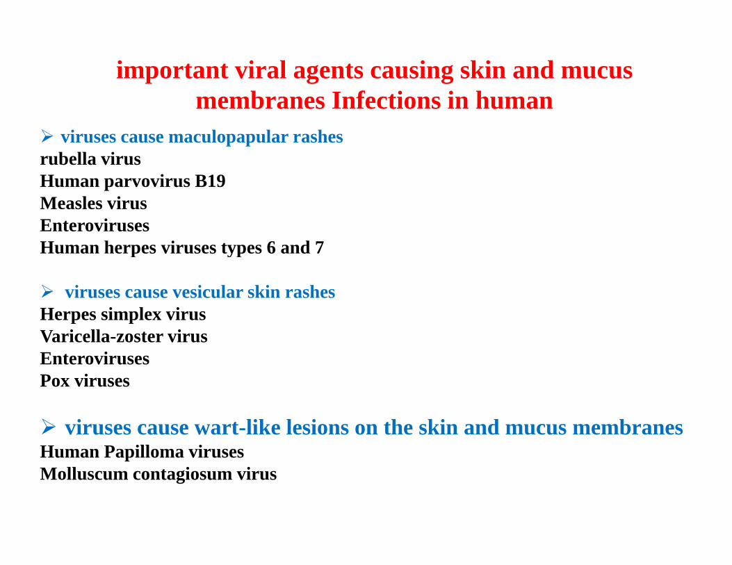
important viral agents causing skin and mucus membranes Infections in human
viruses cause maculopapular rashesrubella virus H i B19
membranes Infections in human
Human parvovirus B19 Measles virus EnterovirusesHuman herpes viruses types 6 and 7Human herpes viruses types 6 and 7
viruses cause vesicular skin rashesHerpes simplex virusHerpes simplex virus Varicella-zoster virus EnterovirusesPox viruses
viruses cause wart-like lesions on the skin and mucus membranesHuman Papilloma viruses pMolluscum contagiosum virus

Diseases of the Skin Caused by yHerpesviruses
The Relationships between the Human Herpesviruses

HSV Viral Structure• Composed of a dsDNA(152kbp) nucleoprotein core( p) p
• Core is surrounded by an icosahedral protein capsidp p
• 100nm Capsid is enclosed in an outer envelope consisting of at least 8 glycoproteins.
• Envelope spikes ~8 nm long• The virus requires a moist environment for survival.

Common NameGenus Subfamily
H i l i 1Si l iAl h h i i Herpes simplex virus - 1 (HSV-1)
SimplexvirusAlphaherpesvirinae
Herpes simplex virus - 2 (HSV-2)
Varicella-zoster virus (VZV)
Varicellovirus
Cytomegalovirus (CMV)CytomegalovirusBetaherpesvirinae
Human herpesvirus 6 Roseolovirus(HHV-6)Human herpesvirus 7 (HHV-7)Epstein-Barr virus (EBV)LymphocryptovirusGammaherpesvirinae
Human herpesvirus 8 Rhadinovirus(HHV-8)

Cold sores are contagious sores caused by HSV‐1.• After primary infection, the viruses become latent in sensory ganglia.
• Recurrence of cold sores occurs when viruses are reactivated• Recurrence of cold sores occurs when viruses are reactivated and move to the epithelium.
Figure 11B: Herpes sore on mouthCourtesy of Dr. Hermann/CDC
Figure 11A: Herpes Simplex Virus© Phototake/Alamy Images


ii. Dermal ‐mainly among the health care workers‐ Herpetic whitlow
‐ painful‐ painful‐ heals without treatment
/‐ no pus/is it necessary to do a stain‐ Herpes gladiatorum – among wrestlers ‐ Eczema herpeticum

Herpetic whitlowp

H l di tHerpes gladiatorum

Eczema herpeticum

• Specimens– aspirate from vesicle– scraping from base of ulcer– serum for antibody

Laboratory diagnosis of HSVLaboratory diagnosis of HSV Direct staining
Tzanck test
g
Immunostaining
HSV isolation
Serology
PCR
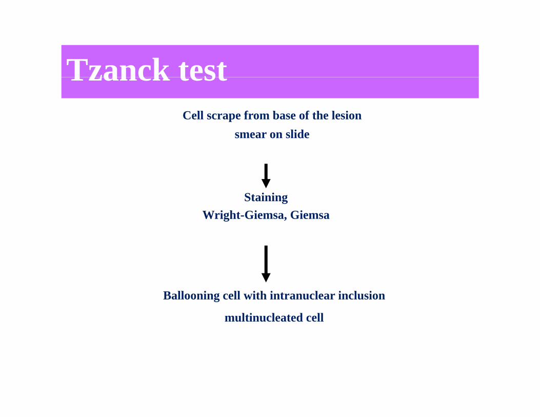
Tzanck testTzanck testCell scrape from base of the lesion
smear on slide
StainingWright-Giemsa, Giemsa
Ballooning cell with intranuclear inclusion
multinucleated cell

Tzanck test
Multinucleated cell
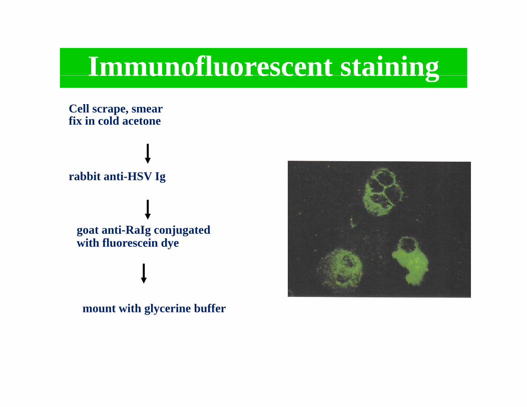
Immunofluorescent stainingImmunofluorescent stainingCell scrape, smear fix in cold acetone
rabbit anti-HSV Ig
goat anti RaIg conjugated
rabbit anti-HSV Ig
goat anti-RaIg conjugated with fluorescein dye
mount with glycerine buffer
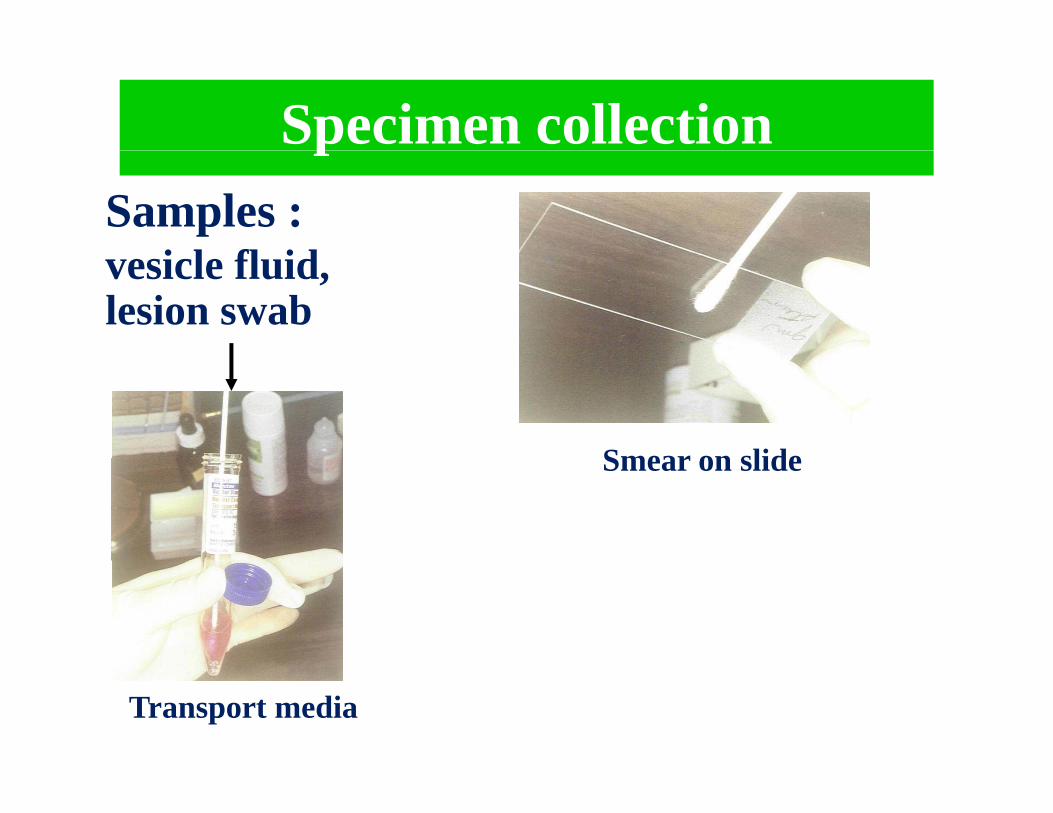
Specimen collectionpSamples :
i l fl idvesicle fluid, lesion swab
Smear on slideSmear on slide
Transport media

Transport mediaTransport media
I t i l ti lt di
Protein b i lb i
Isotonic solution or culture media
Protein bovine serum albuminbovine serum
Antibioticsgentamycinstreptomycin penicillin
A ti f
gentamycinstreptomycin penicillin
h t i i BAnti-fungus amphotericin B

Viral isolationSpecimens Cell culture (human diploid cells, Vero cells, Hela cells)
Cytopathic effect y p(rounded, enlarged and multinucleated cell)
Identification or typing
*Immunofluorescent staining*Immunofluorescent staining

HSV Cytopathic effectHSV Cytopathic effect
Normal cells CPE

Serological test for HSV infectionSerological test for HSV infection
Immunofluorescent staining
Complement fixation test
I G t t
IgM capture test ELISA :
IgG test

HSV serologyHSV serology
Primary infectionPrimary infection Pair serum: acute & convalescent serum
*rising titer > ๔ times
*seroconversion
IgG assay
IgM assay Single serum:
not useful; multiple reactivation
Recurrent infection
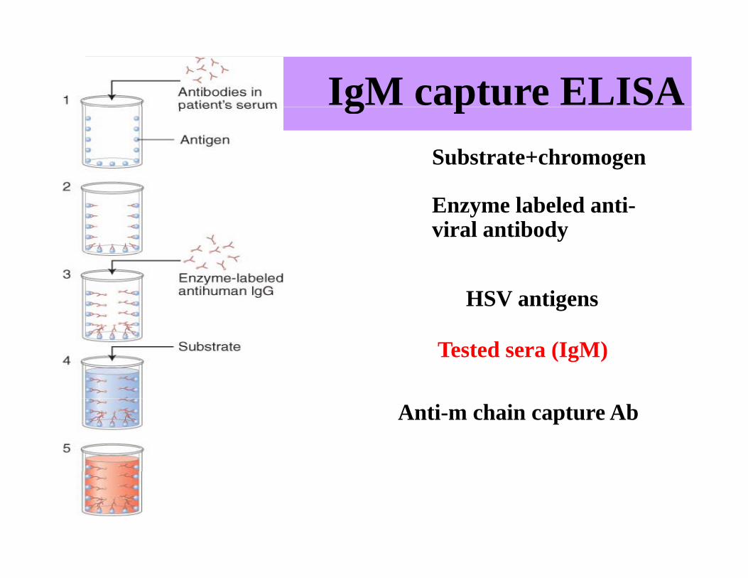
IgM capture ELISAg pSubstrate+chromogen
Enzyme labeled anti-viral antibody
HSV antigens
Tested sera (IgM)
Anti-m chain capture Ab

Polymerase chain reactionSamples
infected cell vesicle fluid CSF Multiplex primers;infected cell, vesicle fluid,CSF
DNA extraction
Multiplex primers; •cutaneous group; HSV, VZV•lymphotropic group; CMV,
PCR solution
(buffer, dNTP,Taq DNA pol, primres) (buffer, dNTP,Taq DNA pol, primres)
A lif l
Detection:•gel electrophoresis
Amplify ๒๐-๓๐ cyclesg p
•dot blot hybridization•*restriction fragment length polymorphismpolymorphism

Common NameGenus Subfamily
H i l i 1Si l iAl h h i i Herpes simplex virus - 1 (HSV-1)
SimplexvirusAlphaherpesvirinae
Herpes simplex virus - 2 (HSV-2)
Varicella-zoster virus (VZV)
Varicellovirus
Cytomegalovirus (CMV)CytomegalovirusBetaherpesvirinae
Human herpesvirus 6 Roseolovirus(HHV-6)Human herpesvirus 7 (HHV-7)Epstein-Barr virus (EBV)LymphocryptovirusGammaherpesvirinae
Human herpesvirus 8 Rhadinovirus(HHV-8)

Varicella‐ Zoster Virus– Chickenpox
• VZV is extremely communicable• Reservoir = infected humans either symptomatic or• Reservoir = infected humans either symptomatic or asymptomatic
• Primary Mode of Transmission = p‐p, direct, respiratory dropletdroplet
• Secondary Route = direct contact with active vesicles
– Shingles• Is a reactivation disease; resulting from previous VZV infection
• Is generally not considered a communicable conditiong y• Exception
– There are a few documented cases of transmission from and adult with shingles to a young childadult with shingles to a young child
• Child developed chickenpox


HERPES ZOSTERf• Reactivation of HVZ
• dermatomal distribution• may recur• can disseminate in immunocompromised patients• complications
– post herpetic pain– ophthalmic zoster ‐corneal scarring and loss of vision
DIAGNOSIS
CLINICALEM of vesicle fluid
SEROLOGYIgM detection


DIAGNOSISDIAGNOSIS
CLINICALCLINICAL
Isolation of virus
EM of vesicle fluid
SEROLOGY (IgM detection)
PCR

Varicella-zoster virus (VZV) infectionVaricella zoster virus (VZV) infection
Chickenpox Zoster Clinical diagnosis
Atypical clinical manifestationImmunocompromised hostImmunocompromised host
*Eye infection
*Brain infection
*Atypical skin rash

Laboratory diagnosis of VZVLaboratory diagnosis of VZV
S l I f t d ll
Direct staining
ballooning cell with intranuclear inclusion
Tzanck test
Samples Infected cell scrape
multinucleated cellsinclusion
fluorescent stainingImmunostaining:

Tzanck test

Serological test of VZVSerological test of VZV
ELISA with VZV specific antigenp g
IgG seroconversion
rising Ab titer > ๔ times
detected both
rising Ab titer > ๔ times
I M detected bothchickenpox & zoster
IgM
sharing some Ag with HSVLimitation:
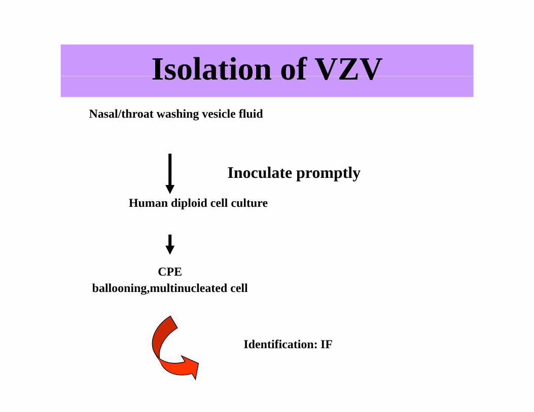
Isolation of VZVIsolation of VZVNasal/throat washing vesicle fluid
Inoculate promptly
Human diploid cell culture
p p y
CPEb ll i l i l d llballooning,multinucleated cell
Identification: IF
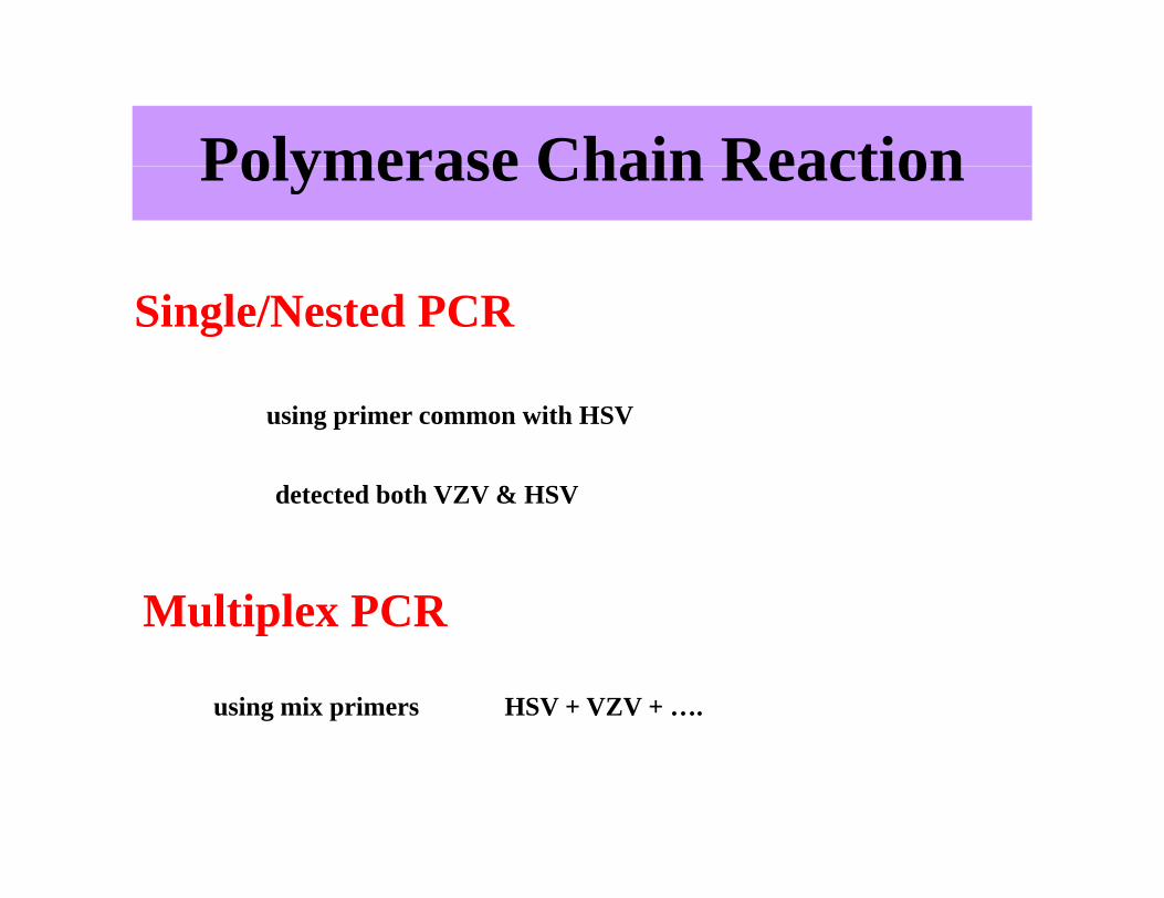
Polymerase Chain ReactionPolymerase Chain Reaction
Single/Nested PCR
using primer common with HSV
detected both VZV & HSV
Multiplex PCR
HSV + VZV + ….using mix primers
Multiplex PCR

PicornavirusesCharacteristics
• pico = small, rna =RNA Viruses• icosahedral 30 nm
• naked nucleocapsid = Nonenveloped• plus strand(+) RNA m‐RNAp ( )
• single stranded and capped for infectivity and packaging• this genome is infectious(should it be introduced into a cell)
• vertices of capsid creates canyon like depressions which contain the• vertices of capsid creates canyon‐like depressions which contain the VAP’s, VAP ‐1, VAP ‐2, VAP ‐3
• most VAP bind to intracellular adhesion molecule ‐1(ICAM‐1) expressed on epithelial cells fibroblasts and endothelial cellsexpressed on epithelial cells, fibroblasts, and endothelial cells

Picornavirus Capsid Structure
Capsid is a pseudo T=3 i h d i i f 60icosahedron consisting of 60 identical asymmetric protomersarranged as 12 pentamers.
Each protomer is composed of a single copy of each of the four capsid proteins, VP1, VP2, VP3 and VP4.
VP4 is located on the inner surface of the protein shell formed by VP1, p yVP2 and VP3.
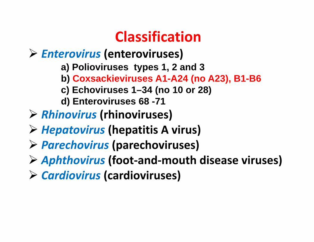
ClassificationEnterovirus (enteroviruses)
a) Polioviruses types 1, 2 and 3b) C ki i A1 A24 ( A23) B1 B6b) Coxsackieviruses A1-A24 (no A23), B1-B6c) Echoviruses 1–34 (no 10 or 28)d) Enteroviruses 68 -71)
Rhinovirus (rhinoviruses)Hepatovirus (hepatitis A virus)Parechovirus (parechoviruses)Aphthovirus (foot‐and‐mouth disease viruses)Cardiovirus (cardioviruses)

Skin and mucus membrane1 Herpangina1. Herpangina
a) Coxsackievirus A
2. Hand‐foot‐and‐mouth diseasea) Coxsackievirus A16
f h hHerpangia = fever, sore throat with painful swallowing, anorexia and
vomitingvomitingvesicular ulcerated lesion on the soft palate and uvulapalate and uvulaetiological agent is Coxsackie virus A, an enterovirusan enterovirusvirus is shed from the lesions, respiratory droplets and in the feces(fecal‐oral)

Herpangina

Hand‐Foot‐Mouth Disease (vesicular exanthem)
vesicular lesions on the hands, feet, mouth, tongue accompanied by mild fever
etiological agent: Coxsackie virus A16etiological agent: Coxsackie virus A16virus is shed/transmitted from lesions and is also shed in the feces(fecal‐oral)


Hand‐Foot‐Mouth DiseaseDisease

Picornaviruses - Diagnosis• Enteroviruses
• Laboratory• Serologygy
• detection of specific viral antibody in IgM fraction• four fold increase in IgG from acute to convelescence
Culture performed only for epidemiological confirmation– Culture performed only for epidemiological confirmation• coxsackie or echoviruses from throat or feces
• monkey kidney tissue culture• human embryo kidney tissue culture
• culture virus is specifically identified with antibody assays
‐ RT‐PCR

• PoxviridaeB i k h d id– Brick‐shaped or ovoid
– Size: 220‐450nm long x 140 260nm wide x140‐260nm wide x 140‐260nm thick– EnvelopedEnveloped– ds DNA– Genome size:130‐375kbs (large!)– Produce skin lesions eg. Small pox and vaccina virus

Poxviruses (continued)(continued)
Figure 1. Structure of the variola virus

GENERA Characteristic MembersPOXVIRIDAE
Orthopoxvirus Variola Major (Smallpox virus) manVariola Minor (Alastrim virus)MonkeypoxVaccinia virus manCowpox virus cattle,cats
Parapoxvirus Pseudocowpox virusParapoxvirus Pseudocowpox virusOrf virus (milker’s nodules)
Leporipoxvirus
Not important to manAvipoxvirus
Capripoxvirus
Suipoxvirus
Molluscipoxvirus Molluscum contagiosum virusMolluscipoxvirus Molluscum contagiosum virusYatapoxvirus Yaba monkey tumor virus
SmallpoxS ll t itt d b i t t f l iSmallpox was transmitted by respiratory route from lesions in the respiratory tract of patients in the early stage of the disease. During the 12 day incubation period, the virus was distributed initially to the internal organs and then to the skin. Variola major caused severe infections with 20‐50% mortality, variola minor with <1% mortality. Management of outbreaks depended on the isolation of infected individuals and the vaccination of close contacts. The vaccine was highly effective. If given during the incubation period, it either prevented or reduced the severity of clinical symptoms. The origin of the vaccine strainseverity of clinical symptoms. The origin of the vaccine strain is not known.
20

SmallpoxSmallpox

SmallpoxSmallpox

Variola Majorj
O di TOrdinary Type
The Eradication of SmallpoxThe Eradication of Smallpox
S ll di d f i i d hSmallpox was eradicated from most countries in Europe and the US by 1940s. By the 1960s, smallpox remained a serious problem in the Indian subcontinent, Indonesia and much of Africa. The
li d ll h h li f di i iWHO listed smallpox as the top on the list for eradication in 1967. The WHO smallpox eradication unit was set up in 1967.Smallpox was officially declared eliminated in 1980.p y
21

Monkeypox
Although Monkeypox was first isolated from monkeys, there is no evidence that African monkeys act as the reservoir. y
The most likely candidate for reservoir is the African squirrel.
One important difference between human Monkeypox and smallpox is the lower capacity for human spreadsmallpox is the lower capacity for human spread.
24

Monkeypox Virus

COWPOXInfection has been described in humans, cows and cats.Infection in humans usually remain localized, often producing a lesion which is similar to that caused by vaccination,a lesion which is similar to that caused by vaccination, although the inflammatory response is greater and general constitutional symptoms such as fever and myalgia may be present in some cases In humans lesions are usuallypresent in some cases. In humans, lesions are usually restricted to the hands, but may also be transferred to the face. EM is generally used for the diagnosis of infection The virusEM is generally used for the diagnosis of infection. The virus will also grow well on CAM.Although cowpox was first isolated form cattle and farm workers. There is no evidence that cattle serve as the reservoir. In fact, cowpox is very rare in cattle. It has been suggested that the reservoir is actually a small rodent but this is not proven.

Cowpox virus
PARAPOXVIRUSESThe laboratory diagnosis is usually made by EM The virus mayThe laboratory diagnosis is usually made by EM. The virus may also be isolated in human, bovine and ovine cells but such investigations are not part of routine diagnostic virology. Parapoxvirus infections occur worldwide and are ofParapoxvirus infections occur worldwide, and are of considerable importance. The lesions are surprisingly painless and thus there is probably substantial under‐reporting. Idoxuridine had occasionally been prescribed for treatment but no trials have been carried out to prove the efficacy of p ytreatment. Prevention of human infection is difficult. Reasonable precautions should be undertaken when handling infected animals.
29

A thumb with two denuded orf lesionsA scabby sore on a human hand caused by orf
Orf Virus in Sheep A sheep infected with Orf disease

milker's nodes in Humanin Human
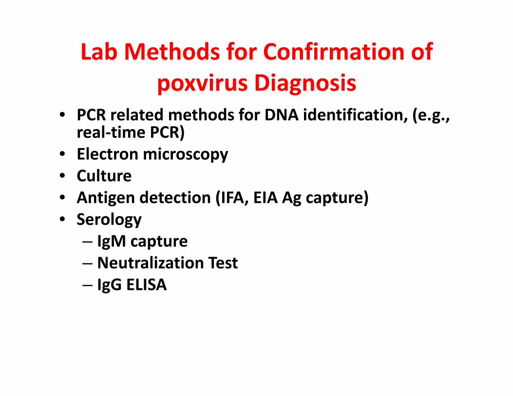
Lab Methods for Confirmation of poxvirus Diagnosis
• PCR related methods for DNA identification, (e.g., ( greal‐time PCR)
• Electron microscopy • Culture• Culture• Antigen detection (IFA, EIA Ag capture)• SerologySerology
– IgM capture – Neutralization Test– IgG ELISA
MolluscipoxvirusMolluscipoxvirusMOLLUSCUM CONTAGIOSUM VIRUS_Molluscum contagiosum is a specifically human disease of worldwide distribution. The incubation period varies from 1 week to 6 months TheThe incubation period varies from 1 week to 6 months. The lesion begins as a small papule and gradually grows into a discrete, waxy, smooth, dome‐shaped, pearly or flesh‐colourednodulenodule.Usually 1‐20 lesions but occasionally they may be present in hundreds.
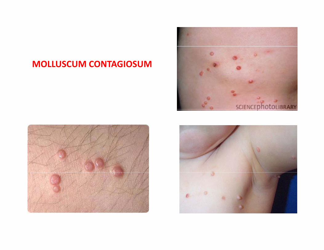
MOLLUSCUM CONTAGIOSUM
Molluscum contagiosum virusg• The disease occurs world‐wide and is spread by direct contactdirect contact.
• In general it tends to occur in children.• MC is transmitted by close personal contact including sexual contact.

Diagnosis• Diagnosis is usually done on clinical grounds alone by the
typical appearance of the lesions.
• Expression of materials stained with Giemsa, Wright or Gram stain reveals molluscum bodies.
• Biopsy, which shows characteristic features of epidermal hyperplasia.
• Polymerase Chain Reaction
Th di i b d b EM• The diagnosis can be supported by EM.
• Unlike other poxviruses, molluscum have notbeen demonstrated to grow in cell culture.
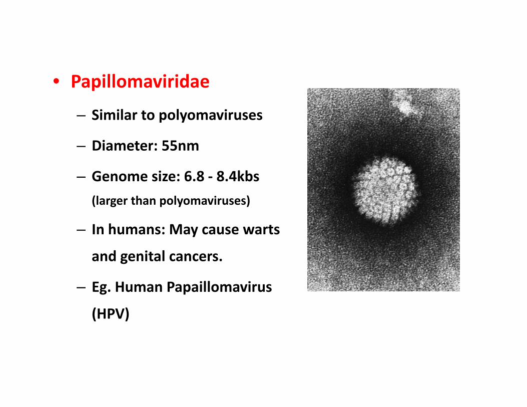
P ill i id• Papillomaviridae
– Similar to polyomaviruses
– Diameter: 55nm
– Genome size: 6 8 ‐ 8 4kbsGenome size: 6.8 ‐ 8.4kbs (larger than polyomaviruses)
In humans: May cause warts– In humans: May cause warts
and genital cancers.
– Eg. Human Papaillomavirus
(HPV)

HPV Infections/ Lesions
• Skins Warts– Hands and Feet HPV 1 ‐ 4– Most common type
• Head and Neck Tumors– oral papillomas benign epithelial tumors of the oral cavityoral papillomas benign epithelial tumors of the oral cavity– laryngeal papilloma HPV 6/11 benign epithelial tumors
• Anogenitial Wartsgenital warts HPV 6/11 exclusively on the squamous– genital warts HPV 6/11 exclusively on the squamousepithelium of the external genitalia and perianal areas rarely malignant
• Cervical dysplasia and neoplasiaCervical dysplasia and neoplasia– malignant changes caused by HPV 16/18 is an intraepithelial cervical
dysplasia– koilocytotic cells observed in Papanicolaou‐stained cervical smearskoilocytotic cells observed in Papanicolaou stained cervical smears
• perinuclear cytoplasmic vacuolization

Replication• Papillomavirus-cell interactions can be classifieds into three main groups: permissive, non-Papillomavirus cell interactions can be classifieds into three main groups: permissive, non
permissive transformable, and non-permissive non-transformable depending on the particular virus and cell8. Sarcoid cells are non-permissive to BPV replication and propagation7.
• BPV targets basal cells5.• Transcriptional states are reg lated b the differentiation of the sq amo s epitheli m9• Transcriptional states are regulated by the differentiation of the squamous epithelium9.
Maturation requires viral transport from the basal layer to the surface epithelium. During this movement, the differentiating keratinocyte undergoes complex changes to provide a correct intracellular environment for viral replication8.
• Viral-infected keratinocytes elicits no immune response5.
www.gsbs.utmb.edu

Human Papilloma Virus Infections
– HPV 16/18 cause cervical papillomasand dysplasia in which the virus DNA is integrated into the genome rather than acting as a plasmid
• E6/E7 genes are oncogenes which produce proteins that bind to and i i ll l hinactivate cellular growth‐suppressor proteins, p53 and pRb
– unsuppressed cells are more prone to mutations and transformationmutations and transformation
– infected cells exhibit nuclear changes with large perinuclear vacuoles
kili t i• kiliocytosis• cause both benign and malignan lesions

Laboratory Diagnosis of HPV InfectionsCytology detects koilocytotic cells
warts are characterized by hyperplasia of the prickle cells and increased keratin production known as hyperkeratosiskeratin production known as hyperkeratosiskoilocytosis of squamous epithelia cells which are rounded and clumped
as observed in a Papanicolaou smear

KOILOCYTEKOILOCYTE
• Hallmark of HPV infection in an epithelium“ d” l• “Raisinoid” nucleus (enlarged, hyperchromatic,hyperchromatic, irregular), perinuclearhalo and cytoplasmicthi k ithickening
• Upper epithelial layer

Laboratory Diagnosis of HPV Infections
• Polymerase chain reaction detects viral nucleic acid
• Southern Blot Hybridization detects viral nucleic acid
• Immunofluorescence detects structural viral antigens
• Electron Microscopy detects intact virus
C lt t f l• Culture: not useful

Associated HPV genotype
lesiong yp
1,2,4Common wartsNon‐malignant lesions
3Flate warts
6,11Genital warts
6.11Laryngeal papilloma
2 3 and otherEpidermodysplasiaPremalignant 2,3 and otherEpidermodysplasiaverrcuiformis
Premalignant lesion
ll 16,18,31,33,35, and others
Cervical cancerMalignant leision

Rapid Genotyping of Human Papillomavirus by Post‐PCR Hybridization
