laboratory analysis of cyanobacterial toxins and bioassays
TRANSCRIPT

745
Chapter 14
Laboratory analysis of cyanobacterial toxins and bioassays
Linda A Lawton James S Metcalf Bojana Žegura Ralf Junek Martin Welker Andrea Toumlroumlkneacute and Luděk Blaacuteha
CONTENTS
Introduction and general considerations 747141 Sample handling storage and shipping 750
1411 Safety 7501412 Sample processing for storage 7501413 Sample storage and shipment 7521414 Traceability 752
142 Generic methodologies used in cyanotoxin analysis 7531421 Sample extraction for analysis 753
14211 Solid-phase extraction (SPE) 7531422 Enzyme-Linked Immunosorbent Assay (ELISA) 7541423 High-performance liquid chromatography (HPLC) 756
14231 Ultra-performance liquid chromatography (UPLC) 758
1424 Liquid chromatography with mass spectrometry (LC-MS) 758
1425 Selecting an analytical system 759143 Quantification of microcystins and nodularins 761
1431 Extraction methods for microcystins and nodularins 76114311 Cyanobacterial cells 76114312 Water samples 76214313 Tissue samples 763
1432 Quantification of microcystins and nodularins by biochemical methods 76414321 Quantification by protein phosphatase
inhibition assay 76414322 Immunoassays for microcystin and
nodularin detection 7651433 Instrumental analytical methods for microcystins
and nodularins 76514331 Analysis of microcystins and nodularins
by HPLC-PDA 765
746 Toxic Cyanobacteria in Water
14332 Analysis of microcystins and nodularins by LC-MS(MS) 767
144 Quantification of CYLINDROSPERMOPSINS 7681441 Extraction of cylindrospermopsins 768
14411 Cyanobacterial cells 76814412 Water samples 76814413 Tissue and urine samples 769
1442 Quantification of cylindrospermopsins by ELISA 7691443 Instrumental analytical methods for
cylindrospermopsins 77014431 Analysis of cylindrospermopsins
by HPLC-PDA 77014432 Analysis of cylindrospermopsins
by LC-MS(MS) 771145 Quantification of ANATOXINS 771
1451 Extraction of anatoxins 77214511 Cyanobacterial cells 77214512 Water samples 77214513 Tissue samples 772
1452 Quantification of anatoxins by ELISA 7721453 Instrumental analytical methods for anatoxins 773
14531 Analysis of anatoxins by HPLC-PDA 77314532 Analysis of anatoxins by LC-MS(MS) 773
146 Quantification of saxitoxins 7741461 Extraction of saxitoxins 774
14611 Cyanobacterial cells 77414612 Water samples 77514613 Tissue samples 775
1462 Quantification of saxitoxins by biochemical methods 77514621 Quantification of saxitoxins by ELISA 775
1463 Instrumental analytical methods for saxitoxins 77614631 Prechromatographic oxidation and
liquid chromatography with fluorescence detection 776
14632 Analysis of saxitoxins by LC-MSMS 776147 Detection and quantification of anatoxin-a(s) 777148 Methods for synchronous detection of multiple types of
cyanotoxin 7771481 Multiplex antibody systems 7771482 Multi-cyanotoxin analytical methods 778
149 Future developments 7781410 Bioassays and their use in the survey of toxic cyanobacteria 779
14101 Insights into interpretation of toxicity results 78014102 Bioassays in the assessment of toxic cyanobacteria 781
141021 Nonmammalian bioassays 782
14 Cyanotoxin analyses and bioassays 747
141022 Mouse bioassay 78414103 In vitro assays for determining toxicity and
genotoxicity 78714104 Summary 789
References 790
INTRODUCTION AND GENERAL CONSIDERATIONS
Cyanobacterial toxins or cyanotoxins are a diverse group of compounds with differing chemistries hence a single analytical method can rarely be used to evaluate all potential compounds The general steps required to detect iden-tify quantify and monitor the different classes of toxins do follow a common approach (Figure 141)
Before committing to a major cyanotoxin sampling campaign it is impor t ant to evaluate the information and level of detail required to make appropriate management decisions (see also Chapters 11 and 12) Planning the work therefore is best done by a group including the different aspects involved for example field samplers laboratory support analysts as well as
Cyanobacterial Alert
Transport to laboratoryImmediately if wet lt 8 hrsDried filter samples gt 8 hrs
LC-MSMS
Sample Collectionchapter 12
chapters 51 52 11
Screening AssayELISAExtraction
HPLC or UPLC
Analytical determination
LC-MS
Figure 141 G eneral scheme from cyanobacteria alert (see Chapter 5) to analytical determination of cyanotoxin concentrations
748 Toxic Cyanobacteria in Water
managers leading the work possibly in contact with the authority responsible for public health This way a range of perspectives come together to design the most suitable and effective approach An audit by others with expertise in the field (in-house or external) serves to ensure that all important areas have been considered and nothing essential is overlooked Some of the key questions and topics which should be covered in planning and auditing are included in Checklist 141 and it is important to take time to make sure that all important requirements can be fulfilled
The available methods for analysing cyanobacterial toxins are very diverse The criteria for selecting the appropriate analytical method for a given monitoring programme include the consideration of reproducibility sensitivity and selectivity (see Box 141) as well as factors such as the time to result required training capital investment and laboratory conditions needed and consumable running costs per sample
BOX 141 PARAMETERS OF
ANALYTICAL PERFORMANCE
Before any analysis is done it is important to understand the nature of the
problem to be addressed and the type of data required A number of terms
are often used to describe the performance of an analytical method Of these
terms the most important are accuracy precision sensitivity repeatability
and reproducibility For any analytical method that is set up to quantify cya-
notoxins these parameters should be determined and well documented to
support the validity of the resulting data
Accuracy defines the closeness of measured amount of a compound in a
sample to the true amount in the sample The actual or true value has to be
determined by a validated method using reference material
Precision defines the closeness of repeated measurements of a single sam-
ple to each other To achieve high accuracy of a method it needs to be highly
precise but high precision does not guarantee high accuracy because measure-
ments could be biased by a systematic error
Sensitivity defines the capability of a method to avoid false-negative
results that is to detect a compound in a sample when it is present but to
not give a result when the compound is absent In practice the higher the
sensitivity the lower the limit of detection
Repeatability characterises the consistency of measured values obtained
from a single sample by one person applying one method on one analytical
system To test repeatability generally includes all sample preparation steps
and usually a predetermined limit of variability is set in which the measured
values should lie for the method to be accepted
14 Cyanotoxin analyses and bioassays 749
Reproducibility characterises the consistency of measured values obtained
from a sample by different persons on different analytical systems Reproducibility
of a method that is applied in different laboratories is preferably validated with
interlaboratory comparison tests where a single sample of known toxin content
is split and analysed by different laboratories
Exact definitions of these and further terms related to analytical chemistry
can be found at httpgoldbookiupacorgindexhtml
Trained staff are needed especially to operate complex analytical sys-tems primarily for the establishment of methods for analytes not yet ana-lysed in the particular laboratory and their initial validation phase For routine analyses modern analytical systems generally offer some degree of automation that can be made use of once a method has been established
As a rule of thumb the most sensitive and specific methods tend to be the most demanding ones in terms of investment (capital method optimisation and validation) required personnel training and running costs ndash but often with a substantial delay of result delivery of hours to days On the other hand methods that are less sensitive and selective may deliver results very fast This can be essential in situations where an analytical result is needed to trigger immediate management actions to mitigate risks for example in Alert Level Frameworks (see Chapters 51 and 52)
CHECKLIST 141 CYANOTOXIN DETECTION
bull Which toxin classes are to be analysed
bull From what type of samples (eg water cells tissue sediments)
bull Does the detection have to be quantitative If so what detection limit
is required
bull Do drinking-water samples need immediate quenching (eg sodium
thiosulphate or ascorbic acid) to eliminate the continued action of
oxidising agents like chlorine
bull What instrumentation and expertise is available (i) in-house or
(ii) external
bull What capacity is available that is which number of samples can currently
be analysed (including laboratory space appropriate sample storage)
bull What training is needed ndash for sampling for sample processing for anal-
ysis and for data interpretation How are the data going to be used and
reported
750 Toxic Cyanobacteria in Water
141 SAMPLE HANDLING STORAGE AND SHIPPING
Following a sampling trip the samples arriving in the laboratory need to be processed further for analysis or storage Three aspects are important for sample handling and storage safety sample processing to ensure stability and traceability
1411 Safety
Laboratory staff handling samples potentially containing toxic cyano-bacteria and cyanobacterial toxins are potentially exposed to health haz-ards (see also section 52) and appropriate protective measures need to be implemented These measures will be based on two aspects implement-ing general safety measures for hazardous material defined in national and international occupational health and safety guidelines and an assessment of the risk of exposure to toxic cyanobacterial material potentially given in the specific procedures to be carried out Any staff member handling potentially toxic cyanobacterial samples has to be accordingly trained and equipped with adequate protective equipment Depending on the work to be done this protective equipment will range from standard laboratory coats gloves and safety glasses to ndash where there is a risk of inhalation exposurendash breathing masks (Stewart et al 2009) For water samples taken in moni-toring programmes the quantities of toxins are generally low that is in the low microgram per litre range likely posing no risk of intoxication Nonetheless skin and eye contact inhalation and ingestion have to be avoided by wearing an appropriate safety wear Risks of exposure tend to be higher if larger quantities of bloom material are handled for example for toxin purification with toxin quantities potentially in the low milli-gram range The highest risk of exposure to toxic material is likely through the handling of dried bloom material that is exposure to dust requiring the wearing of a breathing mask andor the handling of sample material in an exhaust hood Powdery freeze-dried bloom material often statically charged easily escapes containment and this may cause a risk of exposure not only during laboratory work but also for cleaning staff
1412 Sample processing for storage
Samples that are not analysed immediately upon arrival at the laboratory need to be stored properly to avoid degradation of the cyanotoxins to be quantified (see Figure 123) Generally cyanobacterial toxins are rather stable compounds and storage at minus20 degC largely prevents degradation As guidance the stability ndash or breakdown ndash as described in Chapter 2 may serve as a first orientation for conducting stability testing under the actual conditions including all steps in transportation storage and shipping Since
14 Cyanotoxin analyses and bioassays 751
degradation mostly occurs through microbial activity lower temperatures generally increase stability
However simply freezing entire samples may not be appropriate for prac-tical and analytical reasons For example when extracellular and intracel-lular toxins are to be analysed separately this requires prior separation of both fractions which can no longer be done once samples have been frozen Therefore sample processing steps may be required that allow a reliable and efficient preservation of samples or portions of samples for later analyses
If the toxin content in the particulate fraction is to be analysed cyano-bacterial cells and other particulate material (ldquosestonrdquo) are best collected on a filter thus concentrating the sample for space-efficient storage One criterion for the choice of the type of filter (glass fibre membranes of dif-ferent types cellulose) is the appropriate pore size to retain cyanobacteria As most toxigenic taxa occur in colonies or filaments pore sizes lt 2 μm are generally acceptable Smaller pore sizes may slightly increase reten-tion efficiency but at the cost of more rapid clogging of the filters ndash which is a factor that determines to a large extent the time needed to process samples Further it is important that the filter material be compatible with the downstream processing in particular with the extraction procedure to avoid the release of any compounds that interfere with the downstream analysis during the extraction step in organic solvents also it must not be dissolved by these solvents A protocol for the handling of such sam-ples needs to be validated and tested with negative controls that is filters through which pure water has been passed
For the filtration step the water sample is well mixed immediately before measuring a volume to be filtered for example with a calibrated cylinder because even within a very short time (ie minutes) buoyant cyanobac-teria can float to the top (and other phytoplankton can settle) potentially leading to biased analytical results Preferably the total volume to be fil-tered is portioned into several smaller volumes to avoid increasingly lon-ger filtration times on a gradually clogged filter The volume to be filtered for an individual sample depends on the density of cells ndash that can hence be highly variable ndash and on the detection limit of the downstream analyti-cal procedure For the latter the extraction and concentration steps need to be considered
The filter should hold only residual moisture before it is frozen for storage or processed further for analysis respectively as discussed in the following section
Filtrates are used for the analysis of dissolved (ie extracellular) cya-notoxins To produce larger volumes of particle-free samples it may be helpful to use a combination of two filters for example a glass fibre filter to retain larger particles and a second filter with smaller pores to remove small particles
752 Toxic Cyanobacteria in Water
1413 Sample storage and shipment
If filter samples are to be stored they are folded loaded-face on loaded-face and placed in an appropriate container for example chemically inert reac-tion tubes or enveloped in aluminium foil and labelled correctly For their long-term storage or shipment it is advantageous to dry the loaded filters otherwise shipment on dry ice is recommended Drying can be achieved by freeze drying in a vacuum centrifuge or at moderate temperatures (lt80 degC for microcystins and cylindrospermopsins) in a drying oven when lyophilisation is not available (Welker et al 2005) For the latter stability tests are recom-mended to check whether the drying process (that may last for several hours) causes any degradation It is generally good practice to test the stability of the samples under the chosen storage conditions with a series of identical samples that are analysed at the different processing steps and after varying storage times Performing the extraction procedure in the storage tube can allow safe handling reduce processing time and minimise the risk of sample confusion
For the storage of particle-free filtrate or extract toxin adsorption to labware may be relevant A few studies on this issue show that microcystin congeners differ in their tendency to adsorb to materials (Hyenstrand et al 2001 Kamp et al 2016 Altaner et al 2017) These studies cover only some of the possible combinations of materials solvents and toxins and it is recommended to test the material used in an individual laboratory under the actual sample processing procedures to assess suitability Once labware of a particular material and manufacturer has been found appropriate it should not be changed without corresponding verification
Shipping of samples requires consideration not only of stability but also of compliance to legal aspects in particular declaration rules for transbor-der shipments (see Metcalf et al (2006) for an overview)
1414 Traceability
It is critically important to label all field samples arriving in the laboratory in a way that allows the results to be tracked back to the sampling site and sampling date at a later point in time Similar considerations also apply for individual steps in sample processing dilution of standards and quality control (QC) samples For certified laboratories the traceability of samples and materials is essential and generally follows guidelines such as ISO 9001 (ISO 2015) Although a thorough quality management system according to ISO 9001 may not be necessary for non-certified laboratories some prin-ciples can be implemented to ensure proper sample management
As outlined in Chapter 11 a first important point is the labelling of indi-vidual samples Sample names need to be unique not only for a recent set of samples but also with respect to all samples that are expected in the future in a given laboratory and in cooperating laboratories A naming sys-tem is therefore best defined prior to sampling campaigns and followed by
14 Cyanotoxin analyses and bioassays 753
the entire staff Whichever system of sample naming is agreed upon it is important to register all samples received in a laboratory in a central reposi-tory that is in turn backed up regularly As barcode labelling and reading is becoming affordable the introduction of these respective systems is now an option Furthermore labels need to be stable that is lastingly attached to sample containers under the actual storage conditions Especially when samples are stored in the freezer adhesive labels or markings need to be tested for resilience to repeated freezendashthaw cycles
142 GENERIC METHODOLOGIES USED
IN CYANOTOXIN ANALYSIS
1421 Sample extraction for analysis
Most cyanotoxins are retained in the cell-bound fraction of samples taken from waterbodies (Chapter 2) Only when cyanobacterial populations experience cell lysis for example in surface scums exposed to high tem-peratures and light intensities substantial amounts of toxins are released to the surrounding water An exception is cylindrospermopsin that can be released from viable cell and found in large proportions in the cell-free frac-tion (section 22 Box 51 and Chapter 10) Further extraction and sample clean-up is of critical importance for the analysis of cyanotoxins in foods to avoid both under- and overestimating concentrations (see section 534)
To make cell-bound cyanotoxins accessible to chemical analysis they need to be extracted from the cells with an appropriate solvent The solvent needs to be selected to efficiently extract toxins from cells in a few (maxi-mum of three) extraction cycles and needs to be compatible with down-stream analytical methods Extraction procedures for individual classes of toxins will be discussed in the respective sections
There is no single method available that is suitable for extracting all classes of cyanotoxins hence it may be necessary to collect several samples or subdivide samples prior to processing (see Chapter 12)
14211 Solid-phase extraction (SPE)
While some immunoassay (ELISA) methods and LC-MSMS (see below) may be sufficiently sensitive for monitoring in compliance with guideline values trace analysis of cyanotoxins in water (ie concentrations below 1 μgL) typically requires sample concentration using solid-phase extraction (SPE) This processing involves passing a known volume (typically 100ndash500 mL) of a (particle-free) water sample through a solid-phase cartridge to concentrate dissolved cyanotoxins SPE requires a vacuum manifold sys-tem PTFE (polytetrafluorethylen) connectors and tubing (for a minimum adsorption of analytes) and a vacuum pump The equipment for SPE can be
754 Toxic Cyanobacteria in Water
used for different classes of cyanotoxin however specific cartridges need to be used which retain the cyanotoxin of interest A large selection of ready-to-use SPE cartridges is available with different sorbent materials and varying sorbent volumes Sorbent materials primarily differ in the degree of hydrophobicity and have to be selected in correspondence with the cyano-toxin of interest for example C18 for microcystins or graphitised carbon for highly hydrophilic cylindrospermopsins
Filtering water prior to SPE is essential to minimise clogging of the car-tridges Nonetheless during times of blooms the higher load of very small particles may significantly increase loading times In this case it is advised to note the volume of sample that has already passed and discontinue the sample loading Once the water sample has passed through the cartridge a washing step follows before the toxin adsorbed on the cartridge is eluted in a small volume (typically 3ndash5 times the sorbent bed volume) of solvent into a collection tube (Figure 142) This can be analysed directly or further concentrated by drying to enhance the detectability It is tempting to load as large a volume of water sample as possible to allow the detection of low lev-els of cyanotoxin however this is not always appropriate Larger volumes can take many hours to load onto the cartridge and matrix contaminants are also being concentrated hence larger sample volumes increase matrix inter-ference Loading samples onto SPE cartridges can be time-consuming so it is wise to determine the most appropriate volume of water This can be done by processing some test samples of spiking water (preferably similar to the samples) and determining how long the process takes Typically a volume is chosen that takes no more than 3 h to pass through the SPE cartridge allow-ing time for sample preparation elution and processing before analysis
It is useful to validate methods by spiking known amounts of the cyano-toxin of interest into water samples Spiking should not be performed with high-purity water (eg Milli-Q) as this may lead to poor recoveries and as this is in no way representative of the samples being processed Using the typical raw or tap water to be tested is best (if tap water is chlorinated using a chlorine-quenching agent ie sodium thiosulphate or ascorbic acid) as this will provide a good indication of expected performance
1422 Enzyme-Linked Immunosorbent Assay (ELISA)
Immunoassays are based on the binding interaction between a highly spe-cific antibody and the analytes of interest The most common of these assays is the ELISA kit using antibodies raised to specific cyanotoxins The toxins are detected by the modification in the colour reaction with the intensity of the colour being inversely related to the amount of toxin
ELISAs can offer rapid results with a relatively low investment in capital equipment As these assays do not identify specific cyanotoxin variants of a toxin class and give an indication of total toxin concentration ndash total
14 Cyanotoxin analyses and bioassays 755
1
2
3
4
8
6
7
9
10
5
11
Figure 142 S etup for solid-phase extraction (SPE) for concentrating trace amounts of cyanotoxins dissolved in water 1 Filtered water sample in stoppered glass bottle 2 PTFE tubing carrying water under vacuum 3 PTFE tubing 4 SPE cartridge with sorbent 5 PTFE stopcocks used to stop and start flow 6 water flowing to waste while cyanotoxins are adsorbed on the cartridge 7 vacuum manifold system with removable rack 8 reservoir used to intro-duce solvents for conditioning and eluting the cartridge (syringe) 9 pressure gauge with needle valve 10 concentrated sample eluted into a sample col-lection tube or vial 11 vacuum line connection
microcystins for example ndash they are often used as a screening method It is recommended to confirm toxin content and to routinely check for false negatives using instrumental methods (HPLC LCMS Gaget et al 2017) Where such methods are not available periodic shipping of a few selected samples to a support laboratory elsewhere may be an option
ELISA kits are very popular for a rapid straightforward detection of most classes of cyanotoxins although an individual kit is required for each class of cyanotoxin and even different kits may be necessary to cover the variants within one class The kit-based formats provide a straight-forward guidance on how to perform calibrate and interpret the results Multiple samples can be evaluated at one time and results can typically be reported in less than a day As with all biochemical test kits care has to be taken with storage since ambient or elevated temperature during pro-longed transportation can reduce their reliability The 96-well plate for-mat allows samples to be read and quantified in a plate reader facilitating the analysis of many samples and the calibration in a short space of time
756 Toxic Cyanobacteria in Water
Kits often come with a removable strip format so that not all wells need to be used at the time of analysis thus increasing the cost-effectiveness of the assay If only a few samples are to be assayed it is advisable to confirm the format before making a purchase
ELISA kits with sensitivity in the range of the WHO lifetime cyanotoxin guideline values are commercially available for almost all classes of cyano-toxins (see below) However it is important to remember that cell-bound toxins need to be extracted prior to performing the ELISA Also care has to be taken to quench oxidants used in water treatment (chlorine or chlorine dioxide see above) and to ensure the pH of the sample is appropriate for the specifications of the assay Filtration or centrifugation may be required to remove particulates and dilution may prove necessary to ensure that the quantification is in the approved range given in the instructions Where the cost of these kits is a problem and access to producing antibodies is available an option may be to produce antibodies in-house or to have this provided through co-operation with a an external institution (university company etc) as demonstrated in the case study described in Box 151 in Chapter 15
1423 High-performance liquid chromatography (HPLC)
HPLC has become well established for the routine analysis of environmen-tal pollutants These systems consist of a solid-phase chromatography col-umn through which analytes dissolved in liquid solvents are pumped and separated due to differences in the interaction of individual analytes with the solid phase The flow then passes through a detector for example UV absorbance or fluorescence detectors with the absorption proportional to the amount of analyte with data collected on a computer Most systems now include an autosampler to allow a set of samples to be loaded and automatically analysed The number of samples that can be analysed in a given space of time depends primarily on the duration of a single sample run For example the run time for microcystins with a conventional HPLC is around 1 h per sample Analyses with fewer target compounds that is less structural variants such as cylindrospermopsin or anatoxin-a generally require shorter run times and hence allow a higher sample throughput The separation of the analytes can be achieved by isocratic elution this is when the solvent composition remains the same throughout the analysis Isocratic elution is suitable for analyses that target only a few analytes and with a limited matrix interference that is with relatively low amounts of other nontarget compounds To allow for better separation of target analytes gradient elution is commonly applied where the proportions of the solvents change over the run time This allows a wide range of analytes to be sepa-rated such as multiple variants of microcystins To ensure that analytes and contaminants are not carried over to the next sample a washing step with 100 solvent is often included in the analytical run
14 Cyanotoxin analyses and bioassays 757
The most common detector used on HPLC systems is the photodiode array (PDA) which will provide an adsorption spectrum (200ndash600 nm) for the compounds being analysed This is useful for the analysis of cyanotox-ins as many of them have characteristic UV absorption spectra (Figure 144 and 145) thus providing an indication even of cyanotoxins in the sample for which no standard reference material is available (see Box 142)
BOX 142 REFERENCE MATERIAL FOR CALIBRATION
According to IUPAC reference material is defined as ldquoa substance or mixture
of substances the composition of which is known within specified limits []
to be used for the calibration of an apparatusrdquo For cyanobacterial toxins to
be used as reference material for establishing a calibration curve for the quan-
tification of these cyanotoxins two criteria need to be fulfilled
1 Purity defines the share of an individual compound of the total material
Purity is generally expressed in gravimetric percent that should be at
least 95 in reference material
2 Amount is generally defined in gravimetric units and ideally with the
specified limits that is a range of amount that should be as narrow
as possible
In this sense not all cyanotoxins that are commercially available are refer-
ence materials Hence these compounds cannot be used directly to establish
calibration curves In particular the nominal amount in a vial may deviate con-
siderably from the true amount In consequence this means that a calibration
curve established with such a ldquostandardrdquo would introduce a systematic error
to all subsequent analytical quantifications
The true amount hence needs to be determined This can be done either
by weighing with a sufficiently precise and sensitive balance or by spec-
trophotometric analysis (ISO 2005) For the latter wavelength-specific
extinction coefficients need to be available which is the case for some
but not all cyanotoxin variants Extinction coefficients are specific for sol-
vents and temperature that is a compound dissolved in water cannot be
quantified by using an extinction coefficient established for the compound
dissolved in methanol
For cyanobacterial toxins sold as certified reference material the purity
as well as the amount is well defined and its can be used directly as standard
for calibration
758 Toxic Cyanobacteria in Water
The typical capital investment required for an HPLC is around $30000 USD with relatively modest costs for maintenance including replacement UV lamps and columns
Training of staff and adoption of a standard protocol is easily achievable while interpretation of samples and cyanotoxin identification (especially unknowns from their spectra alone) requires more time to develop confi-dence This applies equally to UPLC (ultra-performance liquid chromatog-raphy) discussed below
14231 Ultra-performance liquid chromatography (UPLC)
UPLC offers a considerable advantage over conventional HPLC as it allows very rapid separation of analytes (run times of around 10 min) and a greatly reduced solvent usage typically 03 mLmin compared to 1 mLmin for conventional HPLC systems For example these systems can achieve the separation of multiple microcystin variants in run times little over 10 min thus providing high throughput of samples substantial saving and results on the same day for samples with short extraction times (eg bloom mate-rial and filter discs with cells) For samples requiring longer extraction (SPE of water samples or tissue samples) it can yield results within 24 h
UPLC systems are highly reliable with the response factor for micro-cystins in the UV detector changing little over time The capital invest-ment should typically be around $50 000 USD for the complete system at relatively low levels of maintenance with the main component that needs replacement being the UV lamp (it is useful to have a spare in stock)
1424 Liquid chromatography with mass spectrometry (LC-MS)
The addition of a mass detector to chromatography systems makes a very powerful tool for the analysis of cyanotoxins Mass spectra can provide an indication of the elemental composition and structure of an analyte along with determining the quantity of analytes for which reference materials are available with high sensitivity (see Box 142) A range of differing systems is available and very careful consideration is required to determine which fulfils analytical requirements and is within the budget available Different ion sources are available with positive electrospray ionisation (ESI) most commonly used in the analysis of cyanotoxins The type of mass analyser (Caixach et al 2017) also varies and can have a significant impact on cost and the data obtained hence it is essential that background evaluation is carried out to ensure the system suits the needs defined during planning (see section 1411) In general LC-MS will provide data relating to chromato-graphic retention times the parent ion masses and fragmentation patterns
14 Cyanotoxin analyses and bioassays 759
for each compound as they are eluted More complex LC-MSMS systems combine a series of more than one mass detector (eg a triple quadrupole mass detector) As analyte ions pass through mass analysers the former allow the selection of an analyte based on parent ion mass while the latter allow the selective detection of fragment ions This makes LC-MSMS a highly specific analytical technique
Robust protocols are required for LC-MSMS as the signal from the MS can be either enhanced or suppressed by matrix interference (salts organ-ics etc) Furthermore the response (ie signal strength relative to ana-lyte amount) can drift over a relatively short time necessitating a regular and frequent calibration A routine maintenance protocol is advisable with the interval for cone cleaning determined for different sample matrixes for example in studies for the analysis of mussel tissue cone cleaning was required after 40 samples (Waack 2017) This interval was determined by spiking an extracted sample and then carrying out repeated identical sam-ple injections and determining after how many samples the reliability of the detection and quantification diminished
While LC-MS provides very powerful sensitivity and detection capabili-ties the more advanced systems (LC-MSMS) require a capital investment of around $500 000 USD and an annual running cost of $20ndash40 000 Furthermore a high level of staff training is required to use interpret and maintain these systems but once established they provide unrivalled analyti-cal capabilities It is advisable where possible to see one or several systems in operation and have an opportunity to analyse specific samples from the area to be monitored prior to committing to this significant capital investment
1425 Selecting an analytical system
The lack of suitable analytical equipment is typically a barrier to monitor-ing cyanotoxins and a strong case is often made for capital investment The influence of current scientific publications frequently draws attention to the significant capabilities of very advanced instrumentation However while these systems provide impressive capability a robust evaluation of the analytical requirement running costs and infrastructure should be made to inform purchasing decisions Checklist 142 provides some key points to discuss both in-house and with those who have recently invested in cyanotoxin analysis before making decisions In particular it is advan-tageous to develop a regional network sharing expertise and resources for example through a regional centre of competency This may lead to a decision to use simpler techniques such as ELISA while validating the results periodically by having a small set of samples analysed with advanced techniques elsewhere Where training is required it is often more efficient to invite an expert to provide an in-house workshop as this ensures analyses
760 Toxic Cyanobacteria in Water
are operational and staff develops confidence using the in-house system (Note small grants are often available for this eg through international exchanges and workshop funding) A further benefit of this approach is continued support from experienced international collaborators ensuring ongoing development of monitoring programmes Support provided by the systemrsquos vendor generally is charged for This should be considered in the budget for investment and running costs
CHECKLIST 142 EVALUATING INSTRUMENTAL
ANALYTICAL REQUIREMENTS AND SUITABILITY
bull What information is needed Which class of toxins will be the main focus
Is the main target monitoring compliance of cyanotoxin guideline levels
bull Check the cost of consumables for example vials columns SPE car-
tridges and solvents in relation to the number of samples expected
over time In many countries solvents and even high-purity water can
be prohibitively expensive (the benefits of ultra-performance liquid
chromatography (UPLC) are low flow rates and short runtimes requir-
ing little solvent)
bull What are the costs of waste solvent disposal required by environmen-
tal legislation
bull Is the infrastructure appropriate This includes a stable power supply as
fluctuating or intermittent power can rapidly destroy equipment If not
what are the costs of installing effective power surge protection such as
uninterruptible power supply (UPS) systems
bull Can room temperature be kept within the range needed by the instru-
ments and analyses (results can be affected by high or fluctuating tem-
perature so air conditioning is often required)
bull Do all purchases include installation and initial training ensuring that
there are available engineers in the area
bull What is the cost of a service contract and is it essential
bull For planning to purchase a LCMS (it requires a nitrogen generator and
cannot run efficiently on regular laboratory gas cylinders) ensure that
the contract includes either annual service ndash that is the cost of service
engineers visits ndash or if you have technical expertise to carry out the
service the purchase of a service kit
bull Talk to users of different instrument manufacturers regarding their
experience of service and support especially in your location
bull Consider the benefits of partnering with others rather than buying
own equipment
14 Cyanotoxin analyses and bioassays 761
143 QUANTIFICATION OF MICROCYSTINS
AND NODULARINS
Of all the cyanotoxins most experience exists with the methodology for the extraction and detection of microcystins Furthermore due to its chemical similarity many of the methods for microcystins will also readily detect nodularin (Lawton et al 1994b) hence it will be included in this sec-tion In general the term ldquomicrocystinrdquo will be taken to refer to both these related classes of toxins unless the differentiation is required
Methods range in complexity and sophistication spanning the well-established ldquotried and testedrdquo approaches through to preliminary research findings on novel detection strategies While many of these novel methods offer exciting opportunities for the future this chapter focuses on a few of the most relevant approaches for establishing routine methods suitable for the more widely available resources and common requirements
1431 Extraction methods for microcystins and nodularins
14311 Cyanobacterial cells
All cellbloom samples will require extraction as these toxins tend to be retained inside healthy cells Many extraction protocols for microcystins have been described (eg various solvent combinations cycles of freezethawing sonication freeze drying including combined methods) Among these solvent combinations aqueous methanol (typically 50ndash80 (Barco et al 2005)) has proven to be very effective for extracting microcystins in face of their wide range of polarities This solvent can be used for extracting cell pellets once a sample has been centrifuged (and the super-natant discarded or assayed for extracellular microcystin) as well as for extracting cells concentrated on filters Depending on the volume of cells around 90 recovery of microcystins (Barco et al 2005) can be achieved with the first extraction Often this is sufficient as this has to be bal-anced against the further time required for processing a second extrac-tion as this will typically yield less than 10 of the total microcystin also if the two extractions are combined this reduces the detection limit due to the additional volume of solvent used in the second extraction Extraction time of around 1 h is sufficient for good recovery With the increased availability of dispersive extractor systems (automated vortex-ers that shake samples vigorously at defined speeds and timed duration) however extraction can be achieved in just a few minutes and with high reproducibility Where samples are extracted in centrifuge tubes (typically 15-mL microfuge) these can be spun and the supernatant then directly analysed using instrumental methods
762 Toxic Cyanobacteria in Water
When designing an extraction protocol it is good to keep it as simple as possible as this will limit error and also potential workplace exposure to microcystins for example freeze drying is sometimes reported as a step during sample preparation if a specific dry weight of cells is to be deter-mined but this can produce powders that are difficult to contain and prone to static charge Other methods also reported the use of a sonicator probe which may cause cross-contamination but also produce aerosols
The use of organic solvents (eg methanol) is not compatible with bio-chemical assays such as ELISA and enzyme inhibition tests Some ELISA kit manufacturers provide a cell lysis kit while other analysts have advo-cated aqueous extraction or dilution to limit the concentration of solvent for example a 1 in 10 dilution of a 50 aqueous methanol extract may be tolerated but should be checked with controls for the specific kit used Since microcystins demonstrate high temperature stability a brief exposure (5 min) of a small sample (eg 1 mL) to about 80 degC in a water bath fol-lowed by centrifugation (13 000 times g microfuge) can result in simple solvent-free extraction (Metcalf amp Codd 2000) Extracts can then be diluted in water or buffer as required
Similarly high organic solvent content in extracts to be analysed by chro-matographic systems needs to be tested for compatibility in particular when gradient elution is applied that generally starts with hydrophilic conditions
14312 Water samples
Some very sensitive methods (eg LC-MSMS) may be able to detect micro-cystins at environmental concentrations However even then it may be desirable to carry out solid phase extraction (SPE) to limit matrix effects
The most commonly used SPE material is end-capped C18 cartridges which have demonstrated high recovery and reliability Some users pre-fer newer resins (eg polymeric phases) which are good where MS is the detector of choice however the high recovery of polar compounds by these cartridges can interfere with the more polar microcystins (eg microcystin-RR) if detection is with photodiode array (PDA) Several published meth-ods provide a good detail on establishing SPE extraction of microcystins (Lawton et al 1994b Triantis et al 2017c)
Some researchers have developed online sample concentration for fully automated extraction and analysis of microcystins This is typically an advanced option including LCMS(MS) and a quite specialised approach however it may be desirable particularly for laboratories that need a high throughput such as those of public authorities monitoring compliance to regulations or of drinking-water suppliers
Recoveries are best if sample handling is limited processing time is kept to a minimum and samples are analysed immediately or stored at minus20 degC when this is not possible There is some evidence that samples may change
14 Cyanotoxin analyses and bioassays 763
when stored for longer periods of time even at minus20 degC but further studies are required to clarify the extent of this problem If samples are stored they should be vortexed if a subsample is to be removed after storage
14313 Tissue samples
It is becoming increasingly important to evaluate microcystins in more complex matrixes such as animals that have become intoxicated fish and aquaculture products that may be contaminated or even plant materials Much work is still required to fully understand the efficiency of different extraction and toxin recovery protocols This is particularly challenging for microcystins and nodularins as they are known to bind to proteins fur-thermore microcystins in particular can bind covalently to certain protein phosphatases in living cells Further the recovery of standards spiked to the material to be tested will only represent unbound toxin recovery efficiency
While a range of processing strategies with varying degrees of complexity have been used all of these strategies need to be tested and tailored to the specific requirements of the material to be studied Simple blending of fresh tissue (mussels) followed by a single aqueous methanolic (80 methanol) extraction was found to give good recoveries in the range of 61ndash97 for 11 microcystins and nodularin (Turner et al 2018) Very poor recovery was observed for hydrophobic microcystins when either the samples were acidified or water alone was used The solvent extracts can be directly ana-lysed by instrumental systems In contrast for biochemical tests (ELISA or protein phosphatase inhibition) samples will need to be dried to remove the solvent or sufficiently diluted with water or buffer
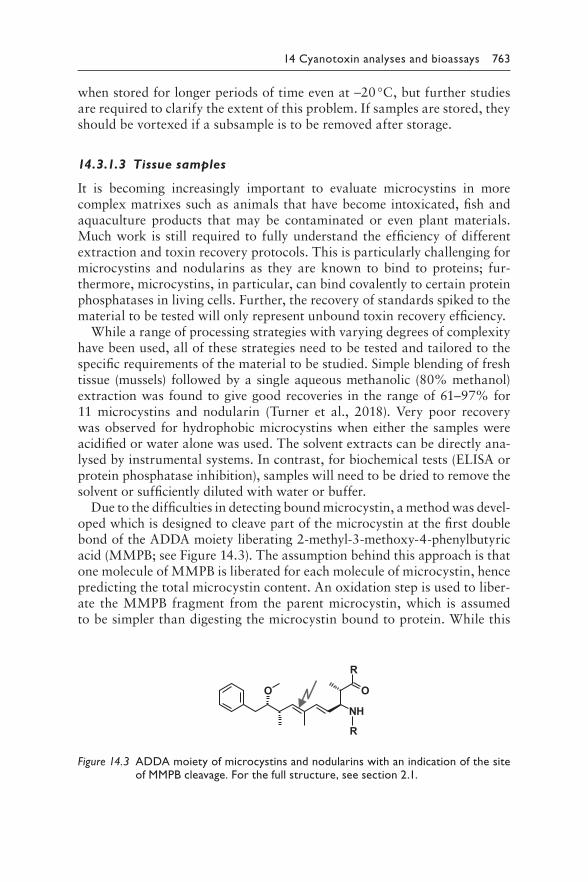
Due to the difficulties in detecting bound microcystin a method was devel-oped which is designed to cleave part of the microcystin at the first double bond of the ADDA moiety liberating 2-methyl-3-methoxy-4-phenylbutyric acid (MMPB see Figure 143) The assumption behind this approach is that one molecule of MMPB is liberated for each molecule of microcystin hence predicting the total microcystin content An oxidation step is used to liber-ate the MMPB fragment from the parent microcystin which is assumed to be simpler than digesting the microcystin bound to protein While this
O
R
R
NH
O
Figure 143 A DDA moiety of microcystins and nodularins with an indication of the site of MMPB cleavage For the full structure see section 21
764 Toxic Cyanobacteria in Water
method has been used in a range of studies it is very difficult to deter-mine the degree of sample recovery as spiking will only represent free toxin Most reported studies currently use MS detection of MMPB (mz 208) however this mass is not unique to this oxidation product (ChemSpider shows gt6300 compound with this or very similar mass) Others have augmented the method to search for a product ion at 131 which again may not provide confident detection However Foss and Aubel (2015) have suc-cessfully used the MMPB method in comparison with the ADDA-ELISA indicating good agreement
In summary the detection of microcystins and nodularins in tissue is important for assessing their possible role in animal poisoning or occur-rence in food (fish shellfish vegetables etc) and while no current method will recover the total amount of microcystins aqueous methanol extraction will give a good indication of whether microcystin is present
1432 Quantification of microcystins and nodularins by biochemical methods
14321 Quantification by protein
phosphatase inhibition assay
Microcystins and nodularins are known to be potent inhibitors of protein phosphatases PP1 and PP2A This activity is central to their toxicity and hence detection of inhibition also indicates the potential biological activity of a sample The assay can be performed relatively easily where the facilities are available for biochemical work All the reagents and enzymes can be purchased for the colorimetric assay which detects the enzymatic hydro-lysis of the substrate (p-nitrophenyl phosphate) that liberates the coloured product p-nitrophenol (detected at 405 nm) The assay can be performed in a microtitre plate with the temperature mixing and timing controlled by the plate reader and relatively straightforward protocol typically resulting in good reproducibility
Some challenges can arise if the sample inhibits the enzyme (depending on pH solvents or other contaminants) or if it contains background colour however this rarely occurs as the enzyme assay is highly sensitive allowing a significant dilution of for example a cell extract Detection limits as low as 00039 μgL have been reported which is well below the WHO guideline value (Sassolas et al 2011) This assay is also available as a commercial kit which has a quantification range between 025 and 25 μgL and an analysis time of only 30 min The manufacturers also provide a tube format that could be used in the field and eliminate the need for investment in a plate reader although users repeatedly analysing multiple samples will benefit from the multiwell plate format as this can be automatically analysed and is more practical for multiple samples and calibration points
14 Cyanotoxin analyses and bioassays 765
14322 Immunoassays for microcystin
and nodularin detection
The ADDA-based ELISA is particularly popular as it has been designed to detect the ADDA moiety which is both very specific to these toxins and present in all variants regardless of other chemical diversity Sensitivity is reported as around 01 μgL with the assay time of 25 h There are a range of ELISA kits (usually with antibodies raised to microcystin-LR) and it is worthwhile checking which would be most appropriate for use in a specific situation Considerations may be cost format location of suppliers and experience of other users in the vicinity ELISA has been used for a range of samples demonstrating a good cross-reactivity for a number of variants and different sample matrices (Heussner et al 2014) however for tissue samples sample processing needs a careful consideration as some studies have reported false positives and concentrations which are greatly in excess of the levels detected by LC-MSMS (Brown et al 2018)
1433 Instrumental analytical methods for microcystins and nodularins
While a range of analytical approaches has been explored over the past 30 years the central method of choice revolves around liquid chromatogra-phy (LC) Microcystins and nodularins can be separated very readily on C18 reverse-phase columns although some closely eluting variants (eg micro-cystin-LR and desmethyl-microcystin-LR) require a careful column selec-tion Different methods of detection have also been evaluated with a general consensus on UV detection andor mass spectrometry including MSMS
14331 Analysis of microcystins and
nodularins by HPLC-PDA
High-performance liquid chromatography with photodiode array (HPLC-PDA) provides an accessible robust method for the detection and quan-tification of all microcystins by virtue of their distinct UV absorption spectra (Lawton et al 1994b) Even in the absence of standards for every microcystin confident quantification of total microcystins can be achieved Most microcystins have very similar absorption spectra (although the over-all characteristics between 200 and 300 nm can vary with concentration) with a maximum at 238 nm from the conjugated double bond in the ADDA moiety (Figure 144) The exception to this are microcystins that contain the variable amino acid tryptophan (eg microcystin-LW) which have an absorption maximum at 223 nm and those that contain the variable amino acid tyrosine (eg microcystin-YR) which have an absorption max-imum at 231 nm (Figure 144) A simple approach is to use microcystin-LR calibration for total microcystin quantification assuming a similar molar
766 Toxic Cyanobacteria in Water
=238 nmmax
rt=165
RR
abso
rptio
n (a
rbitr
ary
units
)
=231 nmmax
rt=201
YR
wavelength [nm]220 220200 240 240260 260280 280 300
LR
rt=218
=238 nmmax LW =223 nmmax
rt=319
retention time [min]
abso
rptio
n at
=2
38 n
m
100 20 30 40
RR YR
LR
LW
(a)
(b)
Figure 144 H PLC-PDA chromatogram of a bloom sample dominated by Microcystis sp (a) and UV-absorption spectra of selected microcystin peaks (b) The individual variants are indicated by the standard two-letter code rt chro-matographic retention time The dotted line in the spectral plots indicates the wavelength λ = 238 nm all microcystins show an absorption band at this wavelength that results in a shoulder in the spectra of variants containing tyrosine (Y) and tryptophan (W) A second absorption band at λ = 246 nm can be seen as shoulder in spectra of all microcystins Both absorption bands are related to the conjugated double bond of the conserved ADDA moiety For analytical details see Welker et al (2003)
14 Cyanotoxin analyses and bioassays 767
absorption coefficient at 238 nm that is a similar response in PDA detec-tion An advantage of HPLC-PDA analysis is the fact that microcystin vari-ants for which no reference material is available can be recognised based on a peakrsquos absorption spectrum Verification can be achieved by collecting the eluting peak and performing an offline analysis for example by MALDI-TOF MS (Welker et al 2002b) Beyond this it is preferable to set up the method with a range of microcystin variants of differing polarities span-ning from early to late retention to provide confidence that a wide range of microcystins can be detected should they occur in samples
The typical analytical set-up will be a gradient separation using high-purity water plus trifluoroacetic acid (TFA 005) and acetonitrile with TFA (005) A gradient from 30 to 70 acetonitrile is usually required to separate all microcystins and a rapid wash to 100 will eliminate car-ryover between runs
14332 Analysis of microcystins and
nodularins by LC-MS(MS)
Both LC-MS and LC-MSMS are powerful instruments for the analysis of microcystins For developing a new method for microcystin analysis by LC-MSMS the Handbook of Cyanobacterial Monitoring and Cyanotoxin Analysis (Meriluoto et al 2017) provides good guidance and a number of standard operating procedures (SOPs) Different approaches are possible including the detection only of microcystins for which standards are available using selected reaction monitoring (SRM) which is very sensitive and accurate (Turner et al 2018) The drawback of this approach however is that a signifi-cant proportion of the microcystins in a sample could go unreported ndash those variants for which no calibration has been established based on standards For example USEPA Method 544 (Shoemaker et al 2015) is limited to six microcystins while other published methods have extended this to over 10 Birbeck et al (2019) reported 40 of samples had more than 20 of their total microcystin variants not detected by the USEPA Method 544 and a num-ber of these variants were the dominant microcystins It is therefore advised to consider the taxonomic composition of the sample that allows a tentative prediction of the structural variants to be present A way forward for routine monitoring is a detailed initial analysis to identify the spectrum of microcys-tins and as long as bloom composition stays stable such SRM can be a robust and sensitive approach provided periodic checks are carried out to confirm that the overall microcystin profile is still covered by the standards used
If microcystin variants are to be quantified for which no purified quan-titative standards are available estimates on the basis of their retention characteristics mass and fragmentation pattern can serve for an initial assessment However as detector response intensities vary substantially between individual variants a quantitation can be at best tentative for
768 Toxic Cyanobacteria in Water
example MC-RR has a several-fold higher response than MC-LR prob-ably due to a higher ionisation efficiency (Krause et al 1999) A valu-able approach is to add a PDA detector to the system along with the MS detection this will greatly enhance confident detection and in particular quantification of microcystins for which no specific calibration could be established Since the PDA is robust and quantification varies little over time it provides an excellent confirmation and quantification
144 QUANTIFICATION OF CYLINDROSPERMOPSINS
Cylindrospermopsin (CYN) including its small number of variants is highly polar due to its zwitterionic nature and hence is readily soluble in water Unlike most of the other cyanotoxins it is often found in significant concentrations outside the cell as well as within the cell (section 22) It appears to be relatively stable and to some extent resistant to a rapid bio-degradation Detection appears to be limited to either ELISA or chromatog-raphy (with photodiode array (PDA) andor MS)
1441 Extraction of cylindrospermopsins
14411 Cyanobacterial cells
Extrtaction of cylindrospermopsins from dried cells can be simply achieved in water (Welker et al 2002a) A known amount of freeze-dried cells can be weighted into a microcentrifuge tube and extracted with 1 mL of water added by vortexing intermittently for 1 h or placing in a dispersive extractor for 2 min at a full speed The sample should then be centrifuged (13 000 times g) and the supernatant can be directly analysed by either ELISA or chroma-tography With fresh cells a similar protocol can be followed by first cen-trifuging the fresh sample and retaining the supernatant for analysis The cell pellet can then be extracted in 50 aqueous methanol although care has to be taken either to remove the methanol by drying the sample or to dilute it since the methanol will interfere with both ELISA and peak shape in chromatography (Metcalf et al 2002a)
14412 Water samples
Low concentrations of cylindrospermopsins in water will require sample concentration by solid phase extraction (SPE Triantis et al 2017a) Due to the polarity of the molecule it is poorly retained by C18 and other solid media typically used for water analysis Good recoveries can be achieved by the specialised cartridges such as graphitised carbon or polymeric res-ins The typical protocol requires the filtration of a water sample (the filter should be extracted for cell-bound cylindrospermopsins) and then passing
14 Cyanotoxin analyses and bioassays 769
the sample through the preconditioned cartridge Recovery can be signifi-cantly affected by the loading speed so this should be carefully controlled and optimised for the protocol being used Cylindrospermopsins are eluted from the cartridge with methanol It is useful to spike some samples of the water to be analysed as well as tap water (quenching any oxidant such as chlorine) to both become familiarised with the process and to define a stan-dard operating procedure (SOP) within the laboratory
14413 Tissue and urine samples
Most accounts of studies investigating the localisation of cylindrospermop-sins in experimental animals have indicated that unaltered cylindrosper-mopsins can be excreted in the urine (Norris et al 2001) hence methods to detect it in this matrix can be useful where human exposure is suspected This has been successfully achieved through salt removal and SPE (carbo-graph or other hydrophobic analyte recovery solid-phase) clean-up and con-centration (Foss amp Aubel 2013) Similarly cylindrospermopsin has been recovered from serum samples although in these samples the focus is on protein precipitation with solvent (methanol) prior to SPE
Good recovery of cylindrospermopsin from tissue (eg fish mussels and vegetables) has been shown in a limited number of studies (Prieto et al 2018) using aqueous solvents (typically methanol aqueous methanol or acetic acid) although these methods may require further testing to deter-mine the optimum protocol Depending on the matrix and concentrations direct analysis without SPE may give satisfactory results
1442 Quantification of cylindrospermopsins by ELISA
Cylindrospermopsin is a protein synthesis inhibitor and as such biologi-cal assays can be relatively slow and nonspecific Therefore the favoured assay is the cylindrospermopsin-specific ELISA kit Several ELISA kits are commercially available for cylindrospermopsin with detection limits well below 1 μgL and these kits can be used directly on water samples As the proportion of extracellular to cell-bound cylindrospermopsin can vary significantly it is important to test for both the cell bound and free toxin Use of kits will require relatively modest investment of a plate reader and the expense of the purchase of the kits Full instructions for performing the assay calibration and validation are provided with each kit When estab-lishing the use of ELISA matching results with HPLC for selected samples and matrices will be valuable for determining the level of confidence as false positives have been shown with low-positive concentrations (Metcalf et al 2017)
770 Toxic Cyanobacteria in Water
1443 Instrumental analytical methods for cylindrospermopsins
As cylindrospermopsin is a very polar analyte it is poorly retained on many C18 columns Some columns have become available which are better suited to the retention and separation of cylindrospermopsin and its vari-ants (eg polar retention C18 graphitised carbon) therefore it is advisable to select a column that is specifically designed for highly polar compounds (de la Cruz et al 2013)
14431 Analysis of cylindrospermopsins by HPLC-PDA
Cylindrospermopsins have a characteristic UV spectrum with an absorption maximum at 262 nm (Figure 145) This spectrum can be used to distinguish cylindrospermopsin from other peaks on the chromatogram in a way similar to that for microcystins Chromatography is either carried out isocratically
0 5 10 15 20Retention time [min]
CYN
CYN
dCYN
dCYN
abso
rptio
n at
=2
62 n
m
=262 nmmax
=262 nmmax
220 220200 320240 240260 260280 280300 300
abso
rptio
n(a
rbitr
ary
units
)
wavelength [nm]
(a)
(b)
Figure 145 HPLC-PDA chromatogram of a bloom sample dominated by Cylindrospermopsis sp (a) and absorption spectra of the two cylindrosper-mopsin variants (b) cylindrospermopsin (CYN) and 7-deoxy-cylindro-spermosin (dCYN) showing an absorption maximum at λ = 262 nm For analytical details see Welker et al (2002a)
14 Cyanotoxin analyses and bioassays 771
(5 organic solvent methanol or acetonitrile with 95 water) or using a slow gradient (eg from 0 to 10 methanol) with 100 solvent wash which limits carryover of other contaminant peaks especially from crude extracts (eg cells tissue) Extracts can be analysed directly while water samples will require SPE-concentration Where samples are in a high proportion of metha-nol this can affect chromatography It is therefore advisable to either dry and resuspend the extract in water or dilute in water (eg 1 in 10 if concentrations in relation to the detection limit are sufficiently high) Using a guard column can eliminate the negative impact of methanol on the chromatography
Hydrophilic interaction liquid chromatography (HILIC) columns have also been evaluated for the analysis of cylindrospermopsin separation due to their suitability for highly polar compounds These columns tend to be less robust and may not separate cylindrospermopsin as well as the high-polar-ity C18 phases however they are continually improving and increasingly becoming the column of choice for the polar cyanotoxins (cylindrospermop-sin anatoxin-a and saxitoxin) allowing the analysis of these cyanotoxins together in one run (Haddad et al 2019)
14432 Analysis of cylindrospermopsins by LC-MS(MS)
Cylindrospermopsin is readily detected by mass spectrometry using chroma-tography conditions similar to those for HPLC-PDA Electrospray in positive ionisation mode yields the parent ion with mz 416 and product ions with mz 194 176 336 and 274 Selected reaction monitoring (SRM) can pro-vide highly specific detection (Triantis et al 2017a) Detection of cylindro-spermopsin in drinking-water by this method with prior SPE concentration gave good recoveries at 001 μgL (67) and 01 μgL (85) Since there are only few other cylindrospermopsin variants it is much less likely that MS detection will miss structural variants as in the case of microcystins US EPA Method 545 based on LC-MSMS has a minimum reporting level of 006 μgL for finished drinking-water (US EPA 2015) for ambient freshwaters Shoemaker and Dietrich (2017) give a minimum reporting level of 023 μgL The analysis of cylindrospermopsin along with other more polar cyanotoxins (deoxycylindrospermopsin anatoxin-a and saxitoxin) has been successfully achieved using HILIC-MS demonstrating a robust detection of these toxins in cultured samples and bloom extracts (DellrsquoAversano et al 2004)
145 QUANTIFICATION OF ANATOXINS
Anatoxin-a and its analogues homoanatoxin-a and dihydro-anatoxin-a can readily be detected by both HPLC and LC-MSMS and while GCMS can also be used this is not commonly done As with many other cyanotox-ins ELISA kits are available for rapid detection (less than 2 h)
772 Toxic Cyanobacteria in Water
1451 Extraction of anatoxins
Anatoxin-a can often be found in benthic cyanobacteria growing on surfaces of for example rocks riverbeds and submerged macrophytes Anatoxins produced by these cyanobacteria has been implicated in animal fatalities Addressing the occurrence of benthic cyanobacteria requires different sam-pling strategies than for pelagic cyanobacteria (Chapter 12)
14511 Cyanobacterial cells
Anatoxin-a has been successfully extracted efficiently from cyanobacterial cells using acidified solvents either just water or methanol or a mixture of both (eg 50 acidified aqueous methanol) This provides good recover-ies and a relatively clean sample although if microcystins are also going to be analysed from the same extract it is advisable to omit the acid as it will adversely affect the recovery of more hydrophobic microcystins Where samples are used for biological tests that would be sensitive to acid andor organic solvents extraction by multiple freezethawing cycles in water is preferable and has been successfully used
14512 Water samples
While anatoxins are largely cell-bound it has also been observed to occur dissolved in water (Wood et al 2018) Anatoxins are increasingly included in drinking-water monitoring during bloom episodes ELISA kits can be used to determine toxin concentration without further extraction merely after filtering the sample for cell removal For instrumental analytical methods particularly HPLC concentration by SPE is required and both C18 and graphitised carbon have been successfully used (Triantis et al 2017b) Where mass spectrometry is going to be used for detection a stable isotope-labelled phenylalanine-d5 can be used as an internal standard to determine the recovery efficiency
14513 Tissue samples
Very few studies have determined the recovery of anatoxins from tissue samples generally applying methods similar to those applied for analysis of anatoxins in cyanobacterial cells Using acidified aqueous methanol can help provide a cleaner extract since both the solvent and acid will precipi-tate proteins Further sample clean-up and concentration can be achieved by an additional SPE step
1452 Quantification of anatoxins by ELISA
ELISA kits are commercially available for the detection and quantification of anatoxin-a and homoanatoxin-a with quantification reported in the range
14 Cyanotoxin analyses and bioassays 773
of 015ndash50 μgL Typically the assay takes around 2 h and is suitable for extracts of cells and toxins dissolved in water These kits have been used to determine anatoxin-a concentrations in field samples (John et al 2019) and throughout the water treatment train (Almuhtaram et al 2018) As always when applying such kits to finished drinking-water care should be taken to quench oxidants when sampling
1453 Instrumental analytical methods for anatoxins
The most commonly used analytical system for the detection identification and quantification of anatoxin-a is HPLC-PDA or LCMS(MS) The advan-tage of ultra-performance liquid chromatography (UPLC) columns and sys-tems is that they allow short retention times providing a rapid analysis One of the main challenges for the analysis of anatoxin-a is the ubiquitous co-occurrence of phenylalanine which has a very similar retention time and mass It is important to ensure that the selected chromatography col-umn and elution profile can separate the two compounds (see Box 53 for an example of this misinterpretation of analytical results)
14531 Analysis of anatoxins by HPLC-PDA
Chromatography for anatoxin analysis is the same for HPLC-PDA and the LC-MS(MS) for example a gradient mobile phase consisting of wateracetonitrile (both acidified with 01 formic acid) where the organic phase is increased from a low proportion of organic solvent (eg 2ndash5 to around 35 over 5 min (for UPLC)) at a flow rate between 03 and 04 mLmin Samples can be separated on a suitable UPLC C18 column typically main-tained at 40 degC (Colas et al 2020) For photodiode array (PDA) detection scanning between 200 and 300 nm can be sufficient with anatoxin-a show-ing a distinct absorption maximum at 227 nm Spiking of separate samples with both anatoxin-a and phenylalanine helps to ensure that both com-pounds are well separated and to establish a specific retention A ldquosimilar retention timerdquo is not sufficient to assign a peak to anatoxin-a as described in Box 53 Notably dihydoanatoxin-a and dihydrohomoanatoxin-a do not show a distinct UV absorption spectrum due to the lack of the double-bond in the molecule which makes it difficult to distinguish these variants from the background matrix by photometric detection without florescence deri-vatisation (James et al 1998)
14532 Analysis of anatoxins by LC-MS(MS)
The same chromatographic conditions are also appropriate for chromato-graphic separation prior to mass spectral detection via positive ESI The par-ent ion [M+H]+ 1661 is identical for anatoxin-a and phenylalanine although
774 Toxic Cyanobacteria in Water
as described above with the suitable column the two compounds can be distinctly separated This has been achieved on both C18 and hydrophilic interaction liquid chromatography (HILIC) columns however not neces-sarily on all brands For anatoxin-a dissolved in water spiking with stable isotope-labelled phenylalanine-d5 allows estimates on recovery during SPE The labelled phenylalanine elutes at the same retention time as phenylalanine occurring in the sample but can be accurately quantified due to its altered mass (mz 171) US EPA drinking and ambient water ATX method based on LC-MSMS has minimum reporting levels below 01 μgL (Shoemaker amp Dietrich 2017) Parent ions of homoanatoxin-a and dihydroanatoxin-a are [M+H]+ 1801 and [M+H]+ 1681 respectively (see section 23)
146 QUANTIFICATION OF SAXITOXINS
The identification and quantification of saxitoxins is challenging although there is a lot to be learned from the analysis of this toxin class in marine harmful algal blooms (HABs) In shellfish monitoring with a mouse bio-assay developed in the 1930s was still the benchmark until the validation of an analytical method in 2005 (Box 144) At least 57 saxitoxin variants have been reported (Wiese et al 2010) but not all of these variants have been found in cyanobacteria (section 24) Furthermore accurate informa-tion on the prevalence of different variants has been hampered by the com-plexity of analysis It is known that saxitoxins can transform into different analogues (Wiese et al 2010) hence care has to be taken to ensure the stability of samples and standard solutions In general acidic solutions (eg HCl) are considered suitable (Alfonso et al 1994)
1461 Extraction of saxitoxins
Saxitoxins are highly polar and extraction protocols tend to use acidic conditions The extraction methods for saxitoxins extensively studied for marine shellfish are also suitable for freshwater saxitoxins A protocol to extract STXs from a range of matrices is available from AOAC (AOAC 2005b) Solid phase extraction (SPE) using graphitised carbon or HILIC resins for example can be used to concentrate STXs for achieving lower detection limits or for sample clean-up (Humpage et al 2010 Testai et al 2016)
14611 Cyanobacterial cells
Saxitoxins typically occur cell bound but up to 40 of the total amount has also been found extracellularly As for cylindrospermopsin it is there-fore important to include both fractions ndash toxin dissolved in water and cell-bound toxin ndash in the determination of total toxin Separation of fractions
14 Cyanotoxin analyses and bioassays 775
can be done either by filtration during sampling or soon afterwards in the laboratory by centrifugation or filtration The cells preferably are extracted in acidified solvent and the supernatant can be directly used for analysis
14612 Water samples
Saxitoxins in water need to be concentrated which can be achieved by SPE (Imhof amp Schmidt 2017) with cartridges suitable for very polar com-pounds (eg porous graphitised carbon) It is recommended to evaluate the suitability of the selected cartridges and protocol by determining recovery using spiked (dechlorinated) tap water or filtrated lake water
14613 Tissue samples
Extraction of saxitoxins has been well described for marine shellfish sam-ples It involves homogenising tissue in a blender with the addition of acidi-fied solvent typically 1 acetic acid solution (AOAC 2005b 2011 Van De Riet et al 2011) Prior to testing in a biological system for example ELISA the pH will need to be adjusted to around 7
1462 Quantification of saxitoxins by biochemical methods
Several biochemical assays have been developed in the past but with the exception of ELISA most have not been widely adopted mainly because of their complexity and specialist expertise required to perform them
14621 Quantification of saxitoxins by ELISA
There are a number of (in 2019 at least six) manufacturers of different ELISA kits for saxitoxins Most have been configured to detect saxitoxin achieving good correlation with analytical results however there are challenges with cross-reactivity with other saxitoxin variants This is of particular concern for neo-saxitoxin and gonyautoxin (GTX) variants since these variants rep-resent as high a risk to health as saxitoxin (Papageorgiou et al 2005) Some innovative methods are available which add an additional sample prepara-tion step (eg incubation in the presence of L-cysteine) to transform most of the GTX variants to detectable saxitoxin or neo-saxitoxin (McCall et al 2019) Some manufacturers produce an ELISA for saxitoxin and another kit for neo-saxitoxin Recently a multiplex ELISA has been demonstrated which detected nine saxitoxins in human plasma (Eangoor et al 2019)
Careful selection of the most appropriate kit for screening purposes is important and should consider regional availability cross-reactivity and the saxitoxin variants prevalent in the area of sampling (Harrison et al 2016) As with all ELISA screening methods for cyanotoxins it is wise to periodically
776 Toxic Cyanobacteria in Water
confirm findings using an established instrumental analytical method This may be achieved through collaboration with a centre of excellence rather than own investment in equipment for instrumental analytical methods and the corresponding expertise as this can be considerable (see below)
1463 Instrumental analytical methods for saxitoxins
The analysis of saxitoxins has been fraught with many difficulties as saxitox-ins do not contain a chromophore (do not adsorb light) nor natural fluores-cence hence typical HPLC detectors cannot be used to identify or quantify them Further they are very polar molecules that are not easily retained by reverse-phase chromatography (eg using C18 columns) Derivatisation to form a fluorescent analyte has proven valuable with both precolumn and postcolumn (ie the saxitoxins are first chromatographically separated and then mixed with the derivatisation reagents before detection) methods developed and the precolumn method becoming an AOAC Official Method (AOAC 2005b 2011) While this method is validated for the analysis of paralytic shellfish poisoning in shellfish it is also suitable for the analysis of saxitoxins from cyanobacteria in cells water and tissues
14631 Prechromatographic oxidation and liquid
chromatography with fluorescence detection
To overcome the difficulties of detecting saxitoxins a preanalysis oxidation method was developed by Lawrence et al (1995) allowing the saxitoxins to be analysed using a fluorescence detector (fluorescence excitation 340 nm and emission 395 nm) Around the same time Oshima (1995) proposed a postcolumn derivatisation method The so-called Lawrence method with precolumn sample oxidation has now been adopted for regulatory monitor-ing purposes and is being used in many laboratories as part of routine mon-itoring programmes for saxitoxins in shellfish The attention this method has received has ensured significant performance testing including inter-laboratory studies (Turner et al 2019) This analysis requires a significant commitment to setting up and maintaining the method including purchas-ing a wide range of standards Where intermittent confirmatory analysis of ELISA results is required it is beneficial to approach a laboratory which is already well established in this field rather than committing to the onerous task of developing this method in-house
14632 Analysis of saxitoxins by LC-MSMS
Ultra-performance liquid chromatography (UPLC) with MSMS provides a very powerful analytical tool for the detection identification and quantifi-cation of saxitoxins at very low limits of quantification in the sub-ngmL
14 Cyanotoxin analyses and bioassays 777
range Several chromatographic approaches can be used one being the more traditional C18 columns with a high proportion of water in the gradient elu-tion Alternatively by using hydrophilic interaction liquid chromatography (HILIC) which is well suited to highly polar analytes a rapid analysis has become possible with fast UPLC columns (eg 11 min compared to a previ-ous 40 min) Furthermore the solvent-rich mobile phase used in HILIC can provide a significant advantage One recent study demonstrated the separa-tion identification and quantification of 14 saxitoxins in less than 10 min (Turner et al 2015)
Another promising approach to the detection of saxitoxins is the use of inline SPE coupled with C18 UPLC-MSMS This approach effectively com-bines sample extraction and concentration from a water sample which is then directly injected onto the analytical system The full automation has many advantages in reducing staff time handling errors and sample loss as well as giving a low limit of quantification (Imhof amp Schmidt 2017)
147 DETECTION AND QUANTIFICATION
OF ANATOXIN-A(S)
Work on anatoxin-a(S) has been hampered by the limited availability of cul-tures which produce this cyanotoxin which subsequently limits the avail-ability of the purified toxin This is further restricted by the difficulty in detecting anatoxin-a(S) in natural samples As an organophosphate inhibi-tor anatoxin-a(S) can potentially be detected with a biochemical screen-ing assay using an acetylcholine esterase inhibition assay However since organophosphate pesticides may be present in environmental samples con-firmation is required Furthermore in the absence of any authentic puri-fied anatoxin-a(S) and confirmatory analytical methods few reports of anatoxin-a(S) have been confirmed
With greater availability and use of HILIC columns along with MSMS (Doumlrr et al 2010) it may be expected that gradually anatoxin-a(S) will become more widely detected isolated and investigated When planning sampling campaigns it may be useful to give consideration to the possible presence of anatoxin-a(S) preferably in collaboration with expert groups able to screen samples with advanced multitoxin methods
148 METHODS FOR SYNCHRONOUS DETECTION
OF MULTIPLE TYPES OF CYANOTOXIN
1481 Multiplex antibody systems
Methods are likely to become available which allow the detection and quan-tification of multiple cyanotoxins in a single system (Eangoor et al 2019) These systems are referred to as bioarray microarray or multiplex systems
778 Toxic Cyanobacteria in Water
One such system using fluorescence detection of antibody binding signals has been found to be very sensitive allowing the detection of microcystins saxitoxins and cylindrospermopsin along with two of the marine shellfish toxins okadaic acid (OA) and domoic acid (McNamee et al 2014) Assay time was around 15 min per sample providing results for all five biotoxins at once and in the sub-μgL range While these systems are yet to become widely available this type of screening is likely to become increasingly adopted in the near future with applications in screening of drinking-water quality and recreational waterbodies
1482 Multi-cyanotoxin analytical methods
An increasing number of instrumental methods which can analyse cya-notoxins in a single analytical chromatography run have been published (DellrsquoAversano et al 2004 Greer et al 2016 Zervou et al 2017) With the use of the optimal UPLC column and MSMS detection multitoxin methods are convenient where advanced analytical systems are routinely employed Pekar et al (2016) demonstrated the separation of anatoxin-a cylindrospermopsin and microcystin variants achieving the analysis of 22 cyanotoxins in both raw water and drinking-water Haddad et al (2019) have added saxitoxin to the analysis allowing the separation of four classes of cyanotoxin in a single analytical method
149 FUTURE DEVELOPMENTS
With increasing legislation and pressure on water resources causing more demand to test for cyanobacterial toxins rapid simple screening tests are increasingly likely to be required These advances will most likely employ immunological biosensors and related technologies to permit a rapid simple assessment of cyanobacterial and water samples after extracting cell-bound toxins Immunological strip detection systems have been commercially developed (eg Kim et al 2013 Weller 2013) and such technologies are being adapted to furthering our understanding of cyanobacteria and their toxins in the environment
Increasingly cyanotoxin analytical methods will be required to analyse a wider complexity of novel materials that may require method development (eg in food see section 53) In the case of cyanotoxins that do not covalently bind to proteins simple extraction and clean-up methods with SPE should per-mit accurate analysis although verification will still be required Combined single-step SPE methods for sample preparation of for example cylindro-spermopsins anatoxin-a and saxitoxins should be developed to extract such cyanotoxins in an effort to reduce sample preparation for their subsequent measurement with for example mass spectrometry (Fayad et al 2015)
14 Cyanotoxin analyses and bioassays 779
Although more specialised analytical methods such as mass spectrometry will continue to be required to verify cyanotoxins future needs will be to develop methods for not yet identified bioactive compounds produced by cyanobacteria and to develop robust multitoxin analytical methods
Further advances may come with the use of biosensors such as with recombinant PP1α showing an increased sensitivity for microcystin-LR (Catanante et al 2015) 3D-graphene-based biosensors (Zhang et al 2017) for the detection of microcystin-LR or DNA-based aptamer systems either for the detection of Microcystis (Tong et al 2015) or for the detec-tion of microcystin (Li et al 2016) Assay systems such as this show good promise and may be useful in future to provide quantitative and toxicologi-cal assessment of cyanobacterial toxins
1410 BIOASSAYS AND THEIR USE IN THE
SURVEY OF TOXIC CYANOBACTERIA
Assessments of potentially toxic environmental samples including blooms of cyanobacteria most commonly rely on chemical monitoring of individ-ual chemicals that is the targeted analytical or bioanalytical identifica-tion and quantification of known toxins as outlined above With respect to quality assurancequality control (QAQC) criteria and straightforward interpretation of the results chemical monitoring is an approach generally applied in all regulatory settings including water quality and safety The obvious limitation of chemical monitoring is the fact that many analytical methods detect only those toxins that they target which could often be only a single or a few structural variants while others remain undetected thus potentially underestimating the samplersquos toxicity
Several studies showed that field cyanobacterial samples may cause stron-ger toxicity in comparison with the effects of pure toxins when tested at equivalent concentrations indicating the presence of other toxic compo-nents (see section 210) These may in addition to diverse cyanobacterial metabolites also be toxic metals or compounds of anthropogenic origin such as pesticides polycyclic aromatic hydrocarbons and other emerg-ing contaminants present in complex environmental samples Based on chemical analysis alone it is not possible to evaluate the overall toxicity of complex mixtures To cope with these limitations some environmental monitoring programmes have implemented toxicity testing with bioassay(s) for example whole effluent toxicity testing in USA and Germany (Escher amp Leusch 2011) or the EU-supported effect-based monitoring programmes (Tousova et al 2017)
This section firstly introduces some toxicology principles with respect to the interpretation of bioassay results then summarises the existing experi-ence and critically discusses current state limitations and recommendations
780 Toxic Cyanobacteria in Water
on the applicability of bioassay for the monitoring of toxic cyanobacterial blooms with respect to possible impacts on human health Other aspects such as testing for ecotoxicity with invertebrates or other aquatic biota are addressed only briefly because of their limitations for assessing human health end-points This chapter does not cover other applications of bio-assays such as the toxicological characterisation of individual toxins The focus of this chapter is on bioassays employing cells tissues or whole ani-mals other subcellular bioanalytical tools (ELISA or enzyme inhibitory assays such as the protein phosphatase assay) do not fit the ldquobioassayrdquo cri-teria and are discussed in section 142
14101 Insights into interpretation of toxicity results
Before discussing examples and the practical applications of bioassays those who plan to implement them in monitoring programmes need a good and common understanding of the terminology and how individual terms are used Firstly there is a central paradigm of toxicology that is ldquoAll things are poison and nothing is without poison Solely the dose deter-mines that a thing is not a poisonrdquo (Paracelsus 1493ndash1541) Whenever ldquotoxicityrdquo or ldquoeffectrdquo is considered (eg animal death due to anatoxin-a neurotoxicity microcystin-induced liver injury or decreased cell viability in vitro through cytotoxicity of cylindrospermopsin) observed effects are related to the defined test conditions Most importantly whether the effect manifests always depends on the dose duration of exposure and biological system (organism) In this sense toxicity of a compound is widely under-stood as causing adverse effects upon exposure as expected under nor-mal conditions For example compounds like vitamins can cause adverse health effects when applied in high doses (hypervitaminoses) but vitamins are generally not considered a toxin because under normal conditions an exposure leading to adverse effects is improbable (Hathcock et al 1990 Vieth 2007)
Current toxicology aims to establish links between the adverse health outcome (ie in vivo manifestation of the toxic effect) with the exposure to a toxicant through a chain of causal events formalised as an ldquoadverse outcome pathwayrdquo (AOP Patlewicz et al 2015) Examples of relevant adverse health outcomes may be for example the death of an animal caused by a high dose of anatoxin-a disruption of neurobehavioral abili-ties after chronic exposures to lower doses of anatoxin-a or eye irritation and skin rash after direct acute exposure to high doses of cyanobacte-rial biomass ldquoToxicityrdquo always starts at the molecular level propagates through cells tissues and organs and eventually becomes apparent in vivo as systemic toxicity The ultimate adverse outcome manifests only under specific preconditions (the toxin can reach the target it is present in suf-ficiently high doses etc) and when the chain of events is not repaired by
14 Cyanotoxin analyses and bioassays 781
detoxification mechanisms (ADME ndash absorption distribution metabolism and excretion) An AOP relevant for cyanobacteria could be for example ldquoinhibition of protein phosphatases (PPase) leading to hepatic hypertrophy and tumour promotion activityrdquo
The interaction between the toxic chemical and its biological counter-parts (molecular initiating event MIE) can be either specific or nonspecific Examples of specific interactions (keyndashlock principle) include binding of MC-LR to PPase preferentially in liver cells cylindrospermopsin inter-ferences with the machinery of protein synthesis with no apparent prefer-ence to the cell type or binding of anatoxin-a to the nicotinic acetylcholine receptor on neuronal cells Nonspecific interactions when a chemical does not have a ldquospecific targetrdquo are common and include for example the disruption of cell membrane function after the accumulation of chemicals also known as narcotic or basal toxicity damage to proteins membrane phospholipids or nucleic acids by reactive oxygen species or denaturation of proteins by acidic chemicals One chemical may act through several modes of action that may lead to a single or multiple different adverse outcomes For example cylindrospermopsin may inhibit protein synthesis react with DNA or induce oxidative stress leading to death or various chronic effects depending on the concentration exposure duration life stage age or sex of the organism (Pichardo et al 2017)
Toxicity of natural samples of cyanobacteria may thus be a complex response to ndash for example ndash unfavourable pH presence of ions and metals saccharides peptides (including toxins amino acids nucleotides phospho-lipids) components of other plankton organisms (Paliacutekovaacute et al 2007a Paliacutekovaacute et al 2007b) or compounds of anthropogenic origin
14102 Bioassays in the assessment of toxic cyanobacteria
Bioassays have primarily been developed for the testing of chemical sub-stances based on different regulatory frameworks Most of these tests went through a validation process with standardisation by ISO or OECD assur-ing good characterisation of the studied chemical and testing conditions Bioassays are mainly based on animal testing but there is an increasing demand to reduce animal experiments and use alternative methodolo-gies such as in silico and in vitro methods often combined into so-called integrated testing strategies (ITS) or integrated approaches to testing and assessment (IATA) Toxicity bioassays have been adapted to assess the tox-icity of cyanobacterial samples However when testing complex samples like cyanobacterial crude extracts the causative agent(s) inducing the toxic-ity cannot be easily identified
Nevertheless many studies explored the use of bioassays in toxicity screenings of natural cyanobacterial samples or explored their potential to
782 Toxic Cyanobacteria in Water
serve as early warning tools Positive bioassay responses could then trigger chemical analysis of cyanotoxins for more precise characterisation of the hazard Testing with bioassays is expected to show whether the sample contains toxic substances and how toxic these substances may potentially be Researchers can combine multiple bioassays to cover various end-points ranging from acute cytotoxicity and mortality to complex organ or systemic effects such as reproduction toxicity Specific in vitro assays have been used to assess mechanisms of action potential genotoxicity or endo-crine disruptive effects Unfortunately complex research approaches can hardly be implemented for routine monitoring or screenings of potential health hazards However at least four cases can be listed in which toxicity testing remains relevant
1 if illness of animals or humans is suspected to have been caused by cyanobacteria but symptoms cannot be attributed to known cyano-toxins found by chemical analyses
2 for testing whether specific cyanobacterial strains show toxicity not attributable to known cyanotoxins
3 to characterise the toxicity andor mechanism of action of newly iden-tified toxins or congeners of previously known cyanotoxins (Fischer et al 2010)
4 to establish the data needed to derive guideline values for the concen-trations of substances to which humans may be exposed for example cyanotoxins in drinking-water or in waterbodies used for recreation
141021 Nonmammalian bioassays
Ecotoxicity assays using bacteria protozoans invertebrates plants or aquatic vertebrates such as fish or amphibians have been used in many studies for detecting cyanotoxins
Bacterial bioassays have been used to screen complex cyanobacterial samples such as the Microtox bioluminescence assay using Aliivibrio (for-merly Vibrio) fischeri or Photobacterium (Vibrio) phosphoreum (Lawton et al 1994a Vezie et al 1996) but with poor correlations between the reduction of the measured end-point (emitted light) and the samplersquos con-tent of cyanotoxins Poor correlations were also revealed in the bioassay with Serratia marcescens despite promising original studies with pure saxi-toxins and microcystins (Lawton et al 1994a)
Cyanobacterial samples were also tested with protozoan assays such as Tetrahymena thermophila (commercially available as Protoxkit-F Protoxkit-F 1998) T pyriformis and T thermophila (Maršaacutelek amp Blaacuteha 2004) or Spirostrum ambiguum (Tarczyńska et al 2001) Further bio-assays with aquatic or terrestrial plants were explored (Koacutes et al 1995 Pflugmacher et al 2001 Vasas et al 2002)
14 Cyanotoxin analyses and bioassays 783
Among the bioassays with aquatic invertebrate animals cladocerans have been widely used due to their easy maintenance small size wide distribu-tion and rapid growth rates These bioassays include standardised 24- and 48-h immobilisation assays with Daphnia magna (OECD 2004 2012) or commercially available test kits (Daphtoxkit-F 1995 Ceriodaphtoxkit-F 1995) Complex cyanobacterial samples have been tested with species of Daphnia (DeMott et al 1991 Okumura et al 2007 Ferratildeo-Filho et al 2009) Ceriodaphnia (Maršaacutelek amp Blaacuteha 2004 Okumura et al 2007) or Moina (Ferratildeo-Filho et al 2009) Further model organisms include Artemia salina (Kiviranta et al 1991 Metcalf et al 2002b Beattie et al 2003 Lindsay et al 2006) or mosquito adults and larvae (Kiviranta et al 1993) Also extensively used was the bioassay with larvae of fairy shrimp Thamnocephalus platyurus commercially available as Thamnotoxkit-F (MicroBioTests Inc Mariakerke Belgium) Box 143 provides more infor-mation and illustrates the difficulties and limitations in the interpretation of results for the Thamnocepalus bioassay and correspondingly for all invertebrate bioassays
BOX 143 THE THAMNOCEPHALUS PLATYURUS BIOASSAY
The bioassay with larvae of fairy shrimp Thamnocephalus platyurus has been
discussed in the past as a potential tool for routine screening of bloom toxic-
ity It has a number of advantages such as allowing for simple and practical
use even in a format of a commercially available kit called Thamnotoxkit It
provides fast 24-h response with a possible reduction of exposure to 1 h
(Toumlroumlkneacute et al 2007) The assay has been standardised (ISO 2011) and it
showed good performance in an interlaboratory test with cyanobacterial
samples (Toumlroumlkneacute et al 2000a)
With respect to individual cyanobacterial metabolites T platyurus was
generally reported to be highly sensitive However the reported IC50 values
were surprisingly within a narrow ndash low micromolar ndash range for all the stud-
ied and structurally diverse cyanobacterial metabolites and toxins (including
microcystins cylindrospermopsin microginin aeruginosins spumigins cyano-
peptolin eucyclamides oscillapeptin J) as well as fluoro-conjugated MCs or
odour compounds such as sesquiterpenes (Blom et al 2001 Blom amp Juumlttner
2005 Portmann et al 2008 Houmlckelmann et al 2009 Gademann et al 2010
Kohler et al 2014 Grundler et al 2015 Mazur-Marzec et al 2015 Scherer
et al 2016 Bober amp Bialczyk 2017) while the assay was reported to be less
sensitive to the neurotoxin anatoxin-a (Sieroslawska 2013) With regard to
screening of complex bloom samples the literature provides a conflicting pic-
ture One study (Tarczyńska et al 2000) compared seven extracts and found
784 Toxic Cyanobacteria in Water
a statistically significant relationship between the observed toxicity and the
microcystin-LR content On the other hand several other studies showed
high toxicity in T platyurus irrespective of the content of major cyanotoxins
(Maršaacutelek et al 2000 Toumlroumlkneacute et al 2000a Toumlroumlkneacute et al 2000b Keil et al
2002 Nał cz-Jawecki et al 2002 Maršaacutelek amp Blaacuteha 2004 Toumlroumlkneacute et al
2007 Aacutecs et al 2013 Sieroslawska 2013)
Despite apparently high sensitivity of the T platyurus assay its implementa-
tion into routine monitoring of bloom toxicity would not provide a major
added value to toxin analyses with chemical or biochemical methods because
it is generally not able to discriminate between toxic (in the sense of ldquocontain-
ing cyanotoxinsrdquo) and nontoxic cyanobacterial samples Further its responses
were not correlated with toxicity observed with other organisms including
mouse in vivo assay (Tarczyńska et al 2000 Tarczyńska et al 2001)
In addition to invertebrates fish and frog bioassays have also been explored With respect to ethical concerns associated with the use of adult fish (namely zebrafish Danio rerio Japanese medaka Oryzias latipes or fat-head minnow Pimephales promelas) assays with fish embryos have become popular (Berry et al 2009) The zebrafish Danio rerio fish embryo toxicity (FET) assay has been standardised (OECD 2013) The embryo fish tests were used in many studies of cyanobacteria (Oberemm et al 1999 Wang et al 2005 Lecoz et al 2008) but some concerns related to the uptake of toxins through the chorion barrier or limited toxicokinetics in develop-ing embryos have been raised With respect to amphibians frog embryo teratogenesis assay Xenopus (FETAX) (ASTM 2017) using Xenopus laevis (African clawed frog) has also been explored but showed rather low cor-relation with the content of known cyanotoxins (Oberemm et al 1999 Fischer amp Dietrich 2000 Buryacuteškovaacute et al 2006)
141022 Mouse bioassay
For many years the mouse in vivo bioassay was used to determine toxicity of cyanobacterial blooms (Carmichael 1992 Fastner et al 2003 Sotero-Santos et al 2006) or to detect phycotoxins in shellfish (Box 144) In testing of toxic cyanobacteria male Swiss Albino mice were the most com-monly used animals Effects are assessed after intraperitoneal injection (ip) of 01ndash10 mL of a lysate of the cyanobacterial sample Mice are observed for 24 h sacrificed by an approved method and submitted to postmortem examination of tissue injury (Falconer 1993) The observation period could be extended when the possibility of protracted symptom manifestation is expected as it may be the case with cylindrospermopsin (see Chapter 22)
14 Cyanotoxin analyses and bioassays 785
When more than one type of cyanotoxin is present the more rapid-acting toxin may mask the symptoms of the others Acute toxicity is expressed as the dose at which one half of the treated animals has died within the deter-mined time period that is usually 24 h (LD50 in mg extract dry weightkg mouse body weight) According to the Globally Harmonized System of Classification and Labelling of Chemicals (GHS UNECE 2017) five acute toxicity categories are recognised based on oral LD50 (mgkg b) The most toxic is Category 1 with oral LD50 lt5 mgkg bw while for Category 5 acute oral LD50 ranges between 2000 and 5000 mgkg LD50 higher than 5000 mgkg b is considered as absence of acute toxicity
BOX 144 MOUSE BIOASSAY IN TOXICITY
TESTING OF MARINE BIVALVES
Biotoxins produced by marine (phyto)plankton which may accumulate in sea-
food remain a major public health issue in some parts of the world Some reg-
ulatory approaches also refer to the use of mouse bioassays but the bioassay
is no longer used very often with regard to recent technology developments
of chemical-specific analytical methods as well as ethical concerns
For example okadaic acid (so-called OA toxins that is OA and its analogues)
the dinophysis toxins (DTX1 DTX2 and DTX3) can be found in tissues of
molluscs such as oysters mussels scallops and clams and cause diarrhetic
shellfish poisoning (DSP) The inhibition of serinethreonine phosphoprotein
phosphatases is their main mode of action similar to for example cyanobac-
terial microcystins To control for DSP the mouse and the rat in vivo bioassays
have been official reference methods in the EU (Commission Regulation (EC)
No 207420054) using the intraperitoneal (ip) injection of mussel tissue
extract followed by 24-h monitoring of test animals Despite the advantages
of the bioassay (whole-organism toxicity response no need for complex ana-
lytical equipment) an official opinion of the European Food Safety Authority
(EFSA 2009) highlighted several disadvantages that is a high variability and
labour demand needs of specialised animal facilities false-positive results
due to interferences with for example free fatty acids not selective for
solely the OA-group toxins not quantitative inappropriate ip administra-
tion route and ethical reasons The EFSA therefore concluded that they are
inappropriate for assessing compliance to the regulatory limit set for seafood
by the EU The same report also concluded that the phosphoprotein phos-
phatase assays and LC-MS(MS)-based methods have the greatest potential
to replace the mammalian assays due to sufficient sensitivity and satisfactory
validation performance
786 Toxic Cyanobacteria in Water
A recommendation to replace the mouse bioassay for the assessment of
broader groups of marine toxins (AZA BTX DA OA PTX SXT YTY) by
alternative chemico-physical methods such as LCMS has been prepared by
German Federal Institute for Risk Assessment (BfR 2005) The mouse bio-
assay is only envisaged as an additional analytical step when a positive result
has been obtained and further clarification is needed in the interests of
consumer protection (suitability of the test results for use in court etc)
The LCMS method has for example been recognised by the New Zealand
Food Safety Authority (FSA) as an official method and successfully tested in
an interlaboratory trial
Nevertheless for control of marine paralytic shellfish poison (PSP) bio-
toxins the mouse bioassay remains a standardised method of the European
Union Reference Laboratory for Marine Biotoxins (EURLMB) at Agencia
Espantildeola de Consumo Seguridad Alimentaria y Nutricioacuten (AECOSAN
2014) which is in line with the Association of Official Analytical Chemists
Official Method (AOAC 2005a) The mouse bioassay is mentioned in the
context of PSP biotoxins in the food of animal origin within the frame of the
European Regulation (EC) Nordm 8532004 and related methodological regulations
(Commission Regulation (EC) Nordm 20742005 amended by EC Nordm 16642006
EC Nordm 12442007 and EU Nordm 152011)
Among the other standardised methods for marine biotoxin detection
HPLC method ndash so-called Lawrence method (Lawrence et al 2005) ndash is men-
tioned in the EU regulation and immunochemistry approaches remain to be
discussed as an alternative for the future after undergoing validation through
interlaboratory exercises (Burrell et al 2016 Dorantes-Aranda et al 2018
Turner et al 2019)
Using this mammalian model observations of the target organs can help extrapolating to the effects in humans However the mouse bioassay is gener-ally done through intraperitoneal injection which may not be the most rele-vant route of exposure for such extrapolation to humans Also other aspects such as low sensitivity and selectivity high rates of false positives variability and ethical concerns created a demand for alternative tests Nevertheless in some countries the mouse bioassays can still provide some guidance for managers for example to determine toxicity of marine bivalves considered for human consumption or when a bloom occurs in the raw water but chemi-cal analyses do not reveal any known cyanotoxins
For deriving WHO guideline values or regulatory standards issued by public authorities worldwide if data on human populations are inadequate or insufficient a preferred basis is chronic exposure studies with rodents
14 Cyanotoxin analyses and bioassays 787
with toxin applied orally that is via food drinking-water or gavage and animal health observed during extended periods of time particularly those based on standardised guidelines such as OECD Test No 408 (Repeated Dose 90-Day Oral Toxicity Study in Rodents) Test No 407 (Repeated Dose 28-day Oral Toxicity Study in Rodents) One-generation reproduc-tion studies (OECD Tests No 415 or No 443) are particularly compre-hensive and thus valuable but rare because of high costs and demands In practice toxicological data from such chronic whole-animal studies may not be available and risk assessors need to include other toxicological data including those from acute oral exposure tests (eg OECD Tests No 420 423 or 425) These and further guidelines are freely available under httpswwwoecd-ilibraryorgenvironment (Book Series)
14103 In vitro assays for determining toxicity and genotoxicity
In vitro bioassays using cell cultures have received wide attention for replac-ing animal tests However a single in vitro bioassay alone cannot cover all of the biological targets or processes found within an organism Therefore a hierarchic in vitro test strategy is necessary for characterising the type of toxicity induced by the unknown toxicants as proposed in different strategic documents and recommendations for water quality assessment (enHealth 2012 Grummt et al 2013) The following paragraphs provide examples as well as a summation of the advantages and disadvantages of in vitro bioassays which could form part of such a hierarchic in vitro test strategy specifically for cyanotoxins
The hepatotoxicity of microcystins triggered the use of hepatocytes (liver cells) from different fish or mammalian species (Aune amp Berg 1986) Freshly isolated hepatocytes may ndash for a certain period ndash retain necessary liver characteristics like active bile acid transport or phase I and II metabo-lising enzymes and many studies showed high sensitivity to cyanotoxins in rat or mouse hepatocytes (Fladmark et al 1998 Li et al 2001 Boaru et al 2006) A need for fresh isolation can be overcome by using cryopre-served hepatocytes preferably of human origin or a specific cell line such as HepaRG (Bazin et al 2010) which maintains most of the in vivo fea-tures A basic prerequisite for microcystin uptake into the cells seems to be the presence of certain organic anion-transporting polypeptides (OATP) within the cell membrane as a study with genetically modified OATP-competent HEK293 cells has shown (Fischer et al 2010) Many other cell lines such as HepG2 CaCo2 and V79 have been used to study cya-nobacterial samples (Lawton et al 1994a Fastner et al 2003 Žegura et al 2003 Lankoff et al 2006 Žegura et al 2008 Fischer et al 2010) Besides having a tumoral origin in most cases these cell lines may lack certain metabolic enzymes important for activation and in particular for
788 Toxic Cyanobacteria in Water
detoxification therefore they are prone to giving misleading results thus limiting their informative value
For assessing neurotoxicity the Neuro-2A neuroblastoma cell test has been developed and used for monitoring of saxitoxins in freshwater cyano-bacteria (Gallacher amp Birkbeck 1992 Humpage et al 2007) Endocrine activity can be examined by oestrogen or androgen receptor-specific reporter gene assays (OECD 2016a b) and steroidogenesis assay in H295R cells (OECD 2011) Within the OECD framework these in vitro tests are part of the first tier and are considered as a screening tool which is not suf-ficient to categorise a substance as an endocrine disruptor
Genotoxicity and mutagenicity are important end-points for human and environmental hazard evaluation and a number of assays often adopted as ISO or OECD guidelines are used in the assessment of toxic cyanobacte-ria Among the bacterial assays the Ames assay (OECD 1997) showed the mutagenicity of various cyanobacterial extracts (Huang et al 2007) while pure cyanotoxins were mostly negative in the assay (Žegura 2016) Palus et al (2007) showed genotoxicity of various extracts or cyanobacterial tox-ins in SOS chromo test with Escherichia coli PQ37 but negative results were reported with the SOSumu-test (ISO 2000) with Salmonella Typhimurium TA1535pSK1002 However the caveat of many publications is that cyto-toxicity (resulting in DNA fragmentation) is not accounted for which may lead to false positives or overestimation of the relevance of genotoxicity
With regard to eukaryotic cell models the mammalian cell gene muta-tion assay (OECD 2016c) demonstrated that MC-LR preferentially induces clastogenic effects on DNA rather than point mutations (Zhan et al 2004) Various cyanobacterial extracts induced micronuclei in the in vitro cyto-kinesis-block micronucleus assay (micronucleus test) (OECD 2016d) in human lymphocytes (Palus et al 2007) Cylindrospermopsin was shown to have clastogenic and aneugenic activities in human WIL2-NS lymphoblas-toids (Humpage et al 2000) and hepatic cells (Bazin et al 2010 Štraser et al 2011) The comet assay also known as the single-cell gel electropho-resis (SCGE) assay ndash which detects DNA damage (in the form of strand breaks or other lesions that are converted into strand breaks under alkaline conditions) and DNA repair activity and gives an indication of the geno-toxic insult ndash has gained broad attention in genetic toxicology of toxic cya-nobacteria (Ding et al 1999 Žegura et al 2003 Humpage et al 2005 Palus et al 2007) Cyanobacterial extracts (Palus et al 2007) and several pure cyanotoxins including MC-LR cylindrospermopsin and nodularin have been shown to induce DNA strand breaks (see Žegura et al 2011 for a review) MC-LR induces transiently present DNA strand breaks that can be repaired and most probably occur indirectly due to oxidative stress (Žegura 2016) while CYN induces DNA strand breaks in metabolically active cells (Humpage et al 2005 Hercog et al 2017)
14 Cyanotoxin analyses and bioassays 789
Since the 2010s hazard identification shifted towards mechanistic assess-ment that enables predictions of adverse outcome pathways (AOPs) (Ankley et al 2010 Schroeder et al 2016) The ldquoomicrdquo biomarker approaches (Li et al 2017) using high-throughput molecular biology and mass spectrom-etry tools allow us to identify biological targets and pathways affected by the toxic compounds including cyanotoxins (Štraser et al 2013 Hercog et al 2017) The ldquoomicrdquo biomarkers complement the standard toxicity and genotoxicity assays but how to use these complex data in the risk assess-ment procedure remains to be clarified
14104 Summary
In summary toxicity testing of complex samples such as cyanobacterial blooms raw or tap water provides rather minor additional value to cur-rent risk assessment of cyanobacterial toxicity particularly where known cyanotoxins are present and sensitive instrumental methods for the detec-tion of multiple toxins as discussed above are available (Meriluoto et al 2017 Zervou et al 2017) Alternatively immunoassays such as ELISA or enzyme-inhibitory assays may serve for semiquantitative and suffi-ciently selective screenings Toxicity testing has its place in bloom situ-ations in which targeted analyses do not reveal any known cyanotoxins and uncertainty about the safety of the water remains as for example in the case of a South Australian water supply with an unidentified toxin from Phormidium (Baker et al 2001) In such a situation the results of bioassays with mammalian cells in vitro or animals in vivo (mouse test) are most likely to provide some immediate guidance for managers regarding the acute toxicity of water
Bioassays are important for further exploring the effects of yet unknown or not sufficiently characterised substances produced by cyanobacteria A wide range of bioassays is available at many different levels from molecular to cell cultures or whole organisms However one single test will rarely be sufficient to fully characterise the toxicity of a cyanobacterial bloom this usually requires a set of assays Bioassays can give rapid responses but a thorough validation process is needed for testing their performances par-ticularly if they are to be applied in the investigation of complex samples such as blooms raw or treated drinking-water In vitro bioassays are useful for developing an understanding of the biochemical processes underlying toxicity whereas in vivo studies despite technical and ethical concerns continue to have a key role in supporting risk assessment including in guideline value derivation For the identification of unknown toxic agents from cyanobacterial blooms effect-based monitoring or effect-directed analyses (EDA) (Escher amp Leusch 2011 Tousova et al 2017) efficiently combine both bioassays and chemical analyses
790 Toxic Cyanobacteria in Water
REFERENCES
Aacutecs A Kovaacutecs AW Csepregi JZ Toumlrő N Kiss G Győri J et al (2013) The eco-toxicological evaluation of Cylindrospermopsis raciborskii from Lake Balaton (Hungary) employing a battery of bioassays and chemical screening Toxicon 7098ndash106
AECOSAN (2014) Standard Operating Procedure for PSP toxins by Mouse Bioassay Vigo Agencia Espantildeola de Consumo Seguridad Alimentaria y Nutricioacuten httpwwwaecosanmsssigobes
Alfonso A Louzao M Vieytes M Botana L (1994) Comparative study of the stabil-ity of saxitoxin and neosaxitoxin in acidic solutions and lyophilized samples Toxicon 321593ndash1598
Almuhtaram H Cui Y Zamyadi A Hofmann R (2018) Cyanotoxins and cyanobac-teria cell accumulations in drinking water treatment plants with a low risk of bloom formation at the source Toxins 10430
Altaner S Puddick J Wood SA Dietrich DR (2017) Adsorption of ten microcys-tin congeners to common laboratory-ware Is solvent and surface dependent Toxins 9129
Ankley GT Bennett RS Erickson RJ Hoff DJ Hornung MW Johnson RD et al (2010) Adverse outcome pathways a conceptual framework to support eco-toxicology research and risk assessment Environ Toxicol Chem 29730ndash741
AOAC (2005a) Official method 95908 paralytic shellfish poison - biological method Gaithersburg (MD) Association of Official Analytical Chemists International httpwwweomaaoacorgmethods
AOAC (2005b) Official method 200506 paralytic shellfish poisoning toxins in shellfish Gaithersburg (MD) Association of Official Analytical Chemists International httpwwweomaaoacorgmethods
AOAC (2011) Official method 201102 determination of paralytic shellfish poisoning toxins in mussels clams oysters and scallops Gaithersburg (MD) Association of Official Analytical Chemists International httpwwweomaaoacorgmethods
ASTM (2017) E1439-12 standard guide for conducting the Frog Embryo Teratogenesis Assay-Xenopus (FETAX) ASTM Volume 1106 Environmental Biological effects and environ fate Industrial biotechnology West Conshohocken (PA) American Society for Testing and Materials
Aune T Berg K (1986) Use of freshly prepared rat hepatocytes to study toxicity of blooms of the blue‐green algae Microcystis aeruginosa and Oscillatoria agard-hii J Toxicol Environ Health Part A Current Issues 19325ndash336
Baker PD Steffensen DA Humpage AR Nicholson BC Falconer IR Lanthois B et al (2001) Preliminary evidence of toxicity asociated with the benthic cyanobace-trium Phormidium in South Australia Environ Toxicol 16506ndash511
Barco M Lawton LA Rivera J Caixach J (2005) Optimization of intracellular microcystin extraction for their subsequent analysis by high-performance liq-uid chromatography J Chromatogr A 107423ndash30
Bazin E Mourot A Humpage AR Fessard V (2010) Genotoxicity of a freshwater cyanotoxin cylindrospermopsin in two human cell lines Caco-2 and HepaRG Environ Mol Mutagen 51251ndash259
Beattie KA Ressler J Wiegand C Krause E Codd GA Steinberg CE et al (2003) Comparative effects and metabolism of two microcystins and nodularin in the brine shrimp Artemia salina Aquat Toxicol 62219ndash226
14 Cyanotoxin analyses and bioassays 791
Berry JP Gibbs PDL Schmale MC Saker ML (2009) Toxicity of cylindrospermopsin and other apparent metabolites from Cylindrospermopsis raciborskii and Aphanizomenon ovalisporum to the zebrafish (Danio rerio) embryo Toxicon 53289ndash299
BfR (2005) Mouse bioassay not suitable as a reference method for the regular analy-sis of algae toxins in mussels Berlin Bundesanstalt fuumlr Risikobewertung7 pp
Birbeck J Westrick J OrsquoNeill G Spies B Szlag D (2019) Comparative analysis of microcystin prevalence in Michigan lakes by online concentration LCMSMS and ELISA Toxins 1113
Blom JF Juumlttner F (2005) High crustacean toxicity of microcystin congeners does not correlate with high protein phosphatase inhibitory activity Toxicon 46465ndash470
Blom JF Robinson JA Juumlttner F (2001) High grazer toxicity of [D-Asp(3) (E)-Dhb(7)]microcystin-RR of Planktothrix rubescens as compared to different microcys-tins Toxicon 391923ndash1932
Boaru DA Dragoş N Schirmer K (2006) Microcystin-LR induced cellular effects in mammalian and fish primary hepatocyte cultures and cell lines a comparative study Toxicology 218134ndash148
Bober B Bialczyk J (2017) Determination of the toxicity of the freshwater cyanobac-terium Woronichinia naegeliana (Unger) Elenkin J Appl Phycol 291355ndash1362
Brown A Foss A Miller MA Gibson Q (2018) Detection of cyanotoxins (micro-cystinsnodularins) in livers from estuarine and coastal bottlenose dolphins (Tursiops truncatus) from Northeast Florida Harmful Algae 7622ndash34
Burrell S Crum S Foley B Turner AD (2016) Proficiency testing of laboratories for paralytic shellfish poisoning toxins in shellfish by QUASIMEME a review Trends Anal Chem 7510ndash23
Buryacuteškovaacute B Hilscherovaacute K Babica P Vrškovaacute D Maršaacutelek B Blaacuteha L (2006) Toxicity of complex cyanobacterial samples and their fractions in Xenopus laevis embryos and the role of microcystins Aquat Toxicol 80346ndash354
Caixach J Flores C Spoof L Meriluoto J Schmidt W Mazur-Marzec H et al (2017) Liquid chromatography-mass spectrometry In Meriluoto J Spoof L Codd G et al editors Handbook of cyanobacterial monitoring and cyanotoxin analy-sis Chichester John Wiley amp Sons218ndash257
Carmichael WW (1992) Cyanobacteria secondary metabolites - the cyanotoxins J Appl Microbiol 72445ndash459
Catanante G Espin L Marty J-L (2015) Sensitive biosensor based on recombinant PP1α for microcystin detection Biosens Bioelectron 67700ndash707
Colas S Duval C Marie B (2020) Toxicity transfer and depuration of anatoxin-a (cyanobacterial neurotoxin) in medaka fish exposed by single-dose gavage Aquatic Toxicol 222105422
de la Cruz AA Hiskia A Kaloudis T Chernoff N Hill D Antoniou MG et al (2013) A review on cylindrospermopsin the global occurrence detection tox-icity and degradation of a potent cyanotoxin Environ Sci Process Impacts 151979ndash2003
DellrsquoAversano C Eaglesham GK Quilliam MA (2004) Analysis of cyanobacterial toxins by hydrophilic interaction liquid chromatographyndashmass spectrometry J Chromatogr A 1028155ndash164
DeMott WR Zhang QX Carmichael WW (1991) Effects of toxic cyanobacteria and purified toxins on the survival and feeding of a copepod and three species of Daphnia Limnol Oceanogr 361346ndash1357
792 Toxic Cyanobacteria in Water
Ding WX Shen HM Zhu HG Lee BL Ong CN (1999) Genotoxicity of microcystic cyanobacteria extract of a water source in China Mut Res Gen Toxicol Environ Mutagen 44269ndash77
Dorantes-Aranda JJ Tan JY Hallegraeff GM Campbell K Ugalde SC Harwood DT et al (2018) Detection of paralytic shellfish toxins in mussels and oys-ters using the qualitative neogen lateral-flow immunoassay an interlaboratory study J AOAC Int 101468ndash479
Doumlrr FA Rodriacuteguez V Molica R Henriksen P Krock B Pinto E (2010) Methods for detection of anatoxin-a (s) by liquid chromatography coupled to electrospray ionization-tandem mass spectrometry Toxicon 5592ndash99
Eangoor P Indapurkar AS Vakkalanka MD Knaack JS (2019) Multiplexed ELISA screening assay for nine paralytic shellfish toxins in human plasma Analyst 1444702ndash4707
EFSA (2009) Scientific opinion marine biotoxins in shellfishndashsaxitoxin group EFSA J 10191ndash76
enHealth (2012) Environmental health risk assessment Canberra Environmental Health Standing Committee244 pp httpswwwhealthgovau
Escher B Leusch F (2011) Bioanalytical tools in water quality assessment London IWA publishing 272 pp
Falconer IR (1993) Measurement of toxins from blue-green algae in water and food-stuffs In Falconer IR editors Algal toxins in seafood and drinking water London Academic Press
Fastner J Heinze R Humpage AR Mischke U Eaglesham GK Chorus I (2003) Cylindrospermopsin occurrence in two German lakes and preliminary assess-ment of toxicity and toxin production of Cylindrospermopsis raciborskii (Cyanobacteria) isolates Toxicon 42313ndash321
Fayad PB Roy-Lachapelle A Duy SV Preacutevost M Sauveacute S (2015) On-line solid-phase extraction coupled to liquid chromatography tandem mass spectrometry for the analysis of cyanotoxins in algal blooms Toxicon 108167ndash175
Ferratildeo-Filho AdS Soares MCS de Freitas Magalhatildees V Azevedo SM (2009) Biomonitoring of cyanotoxins in two tropical reservoirs by cladoceran toxicity bioassays Ecotoxicol Environ Safety 72479ndash489
Fischer A Houmlger SJ Stemmer K Feurstein D Knobeloch D Nussler A et al (2010) The role of organic anion transporting polypeptides (OATPsSLCOs) in the toxicity of different microcystin congeners in vitro a comparison of pri-mary human hepatocytes and OATP-transfected HEK293 cells Toxicol Appl Pharmacol 2459ndash20
Fischer WJ Dietrich DR (2000) Toxicity of the cyanobacterial cyclic heptapeptide toxins microcystin-LR and-RR in early life-stages of the African clawed frog (Xenopus laevis) Aquat Toxicol 49189ndash198
Fladmark KE Serres MH Larsen NL Yasumoto T Aune T Doslashskeland SO (1998) Sensitive detection of apoptogenic toxins in suspension cultures of rat and salmon hepatocytes Toxicon 361101ndash1114
Foss AJ Aubel MT (2013) The extraction and analysis of cylindrospermopsin from human serum and urine Toxicon 7054ndash61
Foss AJ Aubel MT (2015) Using the MMPB technique to confirm microcystin concentrations in water measured by ELISA and HPLC (UV MS MSMS) Toxicon 10491ndash101
14 Cyanotoxin analyses and bioassays 793
Gademann K Portmann C Blom JF Zeder M Juumlttner F (2010) Multiple toxin production in the cyanobacterium Microcystis isolation of the toxic protease inhibitor cyanopeptolin 1020 J Nat Prod 73980ndash984
Gaget V Lau M Sendall B Froscio S Humpage AR (2017) Cyanotoxins which detection technique for an optimum risk assessment Water Res 118227ndash238
Gallacher S Birkbeck T (1992) A tissue culture assay for direct detection of sodium channel blocking toxins in bacterial culture supernates FEMS Microbiol Lett 92101ndash107
Greer B McNamee SE Boots B Cimarelli L Guillebault D Helmi K et al (2016) A validated UPLCndashMSMS method for the surveillance of ten aquatic biotoxins in European brackish and freshwater systems Harmful Algae 5531ndash40
Grummt T Kuckelkorn J Bahlmann A Baumstark-Khan C Brack W Braunbeck T et al (2013) Tox-Box securing drops of life-an enhanced health-related approach for risk assessment of drinking water in Germany Environ Sci Europe 2527
Grundler V Faltermann S Fent K Gademann K (2015) Preparation of fluorescent microcystin derivatives by direct arginine labelling and their biological evalua-tion ChemBioChem 161657ndash1662
Haddad SP Bobbitt JM Taylor RB Lovin LM Conkle JL Chambliss CK et al (2019) Determination of microcystins nodularin anatoxin-a cylindrospermopsin and saxitoxin in water and fish tissue using isotope dilution liquid chromatography tandem mass spectrometry J Chromatogr A 159966ndash74
Harrison K Johnson S Turner AD (2016) Application of rapid test kits for the deter-mination of paralytic shellfish poisoning (PSP) toxins in bivalve molluscs from Great Britain Toxicon 119352ndash361
Hathcock JN Hattan DG Jenkins MY McDonald JT Sundaresan PR Wilkening VL (1990) Evaluation of vitamin A toxicity Am J Clin Nutr 52183ndash202
Hercog K Maisanaba S Filipič M Jos Aacute Cameaacuten AM Žegura B (2017) Genotoxic potential of the binary mixture of cyanotoxins microcystin-LR and cylindro-spermopsin Chemosphere 189319ndash329
Heussner AH Winter I Altaner S Kamp L Rubio F Dietrich DR (2014) Comparison of two ELISA-based methods for the detection of microcystins in blood serum Chem Biol Interact 22310ndash17
Houmlckelmann C Becher PG von Reuszlig SH Juumlttner F (2009) Sesquiterpenes of the geosmin-producing cyanobacterium Calothrix PCC 7507 and their toxicity to invertebrates Zeitschrift fuumlr Naturforschung C 6449ndash55
Huang W-J Lai C-H Cheng Y-L (2007) Evaluation of extracellular products and mutagenicity in cyanobacteria cultures separated from a eutrophic reservoir Sci Tot Environ 377214ndash223
Humpage A Magalhaes V Froscio S (2010) Comparison of analytical tools and bio-logical assays for detection of paralytic shellfish poisoning toxins Anal Bioanal Chem 3971655ndash1671
Humpage AR Fenech M Thomas P Falconer IR (2000) Micronucleus induction and chromosome loss in transformed human white cells indicate clastogenic and aneugenic action of the cyanobacterial toxin cylindrospermopsin Mutat Res 472155ndash161
Humpage AR Fontaine F Froscio S Burcham P Falconer IR (2005) Cylindrospermopsin genotoxicity and cytotoxicity Role of cytochrome P-450 and oxidative stress J Toxicol Environ Health Part A 68739ndash753
794 Toxic Cyanobacteria in Water
Humpage AR Ledreux A Fanok S Bernard C Briand JF Eaglesham G et al (2007) Application of the neuroblastoma assay for paralytic shellfish poisons to neu-rotoxic freshwater cyanobacteria interlaboratory calibration and comparison with other methods of analysis Environ Toxicol Chem 261512ndash1519
Hyenstrand P Metcalf JS Beattie KA Codd GA (2001) Effects of adsorption to plastics and solvent conditions in the analysis of the cyanobacterial toxin microcystin- LR by high performance liquid chromatography Water Res 353508ndash3511
Imhof L Schmidt W (2017) SOP20 extraction and chemical analysis of saxi-toxin and analogues water In Meriluoto J Spoof L Codd GA et al editors Handbook of cyanobacterial monitoring and cyanotoxin analysis Chichester John Wiley amp Sons
ISO (2000) ISO 13829 Determination of the genotoxicity of water and waste water using the umu-test Geneva International Organization for Standardization
ISO (2005) ISO 20179 Determination of microcystins method using solid phase ex traction (SPE) and high performance liquid chromatography (HPLC) with ultra-violet (UV) detection Geneva International Organization for Standardization
ISO (2011) ISO 14380 Determination of the acute toxicity to Thamnocephalus platy urus (Crustacea Anostraca) Geneva International Organization for Standardization
ISO (2015) ISO 9001 Quality management systems - Requirements Geneva International Organization for Standardization
James KJ Furey A Sherlock IR Stack MA Twohig M Caudwell FB et al (1998) Sensitive determination of anatoxin-a homoanatoxin-a and their degradation products by liquid chromatography with fluorimetric detection J Chromatogr A 798147ndash157
John N Baker L Ansell BR Newham S Crosbie ND Jex AR (2019) First report of anatoxin-a producing cyanobacteria in Australia illustrates need to regularly up-date monitoring strategies in a shifting global distribution Sci Rep 91ndash9
Kamp L Church JL Carpino J Faltin-Mara E Rubio F (2016) The effects of water sample treatment preparation and storage prior to cyanotoxin analysis for cyl-indrospermopsin microcystin and saxitoxin Chem-Biol Interact 24645ndash51
Keil C Forchert A Fastner J Szewzyk U Rotard W Chorus I et al (2002) Toxicity and microcystin content of extracts from a Planktothrix bloom and two labo-ratory strains Water Res 362133ndash2139
Kim J Lim J Lee C (2013) Quantitative real-time PCR approaches for microbial community studies in wastewater treatment systems applications and consid-erations Biotechnol Adv 311358ndash1373
Kiviranta J Abdel‐Hameed A Sivonen K Niemelauml S Carlberg G (1993) Toxicity of cyanobacteria to mosquito larvaemdashscreening of active compounds Environ Toxicol 863ndash71
Kiviranta J Sivonen K Niemelauml S Huovinen K (1991) Detection of toxicity of cya-nobacteria by Artemia salina bioassay Environ Toxicol 6423ndash436
Kohler E Grundler V Haumlussinger D Kurmayer R Gademann K Pernthaler J et al (2014) The toxicity and enzyme activity of a chlorine and sulfate containing aeruginosin isolated from a non-microcystin-producing Planktothrix strain Harmful Algae 39154ndash160
Koacutes P Gorzo G Suranyi G Borbely G (1995) Simple and efficient method for isola-tion and measurement of cyanobacterial hepatotoxins by plant tests (Sinapis alba L) Anal Biochem 22549ndash53
14 Cyanotoxin analyses and bioassays 795
Krause E Wenschuh H Jungblut PR (1999) The dominance of arginine-containing peptides in MALDI-derived tryptic mass fingerprints of proteins Anal Chem 714160ndash4165
Lankoff A Wojcik A Fessard V Meriluoto J (2006) Nodularin-induced genotoxicity following oxidative DNA damage and aneuploidy in HepG2 cells Toxicol Lett 164239ndash248
Lawrence J Menard C Cleroux C (1995) Evaluation of prechromatographic oxida-tion for liquid chromatographic determination of paralytic shellfish poisons in shellfish J AOAC Int 78514ndash520
Lawrence JF Niedzwiadek B Menard C (2005) Quantitative determination of para-lytic shellfish poisoning toxins in shellfish using prechromatographic oxidation and liquid chromatography with fluorescence detection collaborative study J AOAC Int 881714ndash1732
Lawton L Beattie K Hawser S Campbell D Codd G (1994a) Evaluation of assay methods for the determination of cyanobacterial hepatotoxicity In Codd GA Jefferies T Keevil C et al editors Detection methods for cynobacterial toxins Cambridge UK The Royal Society of Chemistry111ndash116
Lawton LA Edwards C Codd GA (1994b) Extraction and High-Performance Liquid Chromatography method for the determination of microcystins in raw and treated waters Analyst 1191525ndash1530
Lecoz N Maleacutecot M Quiblier C Puiseux-Dao S Bernard C Crespeau F et al (2008) Effects of cyanobacterial crude extracts from Planktothrix agardhii on embryondashlarval development of medaka fish Oryzias latipes Toxicon 51262ndash269
Li H-H Chen R Hyduke DR Williams A Froumltschl R Ellinger-Ziegelbauer H et al (2017) Development and validation of a high-throughput transcriptomic bio-marker to address 21st century genetic toxicology needs Proc Natl Acad Sci USA 114E10881ndashE10889
Li X Cheng R Shi H Tang B Xiao H Zhao G (2016) A simple highly sensitive and selective aptamer-based colorimetric sensor for environmental toxins microcystin-LR in water samples J Hazard Mat 304474ndash480
Li X Liu Y Song L (2001) Cytological alterations in isolated hepatocytes from com-mon carp (Cyprinus carpio L) exposed to microcystin‐LR Environ Toxicol 16517ndash522
Lindsay J Metcalf J Codd G (2006) Protection against the toxicity of microcystin-LR and cylindrospermopsin in Artemia salina and Daphnia spp by pre-treatment with cyanobacterial lipopolysaccharide (LPS) Toxicon 48995ndash1001
Maršaacutelek B Blaacuteha L (2004) Comparison of 17 biotests for detection of cyanobacterial toxicity Environ Toxicol 19310ndash317
Maršaacutelek B Blaacuteha L Hindaacutek F (2000) Review of toxicity of cyanobacteria in Slovakia Biologia 55645ndash652
Mazur-Marzec H Sutryk K Hebel A Hohlfeld N Pietrasik A Błaszczyk A (2015) Nodularia spumigena peptidesmdashAccumulation and effect on aquatic inverte-brates Toxins 74404ndash4420
McCall JR Holland WC Keeler DM Hardison DR Litaker RW (2019) Improved accuracy of saxitoxin measurement using an optimized enzyme-linked immu-nosorbent assay Toxins 11632
McNamee SE Elliott CT Greer B Lochhead M Campbell K (2014) Development of a planar waveguide microarray for the monitoring and early detection of five harmful algal toxins in water and cultures Environ Sci Technol 4813340ndash13349
796 Toxic Cyanobacteria in Water
Meriluoto J Spoof L Codd GA EU-COST (2017) Handbook of cyanobacterial monitoring and cyanotoxin analysis Chichester John Wiley amp Sons576 pp
Metcalf JS Beattie KA Saker ML Codd GA (2002a) Effects of organic solvents on the high performance liquid chromatographic analysis of the cyanobacte-rial toxin cylindrospermopsin and its recovery from environmental eutrophic waters by solid phase extraction FEMS Microbiol Lett 216159ndash164
Metcalf JS Codd GA (2000) Microwave oven and boiling waterbath extraction of hepatotoxins from cyanobacterial cells FEMS Microbiol Lett 184241ndash246
Metcalf JS Lindsay J Beattie K Birmingham S Saker M Toumlroumlkneacute AK et al (2002b) Toxicity of cylindrospermopsin to the brine shrimp Artemia salina comparisons with protein synthesis inhibitors and microcystins Toxicon 401115ndash1120
Metcalf JS Meriluoto JAO Codd GA (2006) Legal and security requirements for the air transportation of cyanotoxins and toxigenic cyanobacterial cells for legitimate research and analytical purposes Toxicol Lett 16385ndash90
Metcalf JS Young FM Codd GA (2017) Performance assessment of a cylindrosper-mopsin ELISA with purified compounds and cyanobacterial extracts Environ Forensics 18147ndash152
Nał cz-Jawecki G Tarczyńska M Sawicki J (2002) Evaluation of the toxicity of cya-nobacterial blooms in drinking water reservoirs with microbiotests Fresenius Environ Bull 11347ndash351
Norris RL Seawright AA Shaw GR Smith MJ Chiswell RK Moore MR (2001) Distribution of14C cylindrospermopsin in vivo in the mouse Environ Toxicol 16498ndash505
Oberemm A Becker J Codd GA Steinberg C (1999) Effects of cyanobacterial toxins and aqueous crude extracts of cyanobacteria on the development of fish and amphibians Environ Toxicol 1477ndash88
OECD (1997) Test No 471 bacterial reverse mutation test Guidelines for the testing of chemicals Paris Organisation for Economic Cooperation and Development httpsreadoecd-ilibraryorgenvironment
OECD (2004) Test No 202 Daphnia acute immobilisation test and reproduction test Guidelines for the testing of chemicals Paris Organisation for Economic Cooperation and Development httpsreadoecd-ilibraryorgenvironment
OECD (2011) Test No 456 H295R steroidogenesis assay Guidelines for the testing of chemicals Paris Organisation for Economic Cooperation and Development httpsreadoecd-ilibraryorgenvironment
OECD (2012) Test No 211 Daphnia magna reproduction test Guidelines for the testing of chemicals Paris Organisation for Economic Cooperation and Development httpsreadoecd-ilibraryorgenvironment
OECD (2013) Test No 236 Fish Embryo Acute Toxicity (FET) test Guidelines for the testing of chemicals Paris Organisation for Economic Cooperation and Development httpsreadoecd-ilibraryorgenvironment
OECD (2016a) Test No 455 performance-based test guideline for stably transfected transactivation in vitro assays to detect estrogen receptor agonists and antago-nists Guidelines for the testing of chemicals Paris Organisation for Economic Cooperation and Development
OECD (2016b) Test No 458 stably transfected human androgen receptor tran-scriptional activation assay for detection of androgenic agonist and antagonist activity of chemicals Guidelines for the testing of chemicals Paris Organisation for Economic Cooperation and Development httpsreadoecd-ilibraryorgenvironment
14 Cyanotoxin analyses and bioassays 797
OECD (2016c) Test No 476 in vitro mammalian cell gene mutation test Guidelines for the testing of chemicals Paris Organisation for Economic Cooperation and Development httpsreadoecd-ilibraryorgenvironment
OECD (2016d) Test No 487 in vitro mammalian cell micronucleus test Guidelines for the testing of chemicals Paris Organisation for Economic Cooperation and Development httpsreadoecd-ilibraryorgenvironment
Okumura DT Sotero-Santos RB Takenaka RA Rocha O (2007) Evaluation of cya-nobacteria toxicity in tropical reservoirs using crude extracts bioassay with cladocerans Ecotoxicology 16263ndash270
Oshima Y (1995) Postcolumn derivatization liquid chromatographic method for paralytic shellfish toxins J AOAC Int 78528ndash532
Paliacutekovaacute M Krejčiacute R Hilscherovaacute K Babica P Navraacutetil S Kopp R et al (2007a) Effect of different cyanobacterial biomasses and their fractions with variable microcystin content on embryonal development of carp (Cyprinus carpio L) Aquat Toxicol 81312ndash318
Paliacutekovaacute M Krejčiacute R Hilscherovaacute K Buryacuteškovaacute B Babica P Navraacutetil S et al (2007b) Effects of different oxygen saturation on activity of complex biomass and aqueous crude extract of cyanobacteria during embryonal development in carp (Cyprinus carpio L) Acta Veterinaria Brno 76291ndash299
Palus J Dziubałtowska E Stańczyk M Lewińska D Mankiewicz-Boczek J Izydorczyk K et al (2007) Biomonitoring of cyanobacterial blooms in Polish water reser-voir and the cytotoxicity and genotoxicity of selected cyanobacterial extracts Int J Occup Med Environ Health 2048ndash65
Papageorgiou J Nicholson BC Linke TA Kapralos C (2005) Analysis of cyano-bacterial‐derived saxitoxins using high‐performance ion exchange chroma-tography with chemical oxidationfluorescence detection Environ Toxicol 20549ndash559
Patlewicz G Simon TW Rowlands JC Budinsky RA Becker RA (2015) Proposing a scientific confidence framework to help support the application of adverse outcome pathways for regulatory purposes Regul Toxicol Pharmacol 71463ndash477
Pekar H Westerberg E Bruno O Laumlaumlne A Persson KM Sundstroumlm LF et al (2016) Fast rugged and sensitive ultra high pressure liquid chromatography tandem mass spectrometry method for analysis of cyanotoxins in raw water and drink-ing watermdashfirst findings of anatoxins cylindrospermopsins and microcystin variants in Swedish source waters and infiltration ponds J Chromatogr A 1429265ndash276
Pflugmacher S Wiegand C Beattie KA Krause E Steinberg CEW Codd GA (2001) Uptake effects and metabolism of cyanobacterial toxins in the emergent reed plant Phragmites australis (cav) trin ex steud Environ Toxicol Chem 20846ndash852
Pichardo S Cameaacuten AM Jos A (2017) In vitro toxicological assessment of cylindro-spermopsin a review Toxins 9402
Portmann C Blom JF Gademann K Juumlttner F (2008) Aerucyclamides A and B isolation and synthesis of toxic ribosomal heterocyclic peptides from the cya-nobacterium Microcystis aeruginosa PCC 7806 J Nat Prod 711193ndash1196
Prieto AI Guzmaacuten-Guilleacuten R Diacuteez-Quijada L Campos A Vasconcelos V Jos Aacute et al (2018) Validation of a method for cylindrospermopsin determination in vegetables application to real samples such as lettuce (Lactuca sativa L) Toxins 1063
798 Toxic Cyanobacteria in Water
Sassolas A Catanante G Fournier D Marty JL (2011) Development of a colorimetric inhibition assay for microcystin-LR detection comparison of the sensitivity of different protein phosphatases Talanta 852498ndash2503
Scherer M Bezold D Gademann K (2016) Investigating the Toxicity of the Aeruginosin Chlorosulfopeptides by Chemical Synthesis Ang Chem Int Ed 559427ndash9431
Schroeder AL Ankley GT Houck KA Villeneuve DL (2016) Environmental surveil-lance and monitoring mdash The next frontiers for high‐throughput toxicology Environ Toxicol Chem 35513ndash525
Shoemaker J Dietrich W (2017) Single laboratory validated method for determina-tion of cylindrospermopsin and anatoxin-a in ambient water by liquid chroma-tographytandem mass spectrometry (LCMSMS) Washington (DC) United States Environmental Protection Agency Office of Research and Development httpscfpubepagovsiindexcfm
Shoemaker J Tettenhorst D De la Cruz A (2015) Method 544 Determination of microcystins and nodularin in drinking water by solid phase extraction and liq-uid chromatographytandem mass spectrometry (LC-MSMS) Washington (DC) Environmental Protection Agency of the United States70 pp EPA600R-14474
Sieroslawska A (2013) Evaluation of the sensitivity of organisms used in commer-cially available toxkits to selected cyanotoxins Polish J Environ Stud 22
Sotero-Santos RB Silva CRDSE Verani NF Nonaka KO Rocha O (2006) Toxicity of a cyanobacteria bloom in barra bonita reservoir (middle tiete river Sao Paulo Brazil) Ecotoxicol Environ Safety 64163ndash170
Stewart I Carmichael WW Sadler R McGregor GB Reardon K Eaglesham GK et al (2009) Occupational and environmental hazard assessments for the isolation purification and toxicity testing of cyanobacterial toxins Environ Health 852
Štraser A Filipič M Žegura B (2011) Genotoxic effects of the cyanobacterial hepato-toxin cylindrospermopsin in the HepG2 cell line Arch Toxicol 851617ndash1626
Štraser A Filipič M Žegura B (2013) Cylindrospermopsin induced transcriptional responses in human hepatoma HepG2 cells Toxicol in Vitro 271809ndash1819
Tarczyńska M Nał cz-Jawecki G Brzychcy M Zalewski M Sawicki J (2000) The tox-icity of cyanobacterial blooms as determined by microbiotests and mouse assays In Persoone G Janssen C De Coen W editors New microbiotests for routine toxicity screening and biomonitoring New York Kluwer Academic527ndash532
Tarczyńska M Nalecz‐Jawecki G Romanowska‐Duda Z Sawicki J Beattie K Codd G et al (2001) Tests for the toxicity assessment of cyanobacterial bloom samples Environ Toxicol 16383ndash390
Testai E Scardala S Vichi S Buratti FM Funari E (2016) Risk to human health associated with the environmental occurrence of cyanobacterial neurotoxic alkaloids anatoxins and saxitoxins Crit Rev Toxicol 46385ndash419
Tong P Shao Y Chen J He Y Zhang L (2015) A sensitive electrochemical DNA biosensor for Microcystis spp sequence detection based on an Ag Au NP composite film Anal Meth 72993ndash2999
Toumlroumlkneacute AK Laacuteszloacute E Chorus I Fastner J Heinze R Padisaacutek J et al (2000a) Water quality monitoring by Thamnotoxkit Ftrade including cyanobacterial blooms Wat Sci Technol 42381ndash385
Toumlroumlkneacute AK Laacuteszloacute E Chorus I Sivonen K Barbosa FA (2000b) Cyanobacterial toxins detected by Thamnotoxkit (a double blind experiment) Environ Toxicol 15549ndash553
14 Cyanotoxin analyses and bioassays 799
Toumlroumlkneacute AK Vasdinnyei R Asztalos M (2007) A rapid microbiotest for the detection of cyanobacterial toxins Environ Toxicol 2264ndash68
Tousova Z Oswald P Slobodnik J Blaha L Muz M Hu M et al (2017) European demonstration program on the effect-based and chemical identification and monitoring of organic pollutants in European surface waters Sci Tot Environ 6011849ndash1868
Triantis TM Kaloudis T Hiskia A (2017a) SOP16 determination of cylindrosper-mopsin in filtered and drinking water by LC-MSMS In Meriluoto J Spoof L Codd GA et al editors Handbook of cyanobacterial monitoring and cyano-toxin analysis Chichester John Wiley amp Sons400ndash404
Triantis TM Kaloudis T Hiskia A (2017b) SOP17 solid-phase extraction of ana-toxin-a from filtered and drinking water In Meriluoto J Spoof L Codd GA et al editors Handbook of cyanobacterial monitoring and cyanotoxin analy-sis Chichester John Wiley amp Sons405ndash407
Triantis TM Kaloudis T Zervou S-K Hiskia A (2017c) SOP7 solid-phase extraction of microcystins and nodularins from drinking water In Meriluoto J Spoof L Codd GA et al editors Handbook of cyanobacterial monitoring and cyano-toxin analysis Chichester John Wiley amp Sons354ndash357
Turner AD Hatfield RG Maskrey BH Algoet M Lawrence J (2019) Evaluation of the new European Union reference method for paralytic shellfish toxins in shellfish a review of twelve years regulatory monitoring using pre-column oxi-dation LC-FLD TrAC Trends Anal Chem 113124ndash139
Turner AD McNabb PS Harwood DT Selwood AI Boundy MJ (2015) Single-laboratory validation of a multitoxin ultra-performance LC-hydrophilic inter-action LC-MSMS method for quantitation of paralytic shellfish toxins in bivalve shellfish J AOAC Int 98609ndash621
Turner AD Waack J Lewis A Edwards C Lawton L (2018) Development and single-laboratory validation of a UHPLC-MSMS method for quantitation of microcystins and nodularin in natural water cyanobacteria shellfish and algal supplement tablet powders J Chromatogr B 1074111ndash123
UNECE (2017) Globally harmonized system of classification and labelling of chemi-cals (GHS) seventh edition Geneva United Nations Economic Commission for Europe United Nations Publications527 pp
US EPA (2015) Method 545 determination of cylindrospermopsin and anatoxin-a in drinking water by liquid chromatography electrospray ionization tandem mass spectrometry (LCESI-MSMS) Washington (DC) United States Environmental Protection Agency27
Van De Riet J Gibbs RS Muggah PM Rourke WA MacNeil JD Quilliam MA (2011) Liquid chromatography post-column oxidation (PCOX) method for the deter-mination of paralytic shellfish toxins in mussels clams oysters and scallops Collaborative study J AOAC Int 941154ndash1176
Vasas G Gaacutespaacuter A Suraacutenyi G Batta G Gyeacutemaacutent G Maacuterta M et al (2002) Capillary electrophoretic assay and purification of cylindrospermopsin a cyanobacte-rial toxin from Aphanizomenon ovalisporum by plant test (Blue-Green Sinapis Test) Anal Biochem 30295ndash103
Vezie C Benoufella F Sivonen K Bertru G Laplanche A (1996) Detection of toxicity of cyanobacterial strains using Artemia salina and MicrotoxR assays compared with mouse bioassay results Phycologia 35198ndash202
800 Toxic Cyanobacteria in Water
Vieth R (2007) Vitamin D toxicity policy and science J Bone Miner Res 22V64ndashV68Waack J (2017) Uptake and depuration of cyanotoxins in the common blue mussel
Mytilus edulis Aberdeen Institution374 ppWang P-J Chien M-S Wu F-J Chou H-N Lee S-J (2005) Inhibition of embryonic
development by microcystin-LR in zebrafish Danio rerio Toxicon 45303ndash308Welker M Bickel H Fastner J (2002a) HPLC-PDA detection of cylindrospermopsin -
opportunities and limits Water Res 364659ndash4663Welker M Fastner J Erhard M von Doumlhren H (2002b) Application of MALDI-TOF
MS in cyanotoxin research Environ Toxicol 17367ndash374Welker M Khan S Haque MM Islam S Khan NH Chorus I et al (2005)
Microcystins (cyanobacterial toxins) in surface waters of rural Bangladesh ndash pilot study J Water Health 3325ndash337
Welker M von Doumlhren H Taumluscher H Steinberg CEW Erhard M (2003) Toxic Microcystis in shallow lake Muumlggelsee (Germany) - dynamics distribution diversity Arch Hydrobiol 157227ndash248
Weller MG (2013) Immunoassays and biosensors for the detection of cyanobacterial toxins in water Sensors 1315085ndash15112
Wiese M Drsquoagostino PM Mihali TK Moffitt MC Neilan BA (2010) Neurotoxic alkaloids saxitoxin and its analogs Mar Drugs 82185ndash2211
Wood SA Biessy L Puddick J (2018) Anatoxins are consistently released into the water of streams with Microcoleus autumnalis-dominated (cyanobacteria) pro-liferations Harmful Algae 8088ndash95
Žegura B (2016) An overview of the mechanisms of microcystin-LR genotoxicity and potential carcinogenicity Mini Rev Med Chem 161042ndash1062
Žegura B Sedmak B Filipič M (2003) Microcystin-LR induces oxidative DNA dam-age in human hepatoma cell line HepG2 Toxicon 4141ndash48
Žegura B Štraser A Filipič M (2011) Genotoxicity and potential carcinogenicity of cyanobacterial toxins - a review Mutat Res Rev Mutat Res 72716ndash41
Žegura B Volčič M Lah TT Filipič M (2008) Different sensitivities of human colon adenocarcinoma (CaCo-2) astrocytoma (IPDDC-A2) and lymphoblastoid (NCNC) cell lines to microcystin-LR induced reactive oxygen species and DNA damage Toxicon 52518ndash525
Zervou S-K Christophoridis C Kaloudis T Triantis TM Hiskia A (2017) New SPE-LC-MSMS method for simultaneous determination of multi-class cyano-bacterial and algal toxins J Hazard Mat 32356ndash66
Zhan L Sakamoto H Sakuraba M Wu DS Zhang LS Suzuki T et al (2004) Genotoxicity of microcystin-LR in human lymphoblastoid TK6 cells Mutat Res-Genet Toxicol Environ Mutagen 5571ndash6
Zhang W Han C Jia B Saint C Nadagouda M Falaras P et al (2017) A 3D graphene-based biosensor as an early microcystin-LR screening tool in sources of drink-ing water supply Electrochimica Acta 236319ndash327

746 Toxic Cyanobacteria in Water
14332 Analysis of microcystins and nodularins by LC-MS(MS) 767
144 Quantification of CYLINDROSPERMOPSINS 7681441 Extraction of cylindrospermopsins 768
14411 Cyanobacterial cells 76814412 Water samples 76814413 Tissue and urine samples 769
1442 Quantification of cylindrospermopsins by ELISA 7691443 Instrumental analytical methods for
cylindrospermopsins 77014431 Analysis of cylindrospermopsins
by HPLC-PDA 77014432 Analysis of cylindrospermopsins
by LC-MS(MS) 771145 Quantification of ANATOXINS 771
1451 Extraction of anatoxins 77214511 Cyanobacterial cells 77214512 Water samples 77214513 Tissue samples 772
1452 Quantification of anatoxins by ELISA 7721453 Instrumental analytical methods for anatoxins 773
14531 Analysis of anatoxins by HPLC-PDA 77314532 Analysis of anatoxins by LC-MS(MS) 773
146 Quantification of saxitoxins 7741461 Extraction of saxitoxins 774
14611 Cyanobacterial cells 77414612 Water samples 77514613 Tissue samples 775
1462 Quantification of saxitoxins by biochemical methods 77514621 Quantification of saxitoxins by ELISA 775
1463 Instrumental analytical methods for saxitoxins 77614631 Prechromatographic oxidation and
liquid chromatography with fluorescence detection 776
14632 Analysis of saxitoxins by LC-MSMS 776147 Detection and quantification of anatoxin-a(s) 777148 Methods for synchronous detection of multiple types of
cyanotoxin 7771481 Multiplex antibody systems 7771482 Multi-cyanotoxin analytical methods 778
149 Future developments 7781410 Bioassays and their use in the survey of toxic cyanobacteria 779
14101 Insights into interpretation of toxicity results 78014102 Bioassays in the assessment of toxic cyanobacteria 781
141021 Nonmammalian bioassays 782
14 Cyanotoxin analyses and bioassays 747
141022 Mouse bioassay 78414103 In vitro assays for determining toxicity and
genotoxicity 78714104 Summary 789
References 790
INTRODUCTION AND GENERAL CONSIDERATIONS
Cyanobacterial toxins or cyanotoxins are a diverse group of compounds with differing chemistries hence a single analytical method can rarely be used to evaluate all potential compounds The general steps required to detect iden-tify quantify and monitor the different classes of toxins do follow a common approach (Figure 141)
Before committing to a major cyanotoxin sampling campaign it is impor t ant to evaluate the information and level of detail required to make appropriate management decisions (see also Chapters 11 and 12) Planning the work therefore is best done by a group including the different aspects involved for example field samplers laboratory support analysts as well as
Cyanobacterial Alert
Transport to laboratoryImmediately if wet lt 8 hrsDried filter samples gt 8 hrs
LC-MSMS
Sample Collectionchapter 12
chapters 51 52 11
Screening AssayELISAExtraction
HPLC or UPLC
Analytical determination
LC-MS
Figure 141 G eneral scheme from cyanobacteria alert (see Chapter 5) to analytical determination of cyanotoxin concentrations
748 Toxic Cyanobacteria in Water
managers leading the work possibly in contact with the authority responsible for public health This way a range of perspectives come together to design the most suitable and effective approach An audit by others with expertise in the field (in-house or external) serves to ensure that all important areas have been considered and nothing essential is overlooked Some of the key questions and topics which should be covered in planning and auditing are included in Checklist 141 and it is important to take time to make sure that all important requirements can be fulfilled
The available methods for analysing cyanobacterial toxins are very diverse The criteria for selecting the appropriate analytical method for a given monitoring programme include the consideration of reproducibility sensitivity and selectivity (see Box 141) as well as factors such as the time to result required training capital investment and laboratory conditions needed and consumable running costs per sample
BOX 141 PARAMETERS OF
ANALYTICAL PERFORMANCE
Before any analysis is done it is important to understand the nature of the
problem to be addressed and the type of data required A number of terms
are often used to describe the performance of an analytical method Of these
terms the most important are accuracy precision sensitivity repeatability
and reproducibility For any analytical method that is set up to quantify cya-
notoxins these parameters should be determined and well documented to
support the validity of the resulting data
Accuracy defines the closeness of measured amount of a compound in a
sample to the true amount in the sample The actual or true value has to be
determined by a validated method using reference material
Precision defines the closeness of repeated measurements of a single sam-
ple to each other To achieve high accuracy of a method it needs to be highly
precise but high precision does not guarantee high accuracy because measure-
ments could be biased by a systematic error
Sensitivity defines the capability of a method to avoid false-negative
results that is to detect a compound in a sample when it is present but to
not give a result when the compound is absent In practice the higher the
sensitivity the lower the limit of detection
Repeatability characterises the consistency of measured values obtained
from a single sample by one person applying one method on one analytical
system To test repeatability generally includes all sample preparation steps
and usually a predetermined limit of variability is set in which the measured
values should lie for the method to be accepted
14 Cyanotoxin analyses and bioassays 749
Reproducibility characterises the consistency of measured values obtained
from a sample by different persons on different analytical systems Reproducibility
of a method that is applied in different laboratories is preferably validated with
interlaboratory comparison tests where a single sample of known toxin content
is split and analysed by different laboratories
Exact definitions of these and further terms related to analytical chemistry
can be found at httpgoldbookiupacorgindexhtml
Trained staff are needed especially to operate complex analytical sys-tems primarily for the establishment of methods for analytes not yet ana-lysed in the particular laboratory and their initial validation phase For routine analyses modern analytical systems generally offer some degree of automation that can be made use of once a method has been established
As a rule of thumb the most sensitive and specific methods tend to be the most demanding ones in terms of investment (capital method optimisation and validation) required personnel training and running costs ndash but often with a substantial delay of result delivery of hours to days On the other hand methods that are less sensitive and selective may deliver results very fast This can be essential in situations where an analytical result is needed to trigger immediate management actions to mitigate risks for example in Alert Level Frameworks (see Chapters 51 and 52)
CHECKLIST 141 CYANOTOXIN DETECTION
bull Which toxin classes are to be analysed
bull From what type of samples (eg water cells tissue sediments)
bull Does the detection have to be quantitative If so what detection limit
is required
bull Do drinking-water samples need immediate quenching (eg sodium
thiosulphate or ascorbic acid) to eliminate the continued action of
oxidising agents like chlorine
bull What instrumentation and expertise is available (i) in-house or
(ii) external
bull What capacity is available that is which number of samples can currently
be analysed (including laboratory space appropriate sample storage)
bull What training is needed ndash for sampling for sample processing for anal-
ysis and for data interpretation How are the data going to be used and
reported
750 Toxic Cyanobacteria in Water
141 SAMPLE HANDLING STORAGE AND SHIPPING
Following a sampling trip the samples arriving in the laboratory need to be processed further for analysis or storage Three aspects are important for sample handling and storage safety sample processing to ensure stability and traceability
1411 Safety
Laboratory staff handling samples potentially containing toxic cyano-bacteria and cyanobacterial toxins are potentially exposed to health haz-ards (see also section 52) and appropriate protective measures need to be implemented These measures will be based on two aspects implement-ing general safety measures for hazardous material defined in national and international occupational health and safety guidelines and an assessment of the risk of exposure to toxic cyanobacterial material potentially given in the specific procedures to be carried out Any staff member handling potentially toxic cyanobacterial samples has to be accordingly trained and equipped with adequate protective equipment Depending on the work to be done this protective equipment will range from standard laboratory coats gloves and safety glasses to ndash where there is a risk of inhalation exposurendash breathing masks (Stewart et al 2009) For water samples taken in moni-toring programmes the quantities of toxins are generally low that is in the low microgram per litre range likely posing no risk of intoxication Nonetheless skin and eye contact inhalation and ingestion have to be avoided by wearing an appropriate safety wear Risks of exposure tend to be higher if larger quantities of bloom material are handled for example for toxin purification with toxin quantities potentially in the low milli-gram range The highest risk of exposure to toxic material is likely through the handling of dried bloom material that is exposure to dust requiring the wearing of a breathing mask andor the handling of sample material in an exhaust hood Powdery freeze-dried bloom material often statically charged easily escapes containment and this may cause a risk of exposure not only during laboratory work but also for cleaning staff
1412 Sample processing for storage
Samples that are not analysed immediately upon arrival at the laboratory need to be stored properly to avoid degradation of the cyanotoxins to be quantified (see Figure 123) Generally cyanobacterial toxins are rather stable compounds and storage at minus20 degC largely prevents degradation As guidance the stability ndash or breakdown ndash as described in Chapter 2 may serve as a first orientation for conducting stability testing under the actual conditions including all steps in transportation storage and shipping Since
14 Cyanotoxin analyses and bioassays 751
degradation mostly occurs through microbial activity lower temperatures generally increase stability
However simply freezing entire samples may not be appropriate for prac-tical and analytical reasons For example when extracellular and intracel-lular toxins are to be analysed separately this requires prior separation of both fractions which can no longer be done once samples have been frozen Therefore sample processing steps may be required that allow a reliable and efficient preservation of samples or portions of samples for later analyses
If the toxin content in the particulate fraction is to be analysed cyano-bacterial cells and other particulate material (ldquosestonrdquo) are best collected on a filter thus concentrating the sample for space-efficient storage One criterion for the choice of the type of filter (glass fibre membranes of dif-ferent types cellulose) is the appropriate pore size to retain cyanobacteria As most toxigenic taxa occur in colonies or filaments pore sizes lt 2 μm are generally acceptable Smaller pore sizes may slightly increase reten-tion efficiency but at the cost of more rapid clogging of the filters ndash which is a factor that determines to a large extent the time needed to process samples Further it is important that the filter material be compatible with the downstream processing in particular with the extraction procedure to avoid the release of any compounds that interfere with the downstream analysis during the extraction step in organic solvents also it must not be dissolved by these solvents A protocol for the handling of such sam-ples needs to be validated and tested with negative controls that is filters through which pure water has been passed
For the filtration step the water sample is well mixed immediately before measuring a volume to be filtered for example with a calibrated cylinder because even within a very short time (ie minutes) buoyant cyanobac-teria can float to the top (and other phytoplankton can settle) potentially leading to biased analytical results Preferably the total volume to be fil-tered is portioned into several smaller volumes to avoid increasingly lon-ger filtration times on a gradually clogged filter The volume to be filtered for an individual sample depends on the density of cells ndash that can hence be highly variable ndash and on the detection limit of the downstream analyti-cal procedure For the latter the extraction and concentration steps need to be considered
The filter should hold only residual moisture before it is frozen for storage or processed further for analysis respectively as discussed in the following section
Filtrates are used for the analysis of dissolved (ie extracellular) cya-notoxins To produce larger volumes of particle-free samples it may be helpful to use a combination of two filters for example a glass fibre filter to retain larger particles and a second filter with smaller pores to remove small particles
752 Toxic Cyanobacteria in Water
1413 Sample storage and shipment
If filter samples are to be stored they are folded loaded-face on loaded-face and placed in an appropriate container for example chemically inert reac-tion tubes or enveloped in aluminium foil and labelled correctly For their long-term storage or shipment it is advantageous to dry the loaded filters otherwise shipment on dry ice is recommended Drying can be achieved by freeze drying in a vacuum centrifuge or at moderate temperatures (lt80 degC for microcystins and cylindrospermopsins) in a drying oven when lyophilisation is not available (Welker et al 2005) For the latter stability tests are recom-mended to check whether the drying process (that may last for several hours) causes any degradation It is generally good practice to test the stability of the samples under the chosen storage conditions with a series of identical samples that are analysed at the different processing steps and after varying storage times Performing the extraction procedure in the storage tube can allow safe handling reduce processing time and minimise the risk of sample confusion
For the storage of particle-free filtrate or extract toxin adsorption to labware may be relevant A few studies on this issue show that microcystin congeners differ in their tendency to adsorb to materials (Hyenstrand et al 2001 Kamp et al 2016 Altaner et al 2017) These studies cover only some of the possible combinations of materials solvents and toxins and it is recommended to test the material used in an individual laboratory under the actual sample processing procedures to assess suitability Once labware of a particular material and manufacturer has been found appropriate it should not be changed without corresponding verification
Shipping of samples requires consideration not only of stability but also of compliance to legal aspects in particular declaration rules for transbor-der shipments (see Metcalf et al (2006) for an overview)
1414 Traceability
It is critically important to label all field samples arriving in the laboratory in a way that allows the results to be tracked back to the sampling site and sampling date at a later point in time Similar considerations also apply for individual steps in sample processing dilution of standards and quality control (QC) samples For certified laboratories the traceability of samples and materials is essential and generally follows guidelines such as ISO 9001 (ISO 2015) Although a thorough quality management system according to ISO 9001 may not be necessary for non-certified laboratories some prin-ciples can be implemented to ensure proper sample management
As outlined in Chapter 11 a first important point is the labelling of indi-vidual samples Sample names need to be unique not only for a recent set of samples but also with respect to all samples that are expected in the future in a given laboratory and in cooperating laboratories A naming sys-tem is therefore best defined prior to sampling campaigns and followed by
14 Cyanotoxin analyses and bioassays 753
the entire staff Whichever system of sample naming is agreed upon it is important to register all samples received in a laboratory in a central reposi-tory that is in turn backed up regularly As barcode labelling and reading is becoming affordable the introduction of these respective systems is now an option Furthermore labels need to be stable that is lastingly attached to sample containers under the actual storage conditions Especially when samples are stored in the freezer adhesive labels or markings need to be tested for resilience to repeated freezendashthaw cycles
142 GENERIC METHODOLOGIES USED
IN CYANOTOXIN ANALYSIS
1421 Sample extraction for analysis
Most cyanotoxins are retained in the cell-bound fraction of samples taken from waterbodies (Chapter 2) Only when cyanobacterial populations experience cell lysis for example in surface scums exposed to high tem-peratures and light intensities substantial amounts of toxins are released to the surrounding water An exception is cylindrospermopsin that can be released from viable cell and found in large proportions in the cell-free frac-tion (section 22 Box 51 and Chapter 10) Further extraction and sample clean-up is of critical importance for the analysis of cyanotoxins in foods to avoid both under- and overestimating concentrations (see section 534)
To make cell-bound cyanotoxins accessible to chemical analysis they need to be extracted from the cells with an appropriate solvent The solvent needs to be selected to efficiently extract toxins from cells in a few (maxi-mum of three) extraction cycles and needs to be compatible with down-stream analytical methods Extraction procedures for individual classes of toxins will be discussed in the respective sections
There is no single method available that is suitable for extracting all classes of cyanotoxins hence it may be necessary to collect several samples or subdivide samples prior to processing (see Chapter 12)
14211 Solid-phase extraction (SPE)
While some immunoassay (ELISA) methods and LC-MSMS (see below) may be sufficiently sensitive for monitoring in compliance with guideline values trace analysis of cyanotoxins in water (ie concentrations below 1 μgL) typically requires sample concentration using solid-phase extraction (SPE) This processing involves passing a known volume (typically 100ndash500 mL) of a (particle-free) water sample through a solid-phase cartridge to concentrate dissolved cyanotoxins SPE requires a vacuum manifold sys-tem PTFE (polytetrafluorethylen) connectors and tubing (for a minimum adsorption of analytes) and a vacuum pump The equipment for SPE can be
754 Toxic Cyanobacteria in Water
used for different classes of cyanotoxin however specific cartridges need to be used which retain the cyanotoxin of interest A large selection of ready-to-use SPE cartridges is available with different sorbent materials and varying sorbent volumes Sorbent materials primarily differ in the degree of hydrophobicity and have to be selected in correspondence with the cyano-toxin of interest for example C18 for microcystins or graphitised carbon for highly hydrophilic cylindrospermopsins
Filtering water prior to SPE is essential to minimise clogging of the car-tridges Nonetheless during times of blooms the higher load of very small particles may significantly increase loading times In this case it is advised to note the volume of sample that has already passed and discontinue the sample loading Once the water sample has passed through the cartridge a washing step follows before the toxin adsorbed on the cartridge is eluted in a small volume (typically 3ndash5 times the sorbent bed volume) of solvent into a collection tube (Figure 142) This can be analysed directly or further concentrated by drying to enhance the detectability It is tempting to load as large a volume of water sample as possible to allow the detection of low lev-els of cyanotoxin however this is not always appropriate Larger volumes can take many hours to load onto the cartridge and matrix contaminants are also being concentrated hence larger sample volumes increase matrix inter-ference Loading samples onto SPE cartridges can be time-consuming so it is wise to determine the most appropriate volume of water This can be done by processing some test samples of spiking water (preferably similar to the samples) and determining how long the process takes Typically a volume is chosen that takes no more than 3 h to pass through the SPE cartridge allow-ing time for sample preparation elution and processing before analysis
It is useful to validate methods by spiking known amounts of the cyano-toxin of interest into water samples Spiking should not be performed with high-purity water (eg Milli-Q) as this may lead to poor recoveries and as this is in no way representative of the samples being processed Using the typical raw or tap water to be tested is best (if tap water is chlorinated using a chlorine-quenching agent ie sodium thiosulphate or ascorbic acid) as this will provide a good indication of expected performance
1422 Enzyme-Linked Immunosorbent Assay (ELISA)
Immunoassays are based on the binding interaction between a highly spe-cific antibody and the analytes of interest The most common of these assays is the ELISA kit using antibodies raised to specific cyanotoxins The toxins are detected by the modification in the colour reaction with the intensity of the colour being inversely related to the amount of toxin
ELISAs can offer rapid results with a relatively low investment in capital equipment As these assays do not identify specific cyanotoxin variants of a toxin class and give an indication of total toxin concentration ndash total
14 Cyanotoxin analyses and bioassays 755
1
2
3
4
8
6
7
9
10
5
11
Figure 142 S etup for solid-phase extraction (SPE) for concentrating trace amounts of cyanotoxins dissolved in water 1 Filtered water sample in stoppered glass bottle 2 PTFE tubing carrying water under vacuum 3 PTFE tubing 4 SPE cartridge with sorbent 5 PTFE stopcocks used to stop and start flow 6 water flowing to waste while cyanotoxins are adsorbed on the cartridge 7 vacuum manifold system with removable rack 8 reservoir used to intro-duce solvents for conditioning and eluting the cartridge (syringe) 9 pressure gauge with needle valve 10 concentrated sample eluted into a sample col-lection tube or vial 11 vacuum line connection
microcystins for example ndash they are often used as a screening method It is recommended to confirm toxin content and to routinely check for false negatives using instrumental methods (HPLC LCMS Gaget et al 2017) Where such methods are not available periodic shipping of a few selected samples to a support laboratory elsewhere may be an option
ELISA kits are very popular for a rapid straightforward detection of most classes of cyanotoxins although an individual kit is required for each class of cyanotoxin and even different kits may be necessary to cover the variants within one class The kit-based formats provide a straight-forward guidance on how to perform calibrate and interpret the results Multiple samples can be evaluated at one time and results can typically be reported in less than a day As with all biochemical test kits care has to be taken with storage since ambient or elevated temperature during pro-longed transportation can reduce their reliability The 96-well plate for-mat allows samples to be read and quantified in a plate reader facilitating the analysis of many samples and the calibration in a short space of time
756 Toxic Cyanobacteria in Water
Kits often come with a removable strip format so that not all wells need to be used at the time of analysis thus increasing the cost-effectiveness of the assay If only a few samples are to be assayed it is advisable to confirm the format before making a purchase
ELISA kits with sensitivity in the range of the WHO lifetime cyanotoxin guideline values are commercially available for almost all classes of cyano-toxins (see below) However it is important to remember that cell-bound toxins need to be extracted prior to performing the ELISA Also care has to be taken to quench oxidants used in water treatment (chlorine or chlorine dioxide see above) and to ensure the pH of the sample is appropriate for the specifications of the assay Filtration or centrifugation may be required to remove particulates and dilution may prove necessary to ensure that the quantification is in the approved range given in the instructions Where the cost of these kits is a problem and access to producing antibodies is available an option may be to produce antibodies in-house or to have this provided through co-operation with a an external institution (university company etc) as demonstrated in the case study described in Box 151 in Chapter 15
1423 High-performance liquid chromatography (HPLC)
HPLC has become well established for the routine analysis of environmen-tal pollutants These systems consist of a solid-phase chromatography col-umn through which analytes dissolved in liquid solvents are pumped and separated due to differences in the interaction of individual analytes with the solid phase The flow then passes through a detector for example UV absorbance or fluorescence detectors with the absorption proportional to the amount of analyte with data collected on a computer Most systems now include an autosampler to allow a set of samples to be loaded and automatically analysed The number of samples that can be analysed in a given space of time depends primarily on the duration of a single sample run For example the run time for microcystins with a conventional HPLC is around 1 h per sample Analyses with fewer target compounds that is less structural variants such as cylindrospermopsin or anatoxin-a generally require shorter run times and hence allow a higher sample throughput The separation of the analytes can be achieved by isocratic elution this is when the solvent composition remains the same throughout the analysis Isocratic elution is suitable for analyses that target only a few analytes and with a limited matrix interference that is with relatively low amounts of other nontarget compounds To allow for better separation of target analytes gradient elution is commonly applied where the proportions of the solvents change over the run time This allows a wide range of analytes to be sepa-rated such as multiple variants of microcystins To ensure that analytes and contaminants are not carried over to the next sample a washing step with 100 solvent is often included in the analytical run
14 Cyanotoxin analyses and bioassays 757
The most common detector used on HPLC systems is the photodiode array (PDA) which will provide an adsorption spectrum (200ndash600 nm) for the compounds being analysed This is useful for the analysis of cyanotox-ins as many of them have characteristic UV absorption spectra (Figure 144 and 145) thus providing an indication even of cyanotoxins in the sample for which no standard reference material is available (see Box 142)
BOX 142 REFERENCE MATERIAL FOR CALIBRATION
According to IUPAC reference material is defined as ldquoa substance or mixture
of substances the composition of which is known within specified limits []
to be used for the calibration of an apparatusrdquo For cyanobacterial toxins to
be used as reference material for establishing a calibration curve for the quan-
tification of these cyanotoxins two criteria need to be fulfilled
1 Purity defines the share of an individual compound of the total material
Purity is generally expressed in gravimetric percent that should be at
least 95 in reference material
2 Amount is generally defined in gravimetric units and ideally with the
specified limits that is a range of amount that should be as narrow
as possible
In this sense not all cyanotoxins that are commercially available are refer-
ence materials Hence these compounds cannot be used directly to establish
calibration curves In particular the nominal amount in a vial may deviate con-
siderably from the true amount In consequence this means that a calibration
curve established with such a ldquostandardrdquo would introduce a systematic error
to all subsequent analytical quantifications
The true amount hence needs to be determined This can be done either
by weighing with a sufficiently precise and sensitive balance or by spec-
trophotometric analysis (ISO 2005) For the latter wavelength-specific
extinction coefficients need to be available which is the case for some
but not all cyanotoxin variants Extinction coefficients are specific for sol-
vents and temperature that is a compound dissolved in water cannot be
quantified by using an extinction coefficient established for the compound
dissolved in methanol
For cyanobacterial toxins sold as certified reference material the purity
as well as the amount is well defined and its can be used directly as standard
for calibration
758 Toxic Cyanobacteria in Water
The typical capital investment required for an HPLC is around $30000 USD with relatively modest costs for maintenance including replacement UV lamps and columns
Training of staff and adoption of a standard protocol is easily achievable while interpretation of samples and cyanotoxin identification (especially unknowns from their spectra alone) requires more time to develop confi-dence This applies equally to UPLC (ultra-performance liquid chromatog-raphy) discussed below
14231 Ultra-performance liquid chromatography (UPLC)
UPLC offers a considerable advantage over conventional HPLC as it allows very rapid separation of analytes (run times of around 10 min) and a greatly reduced solvent usage typically 03 mLmin compared to 1 mLmin for conventional HPLC systems For example these systems can achieve the separation of multiple microcystin variants in run times little over 10 min thus providing high throughput of samples substantial saving and results on the same day for samples with short extraction times (eg bloom mate-rial and filter discs with cells) For samples requiring longer extraction (SPE of water samples or tissue samples) it can yield results within 24 h
UPLC systems are highly reliable with the response factor for micro-cystins in the UV detector changing little over time The capital invest-ment should typically be around $50 000 USD for the complete system at relatively low levels of maintenance with the main component that needs replacement being the UV lamp (it is useful to have a spare in stock)
1424 Liquid chromatography with mass spectrometry (LC-MS)
The addition of a mass detector to chromatography systems makes a very powerful tool for the analysis of cyanotoxins Mass spectra can provide an indication of the elemental composition and structure of an analyte along with determining the quantity of analytes for which reference materials are available with high sensitivity (see Box 142) A range of differing systems is available and very careful consideration is required to determine which fulfils analytical requirements and is within the budget available Different ion sources are available with positive electrospray ionisation (ESI) most commonly used in the analysis of cyanotoxins The type of mass analyser (Caixach et al 2017) also varies and can have a significant impact on cost and the data obtained hence it is essential that background evaluation is carried out to ensure the system suits the needs defined during planning (see section 1411) In general LC-MS will provide data relating to chromato-graphic retention times the parent ion masses and fragmentation patterns
14 Cyanotoxin analyses and bioassays 759
for each compound as they are eluted More complex LC-MSMS systems combine a series of more than one mass detector (eg a triple quadrupole mass detector) As analyte ions pass through mass analysers the former allow the selection of an analyte based on parent ion mass while the latter allow the selective detection of fragment ions This makes LC-MSMS a highly specific analytical technique
Robust protocols are required for LC-MSMS as the signal from the MS can be either enhanced or suppressed by matrix interference (salts organ-ics etc) Furthermore the response (ie signal strength relative to ana-lyte amount) can drift over a relatively short time necessitating a regular and frequent calibration A routine maintenance protocol is advisable with the interval for cone cleaning determined for different sample matrixes for example in studies for the analysis of mussel tissue cone cleaning was required after 40 samples (Waack 2017) This interval was determined by spiking an extracted sample and then carrying out repeated identical sam-ple injections and determining after how many samples the reliability of the detection and quantification diminished
While LC-MS provides very powerful sensitivity and detection capabili-ties the more advanced systems (LC-MSMS) require a capital investment of around $500 000 USD and an annual running cost of $20ndash40 000 Furthermore a high level of staff training is required to use interpret and maintain these systems but once established they provide unrivalled analyti-cal capabilities It is advisable where possible to see one or several systems in operation and have an opportunity to analyse specific samples from the area to be monitored prior to committing to this significant capital investment
1425 Selecting an analytical system
The lack of suitable analytical equipment is typically a barrier to monitor-ing cyanotoxins and a strong case is often made for capital investment The influence of current scientific publications frequently draws attention to the significant capabilities of very advanced instrumentation However while these systems provide impressive capability a robust evaluation of the analytical requirement running costs and infrastructure should be made to inform purchasing decisions Checklist 142 provides some key points to discuss both in-house and with those who have recently invested in cyanotoxin analysis before making decisions In particular it is advan-tageous to develop a regional network sharing expertise and resources for example through a regional centre of competency This may lead to a decision to use simpler techniques such as ELISA while validating the results periodically by having a small set of samples analysed with advanced techniques elsewhere Where training is required it is often more efficient to invite an expert to provide an in-house workshop as this ensures analyses
760 Toxic Cyanobacteria in Water
are operational and staff develops confidence using the in-house system (Note small grants are often available for this eg through international exchanges and workshop funding) A further benefit of this approach is continued support from experienced international collaborators ensuring ongoing development of monitoring programmes Support provided by the systemrsquos vendor generally is charged for This should be considered in the budget for investment and running costs
CHECKLIST 142 EVALUATING INSTRUMENTAL
ANALYTICAL REQUIREMENTS AND SUITABILITY
bull What information is needed Which class of toxins will be the main focus
Is the main target monitoring compliance of cyanotoxin guideline levels
bull Check the cost of consumables for example vials columns SPE car-
tridges and solvents in relation to the number of samples expected
over time In many countries solvents and even high-purity water can
be prohibitively expensive (the benefits of ultra-performance liquid
chromatography (UPLC) are low flow rates and short runtimes requir-
ing little solvent)
bull What are the costs of waste solvent disposal required by environmen-
tal legislation
bull Is the infrastructure appropriate This includes a stable power supply as
fluctuating or intermittent power can rapidly destroy equipment If not
what are the costs of installing effective power surge protection such as
uninterruptible power supply (UPS) systems
bull Can room temperature be kept within the range needed by the instru-
ments and analyses (results can be affected by high or fluctuating tem-
perature so air conditioning is often required)
bull Do all purchases include installation and initial training ensuring that
there are available engineers in the area
bull What is the cost of a service contract and is it essential
bull For planning to purchase a LCMS (it requires a nitrogen generator and
cannot run efficiently on regular laboratory gas cylinders) ensure that
the contract includes either annual service ndash that is the cost of service
engineers visits ndash or if you have technical expertise to carry out the
service the purchase of a service kit
bull Talk to users of different instrument manufacturers regarding their
experience of service and support especially in your location
bull Consider the benefits of partnering with others rather than buying
own equipment
14 Cyanotoxin analyses and bioassays 761
143 QUANTIFICATION OF MICROCYSTINS
AND NODULARINS
Of all the cyanotoxins most experience exists with the methodology for the extraction and detection of microcystins Furthermore due to its chemical similarity many of the methods for microcystins will also readily detect nodularin (Lawton et al 1994b) hence it will be included in this sec-tion In general the term ldquomicrocystinrdquo will be taken to refer to both these related classes of toxins unless the differentiation is required
Methods range in complexity and sophistication spanning the well-established ldquotried and testedrdquo approaches through to preliminary research findings on novel detection strategies While many of these novel methods offer exciting opportunities for the future this chapter focuses on a few of the most relevant approaches for establishing routine methods suitable for the more widely available resources and common requirements
1431 Extraction methods for microcystins and nodularins
14311 Cyanobacterial cells
All cellbloom samples will require extraction as these toxins tend to be retained inside healthy cells Many extraction protocols for microcystins have been described (eg various solvent combinations cycles of freezethawing sonication freeze drying including combined methods) Among these solvent combinations aqueous methanol (typically 50ndash80 (Barco et al 2005)) has proven to be very effective for extracting microcystins in face of their wide range of polarities This solvent can be used for extracting cell pellets once a sample has been centrifuged (and the super-natant discarded or assayed for extracellular microcystin) as well as for extracting cells concentrated on filters Depending on the volume of cells around 90 recovery of microcystins (Barco et al 2005) can be achieved with the first extraction Often this is sufficient as this has to be bal-anced against the further time required for processing a second extrac-tion as this will typically yield less than 10 of the total microcystin also if the two extractions are combined this reduces the detection limit due to the additional volume of solvent used in the second extraction Extraction time of around 1 h is sufficient for good recovery With the increased availability of dispersive extractor systems (automated vortex-ers that shake samples vigorously at defined speeds and timed duration) however extraction can be achieved in just a few minutes and with high reproducibility Where samples are extracted in centrifuge tubes (typically 15-mL microfuge) these can be spun and the supernatant then directly analysed using instrumental methods
762 Toxic Cyanobacteria in Water
When designing an extraction protocol it is good to keep it as simple as possible as this will limit error and also potential workplace exposure to microcystins for example freeze drying is sometimes reported as a step during sample preparation if a specific dry weight of cells is to be deter-mined but this can produce powders that are difficult to contain and prone to static charge Other methods also reported the use of a sonicator probe which may cause cross-contamination but also produce aerosols
The use of organic solvents (eg methanol) is not compatible with bio-chemical assays such as ELISA and enzyme inhibition tests Some ELISA kit manufacturers provide a cell lysis kit while other analysts have advo-cated aqueous extraction or dilution to limit the concentration of solvent for example a 1 in 10 dilution of a 50 aqueous methanol extract may be tolerated but should be checked with controls for the specific kit used Since microcystins demonstrate high temperature stability a brief exposure (5 min) of a small sample (eg 1 mL) to about 80 degC in a water bath fol-lowed by centrifugation (13 000 times g microfuge) can result in simple solvent-free extraction (Metcalf amp Codd 2000) Extracts can then be diluted in water or buffer as required
Similarly high organic solvent content in extracts to be analysed by chro-matographic systems needs to be tested for compatibility in particular when gradient elution is applied that generally starts with hydrophilic conditions
14312 Water samples
Some very sensitive methods (eg LC-MSMS) may be able to detect micro-cystins at environmental concentrations However even then it may be desirable to carry out solid phase extraction (SPE) to limit matrix effects
The most commonly used SPE material is end-capped C18 cartridges which have demonstrated high recovery and reliability Some users pre-fer newer resins (eg polymeric phases) which are good where MS is the detector of choice however the high recovery of polar compounds by these cartridges can interfere with the more polar microcystins (eg microcystin-RR) if detection is with photodiode array (PDA) Several published meth-ods provide a good detail on establishing SPE extraction of microcystins (Lawton et al 1994b Triantis et al 2017c)
Some researchers have developed online sample concentration for fully automated extraction and analysis of microcystins This is typically an advanced option including LCMS(MS) and a quite specialised approach however it may be desirable particularly for laboratories that need a high throughput such as those of public authorities monitoring compliance to regulations or of drinking-water suppliers
Recoveries are best if sample handling is limited processing time is kept to a minimum and samples are analysed immediately or stored at minus20 degC when this is not possible There is some evidence that samples may change
14 Cyanotoxin analyses and bioassays 763
when stored for longer periods of time even at minus20 degC but further studies are required to clarify the extent of this problem If samples are stored they should be vortexed if a subsample is to be removed after storage
14313 Tissue samples
It is becoming increasingly important to evaluate microcystins in more complex matrixes such as animals that have become intoxicated fish and aquaculture products that may be contaminated or even plant materials Much work is still required to fully understand the efficiency of different extraction and toxin recovery protocols This is particularly challenging for microcystins and nodularins as they are known to bind to proteins fur-thermore microcystins in particular can bind covalently to certain protein phosphatases in living cells Further the recovery of standards spiked to the material to be tested will only represent unbound toxin recovery efficiency
While a range of processing strategies with varying degrees of complexity have been used all of these strategies need to be tested and tailored to the specific requirements of the material to be studied Simple blending of fresh tissue (mussels) followed by a single aqueous methanolic (80 methanol) extraction was found to give good recoveries in the range of 61ndash97 for 11 microcystins and nodularin (Turner et al 2018) Very poor recovery was observed for hydrophobic microcystins when either the samples were acidified or water alone was used The solvent extracts can be directly ana-lysed by instrumental systems In contrast for biochemical tests (ELISA or protein phosphatase inhibition) samples will need to be dried to remove the solvent or sufficiently diluted with water or buffer
Due to the difficulties in detecting bound microcystin a method was devel-oped which is designed to cleave part of the microcystin at the first double bond of the ADDA moiety liberating 2-methyl-3-methoxy-4-phenylbutyric acid (MMPB see Figure 143) The assumption behind this approach is that one molecule of MMPB is liberated for each molecule of microcystin hence predicting the total microcystin content An oxidation step is used to liber-ate the MMPB fragment from the parent microcystin which is assumed to be simpler than digesting the microcystin bound to protein While this
O
R
R
NH
O
Figure 143 A DDA moiety of microcystins and nodularins with an indication of the site of MMPB cleavage For the full structure see section 21
764 Toxic Cyanobacteria in Water
method has been used in a range of studies it is very difficult to deter-mine the degree of sample recovery as spiking will only represent free toxin Most reported studies currently use MS detection of MMPB (mz 208) however this mass is not unique to this oxidation product (ChemSpider shows gt6300 compound with this or very similar mass) Others have augmented the method to search for a product ion at 131 which again may not provide confident detection However Foss and Aubel (2015) have suc-cessfully used the MMPB method in comparison with the ADDA-ELISA indicating good agreement
In summary the detection of microcystins and nodularins in tissue is important for assessing their possible role in animal poisoning or occur-rence in food (fish shellfish vegetables etc) and while no current method will recover the total amount of microcystins aqueous methanol extraction will give a good indication of whether microcystin is present
1432 Quantification of microcystins and nodularins by biochemical methods
14321 Quantification by protein
phosphatase inhibition assay
Microcystins and nodularins are known to be potent inhibitors of protein phosphatases PP1 and PP2A This activity is central to their toxicity and hence detection of inhibition also indicates the potential biological activity of a sample The assay can be performed relatively easily where the facilities are available for biochemical work All the reagents and enzymes can be purchased for the colorimetric assay which detects the enzymatic hydro-lysis of the substrate (p-nitrophenyl phosphate) that liberates the coloured product p-nitrophenol (detected at 405 nm) The assay can be performed in a microtitre plate with the temperature mixing and timing controlled by the plate reader and relatively straightforward protocol typically resulting in good reproducibility
Some challenges can arise if the sample inhibits the enzyme (depending on pH solvents or other contaminants) or if it contains background colour however this rarely occurs as the enzyme assay is highly sensitive allowing a significant dilution of for example a cell extract Detection limits as low as 00039 μgL have been reported which is well below the WHO guideline value (Sassolas et al 2011) This assay is also available as a commercial kit which has a quantification range between 025 and 25 μgL and an analysis time of only 30 min The manufacturers also provide a tube format that could be used in the field and eliminate the need for investment in a plate reader although users repeatedly analysing multiple samples will benefit from the multiwell plate format as this can be automatically analysed and is more practical for multiple samples and calibration points
14 Cyanotoxin analyses and bioassays 765
14322 Immunoassays for microcystin
and nodularin detection
The ADDA-based ELISA is particularly popular as it has been designed to detect the ADDA moiety which is both very specific to these toxins and present in all variants regardless of other chemical diversity Sensitivity is reported as around 01 μgL with the assay time of 25 h There are a range of ELISA kits (usually with antibodies raised to microcystin-LR) and it is worthwhile checking which would be most appropriate for use in a specific situation Considerations may be cost format location of suppliers and experience of other users in the vicinity ELISA has been used for a range of samples demonstrating a good cross-reactivity for a number of variants and different sample matrices (Heussner et al 2014) however for tissue samples sample processing needs a careful consideration as some studies have reported false positives and concentrations which are greatly in excess of the levels detected by LC-MSMS (Brown et al 2018)
1433 Instrumental analytical methods for microcystins and nodularins
While a range of analytical approaches has been explored over the past 30 years the central method of choice revolves around liquid chromatogra-phy (LC) Microcystins and nodularins can be separated very readily on C18 reverse-phase columns although some closely eluting variants (eg micro-cystin-LR and desmethyl-microcystin-LR) require a careful column selec-tion Different methods of detection have also been evaluated with a general consensus on UV detection andor mass spectrometry including MSMS
14331 Analysis of microcystins and
nodularins by HPLC-PDA
High-performance liquid chromatography with photodiode array (HPLC-PDA) provides an accessible robust method for the detection and quan-tification of all microcystins by virtue of their distinct UV absorption spectra (Lawton et al 1994b) Even in the absence of standards for every microcystin confident quantification of total microcystins can be achieved Most microcystins have very similar absorption spectra (although the over-all characteristics between 200 and 300 nm can vary with concentration) with a maximum at 238 nm from the conjugated double bond in the ADDA moiety (Figure 144) The exception to this are microcystins that contain the variable amino acid tryptophan (eg microcystin-LW) which have an absorption maximum at 223 nm and those that contain the variable amino acid tyrosine (eg microcystin-YR) which have an absorption max-imum at 231 nm (Figure 144) A simple approach is to use microcystin-LR calibration for total microcystin quantification assuming a similar molar
766 Toxic Cyanobacteria in Water
=238 nmmax
rt=165
RR
abso
rptio
n (a
rbitr
ary
units
)
=231 nmmax
rt=201
YR
wavelength [nm]220 220200 240 240260 260280 280 300
LR
rt=218
=238 nmmax LW =223 nmmax
rt=319
retention time [min]
abso
rptio
n at
=2
38 n
m
100 20 30 40
RR YR
LR
LW
(a)
(b)
Figure 144 H PLC-PDA chromatogram of a bloom sample dominated by Microcystis sp (a) and UV-absorption spectra of selected microcystin peaks (b) The individual variants are indicated by the standard two-letter code rt chro-matographic retention time The dotted line in the spectral plots indicates the wavelength λ = 238 nm all microcystins show an absorption band at this wavelength that results in a shoulder in the spectra of variants containing tyrosine (Y) and tryptophan (W) A second absorption band at λ = 246 nm can be seen as shoulder in spectra of all microcystins Both absorption bands are related to the conjugated double bond of the conserved ADDA moiety For analytical details see Welker et al (2003)
14 Cyanotoxin analyses and bioassays 767
absorption coefficient at 238 nm that is a similar response in PDA detec-tion An advantage of HPLC-PDA analysis is the fact that microcystin vari-ants for which no reference material is available can be recognised based on a peakrsquos absorption spectrum Verification can be achieved by collecting the eluting peak and performing an offline analysis for example by MALDI-TOF MS (Welker et al 2002b) Beyond this it is preferable to set up the method with a range of microcystin variants of differing polarities span-ning from early to late retention to provide confidence that a wide range of microcystins can be detected should they occur in samples
The typical analytical set-up will be a gradient separation using high-purity water plus trifluoroacetic acid (TFA 005) and acetonitrile with TFA (005) A gradient from 30 to 70 acetonitrile is usually required to separate all microcystins and a rapid wash to 100 will eliminate car-ryover between runs
14332 Analysis of microcystins and
nodularins by LC-MS(MS)
Both LC-MS and LC-MSMS are powerful instruments for the analysis of microcystins For developing a new method for microcystin analysis by LC-MSMS the Handbook of Cyanobacterial Monitoring and Cyanotoxin Analysis (Meriluoto et al 2017) provides good guidance and a number of standard operating procedures (SOPs) Different approaches are possible including the detection only of microcystins for which standards are available using selected reaction monitoring (SRM) which is very sensitive and accurate (Turner et al 2018) The drawback of this approach however is that a signifi-cant proportion of the microcystins in a sample could go unreported ndash those variants for which no calibration has been established based on standards For example USEPA Method 544 (Shoemaker et al 2015) is limited to six microcystins while other published methods have extended this to over 10 Birbeck et al (2019) reported 40 of samples had more than 20 of their total microcystin variants not detected by the USEPA Method 544 and a num-ber of these variants were the dominant microcystins It is therefore advised to consider the taxonomic composition of the sample that allows a tentative prediction of the structural variants to be present A way forward for routine monitoring is a detailed initial analysis to identify the spectrum of microcys-tins and as long as bloom composition stays stable such SRM can be a robust and sensitive approach provided periodic checks are carried out to confirm that the overall microcystin profile is still covered by the standards used
If microcystin variants are to be quantified for which no purified quan-titative standards are available estimates on the basis of their retention characteristics mass and fragmentation pattern can serve for an initial assessment However as detector response intensities vary substantially between individual variants a quantitation can be at best tentative for
768 Toxic Cyanobacteria in Water
example MC-RR has a several-fold higher response than MC-LR prob-ably due to a higher ionisation efficiency (Krause et al 1999) A valu-able approach is to add a PDA detector to the system along with the MS detection this will greatly enhance confident detection and in particular quantification of microcystins for which no specific calibration could be established Since the PDA is robust and quantification varies little over time it provides an excellent confirmation and quantification
144 QUANTIFICATION OF CYLINDROSPERMOPSINS
Cylindrospermopsin (CYN) including its small number of variants is highly polar due to its zwitterionic nature and hence is readily soluble in water Unlike most of the other cyanotoxins it is often found in significant concentrations outside the cell as well as within the cell (section 22) It appears to be relatively stable and to some extent resistant to a rapid bio-degradation Detection appears to be limited to either ELISA or chromatog-raphy (with photodiode array (PDA) andor MS)
1441 Extraction of cylindrospermopsins
14411 Cyanobacterial cells
Extrtaction of cylindrospermopsins from dried cells can be simply achieved in water (Welker et al 2002a) A known amount of freeze-dried cells can be weighted into a microcentrifuge tube and extracted with 1 mL of water added by vortexing intermittently for 1 h or placing in a dispersive extractor for 2 min at a full speed The sample should then be centrifuged (13 000 times g) and the supernatant can be directly analysed by either ELISA or chroma-tography With fresh cells a similar protocol can be followed by first cen-trifuging the fresh sample and retaining the supernatant for analysis The cell pellet can then be extracted in 50 aqueous methanol although care has to be taken either to remove the methanol by drying the sample or to dilute it since the methanol will interfere with both ELISA and peak shape in chromatography (Metcalf et al 2002a)
14412 Water samples
Low concentrations of cylindrospermopsins in water will require sample concentration by solid phase extraction (SPE Triantis et al 2017a) Due to the polarity of the molecule it is poorly retained by C18 and other solid media typically used for water analysis Good recoveries can be achieved by the specialised cartridges such as graphitised carbon or polymeric res-ins The typical protocol requires the filtration of a water sample (the filter should be extracted for cell-bound cylindrospermopsins) and then passing
14 Cyanotoxin analyses and bioassays 769
the sample through the preconditioned cartridge Recovery can be signifi-cantly affected by the loading speed so this should be carefully controlled and optimised for the protocol being used Cylindrospermopsins are eluted from the cartridge with methanol It is useful to spike some samples of the water to be analysed as well as tap water (quenching any oxidant such as chlorine) to both become familiarised with the process and to define a stan-dard operating procedure (SOP) within the laboratory
14413 Tissue and urine samples
Most accounts of studies investigating the localisation of cylindrospermop-sins in experimental animals have indicated that unaltered cylindrosper-mopsins can be excreted in the urine (Norris et al 2001) hence methods to detect it in this matrix can be useful where human exposure is suspected This has been successfully achieved through salt removal and SPE (carbo-graph or other hydrophobic analyte recovery solid-phase) clean-up and con-centration (Foss amp Aubel 2013) Similarly cylindrospermopsin has been recovered from serum samples although in these samples the focus is on protein precipitation with solvent (methanol) prior to SPE
Good recovery of cylindrospermopsin from tissue (eg fish mussels and vegetables) has been shown in a limited number of studies (Prieto et al 2018) using aqueous solvents (typically methanol aqueous methanol or acetic acid) although these methods may require further testing to deter-mine the optimum protocol Depending on the matrix and concentrations direct analysis without SPE may give satisfactory results
1442 Quantification of cylindrospermopsins by ELISA
Cylindrospermopsin is a protein synthesis inhibitor and as such biologi-cal assays can be relatively slow and nonspecific Therefore the favoured assay is the cylindrospermopsin-specific ELISA kit Several ELISA kits are commercially available for cylindrospermopsin with detection limits well below 1 μgL and these kits can be used directly on water samples As the proportion of extracellular to cell-bound cylindrospermopsin can vary significantly it is important to test for both the cell bound and free toxin Use of kits will require relatively modest investment of a plate reader and the expense of the purchase of the kits Full instructions for performing the assay calibration and validation are provided with each kit When estab-lishing the use of ELISA matching results with HPLC for selected samples and matrices will be valuable for determining the level of confidence as false positives have been shown with low-positive concentrations (Metcalf et al 2017)
770 Toxic Cyanobacteria in Water
1443 Instrumental analytical methods for cylindrospermopsins
As cylindrospermopsin is a very polar analyte it is poorly retained on many C18 columns Some columns have become available which are better suited to the retention and separation of cylindrospermopsin and its vari-ants (eg polar retention C18 graphitised carbon) therefore it is advisable to select a column that is specifically designed for highly polar compounds (de la Cruz et al 2013)
14431 Analysis of cylindrospermopsins by HPLC-PDA
Cylindrospermopsins have a characteristic UV spectrum with an absorption maximum at 262 nm (Figure 145) This spectrum can be used to distinguish cylindrospermopsin from other peaks on the chromatogram in a way similar to that for microcystins Chromatography is either carried out isocratically
0 5 10 15 20Retention time [min]
CYN
CYN
dCYN
dCYN
abso
rptio
n at
=2
62 n
m
=262 nmmax
=262 nmmax
220 220200 320240 240260 260280 280300 300
abso
rptio
n(a
rbitr
ary
units
)
wavelength [nm]
(a)
(b)
Figure 145 HPLC-PDA chromatogram of a bloom sample dominated by Cylindrospermopsis sp (a) and absorption spectra of the two cylindrosper-mopsin variants (b) cylindrospermopsin (CYN) and 7-deoxy-cylindro-spermosin (dCYN) showing an absorption maximum at λ = 262 nm For analytical details see Welker et al (2002a)
14 Cyanotoxin analyses and bioassays 771
(5 organic solvent methanol or acetonitrile with 95 water) or using a slow gradient (eg from 0 to 10 methanol) with 100 solvent wash which limits carryover of other contaminant peaks especially from crude extracts (eg cells tissue) Extracts can be analysed directly while water samples will require SPE-concentration Where samples are in a high proportion of metha-nol this can affect chromatography It is therefore advisable to either dry and resuspend the extract in water or dilute in water (eg 1 in 10 if concentrations in relation to the detection limit are sufficiently high) Using a guard column can eliminate the negative impact of methanol on the chromatography
Hydrophilic interaction liquid chromatography (HILIC) columns have also been evaluated for the analysis of cylindrospermopsin separation due to their suitability for highly polar compounds These columns tend to be less robust and may not separate cylindrospermopsin as well as the high-polar-ity C18 phases however they are continually improving and increasingly becoming the column of choice for the polar cyanotoxins (cylindrospermop-sin anatoxin-a and saxitoxin) allowing the analysis of these cyanotoxins together in one run (Haddad et al 2019)
14432 Analysis of cylindrospermopsins by LC-MS(MS)
Cylindrospermopsin is readily detected by mass spectrometry using chroma-tography conditions similar to those for HPLC-PDA Electrospray in positive ionisation mode yields the parent ion with mz 416 and product ions with mz 194 176 336 and 274 Selected reaction monitoring (SRM) can pro-vide highly specific detection (Triantis et al 2017a) Detection of cylindro-spermopsin in drinking-water by this method with prior SPE concentration gave good recoveries at 001 μgL (67) and 01 μgL (85) Since there are only few other cylindrospermopsin variants it is much less likely that MS detection will miss structural variants as in the case of microcystins US EPA Method 545 based on LC-MSMS has a minimum reporting level of 006 μgL for finished drinking-water (US EPA 2015) for ambient freshwaters Shoemaker and Dietrich (2017) give a minimum reporting level of 023 μgL The analysis of cylindrospermopsin along with other more polar cyanotoxins (deoxycylindrospermopsin anatoxin-a and saxitoxin) has been successfully achieved using HILIC-MS demonstrating a robust detection of these toxins in cultured samples and bloom extracts (DellrsquoAversano et al 2004)
145 QUANTIFICATION OF ANATOXINS
Anatoxin-a and its analogues homoanatoxin-a and dihydro-anatoxin-a can readily be detected by both HPLC and LC-MSMS and while GCMS can also be used this is not commonly done As with many other cyanotox-ins ELISA kits are available for rapid detection (less than 2 h)
772 Toxic Cyanobacteria in Water
1451 Extraction of anatoxins
Anatoxin-a can often be found in benthic cyanobacteria growing on surfaces of for example rocks riverbeds and submerged macrophytes Anatoxins produced by these cyanobacteria has been implicated in animal fatalities Addressing the occurrence of benthic cyanobacteria requires different sam-pling strategies than for pelagic cyanobacteria (Chapter 12)
14511 Cyanobacterial cells
Anatoxin-a has been successfully extracted efficiently from cyanobacterial cells using acidified solvents either just water or methanol or a mixture of both (eg 50 acidified aqueous methanol) This provides good recover-ies and a relatively clean sample although if microcystins are also going to be analysed from the same extract it is advisable to omit the acid as it will adversely affect the recovery of more hydrophobic microcystins Where samples are used for biological tests that would be sensitive to acid andor organic solvents extraction by multiple freezethawing cycles in water is preferable and has been successfully used
14512 Water samples
While anatoxins are largely cell-bound it has also been observed to occur dissolved in water (Wood et al 2018) Anatoxins are increasingly included in drinking-water monitoring during bloom episodes ELISA kits can be used to determine toxin concentration without further extraction merely after filtering the sample for cell removal For instrumental analytical methods particularly HPLC concentration by SPE is required and both C18 and graphitised carbon have been successfully used (Triantis et al 2017b) Where mass spectrometry is going to be used for detection a stable isotope-labelled phenylalanine-d5 can be used as an internal standard to determine the recovery efficiency
14513 Tissue samples
Very few studies have determined the recovery of anatoxins from tissue samples generally applying methods similar to those applied for analysis of anatoxins in cyanobacterial cells Using acidified aqueous methanol can help provide a cleaner extract since both the solvent and acid will precipi-tate proteins Further sample clean-up and concentration can be achieved by an additional SPE step
1452 Quantification of anatoxins by ELISA
ELISA kits are commercially available for the detection and quantification of anatoxin-a and homoanatoxin-a with quantification reported in the range
14 Cyanotoxin analyses and bioassays 773
of 015ndash50 μgL Typically the assay takes around 2 h and is suitable for extracts of cells and toxins dissolved in water These kits have been used to determine anatoxin-a concentrations in field samples (John et al 2019) and throughout the water treatment train (Almuhtaram et al 2018) As always when applying such kits to finished drinking-water care should be taken to quench oxidants when sampling
1453 Instrumental analytical methods for anatoxins
The most commonly used analytical system for the detection identification and quantification of anatoxin-a is HPLC-PDA or LCMS(MS) The advan-tage of ultra-performance liquid chromatography (UPLC) columns and sys-tems is that they allow short retention times providing a rapid analysis One of the main challenges for the analysis of anatoxin-a is the ubiquitous co-occurrence of phenylalanine which has a very similar retention time and mass It is important to ensure that the selected chromatography col-umn and elution profile can separate the two compounds (see Box 53 for an example of this misinterpretation of analytical results)
14531 Analysis of anatoxins by HPLC-PDA
Chromatography for anatoxin analysis is the same for HPLC-PDA and the LC-MS(MS) for example a gradient mobile phase consisting of wateracetonitrile (both acidified with 01 formic acid) where the organic phase is increased from a low proportion of organic solvent (eg 2ndash5 to around 35 over 5 min (for UPLC)) at a flow rate between 03 and 04 mLmin Samples can be separated on a suitable UPLC C18 column typically main-tained at 40 degC (Colas et al 2020) For photodiode array (PDA) detection scanning between 200 and 300 nm can be sufficient with anatoxin-a show-ing a distinct absorption maximum at 227 nm Spiking of separate samples with both anatoxin-a and phenylalanine helps to ensure that both com-pounds are well separated and to establish a specific retention A ldquosimilar retention timerdquo is not sufficient to assign a peak to anatoxin-a as described in Box 53 Notably dihydoanatoxin-a and dihydrohomoanatoxin-a do not show a distinct UV absorption spectrum due to the lack of the double-bond in the molecule which makes it difficult to distinguish these variants from the background matrix by photometric detection without florescence deri-vatisation (James et al 1998)
14532 Analysis of anatoxins by LC-MS(MS)
The same chromatographic conditions are also appropriate for chromato-graphic separation prior to mass spectral detection via positive ESI The par-ent ion [M+H]+ 1661 is identical for anatoxin-a and phenylalanine although
774 Toxic Cyanobacteria in Water
as described above with the suitable column the two compounds can be distinctly separated This has been achieved on both C18 and hydrophilic interaction liquid chromatography (HILIC) columns however not neces-sarily on all brands For anatoxin-a dissolved in water spiking with stable isotope-labelled phenylalanine-d5 allows estimates on recovery during SPE The labelled phenylalanine elutes at the same retention time as phenylalanine occurring in the sample but can be accurately quantified due to its altered mass (mz 171) US EPA drinking and ambient water ATX method based on LC-MSMS has minimum reporting levels below 01 μgL (Shoemaker amp Dietrich 2017) Parent ions of homoanatoxin-a and dihydroanatoxin-a are [M+H]+ 1801 and [M+H]+ 1681 respectively (see section 23)
146 QUANTIFICATION OF SAXITOXINS
The identification and quantification of saxitoxins is challenging although there is a lot to be learned from the analysis of this toxin class in marine harmful algal blooms (HABs) In shellfish monitoring with a mouse bio-assay developed in the 1930s was still the benchmark until the validation of an analytical method in 2005 (Box 144) At least 57 saxitoxin variants have been reported (Wiese et al 2010) but not all of these variants have been found in cyanobacteria (section 24) Furthermore accurate informa-tion on the prevalence of different variants has been hampered by the com-plexity of analysis It is known that saxitoxins can transform into different analogues (Wiese et al 2010) hence care has to be taken to ensure the stability of samples and standard solutions In general acidic solutions (eg HCl) are considered suitable (Alfonso et al 1994)
1461 Extraction of saxitoxins
Saxitoxins are highly polar and extraction protocols tend to use acidic conditions The extraction methods for saxitoxins extensively studied for marine shellfish are also suitable for freshwater saxitoxins A protocol to extract STXs from a range of matrices is available from AOAC (AOAC 2005b) Solid phase extraction (SPE) using graphitised carbon or HILIC resins for example can be used to concentrate STXs for achieving lower detection limits or for sample clean-up (Humpage et al 2010 Testai et al 2016)
14611 Cyanobacterial cells
Saxitoxins typically occur cell bound but up to 40 of the total amount has also been found extracellularly As for cylindrospermopsin it is there-fore important to include both fractions ndash toxin dissolved in water and cell-bound toxin ndash in the determination of total toxin Separation of fractions
14 Cyanotoxin analyses and bioassays 775
can be done either by filtration during sampling or soon afterwards in the laboratory by centrifugation or filtration The cells preferably are extracted in acidified solvent and the supernatant can be directly used for analysis
14612 Water samples
Saxitoxins in water need to be concentrated which can be achieved by SPE (Imhof amp Schmidt 2017) with cartridges suitable for very polar com-pounds (eg porous graphitised carbon) It is recommended to evaluate the suitability of the selected cartridges and protocol by determining recovery using spiked (dechlorinated) tap water or filtrated lake water
14613 Tissue samples
Extraction of saxitoxins has been well described for marine shellfish sam-ples It involves homogenising tissue in a blender with the addition of acidi-fied solvent typically 1 acetic acid solution (AOAC 2005b 2011 Van De Riet et al 2011) Prior to testing in a biological system for example ELISA the pH will need to be adjusted to around 7
1462 Quantification of saxitoxins by biochemical methods
Several biochemical assays have been developed in the past but with the exception of ELISA most have not been widely adopted mainly because of their complexity and specialist expertise required to perform them
14621 Quantification of saxitoxins by ELISA
There are a number of (in 2019 at least six) manufacturers of different ELISA kits for saxitoxins Most have been configured to detect saxitoxin achieving good correlation with analytical results however there are challenges with cross-reactivity with other saxitoxin variants This is of particular concern for neo-saxitoxin and gonyautoxin (GTX) variants since these variants rep-resent as high a risk to health as saxitoxin (Papageorgiou et al 2005) Some innovative methods are available which add an additional sample prepara-tion step (eg incubation in the presence of L-cysteine) to transform most of the GTX variants to detectable saxitoxin or neo-saxitoxin (McCall et al 2019) Some manufacturers produce an ELISA for saxitoxin and another kit for neo-saxitoxin Recently a multiplex ELISA has been demonstrated which detected nine saxitoxins in human plasma (Eangoor et al 2019)
Careful selection of the most appropriate kit for screening purposes is important and should consider regional availability cross-reactivity and the saxitoxin variants prevalent in the area of sampling (Harrison et al 2016) As with all ELISA screening methods for cyanotoxins it is wise to periodically
776 Toxic Cyanobacteria in Water
confirm findings using an established instrumental analytical method This may be achieved through collaboration with a centre of excellence rather than own investment in equipment for instrumental analytical methods and the corresponding expertise as this can be considerable (see below)
1463 Instrumental analytical methods for saxitoxins
The analysis of saxitoxins has been fraught with many difficulties as saxitox-ins do not contain a chromophore (do not adsorb light) nor natural fluores-cence hence typical HPLC detectors cannot be used to identify or quantify them Further they are very polar molecules that are not easily retained by reverse-phase chromatography (eg using C18 columns) Derivatisation to form a fluorescent analyte has proven valuable with both precolumn and postcolumn (ie the saxitoxins are first chromatographically separated and then mixed with the derivatisation reagents before detection) methods developed and the precolumn method becoming an AOAC Official Method (AOAC 2005b 2011) While this method is validated for the analysis of paralytic shellfish poisoning in shellfish it is also suitable for the analysis of saxitoxins from cyanobacteria in cells water and tissues
14631 Prechromatographic oxidation and liquid
chromatography with fluorescence detection
To overcome the difficulties of detecting saxitoxins a preanalysis oxidation method was developed by Lawrence et al (1995) allowing the saxitoxins to be analysed using a fluorescence detector (fluorescence excitation 340 nm and emission 395 nm) Around the same time Oshima (1995) proposed a postcolumn derivatisation method The so-called Lawrence method with precolumn sample oxidation has now been adopted for regulatory monitor-ing purposes and is being used in many laboratories as part of routine mon-itoring programmes for saxitoxins in shellfish The attention this method has received has ensured significant performance testing including inter-laboratory studies (Turner et al 2019) This analysis requires a significant commitment to setting up and maintaining the method including purchas-ing a wide range of standards Where intermittent confirmatory analysis of ELISA results is required it is beneficial to approach a laboratory which is already well established in this field rather than committing to the onerous task of developing this method in-house
14632 Analysis of saxitoxins by LC-MSMS
Ultra-performance liquid chromatography (UPLC) with MSMS provides a very powerful analytical tool for the detection identification and quantifi-cation of saxitoxins at very low limits of quantification in the sub-ngmL
14 Cyanotoxin analyses and bioassays 777
range Several chromatographic approaches can be used one being the more traditional C18 columns with a high proportion of water in the gradient elu-tion Alternatively by using hydrophilic interaction liquid chromatography (HILIC) which is well suited to highly polar analytes a rapid analysis has become possible with fast UPLC columns (eg 11 min compared to a previ-ous 40 min) Furthermore the solvent-rich mobile phase used in HILIC can provide a significant advantage One recent study demonstrated the separa-tion identification and quantification of 14 saxitoxins in less than 10 min (Turner et al 2015)
Another promising approach to the detection of saxitoxins is the use of inline SPE coupled with C18 UPLC-MSMS This approach effectively com-bines sample extraction and concentration from a water sample which is then directly injected onto the analytical system The full automation has many advantages in reducing staff time handling errors and sample loss as well as giving a low limit of quantification (Imhof amp Schmidt 2017)
147 DETECTION AND QUANTIFICATION
OF ANATOXIN-A(S)
Work on anatoxin-a(S) has been hampered by the limited availability of cul-tures which produce this cyanotoxin which subsequently limits the avail-ability of the purified toxin This is further restricted by the difficulty in detecting anatoxin-a(S) in natural samples As an organophosphate inhibi-tor anatoxin-a(S) can potentially be detected with a biochemical screen-ing assay using an acetylcholine esterase inhibition assay However since organophosphate pesticides may be present in environmental samples con-firmation is required Furthermore in the absence of any authentic puri-fied anatoxin-a(S) and confirmatory analytical methods few reports of anatoxin-a(S) have been confirmed
With greater availability and use of HILIC columns along with MSMS (Doumlrr et al 2010) it may be expected that gradually anatoxin-a(S) will become more widely detected isolated and investigated When planning sampling campaigns it may be useful to give consideration to the possible presence of anatoxin-a(S) preferably in collaboration with expert groups able to screen samples with advanced multitoxin methods
148 METHODS FOR SYNCHRONOUS DETECTION
OF MULTIPLE TYPES OF CYANOTOXIN
1481 Multiplex antibody systems
Methods are likely to become available which allow the detection and quan-tification of multiple cyanotoxins in a single system (Eangoor et al 2019) These systems are referred to as bioarray microarray or multiplex systems
778 Toxic Cyanobacteria in Water
One such system using fluorescence detection of antibody binding signals has been found to be very sensitive allowing the detection of microcystins saxitoxins and cylindrospermopsin along with two of the marine shellfish toxins okadaic acid (OA) and domoic acid (McNamee et al 2014) Assay time was around 15 min per sample providing results for all five biotoxins at once and in the sub-μgL range While these systems are yet to become widely available this type of screening is likely to become increasingly adopted in the near future with applications in screening of drinking-water quality and recreational waterbodies
1482 Multi-cyanotoxin analytical methods
An increasing number of instrumental methods which can analyse cya-notoxins in a single analytical chromatography run have been published (DellrsquoAversano et al 2004 Greer et al 2016 Zervou et al 2017) With the use of the optimal UPLC column and MSMS detection multitoxin methods are convenient where advanced analytical systems are routinely employed Pekar et al (2016) demonstrated the separation of anatoxin-a cylindrospermopsin and microcystin variants achieving the analysis of 22 cyanotoxins in both raw water and drinking-water Haddad et al (2019) have added saxitoxin to the analysis allowing the separation of four classes of cyanotoxin in a single analytical method
149 FUTURE DEVELOPMENTS
With increasing legislation and pressure on water resources causing more demand to test for cyanobacterial toxins rapid simple screening tests are increasingly likely to be required These advances will most likely employ immunological biosensors and related technologies to permit a rapid simple assessment of cyanobacterial and water samples after extracting cell-bound toxins Immunological strip detection systems have been commercially developed (eg Kim et al 2013 Weller 2013) and such technologies are being adapted to furthering our understanding of cyanobacteria and their toxins in the environment
Increasingly cyanotoxin analytical methods will be required to analyse a wider complexity of novel materials that may require method development (eg in food see section 53) In the case of cyanotoxins that do not covalently bind to proteins simple extraction and clean-up methods with SPE should per-mit accurate analysis although verification will still be required Combined single-step SPE methods for sample preparation of for example cylindro-spermopsins anatoxin-a and saxitoxins should be developed to extract such cyanotoxins in an effort to reduce sample preparation for their subsequent measurement with for example mass spectrometry (Fayad et al 2015)
14 Cyanotoxin analyses and bioassays 779
Although more specialised analytical methods such as mass spectrometry will continue to be required to verify cyanotoxins future needs will be to develop methods for not yet identified bioactive compounds produced by cyanobacteria and to develop robust multitoxin analytical methods
Further advances may come with the use of biosensors such as with recombinant PP1α showing an increased sensitivity for microcystin-LR (Catanante et al 2015) 3D-graphene-based biosensors (Zhang et al 2017) for the detection of microcystin-LR or DNA-based aptamer systems either for the detection of Microcystis (Tong et al 2015) or for the detec-tion of microcystin (Li et al 2016) Assay systems such as this show good promise and may be useful in future to provide quantitative and toxicologi-cal assessment of cyanobacterial toxins
1410 BIOASSAYS AND THEIR USE IN THE
SURVEY OF TOXIC CYANOBACTERIA
Assessments of potentially toxic environmental samples including blooms of cyanobacteria most commonly rely on chemical monitoring of individ-ual chemicals that is the targeted analytical or bioanalytical identifica-tion and quantification of known toxins as outlined above With respect to quality assurancequality control (QAQC) criteria and straightforward interpretation of the results chemical monitoring is an approach generally applied in all regulatory settings including water quality and safety The obvious limitation of chemical monitoring is the fact that many analytical methods detect only those toxins that they target which could often be only a single or a few structural variants while others remain undetected thus potentially underestimating the samplersquos toxicity
Several studies showed that field cyanobacterial samples may cause stron-ger toxicity in comparison with the effects of pure toxins when tested at equivalent concentrations indicating the presence of other toxic compo-nents (see section 210) These may in addition to diverse cyanobacterial metabolites also be toxic metals or compounds of anthropogenic origin such as pesticides polycyclic aromatic hydrocarbons and other emerg-ing contaminants present in complex environmental samples Based on chemical analysis alone it is not possible to evaluate the overall toxicity of complex mixtures To cope with these limitations some environmental monitoring programmes have implemented toxicity testing with bioassay(s) for example whole effluent toxicity testing in USA and Germany (Escher amp Leusch 2011) or the EU-supported effect-based monitoring programmes (Tousova et al 2017)
This section firstly introduces some toxicology principles with respect to the interpretation of bioassay results then summarises the existing experi-ence and critically discusses current state limitations and recommendations
780 Toxic Cyanobacteria in Water
on the applicability of bioassay for the monitoring of toxic cyanobacterial blooms with respect to possible impacts on human health Other aspects such as testing for ecotoxicity with invertebrates or other aquatic biota are addressed only briefly because of their limitations for assessing human health end-points This chapter does not cover other applications of bio-assays such as the toxicological characterisation of individual toxins The focus of this chapter is on bioassays employing cells tissues or whole ani-mals other subcellular bioanalytical tools (ELISA or enzyme inhibitory assays such as the protein phosphatase assay) do not fit the ldquobioassayrdquo cri-teria and are discussed in section 142
14101 Insights into interpretation of toxicity results
Before discussing examples and the practical applications of bioassays those who plan to implement them in monitoring programmes need a good and common understanding of the terminology and how individual terms are used Firstly there is a central paradigm of toxicology that is ldquoAll things are poison and nothing is without poison Solely the dose deter-mines that a thing is not a poisonrdquo (Paracelsus 1493ndash1541) Whenever ldquotoxicityrdquo or ldquoeffectrdquo is considered (eg animal death due to anatoxin-a neurotoxicity microcystin-induced liver injury or decreased cell viability in vitro through cytotoxicity of cylindrospermopsin) observed effects are related to the defined test conditions Most importantly whether the effect manifests always depends on the dose duration of exposure and biological system (organism) In this sense toxicity of a compound is widely under-stood as causing adverse effects upon exposure as expected under nor-mal conditions For example compounds like vitamins can cause adverse health effects when applied in high doses (hypervitaminoses) but vitamins are generally not considered a toxin because under normal conditions an exposure leading to adverse effects is improbable (Hathcock et al 1990 Vieth 2007)
Current toxicology aims to establish links between the adverse health outcome (ie in vivo manifestation of the toxic effect) with the exposure to a toxicant through a chain of causal events formalised as an ldquoadverse outcome pathwayrdquo (AOP Patlewicz et al 2015) Examples of relevant adverse health outcomes may be for example the death of an animal caused by a high dose of anatoxin-a disruption of neurobehavioral abili-ties after chronic exposures to lower doses of anatoxin-a or eye irritation and skin rash after direct acute exposure to high doses of cyanobacte-rial biomass ldquoToxicityrdquo always starts at the molecular level propagates through cells tissues and organs and eventually becomes apparent in vivo as systemic toxicity The ultimate adverse outcome manifests only under specific preconditions (the toxin can reach the target it is present in suf-ficiently high doses etc) and when the chain of events is not repaired by
14 Cyanotoxin analyses and bioassays 781
detoxification mechanisms (ADME ndash absorption distribution metabolism and excretion) An AOP relevant for cyanobacteria could be for example ldquoinhibition of protein phosphatases (PPase) leading to hepatic hypertrophy and tumour promotion activityrdquo
The interaction between the toxic chemical and its biological counter-parts (molecular initiating event MIE) can be either specific or nonspecific Examples of specific interactions (keyndashlock principle) include binding of MC-LR to PPase preferentially in liver cells cylindrospermopsin inter-ferences with the machinery of protein synthesis with no apparent prefer-ence to the cell type or binding of anatoxin-a to the nicotinic acetylcholine receptor on neuronal cells Nonspecific interactions when a chemical does not have a ldquospecific targetrdquo are common and include for example the disruption of cell membrane function after the accumulation of chemicals also known as narcotic or basal toxicity damage to proteins membrane phospholipids or nucleic acids by reactive oxygen species or denaturation of proteins by acidic chemicals One chemical may act through several modes of action that may lead to a single or multiple different adverse outcomes For example cylindrospermopsin may inhibit protein synthesis react with DNA or induce oxidative stress leading to death or various chronic effects depending on the concentration exposure duration life stage age or sex of the organism (Pichardo et al 2017)
Toxicity of natural samples of cyanobacteria may thus be a complex response to ndash for example ndash unfavourable pH presence of ions and metals saccharides peptides (including toxins amino acids nucleotides phospho-lipids) components of other plankton organisms (Paliacutekovaacute et al 2007a Paliacutekovaacute et al 2007b) or compounds of anthropogenic origin
14102 Bioassays in the assessment of toxic cyanobacteria
Bioassays have primarily been developed for the testing of chemical sub-stances based on different regulatory frameworks Most of these tests went through a validation process with standardisation by ISO or OECD assur-ing good characterisation of the studied chemical and testing conditions Bioassays are mainly based on animal testing but there is an increasing demand to reduce animal experiments and use alternative methodolo-gies such as in silico and in vitro methods often combined into so-called integrated testing strategies (ITS) or integrated approaches to testing and assessment (IATA) Toxicity bioassays have been adapted to assess the tox-icity of cyanobacterial samples However when testing complex samples like cyanobacterial crude extracts the causative agent(s) inducing the toxic-ity cannot be easily identified
Nevertheless many studies explored the use of bioassays in toxicity screenings of natural cyanobacterial samples or explored their potential to
782 Toxic Cyanobacteria in Water
serve as early warning tools Positive bioassay responses could then trigger chemical analysis of cyanotoxins for more precise characterisation of the hazard Testing with bioassays is expected to show whether the sample contains toxic substances and how toxic these substances may potentially be Researchers can combine multiple bioassays to cover various end-points ranging from acute cytotoxicity and mortality to complex organ or systemic effects such as reproduction toxicity Specific in vitro assays have been used to assess mechanisms of action potential genotoxicity or endo-crine disruptive effects Unfortunately complex research approaches can hardly be implemented for routine monitoring or screenings of potential health hazards However at least four cases can be listed in which toxicity testing remains relevant
1 if illness of animals or humans is suspected to have been caused by cyanobacteria but symptoms cannot be attributed to known cyano-toxins found by chemical analyses
2 for testing whether specific cyanobacterial strains show toxicity not attributable to known cyanotoxins
3 to characterise the toxicity andor mechanism of action of newly iden-tified toxins or congeners of previously known cyanotoxins (Fischer et al 2010)
4 to establish the data needed to derive guideline values for the concen-trations of substances to which humans may be exposed for example cyanotoxins in drinking-water or in waterbodies used for recreation
141021 Nonmammalian bioassays
Ecotoxicity assays using bacteria protozoans invertebrates plants or aquatic vertebrates such as fish or amphibians have been used in many studies for detecting cyanotoxins
Bacterial bioassays have been used to screen complex cyanobacterial samples such as the Microtox bioluminescence assay using Aliivibrio (for-merly Vibrio) fischeri or Photobacterium (Vibrio) phosphoreum (Lawton et al 1994a Vezie et al 1996) but with poor correlations between the reduction of the measured end-point (emitted light) and the samplersquos con-tent of cyanotoxins Poor correlations were also revealed in the bioassay with Serratia marcescens despite promising original studies with pure saxi-toxins and microcystins (Lawton et al 1994a)
Cyanobacterial samples were also tested with protozoan assays such as Tetrahymena thermophila (commercially available as Protoxkit-F Protoxkit-F 1998) T pyriformis and T thermophila (Maršaacutelek amp Blaacuteha 2004) or Spirostrum ambiguum (Tarczyńska et al 2001) Further bio-assays with aquatic or terrestrial plants were explored (Koacutes et al 1995 Pflugmacher et al 2001 Vasas et al 2002)
14 Cyanotoxin analyses and bioassays 783
Among the bioassays with aquatic invertebrate animals cladocerans have been widely used due to their easy maintenance small size wide distribu-tion and rapid growth rates These bioassays include standardised 24- and 48-h immobilisation assays with Daphnia magna (OECD 2004 2012) or commercially available test kits (Daphtoxkit-F 1995 Ceriodaphtoxkit-F 1995) Complex cyanobacterial samples have been tested with species of Daphnia (DeMott et al 1991 Okumura et al 2007 Ferratildeo-Filho et al 2009) Ceriodaphnia (Maršaacutelek amp Blaacuteha 2004 Okumura et al 2007) or Moina (Ferratildeo-Filho et al 2009) Further model organisms include Artemia salina (Kiviranta et al 1991 Metcalf et al 2002b Beattie et al 2003 Lindsay et al 2006) or mosquito adults and larvae (Kiviranta et al 1993) Also extensively used was the bioassay with larvae of fairy shrimp Thamnocephalus platyurus commercially available as Thamnotoxkit-F (MicroBioTests Inc Mariakerke Belgium) Box 143 provides more infor-mation and illustrates the difficulties and limitations in the interpretation of results for the Thamnocepalus bioassay and correspondingly for all invertebrate bioassays
BOX 143 THE THAMNOCEPHALUS PLATYURUS BIOASSAY
The bioassay with larvae of fairy shrimp Thamnocephalus platyurus has been
discussed in the past as a potential tool for routine screening of bloom toxic-
ity It has a number of advantages such as allowing for simple and practical
use even in a format of a commercially available kit called Thamnotoxkit It
provides fast 24-h response with a possible reduction of exposure to 1 h
(Toumlroumlkneacute et al 2007) The assay has been standardised (ISO 2011) and it
showed good performance in an interlaboratory test with cyanobacterial
samples (Toumlroumlkneacute et al 2000a)
With respect to individual cyanobacterial metabolites T platyurus was
generally reported to be highly sensitive However the reported IC50 values
were surprisingly within a narrow ndash low micromolar ndash range for all the stud-
ied and structurally diverse cyanobacterial metabolites and toxins (including
microcystins cylindrospermopsin microginin aeruginosins spumigins cyano-
peptolin eucyclamides oscillapeptin J) as well as fluoro-conjugated MCs or
odour compounds such as sesquiterpenes (Blom et al 2001 Blom amp Juumlttner
2005 Portmann et al 2008 Houmlckelmann et al 2009 Gademann et al 2010
Kohler et al 2014 Grundler et al 2015 Mazur-Marzec et al 2015 Scherer
et al 2016 Bober amp Bialczyk 2017) while the assay was reported to be less
sensitive to the neurotoxin anatoxin-a (Sieroslawska 2013) With regard to
screening of complex bloom samples the literature provides a conflicting pic-
ture One study (Tarczyńska et al 2000) compared seven extracts and found
784 Toxic Cyanobacteria in Water
a statistically significant relationship between the observed toxicity and the
microcystin-LR content On the other hand several other studies showed
high toxicity in T platyurus irrespective of the content of major cyanotoxins
(Maršaacutelek et al 2000 Toumlroumlkneacute et al 2000a Toumlroumlkneacute et al 2000b Keil et al
2002 Nał cz-Jawecki et al 2002 Maršaacutelek amp Blaacuteha 2004 Toumlroumlkneacute et al
2007 Aacutecs et al 2013 Sieroslawska 2013)
Despite apparently high sensitivity of the T platyurus assay its implementa-
tion into routine monitoring of bloom toxicity would not provide a major
added value to toxin analyses with chemical or biochemical methods because
it is generally not able to discriminate between toxic (in the sense of ldquocontain-
ing cyanotoxinsrdquo) and nontoxic cyanobacterial samples Further its responses
were not correlated with toxicity observed with other organisms including
mouse in vivo assay (Tarczyńska et al 2000 Tarczyńska et al 2001)
In addition to invertebrates fish and frog bioassays have also been explored With respect to ethical concerns associated with the use of adult fish (namely zebrafish Danio rerio Japanese medaka Oryzias latipes or fat-head minnow Pimephales promelas) assays with fish embryos have become popular (Berry et al 2009) The zebrafish Danio rerio fish embryo toxicity (FET) assay has been standardised (OECD 2013) The embryo fish tests were used in many studies of cyanobacteria (Oberemm et al 1999 Wang et al 2005 Lecoz et al 2008) but some concerns related to the uptake of toxins through the chorion barrier or limited toxicokinetics in develop-ing embryos have been raised With respect to amphibians frog embryo teratogenesis assay Xenopus (FETAX) (ASTM 2017) using Xenopus laevis (African clawed frog) has also been explored but showed rather low cor-relation with the content of known cyanotoxins (Oberemm et al 1999 Fischer amp Dietrich 2000 Buryacuteškovaacute et al 2006)
141022 Mouse bioassay
For many years the mouse in vivo bioassay was used to determine toxicity of cyanobacterial blooms (Carmichael 1992 Fastner et al 2003 Sotero-Santos et al 2006) or to detect phycotoxins in shellfish (Box 144) In testing of toxic cyanobacteria male Swiss Albino mice were the most com-monly used animals Effects are assessed after intraperitoneal injection (ip) of 01ndash10 mL of a lysate of the cyanobacterial sample Mice are observed for 24 h sacrificed by an approved method and submitted to postmortem examination of tissue injury (Falconer 1993) The observation period could be extended when the possibility of protracted symptom manifestation is expected as it may be the case with cylindrospermopsin (see Chapter 22)
14 Cyanotoxin analyses and bioassays 785
When more than one type of cyanotoxin is present the more rapid-acting toxin may mask the symptoms of the others Acute toxicity is expressed as the dose at which one half of the treated animals has died within the deter-mined time period that is usually 24 h (LD50 in mg extract dry weightkg mouse body weight) According to the Globally Harmonized System of Classification and Labelling of Chemicals (GHS UNECE 2017) five acute toxicity categories are recognised based on oral LD50 (mgkg b) The most toxic is Category 1 with oral LD50 lt5 mgkg bw while for Category 5 acute oral LD50 ranges between 2000 and 5000 mgkg LD50 higher than 5000 mgkg b is considered as absence of acute toxicity
BOX 144 MOUSE BIOASSAY IN TOXICITY
TESTING OF MARINE BIVALVES
Biotoxins produced by marine (phyto)plankton which may accumulate in sea-
food remain a major public health issue in some parts of the world Some reg-
ulatory approaches also refer to the use of mouse bioassays but the bioassay
is no longer used very often with regard to recent technology developments
of chemical-specific analytical methods as well as ethical concerns
For example okadaic acid (so-called OA toxins that is OA and its analogues)
the dinophysis toxins (DTX1 DTX2 and DTX3) can be found in tissues of
molluscs such as oysters mussels scallops and clams and cause diarrhetic
shellfish poisoning (DSP) The inhibition of serinethreonine phosphoprotein
phosphatases is their main mode of action similar to for example cyanobac-
terial microcystins To control for DSP the mouse and the rat in vivo bioassays
have been official reference methods in the EU (Commission Regulation (EC)
No 207420054) using the intraperitoneal (ip) injection of mussel tissue
extract followed by 24-h monitoring of test animals Despite the advantages
of the bioassay (whole-organism toxicity response no need for complex ana-
lytical equipment) an official opinion of the European Food Safety Authority
(EFSA 2009) highlighted several disadvantages that is a high variability and
labour demand needs of specialised animal facilities false-positive results
due to interferences with for example free fatty acids not selective for
solely the OA-group toxins not quantitative inappropriate ip administra-
tion route and ethical reasons The EFSA therefore concluded that they are
inappropriate for assessing compliance to the regulatory limit set for seafood
by the EU The same report also concluded that the phosphoprotein phos-
phatase assays and LC-MS(MS)-based methods have the greatest potential
to replace the mammalian assays due to sufficient sensitivity and satisfactory
validation performance
786 Toxic Cyanobacteria in Water
A recommendation to replace the mouse bioassay for the assessment of
broader groups of marine toxins (AZA BTX DA OA PTX SXT YTY) by
alternative chemico-physical methods such as LCMS has been prepared by
German Federal Institute for Risk Assessment (BfR 2005) The mouse bio-
assay is only envisaged as an additional analytical step when a positive result
has been obtained and further clarification is needed in the interests of
consumer protection (suitability of the test results for use in court etc)
The LCMS method has for example been recognised by the New Zealand
Food Safety Authority (FSA) as an official method and successfully tested in
an interlaboratory trial
Nevertheless for control of marine paralytic shellfish poison (PSP) bio-
toxins the mouse bioassay remains a standardised method of the European
Union Reference Laboratory for Marine Biotoxins (EURLMB) at Agencia
Espantildeola de Consumo Seguridad Alimentaria y Nutricioacuten (AECOSAN
2014) which is in line with the Association of Official Analytical Chemists
Official Method (AOAC 2005a) The mouse bioassay is mentioned in the
context of PSP biotoxins in the food of animal origin within the frame of the
European Regulation (EC) Nordm 8532004 and related methodological regulations
(Commission Regulation (EC) Nordm 20742005 amended by EC Nordm 16642006
EC Nordm 12442007 and EU Nordm 152011)
Among the other standardised methods for marine biotoxin detection
HPLC method ndash so-called Lawrence method (Lawrence et al 2005) ndash is men-
tioned in the EU regulation and immunochemistry approaches remain to be
discussed as an alternative for the future after undergoing validation through
interlaboratory exercises (Burrell et al 2016 Dorantes-Aranda et al 2018
Turner et al 2019)
Using this mammalian model observations of the target organs can help extrapolating to the effects in humans However the mouse bioassay is gener-ally done through intraperitoneal injection which may not be the most rele-vant route of exposure for such extrapolation to humans Also other aspects such as low sensitivity and selectivity high rates of false positives variability and ethical concerns created a demand for alternative tests Nevertheless in some countries the mouse bioassays can still provide some guidance for managers for example to determine toxicity of marine bivalves considered for human consumption or when a bloom occurs in the raw water but chemi-cal analyses do not reveal any known cyanotoxins
For deriving WHO guideline values or regulatory standards issued by public authorities worldwide if data on human populations are inadequate or insufficient a preferred basis is chronic exposure studies with rodents
14 Cyanotoxin analyses and bioassays 787
with toxin applied orally that is via food drinking-water or gavage and animal health observed during extended periods of time particularly those based on standardised guidelines such as OECD Test No 408 (Repeated Dose 90-Day Oral Toxicity Study in Rodents) Test No 407 (Repeated Dose 28-day Oral Toxicity Study in Rodents) One-generation reproduc-tion studies (OECD Tests No 415 or No 443) are particularly compre-hensive and thus valuable but rare because of high costs and demands In practice toxicological data from such chronic whole-animal studies may not be available and risk assessors need to include other toxicological data including those from acute oral exposure tests (eg OECD Tests No 420 423 or 425) These and further guidelines are freely available under httpswwwoecd-ilibraryorgenvironment (Book Series)
14103 In vitro assays for determining toxicity and genotoxicity
In vitro bioassays using cell cultures have received wide attention for replac-ing animal tests However a single in vitro bioassay alone cannot cover all of the biological targets or processes found within an organism Therefore a hierarchic in vitro test strategy is necessary for characterising the type of toxicity induced by the unknown toxicants as proposed in different strategic documents and recommendations for water quality assessment (enHealth 2012 Grummt et al 2013) The following paragraphs provide examples as well as a summation of the advantages and disadvantages of in vitro bioassays which could form part of such a hierarchic in vitro test strategy specifically for cyanotoxins
The hepatotoxicity of microcystins triggered the use of hepatocytes (liver cells) from different fish or mammalian species (Aune amp Berg 1986) Freshly isolated hepatocytes may ndash for a certain period ndash retain necessary liver characteristics like active bile acid transport or phase I and II metabo-lising enzymes and many studies showed high sensitivity to cyanotoxins in rat or mouse hepatocytes (Fladmark et al 1998 Li et al 2001 Boaru et al 2006) A need for fresh isolation can be overcome by using cryopre-served hepatocytes preferably of human origin or a specific cell line such as HepaRG (Bazin et al 2010) which maintains most of the in vivo fea-tures A basic prerequisite for microcystin uptake into the cells seems to be the presence of certain organic anion-transporting polypeptides (OATP) within the cell membrane as a study with genetically modified OATP-competent HEK293 cells has shown (Fischer et al 2010) Many other cell lines such as HepG2 CaCo2 and V79 have been used to study cya-nobacterial samples (Lawton et al 1994a Fastner et al 2003 Žegura et al 2003 Lankoff et al 2006 Žegura et al 2008 Fischer et al 2010) Besides having a tumoral origin in most cases these cell lines may lack certain metabolic enzymes important for activation and in particular for
788 Toxic Cyanobacteria in Water
detoxification therefore they are prone to giving misleading results thus limiting their informative value
For assessing neurotoxicity the Neuro-2A neuroblastoma cell test has been developed and used for monitoring of saxitoxins in freshwater cyano-bacteria (Gallacher amp Birkbeck 1992 Humpage et al 2007) Endocrine activity can be examined by oestrogen or androgen receptor-specific reporter gene assays (OECD 2016a b) and steroidogenesis assay in H295R cells (OECD 2011) Within the OECD framework these in vitro tests are part of the first tier and are considered as a screening tool which is not suf-ficient to categorise a substance as an endocrine disruptor
Genotoxicity and mutagenicity are important end-points for human and environmental hazard evaluation and a number of assays often adopted as ISO or OECD guidelines are used in the assessment of toxic cyanobacte-ria Among the bacterial assays the Ames assay (OECD 1997) showed the mutagenicity of various cyanobacterial extracts (Huang et al 2007) while pure cyanotoxins were mostly negative in the assay (Žegura 2016) Palus et al (2007) showed genotoxicity of various extracts or cyanobacterial tox-ins in SOS chromo test with Escherichia coli PQ37 but negative results were reported with the SOSumu-test (ISO 2000) with Salmonella Typhimurium TA1535pSK1002 However the caveat of many publications is that cyto-toxicity (resulting in DNA fragmentation) is not accounted for which may lead to false positives or overestimation of the relevance of genotoxicity
With regard to eukaryotic cell models the mammalian cell gene muta-tion assay (OECD 2016c) demonstrated that MC-LR preferentially induces clastogenic effects on DNA rather than point mutations (Zhan et al 2004) Various cyanobacterial extracts induced micronuclei in the in vitro cyto-kinesis-block micronucleus assay (micronucleus test) (OECD 2016d) in human lymphocytes (Palus et al 2007) Cylindrospermopsin was shown to have clastogenic and aneugenic activities in human WIL2-NS lymphoblas-toids (Humpage et al 2000) and hepatic cells (Bazin et al 2010 Štraser et al 2011) The comet assay also known as the single-cell gel electropho-resis (SCGE) assay ndash which detects DNA damage (in the form of strand breaks or other lesions that are converted into strand breaks under alkaline conditions) and DNA repair activity and gives an indication of the geno-toxic insult ndash has gained broad attention in genetic toxicology of toxic cya-nobacteria (Ding et al 1999 Žegura et al 2003 Humpage et al 2005 Palus et al 2007) Cyanobacterial extracts (Palus et al 2007) and several pure cyanotoxins including MC-LR cylindrospermopsin and nodularin have been shown to induce DNA strand breaks (see Žegura et al 2011 for a review) MC-LR induces transiently present DNA strand breaks that can be repaired and most probably occur indirectly due to oxidative stress (Žegura 2016) while CYN induces DNA strand breaks in metabolically active cells (Humpage et al 2005 Hercog et al 2017)
14 Cyanotoxin analyses and bioassays 789
Since the 2010s hazard identification shifted towards mechanistic assess-ment that enables predictions of adverse outcome pathways (AOPs) (Ankley et al 2010 Schroeder et al 2016) The ldquoomicrdquo biomarker approaches (Li et al 2017) using high-throughput molecular biology and mass spectrom-etry tools allow us to identify biological targets and pathways affected by the toxic compounds including cyanotoxins (Štraser et al 2013 Hercog et al 2017) The ldquoomicrdquo biomarkers complement the standard toxicity and genotoxicity assays but how to use these complex data in the risk assess-ment procedure remains to be clarified
14104 Summary
In summary toxicity testing of complex samples such as cyanobacterial blooms raw or tap water provides rather minor additional value to cur-rent risk assessment of cyanobacterial toxicity particularly where known cyanotoxins are present and sensitive instrumental methods for the detec-tion of multiple toxins as discussed above are available (Meriluoto et al 2017 Zervou et al 2017) Alternatively immunoassays such as ELISA or enzyme-inhibitory assays may serve for semiquantitative and suffi-ciently selective screenings Toxicity testing has its place in bloom situ-ations in which targeted analyses do not reveal any known cyanotoxins and uncertainty about the safety of the water remains as for example in the case of a South Australian water supply with an unidentified toxin from Phormidium (Baker et al 2001) In such a situation the results of bioassays with mammalian cells in vitro or animals in vivo (mouse test) are most likely to provide some immediate guidance for managers regarding the acute toxicity of water
Bioassays are important for further exploring the effects of yet unknown or not sufficiently characterised substances produced by cyanobacteria A wide range of bioassays is available at many different levels from molecular to cell cultures or whole organisms However one single test will rarely be sufficient to fully characterise the toxicity of a cyanobacterial bloom this usually requires a set of assays Bioassays can give rapid responses but a thorough validation process is needed for testing their performances par-ticularly if they are to be applied in the investigation of complex samples such as blooms raw or treated drinking-water In vitro bioassays are useful for developing an understanding of the biochemical processes underlying toxicity whereas in vivo studies despite technical and ethical concerns continue to have a key role in supporting risk assessment including in guideline value derivation For the identification of unknown toxic agents from cyanobacterial blooms effect-based monitoring or effect-directed analyses (EDA) (Escher amp Leusch 2011 Tousova et al 2017) efficiently combine both bioassays and chemical analyses
790 Toxic Cyanobacteria in Water
REFERENCES
Aacutecs A Kovaacutecs AW Csepregi JZ Toumlrő N Kiss G Győri J et al (2013) The eco-toxicological evaluation of Cylindrospermopsis raciborskii from Lake Balaton (Hungary) employing a battery of bioassays and chemical screening Toxicon 7098ndash106
AECOSAN (2014) Standard Operating Procedure for PSP toxins by Mouse Bioassay Vigo Agencia Espantildeola de Consumo Seguridad Alimentaria y Nutricioacuten httpwwwaecosanmsssigobes
Alfonso A Louzao M Vieytes M Botana L (1994) Comparative study of the stabil-ity of saxitoxin and neosaxitoxin in acidic solutions and lyophilized samples Toxicon 321593ndash1598
Almuhtaram H Cui Y Zamyadi A Hofmann R (2018) Cyanotoxins and cyanobac-teria cell accumulations in drinking water treatment plants with a low risk of bloom formation at the source Toxins 10430
Altaner S Puddick J Wood SA Dietrich DR (2017) Adsorption of ten microcys-tin congeners to common laboratory-ware Is solvent and surface dependent Toxins 9129
Ankley GT Bennett RS Erickson RJ Hoff DJ Hornung MW Johnson RD et al (2010) Adverse outcome pathways a conceptual framework to support eco-toxicology research and risk assessment Environ Toxicol Chem 29730ndash741
AOAC (2005a) Official method 95908 paralytic shellfish poison - biological method Gaithersburg (MD) Association of Official Analytical Chemists International httpwwweomaaoacorgmethods
AOAC (2005b) Official method 200506 paralytic shellfish poisoning toxins in shellfish Gaithersburg (MD) Association of Official Analytical Chemists International httpwwweomaaoacorgmethods
AOAC (2011) Official method 201102 determination of paralytic shellfish poisoning toxins in mussels clams oysters and scallops Gaithersburg (MD) Association of Official Analytical Chemists International httpwwweomaaoacorgmethods
ASTM (2017) E1439-12 standard guide for conducting the Frog Embryo Teratogenesis Assay-Xenopus (FETAX) ASTM Volume 1106 Environmental Biological effects and environ fate Industrial biotechnology West Conshohocken (PA) American Society for Testing and Materials
Aune T Berg K (1986) Use of freshly prepared rat hepatocytes to study toxicity of blooms of the blue‐green algae Microcystis aeruginosa and Oscillatoria agard-hii J Toxicol Environ Health Part A Current Issues 19325ndash336
Baker PD Steffensen DA Humpage AR Nicholson BC Falconer IR Lanthois B et al (2001) Preliminary evidence of toxicity asociated with the benthic cyanobace-trium Phormidium in South Australia Environ Toxicol 16506ndash511
Barco M Lawton LA Rivera J Caixach J (2005) Optimization of intracellular microcystin extraction for their subsequent analysis by high-performance liq-uid chromatography J Chromatogr A 107423ndash30
Bazin E Mourot A Humpage AR Fessard V (2010) Genotoxicity of a freshwater cyanotoxin cylindrospermopsin in two human cell lines Caco-2 and HepaRG Environ Mol Mutagen 51251ndash259
Beattie KA Ressler J Wiegand C Krause E Codd GA Steinberg CE et al (2003) Comparative effects and metabolism of two microcystins and nodularin in the brine shrimp Artemia salina Aquat Toxicol 62219ndash226
14 Cyanotoxin analyses and bioassays 791
Berry JP Gibbs PDL Schmale MC Saker ML (2009) Toxicity of cylindrospermopsin and other apparent metabolites from Cylindrospermopsis raciborskii and Aphanizomenon ovalisporum to the zebrafish (Danio rerio) embryo Toxicon 53289ndash299
BfR (2005) Mouse bioassay not suitable as a reference method for the regular analy-sis of algae toxins in mussels Berlin Bundesanstalt fuumlr Risikobewertung7 pp
Birbeck J Westrick J OrsquoNeill G Spies B Szlag D (2019) Comparative analysis of microcystin prevalence in Michigan lakes by online concentration LCMSMS and ELISA Toxins 1113
Blom JF Juumlttner F (2005) High crustacean toxicity of microcystin congeners does not correlate with high protein phosphatase inhibitory activity Toxicon 46465ndash470
Blom JF Robinson JA Juumlttner F (2001) High grazer toxicity of [D-Asp(3) (E)-Dhb(7)]microcystin-RR of Planktothrix rubescens as compared to different microcys-tins Toxicon 391923ndash1932
Boaru DA Dragoş N Schirmer K (2006) Microcystin-LR induced cellular effects in mammalian and fish primary hepatocyte cultures and cell lines a comparative study Toxicology 218134ndash148
Bober B Bialczyk J (2017) Determination of the toxicity of the freshwater cyanobac-terium Woronichinia naegeliana (Unger) Elenkin J Appl Phycol 291355ndash1362
Brown A Foss A Miller MA Gibson Q (2018) Detection of cyanotoxins (micro-cystinsnodularins) in livers from estuarine and coastal bottlenose dolphins (Tursiops truncatus) from Northeast Florida Harmful Algae 7622ndash34
Burrell S Crum S Foley B Turner AD (2016) Proficiency testing of laboratories for paralytic shellfish poisoning toxins in shellfish by QUASIMEME a review Trends Anal Chem 7510ndash23
Buryacuteškovaacute B Hilscherovaacute K Babica P Vrškovaacute D Maršaacutelek B Blaacuteha L (2006) Toxicity of complex cyanobacterial samples and their fractions in Xenopus laevis embryos and the role of microcystins Aquat Toxicol 80346ndash354
Caixach J Flores C Spoof L Meriluoto J Schmidt W Mazur-Marzec H et al (2017) Liquid chromatography-mass spectrometry In Meriluoto J Spoof L Codd G et al editors Handbook of cyanobacterial monitoring and cyanotoxin analy-sis Chichester John Wiley amp Sons218ndash257
Carmichael WW (1992) Cyanobacteria secondary metabolites - the cyanotoxins J Appl Microbiol 72445ndash459
Catanante G Espin L Marty J-L (2015) Sensitive biosensor based on recombinant PP1α for microcystin detection Biosens Bioelectron 67700ndash707
Colas S Duval C Marie B (2020) Toxicity transfer and depuration of anatoxin-a (cyanobacterial neurotoxin) in medaka fish exposed by single-dose gavage Aquatic Toxicol 222105422
de la Cruz AA Hiskia A Kaloudis T Chernoff N Hill D Antoniou MG et al (2013) A review on cylindrospermopsin the global occurrence detection tox-icity and degradation of a potent cyanotoxin Environ Sci Process Impacts 151979ndash2003
DellrsquoAversano C Eaglesham GK Quilliam MA (2004) Analysis of cyanobacterial toxins by hydrophilic interaction liquid chromatographyndashmass spectrometry J Chromatogr A 1028155ndash164
DeMott WR Zhang QX Carmichael WW (1991) Effects of toxic cyanobacteria and purified toxins on the survival and feeding of a copepod and three species of Daphnia Limnol Oceanogr 361346ndash1357
792 Toxic Cyanobacteria in Water
Ding WX Shen HM Zhu HG Lee BL Ong CN (1999) Genotoxicity of microcystic cyanobacteria extract of a water source in China Mut Res Gen Toxicol Environ Mutagen 44269ndash77
Dorantes-Aranda JJ Tan JY Hallegraeff GM Campbell K Ugalde SC Harwood DT et al (2018) Detection of paralytic shellfish toxins in mussels and oys-ters using the qualitative neogen lateral-flow immunoassay an interlaboratory study J AOAC Int 101468ndash479
Doumlrr FA Rodriacuteguez V Molica R Henriksen P Krock B Pinto E (2010) Methods for detection of anatoxin-a (s) by liquid chromatography coupled to electrospray ionization-tandem mass spectrometry Toxicon 5592ndash99
Eangoor P Indapurkar AS Vakkalanka MD Knaack JS (2019) Multiplexed ELISA screening assay for nine paralytic shellfish toxins in human plasma Analyst 1444702ndash4707
EFSA (2009) Scientific opinion marine biotoxins in shellfishndashsaxitoxin group EFSA J 10191ndash76
enHealth (2012) Environmental health risk assessment Canberra Environmental Health Standing Committee244 pp httpswwwhealthgovau
Escher B Leusch F (2011) Bioanalytical tools in water quality assessment London IWA publishing 272 pp
Falconer IR (1993) Measurement of toxins from blue-green algae in water and food-stuffs In Falconer IR editors Algal toxins in seafood and drinking water London Academic Press
Fastner J Heinze R Humpage AR Mischke U Eaglesham GK Chorus I (2003) Cylindrospermopsin occurrence in two German lakes and preliminary assess-ment of toxicity and toxin production of Cylindrospermopsis raciborskii (Cyanobacteria) isolates Toxicon 42313ndash321
Fayad PB Roy-Lachapelle A Duy SV Preacutevost M Sauveacute S (2015) On-line solid-phase extraction coupled to liquid chromatography tandem mass spectrometry for the analysis of cyanotoxins in algal blooms Toxicon 108167ndash175
Ferratildeo-Filho AdS Soares MCS de Freitas Magalhatildees V Azevedo SM (2009) Biomonitoring of cyanotoxins in two tropical reservoirs by cladoceran toxicity bioassays Ecotoxicol Environ Safety 72479ndash489
Fischer A Houmlger SJ Stemmer K Feurstein D Knobeloch D Nussler A et al (2010) The role of organic anion transporting polypeptides (OATPsSLCOs) in the toxicity of different microcystin congeners in vitro a comparison of pri-mary human hepatocytes and OATP-transfected HEK293 cells Toxicol Appl Pharmacol 2459ndash20
Fischer WJ Dietrich DR (2000) Toxicity of the cyanobacterial cyclic heptapeptide toxins microcystin-LR and-RR in early life-stages of the African clawed frog (Xenopus laevis) Aquat Toxicol 49189ndash198
Fladmark KE Serres MH Larsen NL Yasumoto T Aune T Doslashskeland SO (1998) Sensitive detection of apoptogenic toxins in suspension cultures of rat and salmon hepatocytes Toxicon 361101ndash1114
Foss AJ Aubel MT (2013) The extraction and analysis of cylindrospermopsin from human serum and urine Toxicon 7054ndash61
Foss AJ Aubel MT (2015) Using the MMPB technique to confirm microcystin concentrations in water measured by ELISA and HPLC (UV MS MSMS) Toxicon 10491ndash101
14 Cyanotoxin analyses and bioassays 793
Gademann K Portmann C Blom JF Zeder M Juumlttner F (2010) Multiple toxin production in the cyanobacterium Microcystis isolation of the toxic protease inhibitor cyanopeptolin 1020 J Nat Prod 73980ndash984
Gaget V Lau M Sendall B Froscio S Humpage AR (2017) Cyanotoxins which detection technique for an optimum risk assessment Water Res 118227ndash238
Gallacher S Birkbeck T (1992) A tissue culture assay for direct detection of sodium channel blocking toxins in bacterial culture supernates FEMS Microbiol Lett 92101ndash107
Greer B McNamee SE Boots B Cimarelli L Guillebault D Helmi K et al (2016) A validated UPLCndashMSMS method for the surveillance of ten aquatic biotoxins in European brackish and freshwater systems Harmful Algae 5531ndash40
Grummt T Kuckelkorn J Bahlmann A Baumstark-Khan C Brack W Braunbeck T et al (2013) Tox-Box securing drops of life-an enhanced health-related approach for risk assessment of drinking water in Germany Environ Sci Europe 2527
Grundler V Faltermann S Fent K Gademann K (2015) Preparation of fluorescent microcystin derivatives by direct arginine labelling and their biological evalua-tion ChemBioChem 161657ndash1662
Haddad SP Bobbitt JM Taylor RB Lovin LM Conkle JL Chambliss CK et al (2019) Determination of microcystins nodularin anatoxin-a cylindrospermopsin and saxitoxin in water and fish tissue using isotope dilution liquid chromatography tandem mass spectrometry J Chromatogr A 159966ndash74
Harrison K Johnson S Turner AD (2016) Application of rapid test kits for the deter-mination of paralytic shellfish poisoning (PSP) toxins in bivalve molluscs from Great Britain Toxicon 119352ndash361
Hathcock JN Hattan DG Jenkins MY McDonald JT Sundaresan PR Wilkening VL (1990) Evaluation of vitamin A toxicity Am J Clin Nutr 52183ndash202
Hercog K Maisanaba S Filipič M Jos Aacute Cameaacuten AM Žegura B (2017) Genotoxic potential of the binary mixture of cyanotoxins microcystin-LR and cylindro-spermopsin Chemosphere 189319ndash329
Heussner AH Winter I Altaner S Kamp L Rubio F Dietrich DR (2014) Comparison of two ELISA-based methods for the detection of microcystins in blood serum Chem Biol Interact 22310ndash17
Houmlckelmann C Becher PG von Reuszlig SH Juumlttner F (2009) Sesquiterpenes of the geosmin-producing cyanobacterium Calothrix PCC 7507 and their toxicity to invertebrates Zeitschrift fuumlr Naturforschung C 6449ndash55
Huang W-J Lai C-H Cheng Y-L (2007) Evaluation of extracellular products and mutagenicity in cyanobacteria cultures separated from a eutrophic reservoir Sci Tot Environ 377214ndash223
Humpage A Magalhaes V Froscio S (2010) Comparison of analytical tools and bio-logical assays for detection of paralytic shellfish poisoning toxins Anal Bioanal Chem 3971655ndash1671
Humpage AR Fenech M Thomas P Falconer IR (2000) Micronucleus induction and chromosome loss in transformed human white cells indicate clastogenic and aneugenic action of the cyanobacterial toxin cylindrospermopsin Mutat Res 472155ndash161
Humpage AR Fontaine F Froscio S Burcham P Falconer IR (2005) Cylindrospermopsin genotoxicity and cytotoxicity Role of cytochrome P-450 and oxidative stress J Toxicol Environ Health Part A 68739ndash753
794 Toxic Cyanobacteria in Water
Humpage AR Ledreux A Fanok S Bernard C Briand JF Eaglesham G et al (2007) Application of the neuroblastoma assay for paralytic shellfish poisons to neu-rotoxic freshwater cyanobacteria interlaboratory calibration and comparison with other methods of analysis Environ Toxicol Chem 261512ndash1519
Hyenstrand P Metcalf JS Beattie KA Codd GA (2001) Effects of adsorption to plastics and solvent conditions in the analysis of the cyanobacterial toxin microcystin- LR by high performance liquid chromatography Water Res 353508ndash3511
Imhof L Schmidt W (2017) SOP20 extraction and chemical analysis of saxi-toxin and analogues water In Meriluoto J Spoof L Codd GA et al editors Handbook of cyanobacterial monitoring and cyanotoxin analysis Chichester John Wiley amp Sons
ISO (2000) ISO 13829 Determination of the genotoxicity of water and waste water using the umu-test Geneva International Organization for Standardization
ISO (2005) ISO 20179 Determination of microcystins method using solid phase ex traction (SPE) and high performance liquid chromatography (HPLC) with ultra-violet (UV) detection Geneva International Organization for Standardization
ISO (2011) ISO 14380 Determination of the acute toxicity to Thamnocephalus platy urus (Crustacea Anostraca) Geneva International Organization for Standardization
ISO (2015) ISO 9001 Quality management systems - Requirements Geneva International Organization for Standardization
James KJ Furey A Sherlock IR Stack MA Twohig M Caudwell FB et al (1998) Sensitive determination of anatoxin-a homoanatoxin-a and their degradation products by liquid chromatography with fluorimetric detection J Chromatogr A 798147ndash157
John N Baker L Ansell BR Newham S Crosbie ND Jex AR (2019) First report of anatoxin-a producing cyanobacteria in Australia illustrates need to regularly up-date monitoring strategies in a shifting global distribution Sci Rep 91ndash9
Kamp L Church JL Carpino J Faltin-Mara E Rubio F (2016) The effects of water sample treatment preparation and storage prior to cyanotoxin analysis for cyl-indrospermopsin microcystin and saxitoxin Chem-Biol Interact 24645ndash51
Keil C Forchert A Fastner J Szewzyk U Rotard W Chorus I et al (2002) Toxicity and microcystin content of extracts from a Planktothrix bloom and two labo-ratory strains Water Res 362133ndash2139
Kim J Lim J Lee C (2013) Quantitative real-time PCR approaches for microbial community studies in wastewater treatment systems applications and consid-erations Biotechnol Adv 311358ndash1373
Kiviranta J Abdel‐Hameed A Sivonen K Niemelauml S Carlberg G (1993) Toxicity of cyanobacteria to mosquito larvaemdashscreening of active compounds Environ Toxicol 863ndash71
Kiviranta J Sivonen K Niemelauml S Huovinen K (1991) Detection of toxicity of cya-nobacteria by Artemia salina bioassay Environ Toxicol 6423ndash436
Kohler E Grundler V Haumlussinger D Kurmayer R Gademann K Pernthaler J et al (2014) The toxicity and enzyme activity of a chlorine and sulfate containing aeruginosin isolated from a non-microcystin-producing Planktothrix strain Harmful Algae 39154ndash160
Koacutes P Gorzo G Suranyi G Borbely G (1995) Simple and efficient method for isola-tion and measurement of cyanobacterial hepatotoxins by plant tests (Sinapis alba L) Anal Biochem 22549ndash53
14 Cyanotoxin analyses and bioassays 795
Krause E Wenschuh H Jungblut PR (1999) The dominance of arginine-containing peptides in MALDI-derived tryptic mass fingerprints of proteins Anal Chem 714160ndash4165
Lankoff A Wojcik A Fessard V Meriluoto J (2006) Nodularin-induced genotoxicity following oxidative DNA damage and aneuploidy in HepG2 cells Toxicol Lett 164239ndash248
Lawrence J Menard C Cleroux C (1995) Evaluation of prechromatographic oxida-tion for liquid chromatographic determination of paralytic shellfish poisons in shellfish J AOAC Int 78514ndash520
Lawrence JF Niedzwiadek B Menard C (2005) Quantitative determination of para-lytic shellfish poisoning toxins in shellfish using prechromatographic oxidation and liquid chromatography with fluorescence detection collaborative study J AOAC Int 881714ndash1732
Lawton L Beattie K Hawser S Campbell D Codd G (1994a) Evaluation of assay methods for the determination of cyanobacterial hepatotoxicity In Codd GA Jefferies T Keevil C et al editors Detection methods for cynobacterial toxins Cambridge UK The Royal Society of Chemistry111ndash116
Lawton LA Edwards C Codd GA (1994b) Extraction and High-Performance Liquid Chromatography method for the determination of microcystins in raw and treated waters Analyst 1191525ndash1530
Lecoz N Maleacutecot M Quiblier C Puiseux-Dao S Bernard C Crespeau F et al (2008) Effects of cyanobacterial crude extracts from Planktothrix agardhii on embryondashlarval development of medaka fish Oryzias latipes Toxicon 51262ndash269
Li H-H Chen R Hyduke DR Williams A Froumltschl R Ellinger-Ziegelbauer H et al (2017) Development and validation of a high-throughput transcriptomic bio-marker to address 21st century genetic toxicology needs Proc Natl Acad Sci USA 114E10881ndashE10889
Li X Cheng R Shi H Tang B Xiao H Zhao G (2016) A simple highly sensitive and selective aptamer-based colorimetric sensor for environmental toxins microcystin-LR in water samples J Hazard Mat 304474ndash480
Li X Liu Y Song L (2001) Cytological alterations in isolated hepatocytes from com-mon carp (Cyprinus carpio L) exposed to microcystin‐LR Environ Toxicol 16517ndash522
Lindsay J Metcalf J Codd G (2006) Protection against the toxicity of microcystin-LR and cylindrospermopsin in Artemia salina and Daphnia spp by pre-treatment with cyanobacterial lipopolysaccharide (LPS) Toxicon 48995ndash1001
Maršaacutelek B Blaacuteha L (2004) Comparison of 17 biotests for detection of cyanobacterial toxicity Environ Toxicol 19310ndash317
Maršaacutelek B Blaacuteha L Hindaacutek F (2000) Review of toxicity of cyanobacteria in Slovakia Biologia 55645ndash652
Mazur-Marzec H Sutryk K Hebel A Hohlfeld N Pietrasik A Błaszczyk A (2015) Nodularia spumigena peptidesmdashAccumulation and effect on aquatic inverte-brates Toxins 74404ndash4420
McCall JR Holland WC Keeler DM Hardison DR Litaker RW (2019) Improved accuracy of saxitoxin measurement using an optimized enzyme-linked immu-nosorbent assay Toxins 11632
McNamee SE Elliott CT Greer B Lochhead M Campbell K (2014) Development of a planar waveguide microarray for the monitoring and early detection of five harmful algal toxins in water and cultures Environ Sci Technol 4813340ndash13349
796 Toxic Cyanobacteria in Water
Meriluoto J Spoof L Codd GA EU-COST (2017) Handbook of cyanobacterial monitoring and cyanotoxin analysis Chichester John Wiley amp Sons576 pp
Metcalf JS Beattie KA Saker ML Codd GA (2002a) Effects of organic solvents on the high performance liquid chromatographic analysis of the cyanobacte-rial toxin cylindrospermopsin and its recovery from environmental eutrophic waters by solid phase extraction FEMS Microbiol Lett 216159ndash164
Metcalf JS Codd GA (2000) Microwave oven and boiling waterbath extraction of hepatotoxins from cyanobacterial cells FEMS Microbiol Lett 184241ndash246
Metcalf JS Lindsay J Beattie K Birmingham S Saker M Toumlroumlkneacute AK et al (2002b) Toxicity of cylindrospermopsin to the brine shrimp Artemia salina comparisons with protein synthesis inhibitors and microcystins Toxicon 401115ndash1120
Metcalf JS Meriluoto JAO Codd GA (2006) Legal and security requirements for the air transportation of cyanotoxins and toxigenic cyanobacterial cells for legitimate research and analytical purposes Toxicol Lett 16385ndash90
Metcalf JS Young FM Codd GA (2017) Performance assessment of a cylindrosper-mopsin ELISA with purified compounds and cyanobacterial extracts Environ Forensics 18147ndash152
Nał cz-Jawecki G Tarczyńska M Sawicki J (2002) Evaluation of the toxicity of cya-nobacterial blooms in drinking water reservoirs with microbiotests Fresenius Environ Bull 11347ndash351
Norris RL Seawright AA Shaw GR Smith MJ Chiswell RK Moore MR (2001) Distribution of14C cylindrospermopsin in vivo in the mouse Environ Toxicol 16498ndash505
Oberemm A Becker J Codd GA Steinberg C (1999) Effects of cyanobacterial toxins and aqueous crude extracts of cyanobacteria on the development of fish and amphibians Environ Toxicol 1477ndash88
OECD (1997) Test No 471 bacterial reverse mutation test Guidelines for the testing of chemicals Paris Organisation for Economic Cooperation and Development httpsreadoecd-ilibraryorgenvironment
OECD (2004) Test No 202 Daphnia acute immobilisation test and reproduction test Guidelines for the testing of chemicals Paris Organisation for Economic Cooperation and Development httpsreadoecd-ilibraryorgenvironment
OECD (2011) Test No 456 H295R steroidogenesis assay Guidelines for the testing of chemicals Paris Organisation for Economic Cooperation and Development httpsreadoecd-ilibraryorgenvironment
OECD (2012) Test No 211 Daphnia magna reproduction test Guidelines for the testing of chemicals Paris Organisation for Economic Cooperation and Development httpsreadoecd-ilibraryorgenvironment
OECD (2013) Test No 236 Fish Embryo Acute Toxicity (FET) test Guidelines for the testing of chemicals Paris Organisation for Economic Cooperation and Development httpsreadoecd-ilibraryorgenvironment
OECD (2016a) Test No 455 performance-based test guideline for stably transfected transactivation in vitro assays to detect estrogen receptor agonists and antago-nists Guidelines for the testing of chemicals Paris Organisation for Economic Cooperation and Development
OECD (2016b) Test No 458 stably transfected human androgen receptor tran-scriptional activation assay for detection of androgenic agonist and antagonist activity of chemicals Guidelines for the testing of chemicals Paris Organisation for Economic Cooperation and Development httpsreadoecd-ilibraryorgenvironment
14 Cyanotoxin analyses and bioassays 797
OECD (2016c) Test No 476 in vitro mammalian cell gene mutation test Guidelines for the testing of chemicals Paris Organisation for Economic Cooperation and Development httpsreadoecd-ilibraryorgenvironment
OECD (2016d) Test No 487 in vitro mammalian cell micronucleus test Guidelines for the testing of chemicals Paris Organisation for Economic Cooperation and Development httpsreadoecd-ilibraryorgenvironment
Okumura DT Sotero-Santos RB Takenaka RA Rocha O (2007) Evaluation of cya-nobacteria toxicity in tropical reservoirs using crude extracts bioassay with cladocerans Ecotoxicology 16263ndash270
Oshima Y (1995) Postcolumn derivatization liquid chromatographic method for paralytic shellfish toxins J AOAC Int 78528ndash532
Paliacutekovaacute M Krejčiacute R Hilscherovaacute K Babica P Navraacutetil S Kopp R et al (2007a) Effect of different cyanobacterial biomasses and their fractions with variable microcystin content on embryonal development of carp (Cyprinus carpio L) Aquat Toxicol 81312ndash318
Paliacutekovaacute M Krejčiacute R Hilscherovaacute K Buryacuteškovaacute B Babica P Navraacutetil S et al (2007b) Effects of different oxygen saturation on activity of complex biomass and aqueous crude extract of cyanobacteria during embryonal development in carp (Cyprinus carpio L) Acta Veterinaria Brno 76291ndash299
Palus J Dziubałtowska E Stańczyk M Lewińska D Mankiewicz-Boczek J Izydorczyk K et al (2007) Biomonitoring of cyanobacterial blooms in Polish water reser-voir and the cytotoxicity and genotoxicity of selected cyanobacterial extracts Int J Occup Med Environ Health 2048ndash65
Papageorgiou J Nicholson BC Linke TA Kapralos C (2005) Analysis of cyano-bacterial‐derived saxitoxins using high‐performance ion exchange chroma-tography with chemical oxidationfluorescence detection Environ Toxicol 20549ndash559
Patlewicz G Simon TW Rowlands JC Budinsky RA Becker RA (2015) Proposing a scientific confidence framework to help support the application of adverse outcome pathways for regulatory purposes Regul Toxicol Pharmacol 71463ndash477
Pekar H Westerberg E Bruno O Laumlaumlne A Persson KM Sundstroumlm LF et al (2016) Fast rugged and sensitive ultra high pressure liquid chromatography tandem mass spectrometry method for analysis of cyanotoxins in raw water and drink-ing watermdashfirst findings of anatoxins cylindrospermopsins and microcystin variants in Swedish source waters and infiltration ponds J Chromatogr A 1429265ndash276
Pflugmacher S Wiegand C Beattie KA Krause E Steinberg CEW Codd GA (2001) Uptake effects and metabolism of cyanobacterial toxins in the emergent reed plant Phragmites australis (cav) trin ex steud Environ Toxicol Chem 20846ndash852
Pichardo S Cameaacuten AM Jos A (2017) In vitro toxicological assessment of cylindro-spermopsin a review Toxins 9402
Portmann C Blom JF Gademann K Juumlttner F (2008) Aerucyclamides A and B isolation and synthesis of toxic ribosomal heterocyclic peptides from the cya-nobacterium Microcystis aeruginosa PCC 7806 J Nat Prod 711193ndash1196
Prieto AI Guzmaacuten-Guilleacuten R Diacuteez-Quijada L Campos A Vasconcelos V Jos Aacute et al (2018) Validation of a method for cylindrospermopsin determination in vegetables application to real samples such as lettuce (Lactuca sativa L) Toxins 1063
798 Toxic Cyanobacteria in Water
Sassolas A Catanante G Fournier D Marty JL (2011) Development of a colorimetric inhibition assay for microcystin-LR detection comparison of the sensitivity of different protein phosphatases Talanta 852498ndash2503
Scherer M Bezold D Gademann K (2016) Investigating the Toxicity of the Aeruginosin Chlorosulfopeptides by Chemical Synthesis Ang Chem Int Ed 559427ndash9431
Schroeder AL Ankley GT Houck KA Villeneuve DL (2016) Environmental surveil-lance and monitoring mdash The next frontiers for high‐throughput toxicology Environ Toxicol Chem 35513ndash525
Shoemaker J Dietrich W (2017) Single laboratory validated method for determina-tion of cylindrospermopsin and anatoxin-a in ambient water by liquid chroma-tographytandem mass spectrometry (LCMSMS) Washington (DC) United States Environmental Protection Agency Office of Research and Development httpscfpubepagovsiindexcfm
Shoemaker J Tettenhorst D De la Cruz A (2015) Method 544 Determination of microcystins and nodularin in drinking water by solid phase extraction and liq-uid chromatographytandem mass spectrometry (LC-MSMS) Washington (DC) Environmental Protection Agency of the United States70 pp EPA600R-14474
Sieroslawska A (2013) Evaluation of the sensitivity of organisms used in commer-cially available toxkits to selected cyanotoxins Polish J Environ Stud 22
Sotero-Santos RB Silva CRDSE Verani NF Nonaka KO Rocha O (2006) Toxicity of a cyanobacteria bloom in barra bonita reservoir (middle tiete river Sao Paulo Brazil) Ecotoxicol Environ Safety 64163ndash170
Stewart I Carmichael WW Sadler R McGregor GB Reardon K Eaglesham GK et al (2009) Occupational and environmental hazard assessments for the isolation purification and toxicity testing of cyanobacterial toxins Environ Health 852
Štraser A Filipič M Žegura B (2011) Genotoxic effects of the cyanobacterial hepato-toxin cylindrospermopsin in the HepG2 cell line Arch Toxicol 851617ndash1626
Štraser A Filipič M Žegura B (2013) Cylindrospermopsin induced transcriptional responses in human hepatoma HepG2 cells Toxicol in Vitro 271809ndash1819
Tarczyńska M Nał cz-Jawecki G Brzychcy M Zalewski M Sawicki J (2000) The tox-icity of cyanobacterial blooms as determined by microbiotests and mouse assays In Persoone G Janssen C De Coen W editors New microbiotests for routine toxicity screening and biomonitoring New York Kluwer Academic527ndash532
Tarczyńska M Nalecz‐Jawecki G Romanowska‐Duda Z Sawicki J Beattie K Codd G et al (2001) Tests for the toxicity assessment of cyanobacterial bloom samples Environ Toxicol 16383ndash390
Testai E Scardala S Vichi S Buratti FM Funari E (2016) Risk to human health associated with the environmental occurrence of cyanobacterial neurotoxic alkaloids anatoxins and saxitoxins Crit Rev Toxicol 46385ndash419
Tong P Shao Y Chen J He Y Zhang L (2015) A sensitive electrochemical DNA biosensor for Microcystis spp sequence detection based on an Ag Au NP composite film Anal Meth 72993ndash2999
Toumlroumlkneacute AK Laacuteszloacute E Chorus I Fastner J Heinze R Padisaacutek J et al (2000a) Water quality monitoring by Thamnotoxkit Ftrade including cyanobacterial blooms Wat Sci Technol 42381ndash385
Toumlroumlkneacute AK Laacuteszloacute E Chorus I Sivonen K Barbosa FA (2000b) Cyanobacterial toxins detected by Thamnotoxkit (a double blind experiment) Environ Toxicol 15549ndash553
14 Cyanotoxin analyses and bioassays 799
Toumlroumlkneacute AK Vasdinnyei R Asztalos M (2007) A rapid microbiotest for the detection of cyanobacterial toxins Environ Toxicol 2264ndash68
Tousova Z Oswald P Slobodnik J Blaha L Muz M Hu M et al (2017) European demonstration program on the effect-based and chemical identification and monitoring of organic pollutants in European surface waters Sci Tot Environ 6011849ndash1868
Triantis TM Kaloudis T Hiskia A (2017a) SOP16 determination of cylindrosper-mopsin in filtered and drinking water by LC-MSMS In Meriluoto J Spoof L Codd GA et al editors Handbook of cyanobacterial monitoring and cyano-toxin analysis Chichester John Wiley amp Sons400ndash404
Triantis TM Kaloudis T Hiskia A (2017b) SOP17 solid-phase extraction of ana-toxin-a from filtered and drinking water In Meriluoto J Spoof L Codd GA et al editors Handbook of cyanobacterial monitoring and cyanotoxin analy-sis Chichester John Wiley amp Sons405ndash407
Triantis TM Kaloudis T Zervou S-K Hiskia A (2017c) SOP7 solid-phase extraction of microcystins and nodularins from drinking water In Meriluoto J Spoof L Codd GA et al editors Handbook of cyanobacterial monitoring and cyano-toxin analysis Chichester John Wiley amp Sons354ndash357
Turner AD Hatfield RG Maskrey BH Algoet M Lawrence J (2019) Evaluation of the new European Union reference method for paralytic shellfish toxins in shellfish a review of twelve years regulatory monitoring using pre-column oxi-dation LC-FLD TrAC Trends Anal Chem 113124ndash139
Turner AD McNabb PS Harwood DT Selwood AI Boundy MJ (2015) Single-laboratory validation of a multitoxin ultra-performance LC-hydrophilic inter-action LC-MSMS method for quantitation of paralytic shellfish toxins in bivalve shellfish J AOAC Int 98609ndash621
Turner AD Waack J Lewis A Edwards C Lawton L (2018) Development and single-laboratory validation of a UHPLC-MSMS method for quantitation of microcystins and nodularin in natural water cyanobacteria shellfish and algal supplement tablet powders J Chromatogr B 1074111ndash123
UNECE (2017) Globally harmonized system of classification and labelling of chemi-cals (GHS) seventh edition Geneva United Nations Economic Commission for Europe United Nations Publications527 pp
US EPA (2015) Method 545 determination of cylindrospermopsin and anatoxin-a in drinking water by liquid chromatography electrospray ionization tandem mass spectrometry (LCESI-MSMS) Washington (DC) United States Environmental Protection Agency27
Van De Riet J Gibbs RS Muggah PM Rourke WA MacNeil JD Quilliam MA (2011) Liquid chromatography post-column oxidation (PCOX) method for the deter-mination of paralytic shellfish toxins in mussels clams oysters and scallops Collaborative study J AOAC Int 941154ndash1176
Vasas G Gaacutespaacuter A Suraacutenyi G Batta G Gyeacutemaacutent G Maacuterta M et al (2002) Capillary electrophoretic assay and purification of cylindrospermopsin a cyanobacte-rial toxin from Aphanizomenon ovalisporum by plant test (Blue-Green Sinapis Test) Anal Biochem 30295ndash103
Vezie C Benoufella F Sivonen K Bertru G Laplanche A (1996) Detection of toxicity of cyanobacterial strains using Artemia salina and MicrotoxR assays compared with mouse bioassay results Phycologia 35198ndash202
800 Toxic Cyanobacteria in Water
Vieth R (2007) Vitamin D toxicity policy and science J Bone Miner Res 22V64ndashV68Waack J (2017) Uptake and depuration of cyanotoxins in the common blue mussel
Mytilus edulis Aberdeen Institution374 ppWang P-J Chien M-S Wu F-J Chou H-N Lee S-J (2005) Inhibition of embryonic
development by microcystin-LR in zebrafish Danio rerio Toxicon 45303ndash308Welker M Bickel H Fastner J (2002a) HPLC-PDA detection of cylindrospermopsin -
opportunities and limits Water Res 364659ndash4663Welker M Fastner J Erhard M von Doumlhren H (2002b) Application of MALDI-TOF
MS in cyanotoxin research Environ Toxicol 17367ndash374Welker M Khan S Haque MM Islam S Khan NH Chorus I et al (2005)
Microcystins (cyanobacterial toxins) in surface waters of rural Bangladesh ndash pilot study J Water Health 3325ndash337
Welker M von Doumlhren H Taumluscher H Steinberg CEW Erhard M (2003) Toxic Microcystis in shallow lake Muumlggelsee (Germany) - dynamics distribution diversity Arch Hydrobiol 157227ndash248
Weller MG (2013) Immunoassays and biosensors for the detection of cyanobacterial toxins in water Sensors 1315085ndash15112
Wiese M Drsquoagostino PM Mihali TK Moffitt MC Neilan BA (2010) Neurotoxic alkaloids saxitoxin and its analogs Mar Drugs 82185ndash2211
Wood SA Biessy L Puddick J (2018) Anatoxins are consistently released into the water of streams with Microcoleus autumnalis-dominated (cyanobacteria) pro-liferations Harmful Algae 8088ndash95
Žegura B (2016) An overview of the mechanisms of microcystin-LR genotoxicity and potential carcinogenicity Mini Rev Med Chem 161042ndash1062
Žegura B Sedmak B Filipič M (2003) Microcystin-LR induces oxidative DNA dam-age in human hepatoma cell line HepG2 Toxicon 4141ndash48
Žegura B Štraser A Filipič M (2011) Genotoxicity and potential carcinogenicity of cyanobacterial toxins - a review Mutat Res Rev Mutat Res 72716ndash41
Žegura B Volčič M Lah TT Filipič M (2008) Different sensitivities of human colon adenocarcinoma (CaCo-2) astrocytoma (IPDDC-A2) and lymphoblastoid (NCNC) cell lines to microcystin-LR induced reactive oxygen species and DNA damage Toxicon 52518ndash525
Zervou S-K Christophoridis C Kaloudis T Triantis TM Hiskia A (2017) New SPE-LC-MSMS method for simultaneous determination of multi-class cyano-bacterial and algal toxins J Hazard Mat 32356ndash66
Zhan L Sakamoto H Sakuraba M Wu DS Zhang LS Suzuki T et al (2004) Genotoxicity of microcystin-LR in human lymphoblastoid TK6 cells Mutat Res-Genet Toxicol Environ Mutagen 5571ndash6
Zhang W Han C Jia B Saint C Nadagouda M Falaras P et al (2017) A 3D graphene-based biosensor as an early microcystin-LR screening tool in sources of drink-ing water supply Electrochimica Acta 236319ndash327