k. kaya graduate school of environmental studies tohoku university chemistry and toxicology of...
TRANSCRIPT
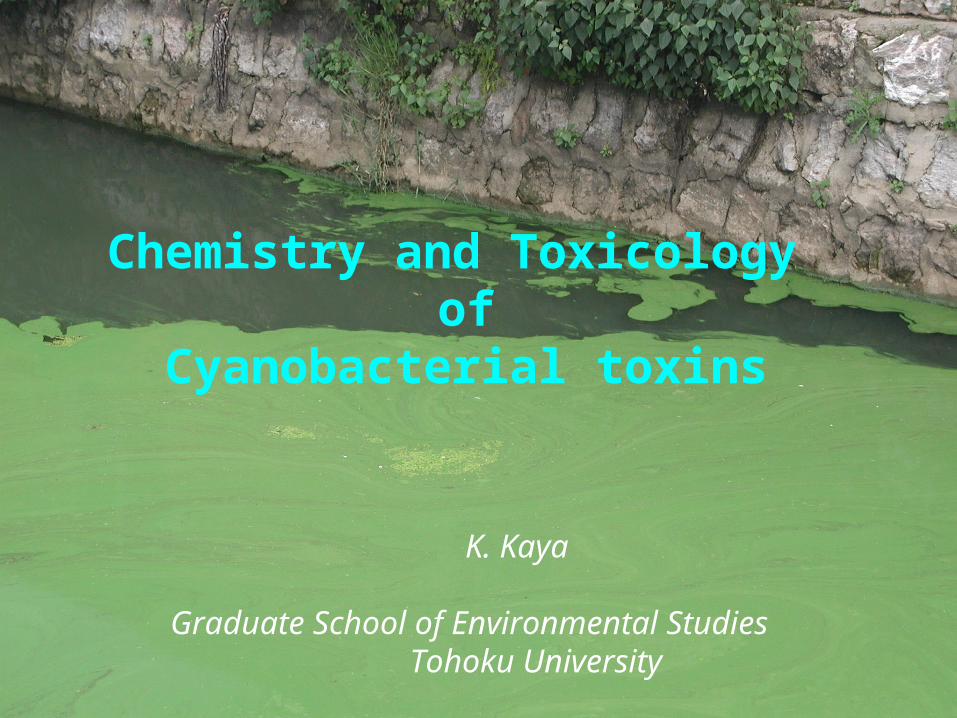
K. Kaya
Graduate School of Environmental Studies Tohoku University
Chemistry and Toxicology of
Cyanobacterial toxins
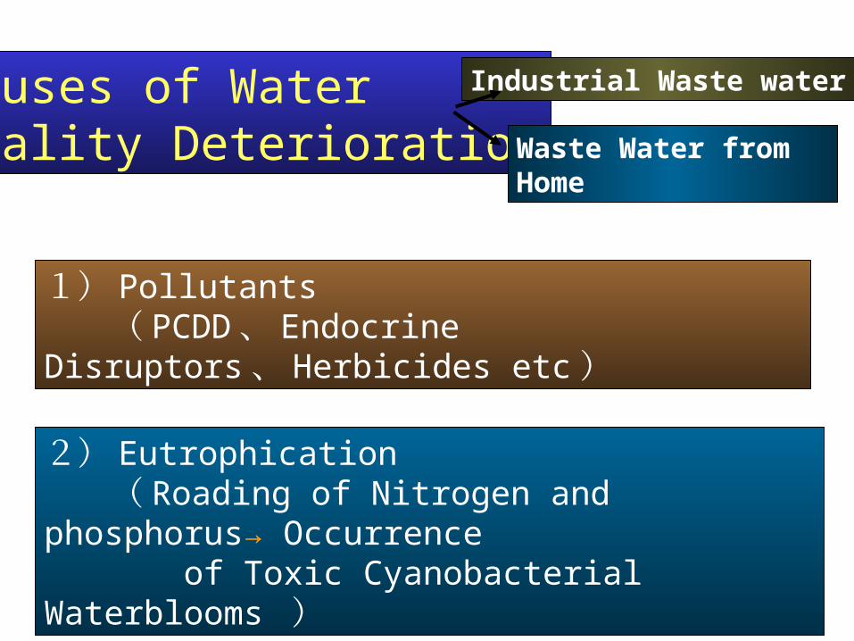
Causes of WaterQuality Deterioration
Industrial Waste water
Waste Water from Home
1) Pollutants ( PCDD 、 Endocrine Disruptors 、 Herbicides etc )
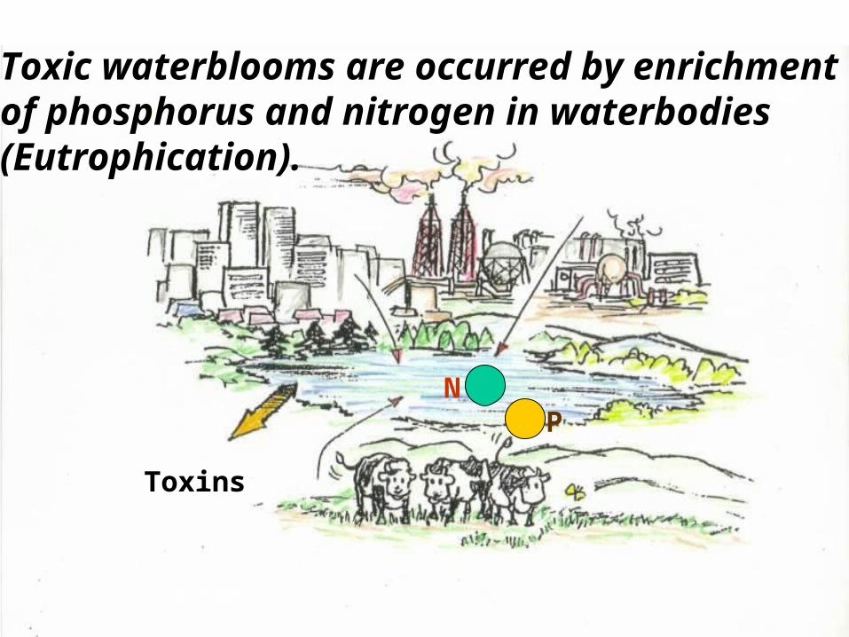
2) Eutrophication ( Roading of Nitrogen and phosphorus→ Occurrence of Toxic Cyanobacterial Waterblooms )
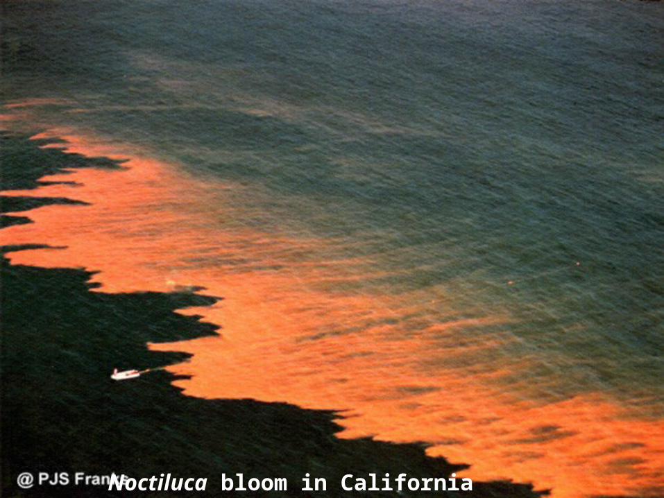
Noctiluca bloom in California
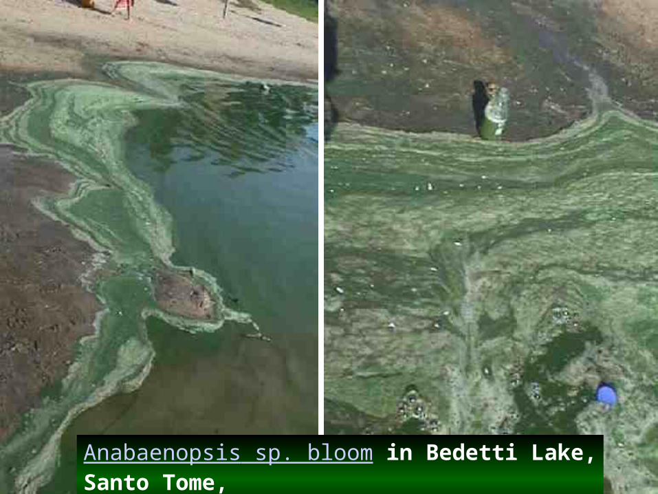
Anabaenopsis sp. bloom in Bedetti Lake, Santo Tome, Santa Fe, Argentina
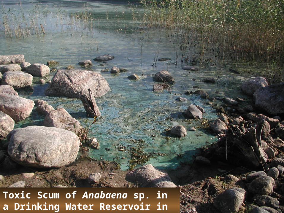
Toxic Scum of Anabaena sp. in a Drinking Water Reservoir in Finland
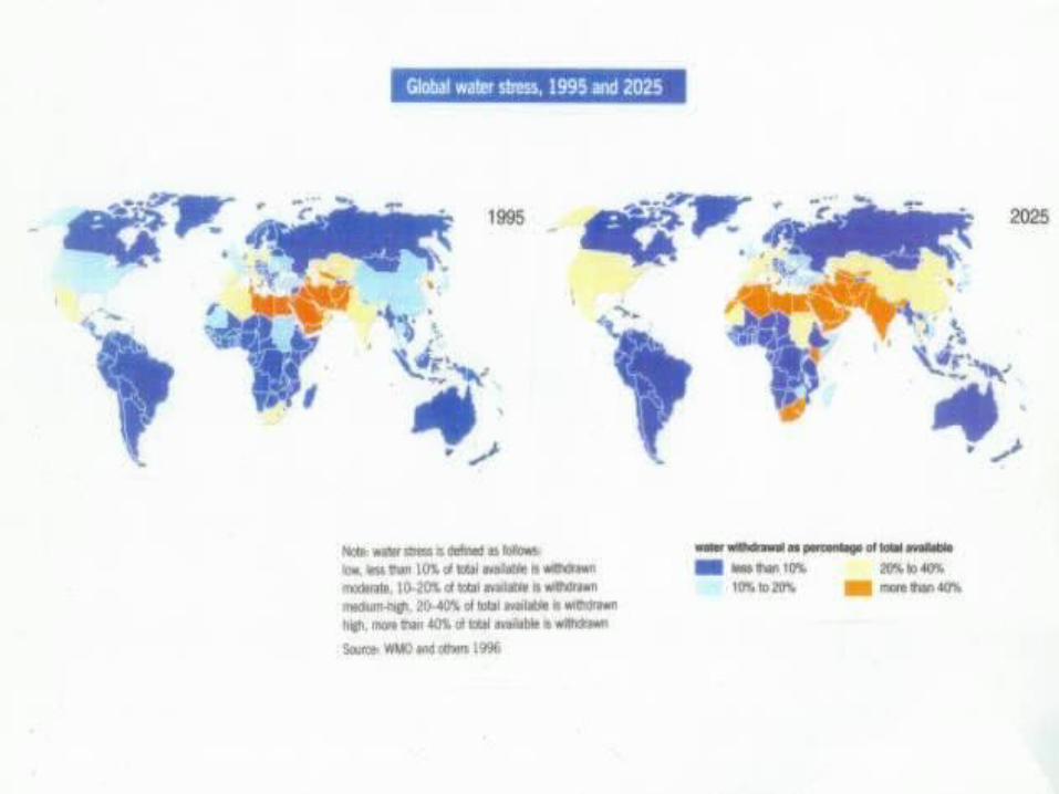

Estimation of Asian Water Resource in 21 Century by UNEP
Increases in Population in Asia ( 1/3 of world population), Food Production and Industrial Activities
Increase in Water DemandScarcity of Freshwater ResourceLocalized Torrential Downpour by Global Warmingand Reducing of Forest
Scarcity of Freshwater and Eutrophication
N P
Toxic waterblooms are occurred by enrichment of phosphorus and nitrogen in waterbodies(Eutrophication).
Toxins
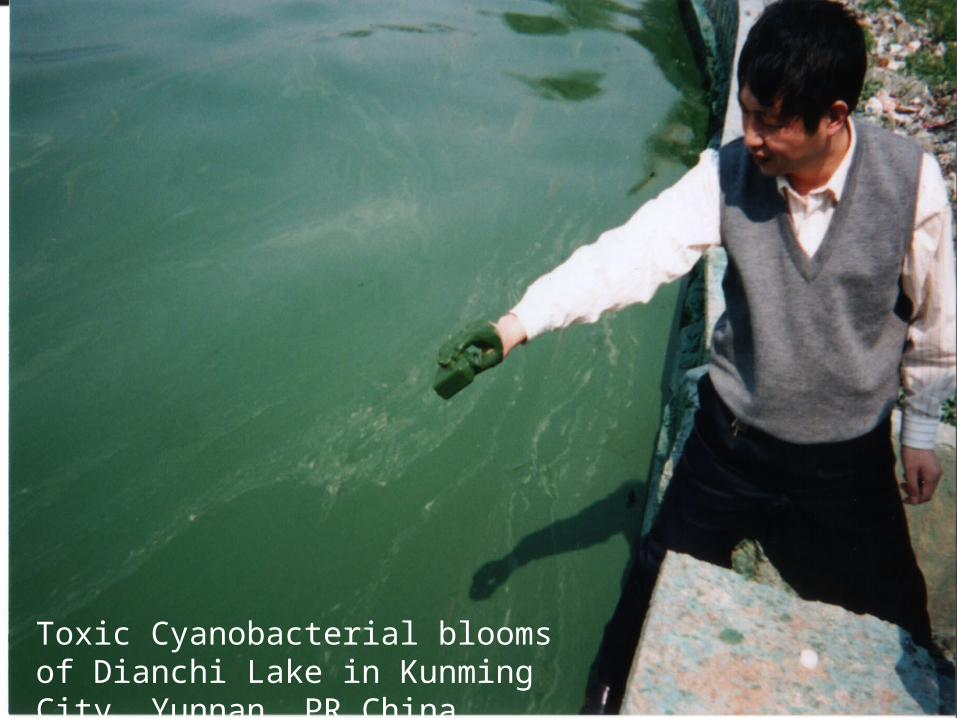
Toxic Cyanobacterial blooms of Dianchi Lake in Kunming City, Yunnan, PR China
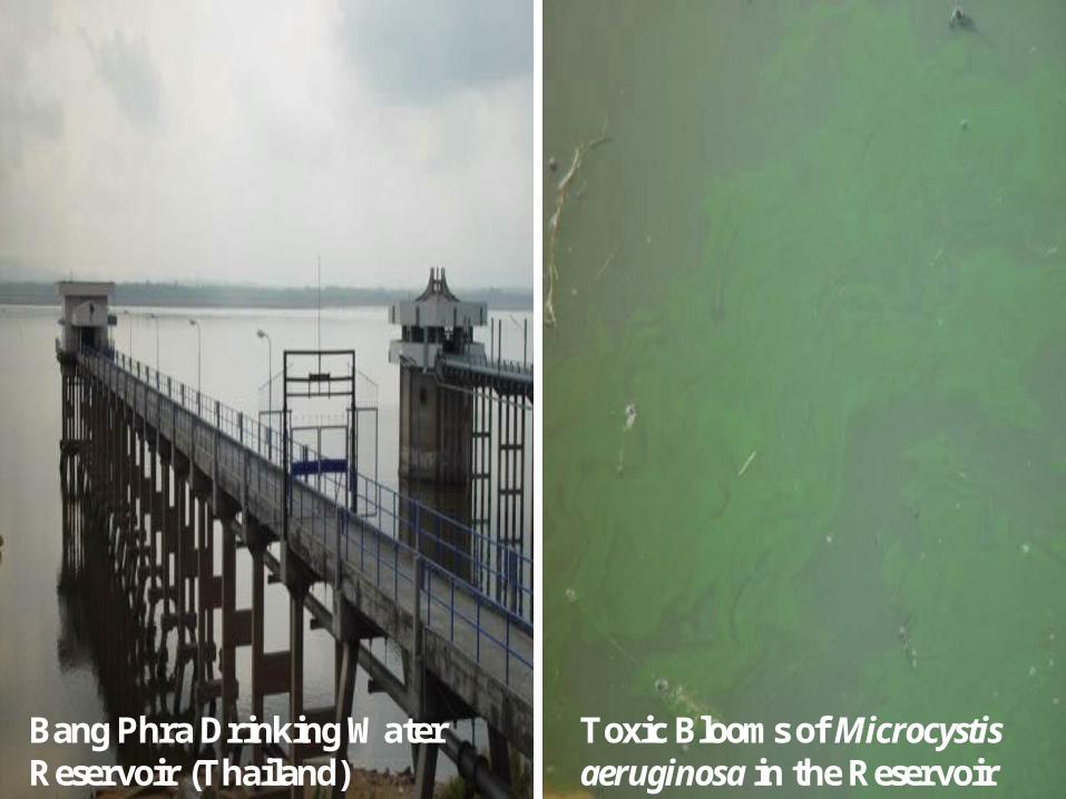
Bang Phra Drinking Water Reservoir (Thailand)
Toxic Blooms of Microcystis aeruginosa in the Reservoir
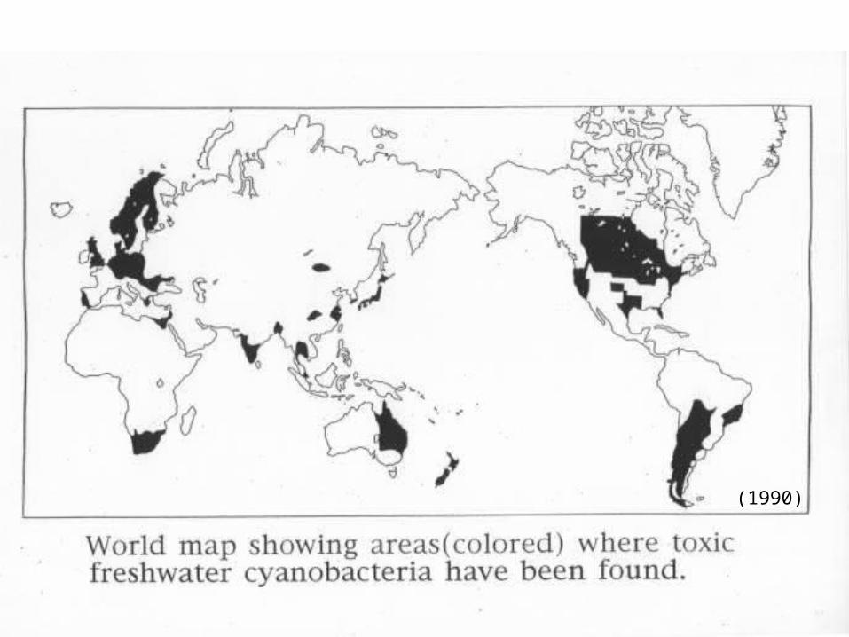
(1990)
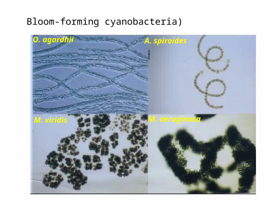
Bloom-forming cyanobacteria)
O. agardhii A. spiroides
M. viridis M. aeruginosa
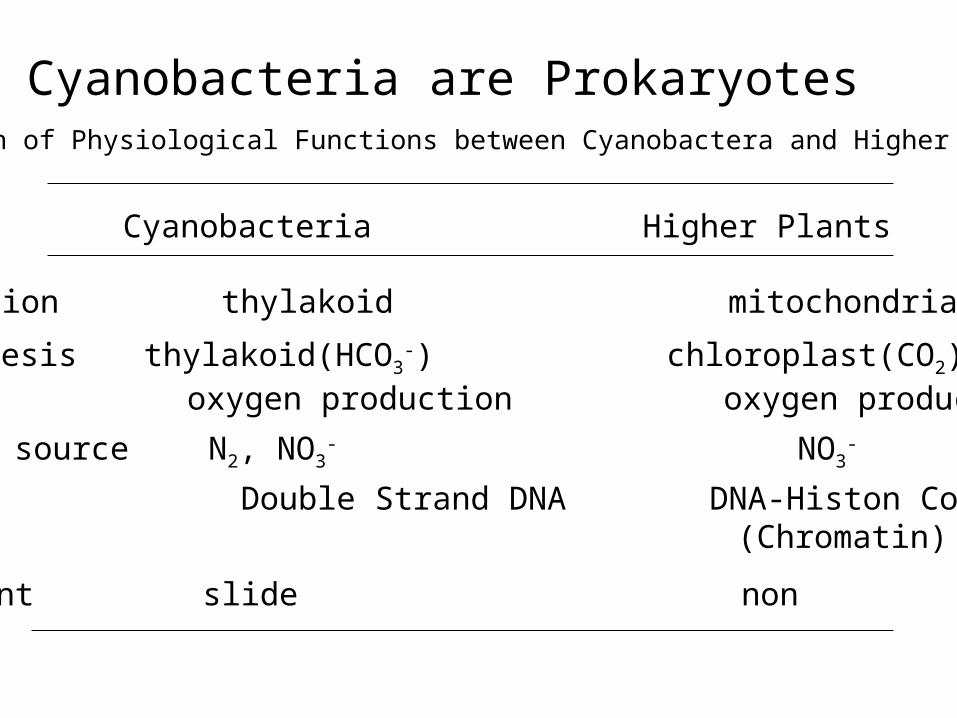
Cyanobacteria are ProkaryotesComparison of Physiological Functions between Cyanobactera and Higher Plants
Cyanobacteria Higher Plants
Respiration thylakoid mitochondria
Photosynthesis thylakoid(HCO3-) chloroplast(CO2)
oxygen production oxygen production
Nitrogen source N2, NO3- NO3
-
Genetic Double Strand DNA DNA-Histon Complex (Chromatin)
movement slide non
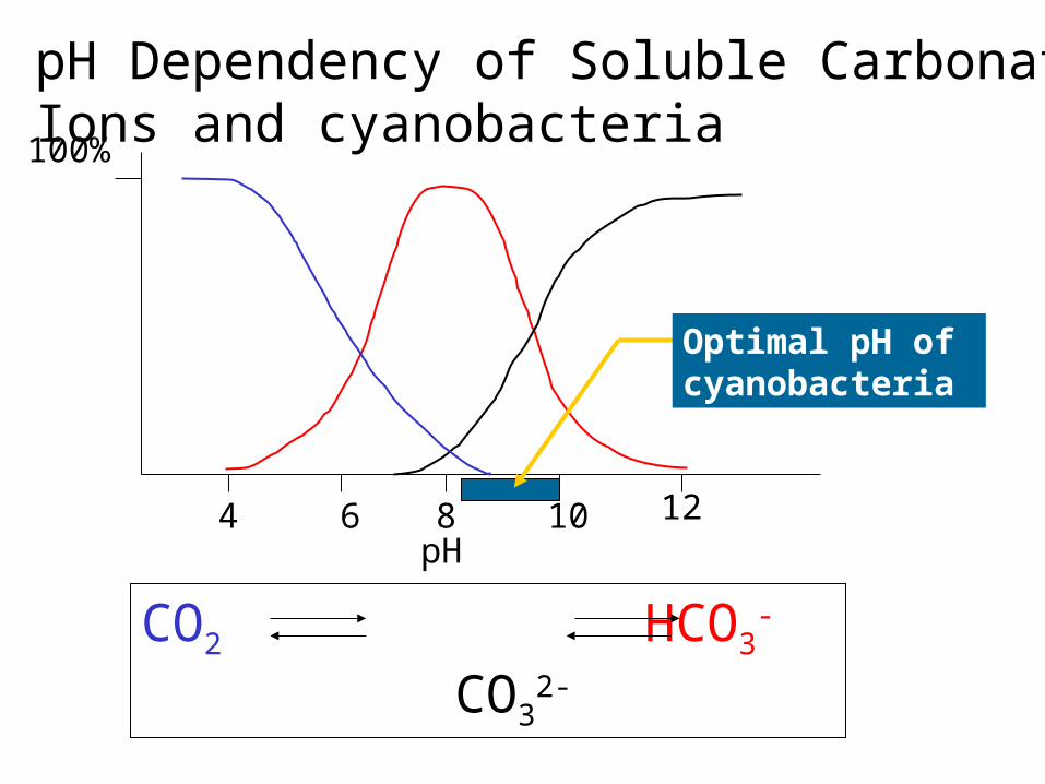
CO2 HCO3-
CO32-
84 6 10 12pH
100%
pH Dependency of Soluble Carbonate Ions and cyanobacteria
Optimal pH of cyanobacteria
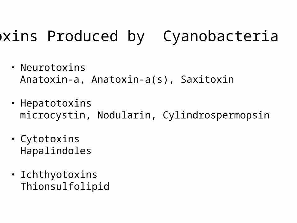
Toxins Produced by Cyanobacteria
・ Neurotoxins Anatoxin-a, Anatoxin-a(s), Saxitoxin
・ Hepatotoxins microcystin, Nodularin, Cylindrospermopsin
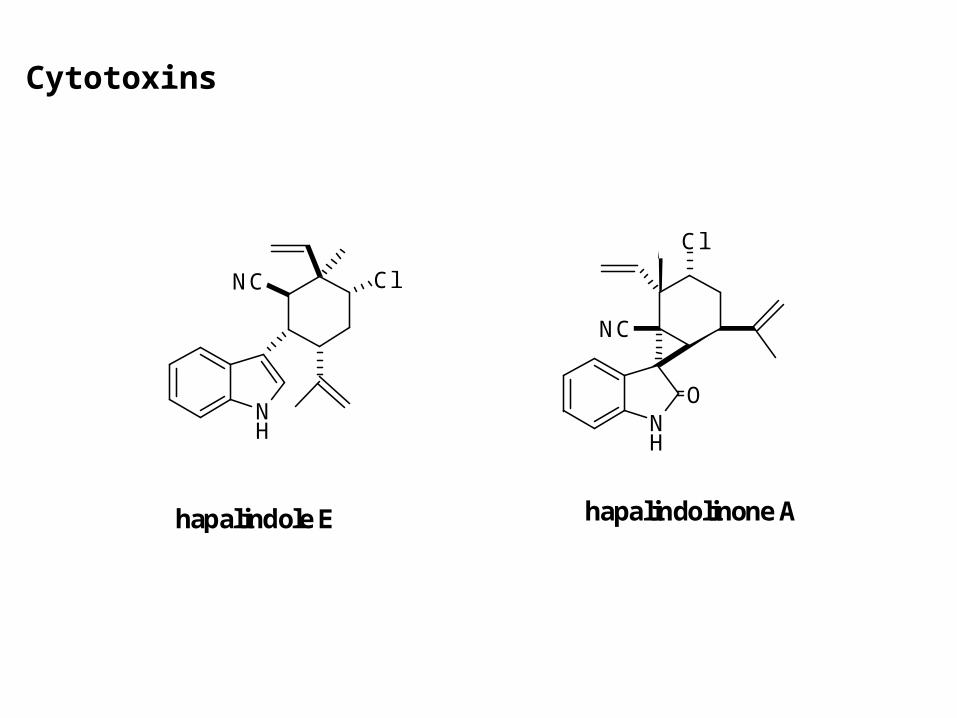
・ Cytotoxins Hapalindoles
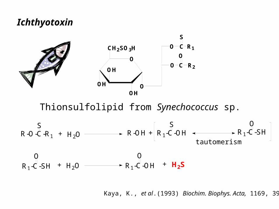
・ Ichthyotoxins Thionsulfolipid

HepatotoxinsOCH3
CH3CH3
HNN CH2
O
HOOC
NH
H3CO NH
HN
O
O
H3C
HN
O
CH3
CH3
HN
COOH
CH3
CH3
O
O
NHH2N
HN
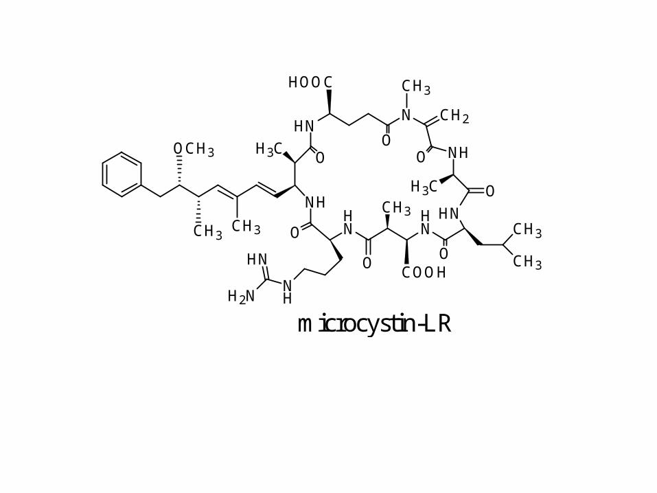
microcystin-LR
OCH3
CH3 CH3
HNN CH
O
COOH
H3CO
CH3 CH3
NH
COOH
O
NHHN
H3C
OO
NHH2N
HN
N NH HN NH
O3SO
H3CNHH
H HO
O
OH
cylindrospermopsin
+
-
nodularin
-
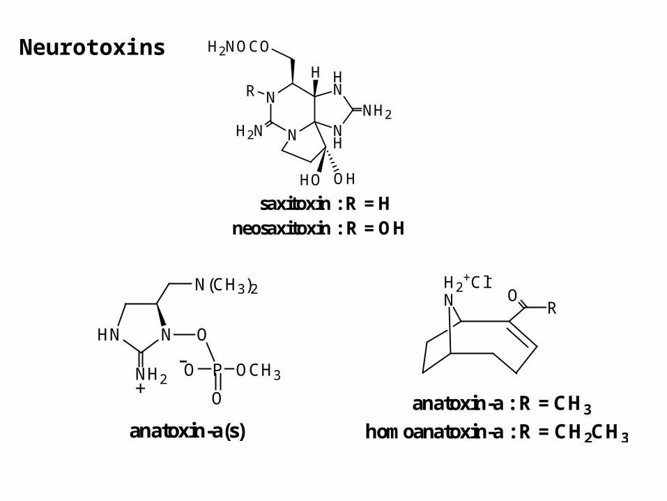
Neurotoxins
N
N
NH
HN
H2NOCO
H2N
HO OH
NH2
RH
saxitoxin : R = Hneosaxitoxin : R = OH
HN N
NH2
N(CH3)2
O
P OCH3O
O
anatoxin-a(s)
+-
H2+Cl-
N RO
anatoxin-a : R = CH3
homoanatoxin-a : R = CH2CH3
NH
ClNC
hapalindole E
NH
Cl
NC
O
hapalindolinone A
Cytotoxins
O
OH
OH
OH
CH2SO3H
O
O
O
C
C
R1
R2
S
O
R-O-C-R1
S+ H2O R-OH + R1-C-OH
SR1-C-SH
O
R1-C-SHO
+ H2O R1-C-OHO
+ H2S
Thionsulfolipid from Synechococcus sp.
Ichthyotoxin
Kaya, K., et al.(1993) Biochim. Biophys. Acta, 1169, 39-45.
tautomerism

H3CNH
COOH
NH2
L--methylaminoalanine (BMAA)
ALS/PDC(The amyotrophic lateral sclerosis / parkinsonism-dementia complex)
The rate of ALS/PDC in Chamorro people in Guam is higher than those of other people. ALS/PDC is related with fruit-bat soup as a domestic food of the Chamorro.
Neurotoxin (chronic)
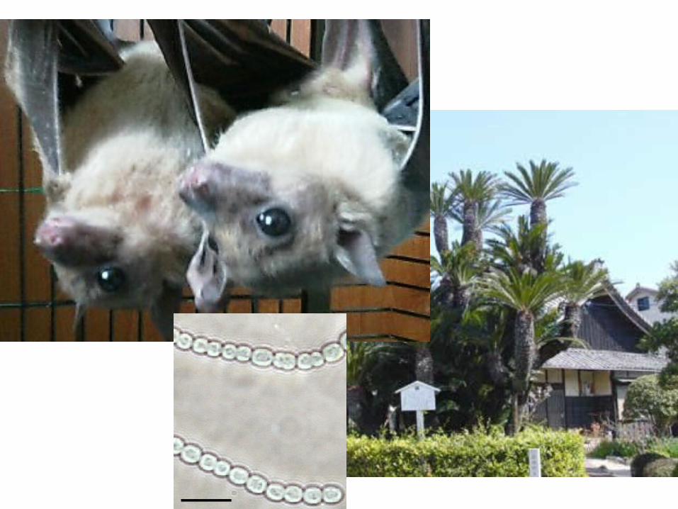
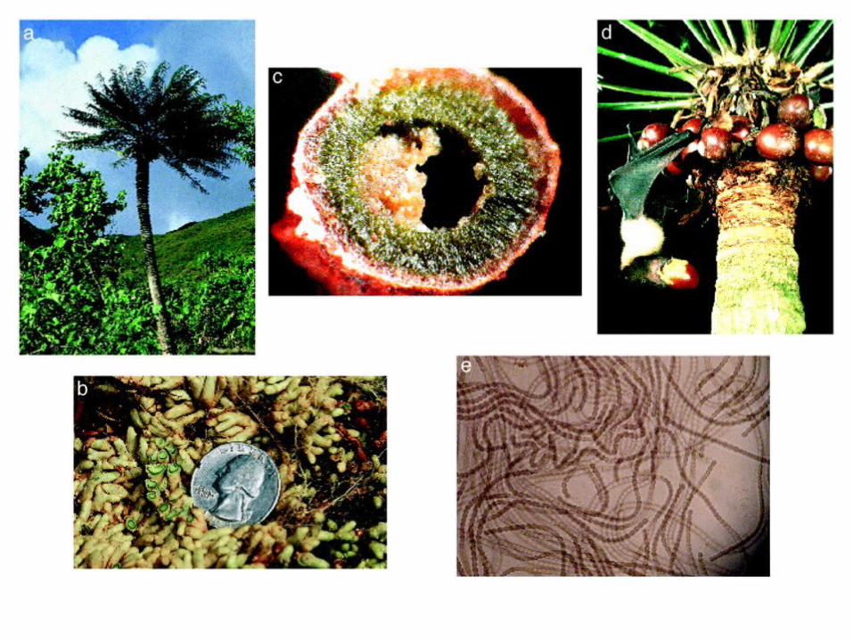
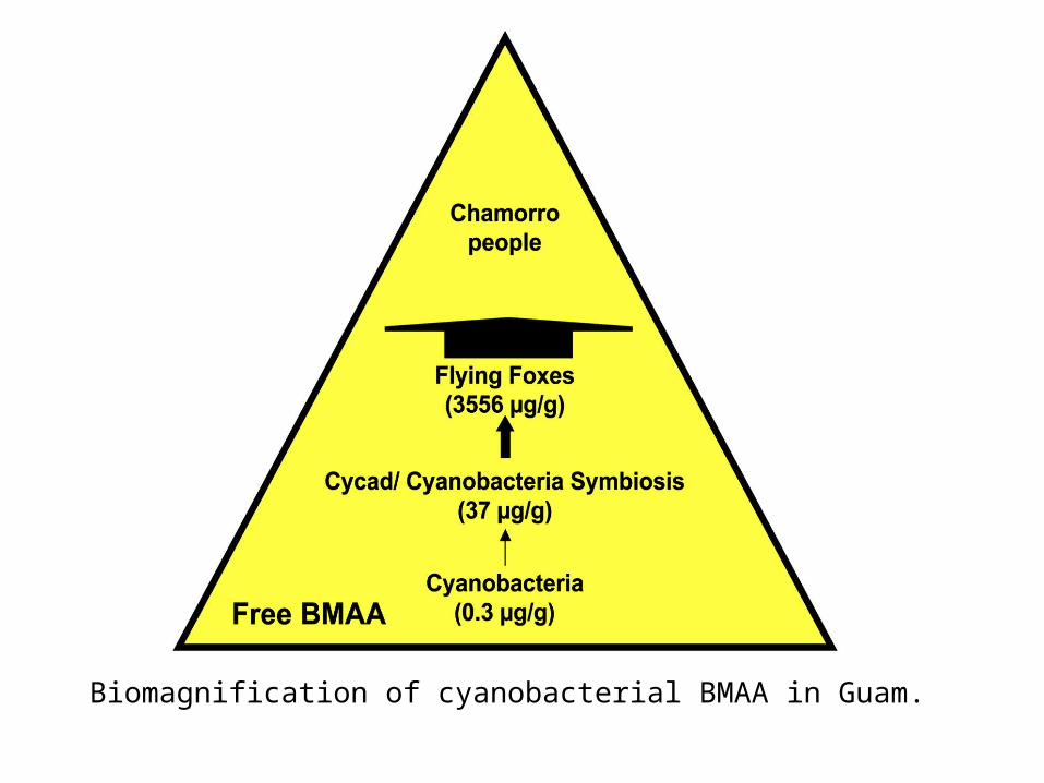
Biomagnification of cyanobacterial BMAA in Guam.
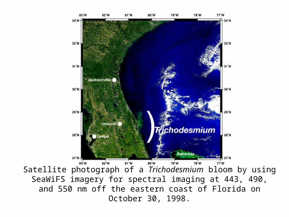
Satellite photograph of a Trichodesmium bloom by using SeaWiFS imagery for spectral imaging at 443, 490, and 550 nm off the eastern coast of Florida
on October 30, 1998.
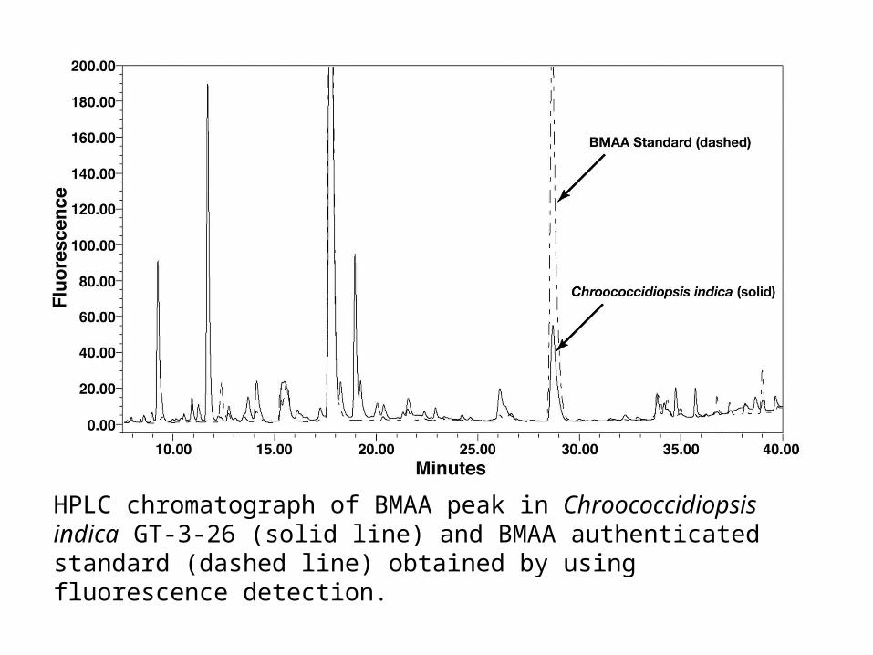
HPLC chromatograph of BMAA peak in Chroococcidiopsis indica GT-3-26 (solid line) and BMAA authenticated standard (dashed line) obtained by using fluorescence detection.
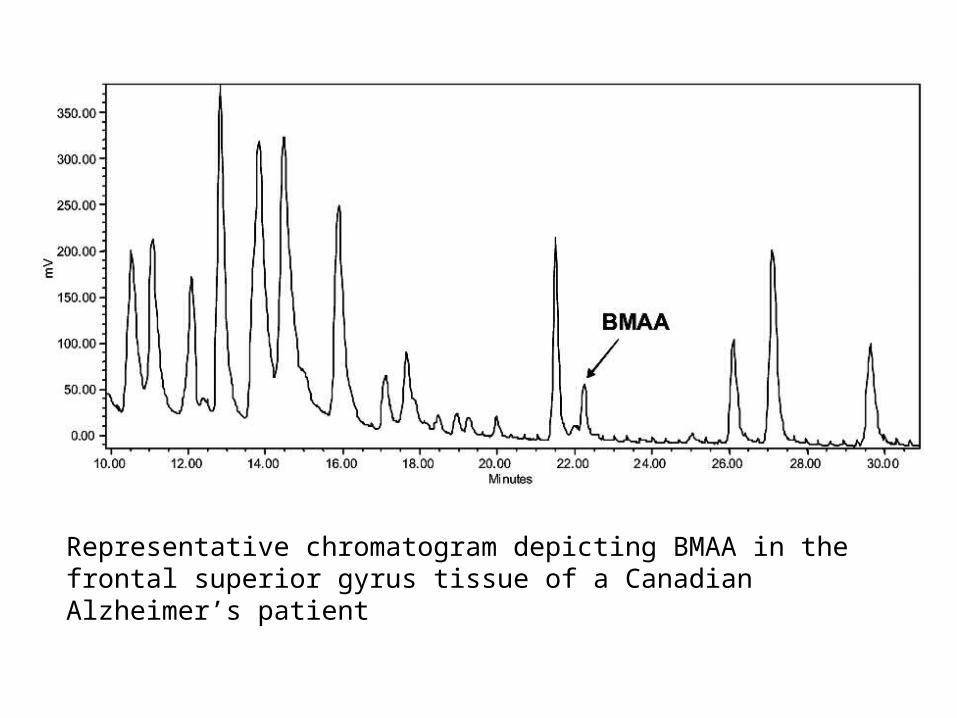
Representative chromatogram depicting BMAA in the frontal superior gyrus tissue of a Canadian Alzheimer’s patient
Microcystins, Nodularins
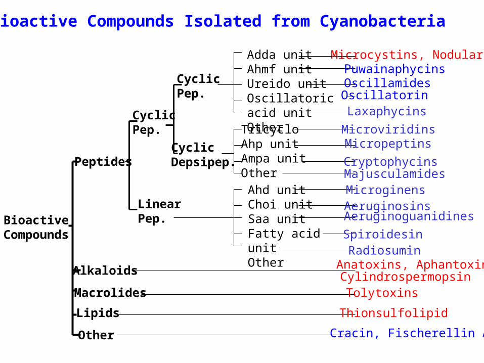
Bioactive Compounds Isolated from Cyanobacteria
Adda unitAhmf unitUreido unitOscillatoric acid unitOtherTricycloAhp unitAmpa unitOther
Ahd unitChoi unitSaa unitFatty acid unitOther
CyclicPep.
CyclicDepsipep.
LinearPep.
CyclicPep.
Peptides
Alkaloids
Macrolides
Lipids
Other
PuwainaphycinsOscillamidesOscillatorinLaxaphycins
MicroviridinsMicropeptins
CryptophycinsMajusculamidesMicroginensAeruginosinsAeruginoguanidines
SpiroidesinRadiosuminAnatoxins, AphantoxinCylindrospermopsinTolytoxins
Thionsulfolipid
Cracin, Fischerellin A
BioactiveCompounds
H3C NH
NHNH
NCH3
CH3
NHN
H3C CH3
OH3C
O
O
O
OO
OH
OH3C
O
O
NH
NH2
HN
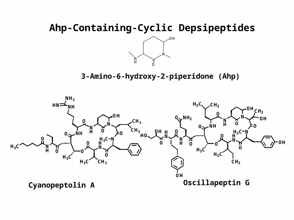
Cyanopeptolin A Oscillapeptin G
3-Amino-6-hydroxy-2-piperidone (Ahp)
Ahp-Containing-Cyclic Depsipeptides
N
O
OH
NH
HN
NH
NHNH
N OH
NHN
H3C
OH3C
O
O
O
OO
OH
OH3C
O
O
CH3
H3C CH3
OH
CH3
NH2O
OH
O
OHHO
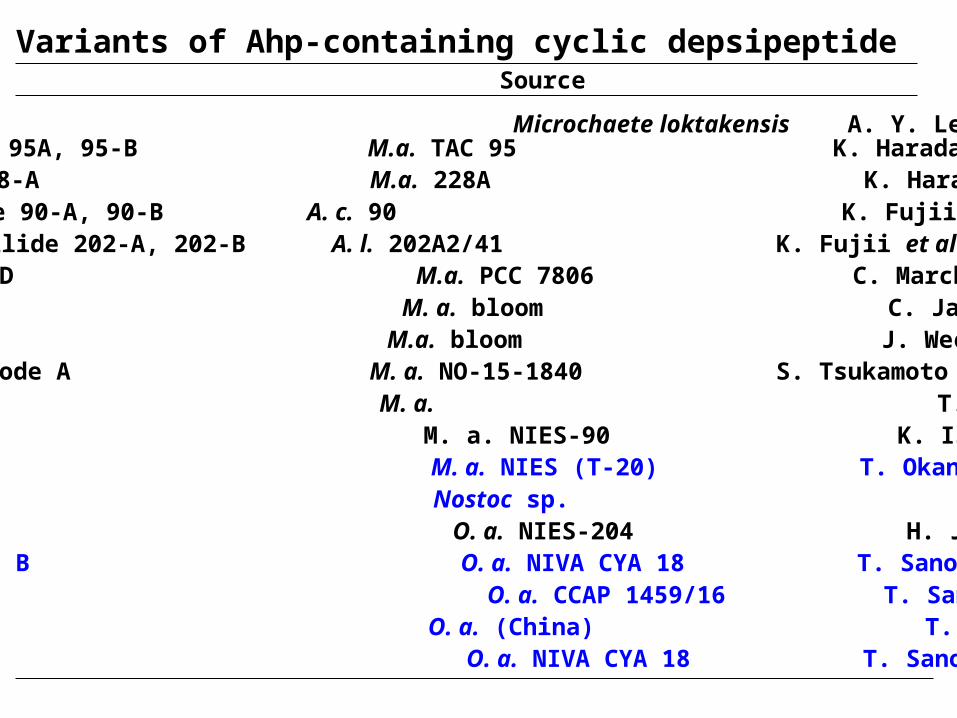
Micropeptin A, B M. a. T. Okino et al (1993)Micropeptin 90 M. a. NIES-90 K. Ishida et al (1995)Micropeptin T-20 M. a. NIES (T-20) T. Okano et al (1999)
Oscillapeptin O. a. NIES-204 H. J. Shin et al (1995)Oscillapeptin A, B O. a. NIVA CYA 18 T. Sano & K. Kaya (1998)Oscillapeptin C O. a. CCAP 1459/16 T. Sano (1996)Oscillapeptin D O. a. (China) T. Sano et al (1998)
Nostocyclin Nostoc sp. K. Kaya et al (1996)
Aeruginopeptin 95A, 95-B M.a. TAC 95 K. Harada et al (1993)Aeruginopeptin 228-A M.a. 228A K. Harada et al (1993)
Cyanopeptolin A-D M.a. PCC 7806 C. Marchin et al (1993)
Microcystilode A M. a. NO-15-1840 S. Tsukamoto et al (1993)
Cyanopeptolin S M. a. bloom C. Jakobi et al (1993)Cyanopeptolin SS M.a. bloom J. Weckesser et al (1996)
Anabaenopeptilide 90-A, 90-B A. c. 90 K. Fujii et al (1995) Anabaenopeptilide 202-A, 202-B A. l. 202A2/41 K. Fujii et al (1995)
A90720A Microchaete loktakensis A. Y. Lee et al (1994)
Variants of Ahp-containing cyclic depsipeptideVariants Source Reference
Oscillapeptin G O. a. NIVA CYA 18 T. Sano & K. Kaya (1996)
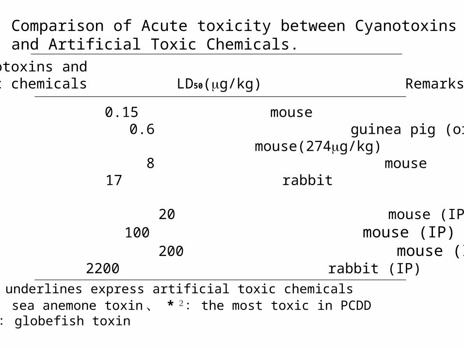
Comparison of Acute toxicity between Cyanotoxins and Artificial Toxic Chemicals.
Cyanotoxins andToxic chemicals LD50(g/kg) Remarks
Palytoxin*1 0.15 mouse2,3,7,8-TCDD *2 0.6 guinea pig (oral adm.) mouse(274g/kg)Tetrodotoxin*3 8 mouseSarin 17 rabbit mouse (170g/kg)Anatoxin-a(s) 20 mouse (IP)Microcystin-LR 100 mouse (IP)Anatoxin-a 200 mouse (IP)Sodium cyanate 2200 rabbit (IP)
The underlines express artificial toxic chemicals*1 : sea anemone toxin 、 * 2: the most toxic in PCDD* 3: globefish toxin
OCH3
CH3CH3
HNN CH2
O
HOOC
NH
H3CO NH
HN
O
O
H3C
HN
O
CH3
CH3
HN
COOH
CH3
CH3
O
O
NHH2N
HN
microcystin-LR
HN
H3C
O
N
R2
NH
N
O
NH
H3COCH3
CH3
R1
COOH
CH2
COOH
CH3
O
OO
XZ
1
23
4
5
67
* L-aminoisobutyric acid
1023
966
980
980
923
1028
1001
959
1019
1037
1044
994
909
Aba*
Arg
Tyr
Ala
Met
Arg
Arg
Arg
Arg
Arg
Arg
Arg
Arg
Leu
Leu
Leu
Leu
Phe
Leu
Tyr
Tyr
Leu
Arg
Tyr
AlaLeu
Z
[D-Asp3,Dha7]microcystin LR
[Dha7]microcystin LR
[D-Asp3]microcystin LR
microcystin LAba
microcystin FR
microcystin LY
microcystin YA
microcystin YM
microcystin RR
microcystin YR
microcystin LR
microcystin LA
[D-Asp3]microcystinRR
H
H
H
CH3
CH3CH3
CH3
CH3
CH3CH3
CH3
CH3CH3
CH3
CH3CH3
CH3
CH3CH3
H
CH3CH3
H
CH3CH3
CH3
CH3CH3
CH3
CH3CH3
CH3
CH3
CH3CH3
CH3
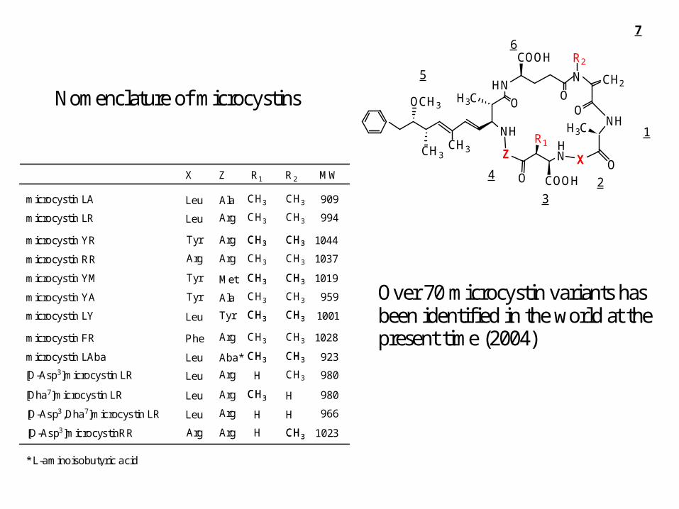
MWX R1 R2
H
Nomenclature of microcystins
Over 70 microcystin variants has been identified in the world at the present time (2004)
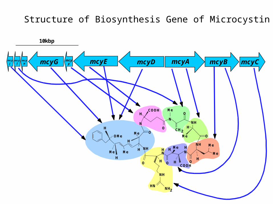
ミクロシスチン生合成遺伝子の構造
10kbp
ミクロシスチン
O
NH2
N
ON
N
O
O
OO
H
H HH
H
H
H
O
H
H
CH2H
mcyE mcyD mcyA mcyB mcyCmcyGmcyJ
mcyI
mcyH
mcyF
Me
Me
Me
Me
MeMe
OMe
COOH
NH
Me
NH
HN
NH
H
NH
COOH
NHMe
Structure of Biosynthesis Gene of Microcystin
mcyE mcyDmcyGmcyJ
mcyI
mcyH
mcyF
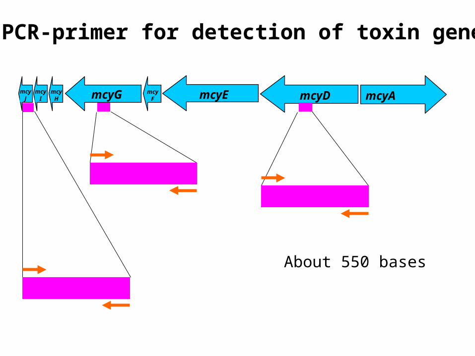
PCR-primer for detection of toxin gene
mcyA
About 550 bases
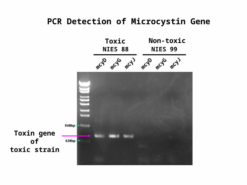
NIES 88
mcy
Dm
cyG
mcy
J
mcy
Dm
cyG
mcy
J
NIES 99Toxic Non-toxic
Toxin gene oftoxic strain
940bp
420bp
PCR Detection of Microcystin Gene
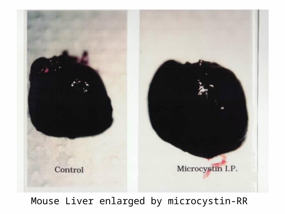
Mouse Liver enlarged by microcystin-RR
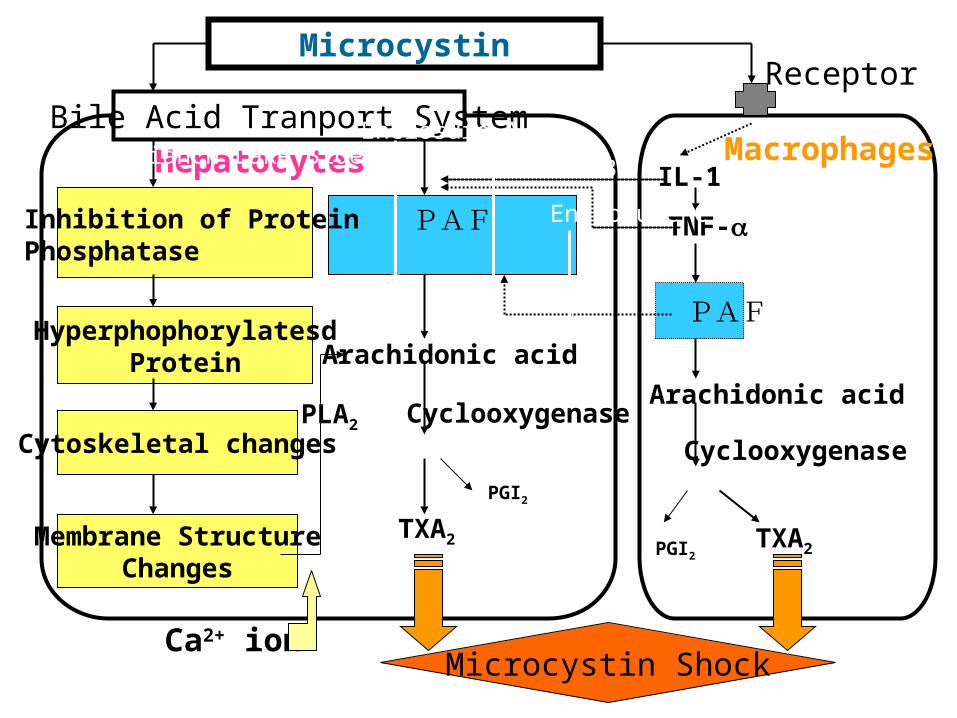
Microcystin Shock
Microcystin
Bile Acid Tranport SystemReceptor
HyperphophorylatesdProtein
Cytoskeletal changes
Membrane StructureChanges
PAFInhibition of ProteinPhosphatase
Arachidonic acid
CyclooxygenasePLA2
TXA2
IL-1
TNF-
PAF
Arachidonic acid
Cyclooxygenase
TXA2
Hepatocytes Macrophages
Ca2+ ion
PGI2
PGI2
Enclosure A
Enclosure B
Enclosure C
Dianch Lake Side
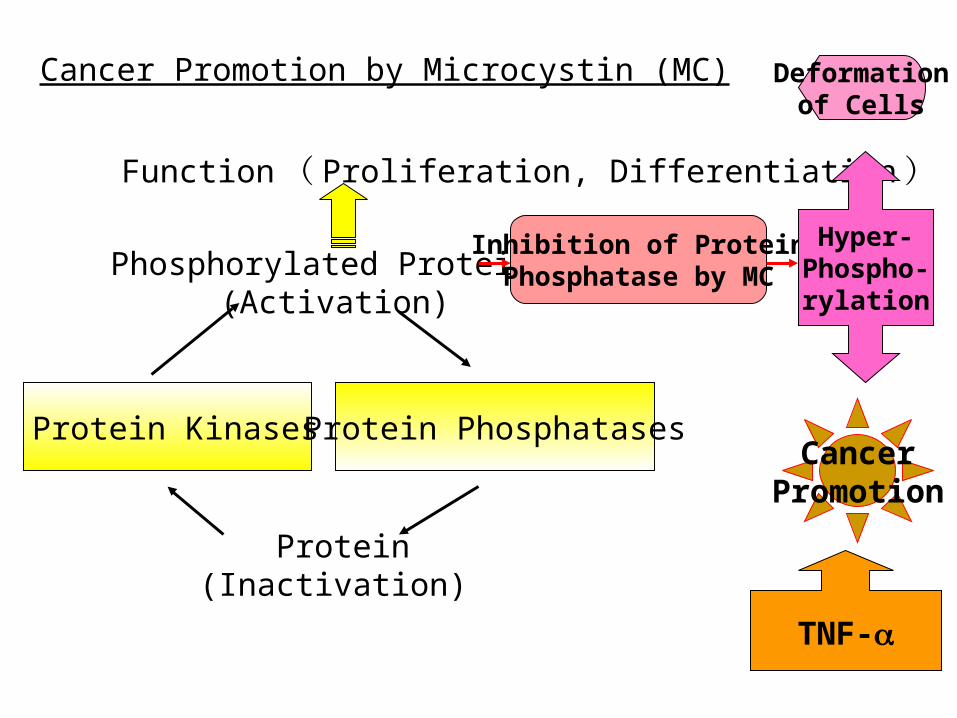
Cancer Promotion by Microcystin (MC)
Phosphorylated Protein (Activation)
Function ( Proliferation, Differentiation )
Protein Kinases Protein Phosphatases
Protein (Inactivation)
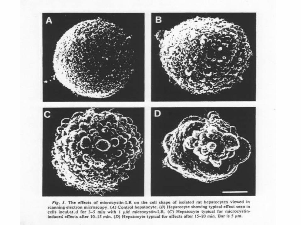
Inhibition of ProteinPhosphatase by MC
Hyper-Phospho-rylation
Deformationof Cells
CancerPromotion
TNF-
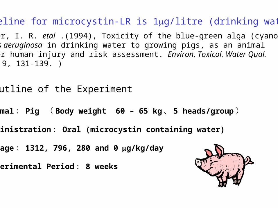
WHO Guideline for microcystin-LR is 1g/litre (drinking water), 1997
(Falconer, I. R. etal .(1994), Toxicity of the blue-green alga (cyanobacterium)Microcystis aeruginosa in drinking water to growing pigs, as an animal model for human injury and risk assessment. Environ. Toxicol. Water Qual.Intern. J., 9, 131-139. )
Outline of the Experiment
1) Animal : Pig ( Body weight 60 – 65 kg 、 5 heads/group )
2) Administration : Oral (microcystin containing water)
3) Dosage : 1312, 796, 280 and 0g/kg/day
4) experimental Period : 8 weeks
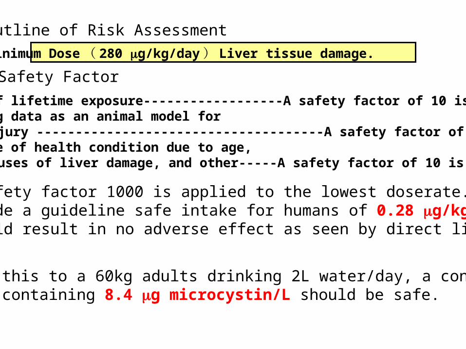
Outline of Risk Assessment
Minimum Dose ( 280 g/kg/day ) Liver tissue damage.
Safety Factor
1) Only 1% of lifetime exposure------------------A safety factor of 10 is applied2) Use of pig data as an animal model for human injury -------------------------------------A safety factor of 10 is applied3) Difference of health condition due to age, other causes of liver damage, and other-----A safety factor of 10 is applied
Thus a safety factor 1000 is applied to the lowest doserate.This provide a guideline safe intake for humans of 0.28 g/kg/day, Which should result in no adverse effect as seen by direct liver injury
To apply this to a 60kg adults drinking 2L water/day, a consumption,Of water containing 8.4 g microcystin/L should be safe.
4) For tumor prmortion, additional safety factor of 5 or 10 is required
Thus a conservative estimate for water safety is 0.84 g microcystin/ L or approximately 1 g/L.
Determination Methods for Total Microcystin
1)Molecular biological method i) PCR of Toxin gene
2) Biochemical methods i) Protein Phosphatase Inhibition ii) ELISA
3) Physical methods i) HPLC/UV or MS
4) Chemical i) MMPB metho ii) GSH method
1) Inhibition of Protein Phosphatase 2A
2) Enzyme-Linked Immunosorbent Assay (ELISA)
Biochemical Determination
Microcystin
Bile Acid Tranport SystemReceptor
HyperphophorylatesdProtein
Cytoskeletal changes
Membrane StructureChanges
PAFInhibition of ProteinPhosphatase
Arachidonic acid
CyclooxygenasePLA2
TXA2
IL-1
TNF-
PAF
Arachidonic acid
Cyclooxygenase
TXA2
Hepatocytes Macrophages
Caイオン
PGI2
PGI2
Inhibitory Activity
Not only microcystinbut also other compounds inhibit
ELISA
Secondary antibody
HRP
HRP
Substrate
Color
MCLR-BSA
Perimary antibody
microcystin
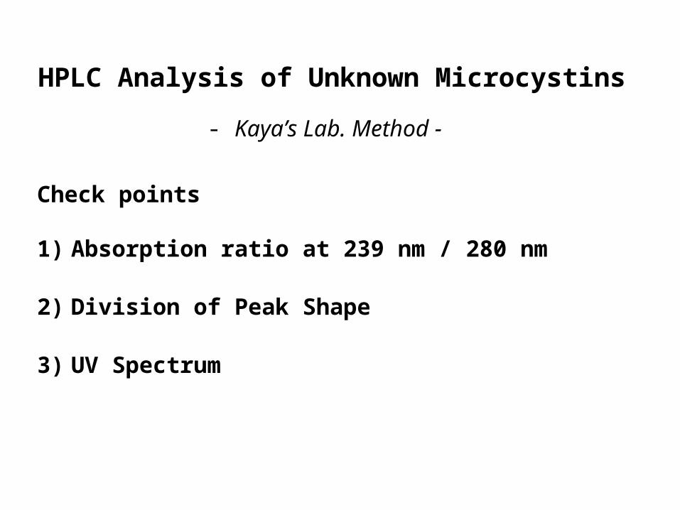
HPLC Analysis of Unknown Microcystins
- Kaya’s Lab. Method -
Check points
1) Absorption ratio at 239 nm / 280 nm
2) Division of Peak Shape
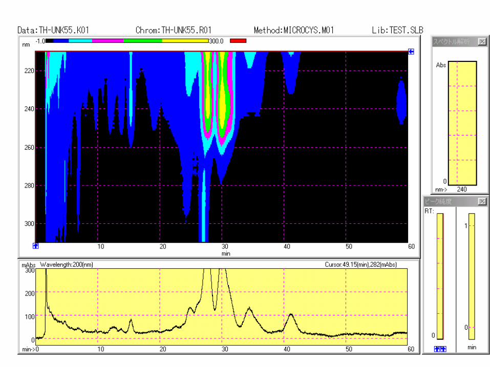
3) UV Spectrum
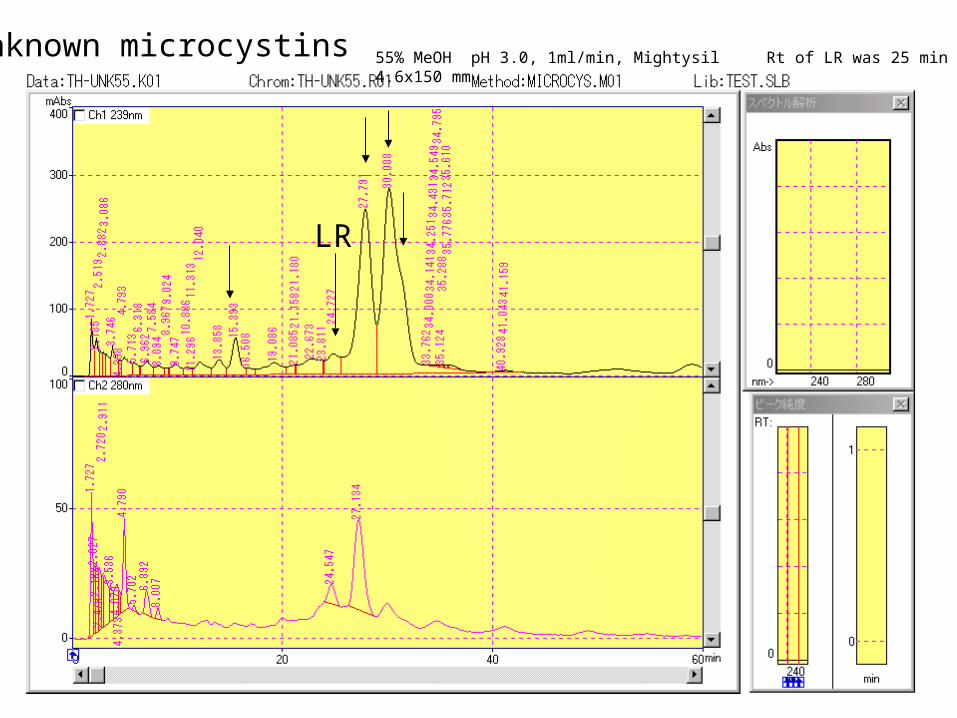
55% MeOH pH 3.0, 1ml/min, Mightysil 4.6x150 mm Rt of LR was 25 minUnknown microcystins
LR

Rt, 15.4 min Rt, 28 min
Rt, 30 min Rt, 31 min (shoulder)
HN
H3C
O
N
CH3
NH
N
O
NH
H3COCH3
CH3
CH3
COOH
R
COOH
CH3
O
O
O
XZ
H
COOH
OCH3
CH3
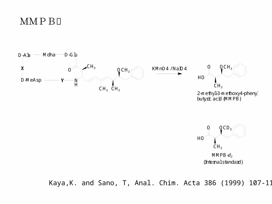
MMPB method for total microcystin determination
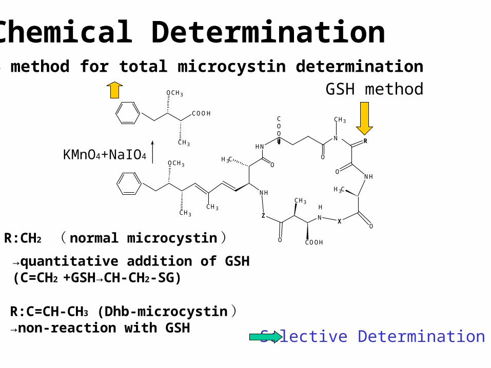
Chemical Determination
R:CH2 ( normal microcystin )
→quantitative addition of GSH (C=CH2 +GSH→CH-CH2-SG)
R:C=CH-CH3 (Dhb-microcystin )→non-reaction with GSH
Selective Determination
KMnO4+NaIO4
GSH method
NH
CH3 CH3
OCH3
CH3O
Mdha
D-MeAsp
D-Ala D-Glu
CH3
OCH3
HO
OKMnO4 / NaIO4
CH3
OCD3
HO
O
2-methyl-3-methoxy4-phenylbutyric acid (MMPB)
MMPB-d3(Internal standard)
Y
X
MMPB法
Kaya,K. and Sano, T, Anal. Chim. Acta 386 (1999) 107-112
0.5 1.0 1.5 2.0 2.5 3.0 3.5 4.0 4.5 5.0 5.5 6.0 6.5 min
10000
15000
20000
25000
30000
35000
40000
45000
50000
55000Int.
210.00(1.07)207.00(1.00)
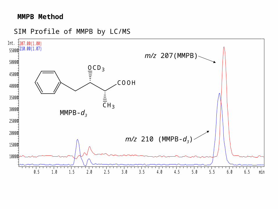
SIM Profile of MMPB by LC/MS
COOH
CH3
OCD3
m/z 207(MMPB)
m/z 210 (MMPB-d3)
MMPB-d3
MMPB Method
HN
H3C
O
N
NH
HN
O
NH
H3COCH3
CH3
COOH
COOH
CH3
O
OO
XZ
HN
H3C
O
N
R
NH
HN
O
NH
H3COCH3
CH3CH3
COOH
CH2
COOH
CH3
O
OO
XZ
1
23
4
5
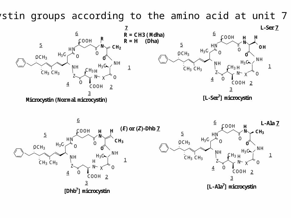
6 R = CH3 (Mdha) R = H (Dha)
H
CH3
H
1
23
4
5
6
(E) or (Z)-Dhb 7
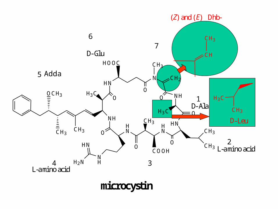
Microcystin (Normal microcystin)
[Dhb7] microcystin
HN
H3C
O
N
NH
HN
O
NH
H3COCH3
CH3
COOH
COOH
CH3
O
OO
XZ
H
OH
H
1
23
4
5
[L-Ser7] microcystin
6L-Ser 7
HN
H3C
O
N
NH
HN
O
NH
H3COCH3
CH3
COOH
COOH
CH3
O
OO
XZ
HCH3
1
23
4
5
[L-Ala7] microcystin
6 L-Ala 7
7
CH3
CH3
Microcystin groups according to the amino acid at unit 7
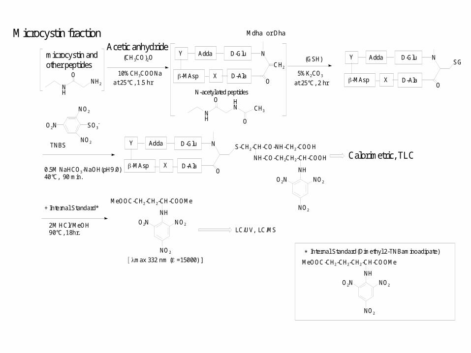
(GSH)
5%K2CO3
TNBS
0.5M NaHCO3-NaOH(pH9.0)40°C, 90 min.
SO3-O2N
NO2
NO2S-CH2-CH-CO-NH-CH2-COOH
NH-CO-CH2CH2-CH-COOH
NH
NO2O2N
NO2
Calorimetric, TLC
2M HCl/ MeOH90°C, 18hr.
+ Internal Standard*MeOOC-CH2-CH2-CH-COOMe
NH
NO2O2N
NO2
D-AlaX
Y D-Glu N
CH2
O-MAsp
Adda
Mdha or Dha
D-AlaX
Y D-Glu N
O-MAsp
Adda
D-AlaX
Y D-Glu N
O-MAsp
Adda
SG
LC/UV, LC/MS
max 332 nm (=15000) ]*
MeOOC-CH2-CH2-CH2-CH-COOMe
NH
NO2O2N
NO2
Internal Standard(Dimethyl 2-TNBaminoadipate)
NH
NH2
O
(CH3CO)2O
10%CH3COONa
NH
HN
OCH3
O
at 25°C, 1.5 hr at 25°C, 2 hr
N-acetylated peptides
microcystin and other peptides
Acetic anhydrideMicrocystin fraction
2000
1800
1600
1400
1200
1000
800
2000
1800
1600
1400
1200
1000
800
2 4 6 8 10 12 14 Rt (min)
2 4 6 8 10 12 14 Rt (min)
O2N
NO2
NO2
NH CHCOOMe
CH2
CH2
COOMe
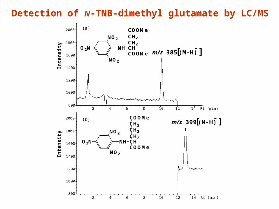
m/z 385 [(M-H)- ]
O2N
NO2
NO2
NH CHCOOMe
CH2
CH2
CH2m/z 399 [(M-H)
- ]COOMe(b)
(a)
Inte
ns
ity
Inte
ns
ity
Detection of N-TNB-dimethyl glutamate by LC/MS
D-AlaX
Y D-Glu N
O-MAsp
AddaSG
S-CH2-CH-CO-NH-CH2-COOH
NH-CO-CH2CH2-CH-COOH
NH2D-AlaX
Y D-Glu N
O-MAsp
Adda
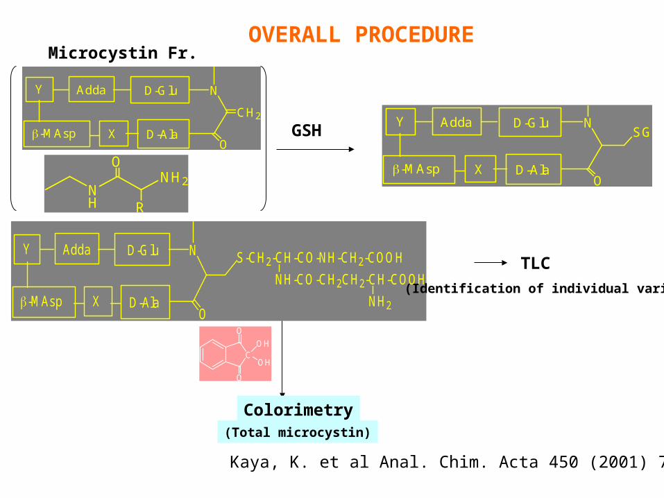
Microcystin Fr.
NH
NH2
O
R
D-AlaX
Y D-Glu N
CH2
O-MAsp
Adda
TLC
(Identification of individual variants)
C
O
O
OH
OH
(Total microcystin)
Colorimetry
OVERALL PROCEDURE
GSH
Kaya, K. et al Anal. Chim. Acta 450 (2001) 73-80
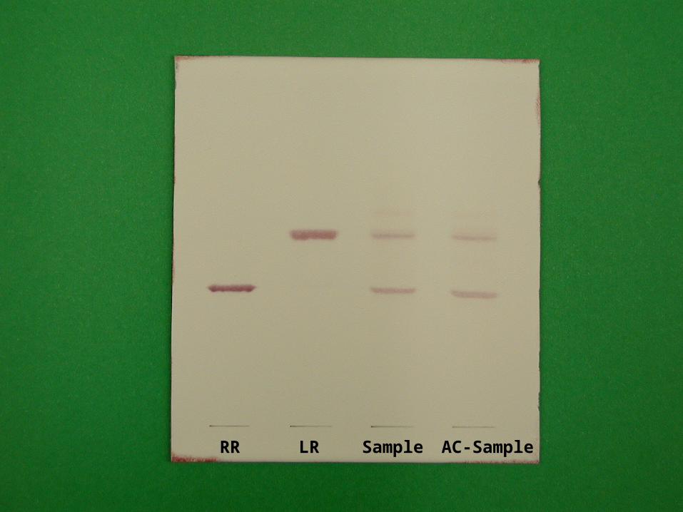
RR SampleLR AC-Sample
OCH3
CH3CH3
HNN CH2
O
HOOC
NH
H3CO NH
HN
O
O
H3C
HN
O
CH3
CH3
HN
COOH
CH3
CH3
O
O
NHH2N
HN
microcystin
CH
CH3
Dhb-
H3C
CH3
1
2
34
5
67
L-amino acid
L-amino acid
D-Ala
D-Leu
D-Glu
(Z) and (E)
Adda
OCH3
CH3CH3
HNN CH2
O
HOOC
NH
H3C O NH
HNO
O
H3C
HN
OCH3
CH3
HN
COOH
CH3
CH3
O
O
NHH2N
HN
Dhb-microcystinC
CH3
1
2
34
5
67
L-amino acid
L-amino acid
D-Ala
D-Glu Mdh
HN
H
NHO
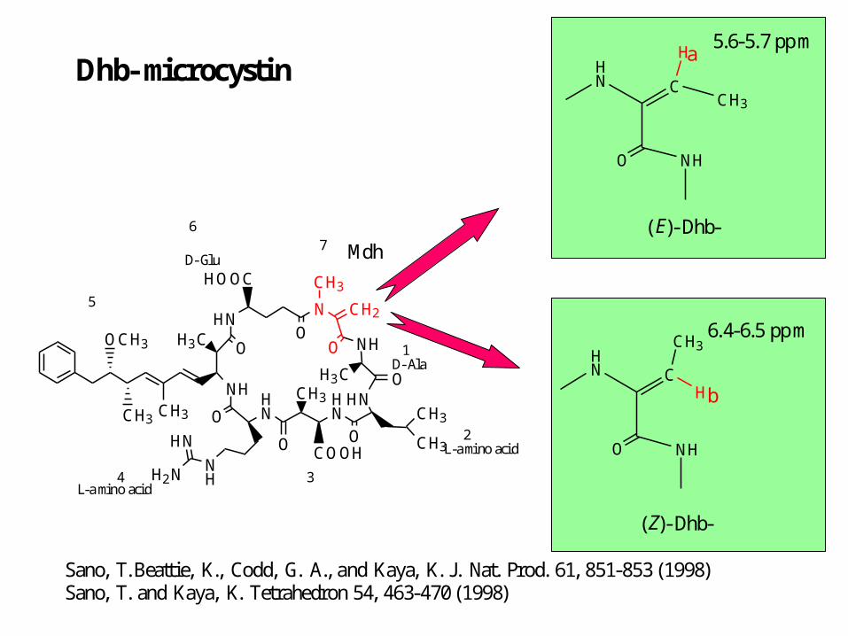
a5.6-5.7 ppm
(E)-Dhb-
C
CH3HN
H
NHO
b
6.4-6.5 ppm
(Z)-Dhb-
Sano, T.Beattie, K., Codd, G. A., and Kaya, K. J. Nat. Prod. 61, 851-853 (1998)Sano, T. and Kaya, K. Tetrahedron 54, 463-470 (1998)
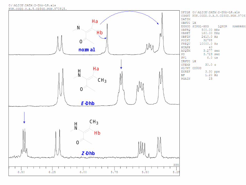
normal
E-Dhb
Z-Dhb
HN
CH3
Ha
NHb
Ha
HN
Hb
CH3
O
O
O
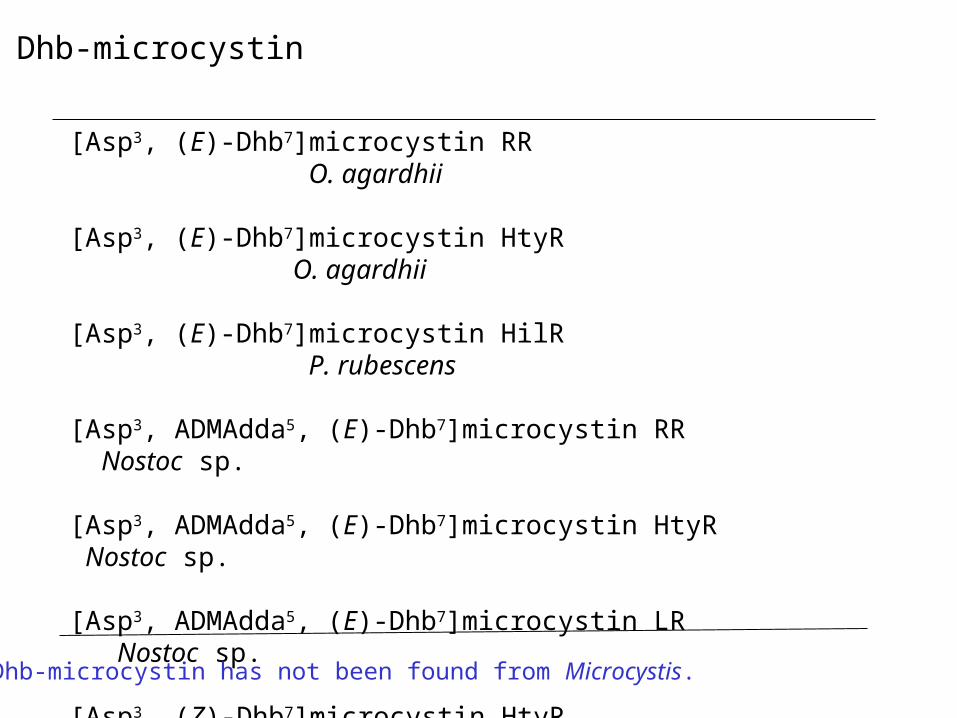
[Asp3, (E)-Dhb7]microcystin RR O. agardhii
[Asp3, (E)-Dhb7]microcystin HtyR O. agardhii
[Asp3, (E)-Dhb7]microcystin HilR P. rubescens
[Asp3, ADMAdda5, (E)-Dhb7]microcystin RR Nostoc sp.
[Asp3, ADMAdda5, (E)-Dhb7]microcystin HtyR Nostoc sp.
[Asp3, ADMAdda5, (E)-Dhb7]microcystin LR Nostoc sp.
[Asp3, (Z)-Dhb7]microcystin HtyR O. agardhii
[Asp3, (Z)-Dhb7]microcystin LR O. agardhii
Dhb-microcystin
Dhb-microcystin has not been found from Microcystis.
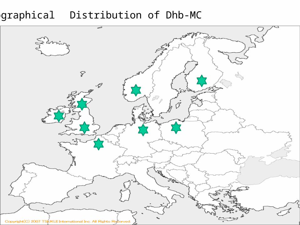
世界地図: http://www.sekaichizu.jp/
Geographical Distribution of Dhb-MC
OCH3
CH3CH3
HNN CH2
O
HOOC
NH
H3CO NH
HN
O
O
H3C
HN
O
CH3
CH3
HN
COOH
CH3
CH3
O
O
NHH2N
HN
microcystin
CH
CH3
Dhb-
H3C
CH3
1
2
34
5
67
L-amino acid
L-amino acid
D-Ala
D-Leu
D-Glu
(Z) and (E)
Adda
D-[D-Leu1] microcystin LR
[D-Leu1]microcystin LR foundfrom Microcystis aeruginosaisolated from Brazil and Canada.
Summary
1)Dhb-microcystins were found from cells of O. agardhii, P. rubescens,and Nostoc sp. isolated from North European countries.
2)[D-Leu1]microcystin was isolated from cells of M. aeruginosa collected from Brazil and Canada, but has not been found any other area.
Problems Are toxin genes in cyanobacteria localized ? Do migratory birds carry cyanobacteria ?
SELECTIVE CONTROL OF TOXIC MICROCYSTIS WATERBLOOMS USING LYSINE AND
MALONIC ACID
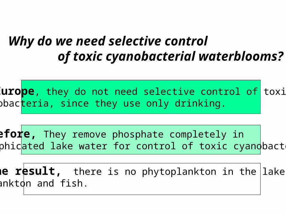
Why do we need selective control of toxic cyanobacterial waterblooms?
In Europe, they do not need selective control of toxiccyanobacteria, since they use only drinking.
Therefore, They remove phosphate completely in eutrophicated lake water for control of toxic cyanobacteria.
As the result, there is no phytoplankton in the lake, alsoZooplankton and fish.

In Asia, inland residents have utilized freshwater fish for a major protein source.
Therefore, aquaculture is important, and eutrophication is necessary for growth of phytoplankton, zooplankton and fish, but exclusion of toxic cyanobacteria is necessary for human health and aquaculture.
As the result, we need to develop a method of selective control of toxic cyanobacteria.
As the opposite situation of the European,
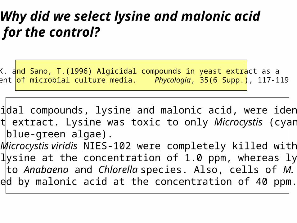
Kaya, K. and Sano, T.(1996) Algicidal compounds in yeast extract as a component of microbial culture media. Phycologia, 35(6 Supp.), 117-119
Two algicidal compounds, lysine and malonic acid, were identified from Yeast extract. Lysine was toxic to only Microcystis (cyano-Bacteria, blue-green algae).Cells of Microcystis viridis NIES-102 were completely killed within 48 hr by lysine at the concentration of 1.0 ppm, whereas lysine was non-toxic to Anabaena and Chlorella species. Also, cells of M. viridis were killed by malonic acid at the concentration of 40 ppm.
Why did we select lysine and malonic acid for the control?
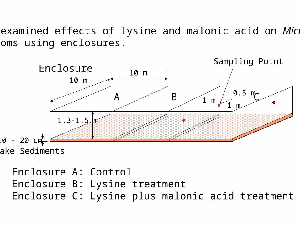
We examined effects of lysine and malonic acid on MicrocystisBlooms using enclosures.
Enclosure 10 m10 m
1.3-1.5 m
10 – 20 cm
A B C
Lake Sediments
Enclosure A: ControlEnclosure B: Lysine treatmentEnclosure C: Lysine plus malonic acid treatment
1 m1 m
0.5 m
Sampling Point
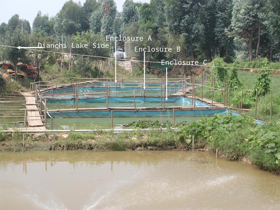
Enclosure A
Enclosure B
Enclosure C
Dianchi Lake Side
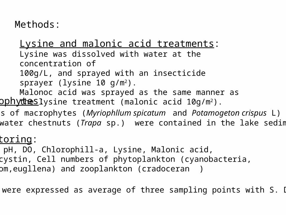
Macrophytes: Seeds of macrophytes (Myriophllum spicatum and Potamogeton crispus L) and water chestnuts (Trapa sp.) were contained in the lake sediment.
Monitoring:Water pH, DO, Chlorophill-a, Lysine, Malonic acid, Microcystin, Cell numbers of phytoplankton (cyanobacteria, dyatom,eugllena) and zooplankton (cradoceran )
Results were expressed as average of three sampling points with S. D.
Lysine and malonic acid treatments:Lysine was dissolved with water at the concentration of 100g/L, and sprayed with an insecticide sprayer (lysine 10 g/m2).Malonoc acid was sprayed as the same manner as the lysine treatment (malonic acid 10g/m2).
Methods:
1
B
3
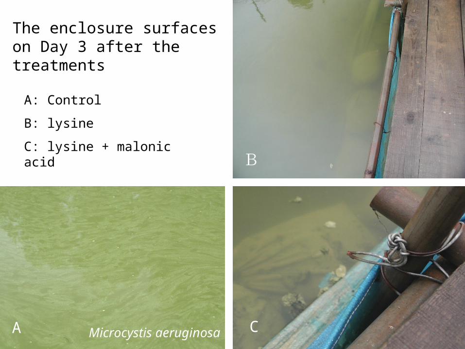
The enclosure surfaces on Day 3 after the treatments
A: Control
B: lysine
C: lysine + malonic acid
A CMicrocystis aeruginosa
6
8
10L
ys
in
e
[ m
g/L]
10 7 140
Days after Treatment
2
4
3 5
Enclosure C
Enclosure B
Enclosure A
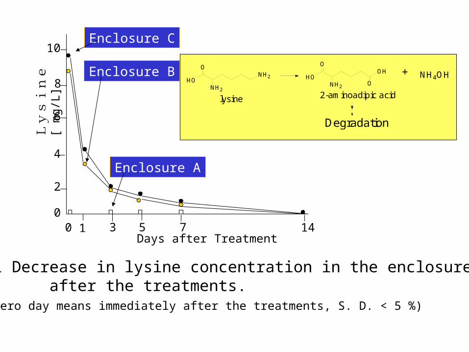
Fig.1 Decrease in lysine concentration in the enclosures after the treatments. (The zero day means immediately after the treatments, S. D. < 5 %)
HO
O
NH2
NH2 HO
O
NH2
OH
O
+ NH4OH
Degradation
lysine 2-aminoadipic acid

8.0
8.5
9.0
9.5
pH
7.5
20 7 14 21 280
Days after Treatment
Enclosure A
Enclosure B
Enclosure C
Fig.2 Changes in pH in the enclosures after the treatments
(S. D. < 5%; *p < 0.05 )
**
60
80
100
120
Ch
lo
ro
ph
yl
l
a
[g
/L]
20 7 14 21 280
20
40
Days after Treatment
Enclosure A
Enclosure B
Enclosure C
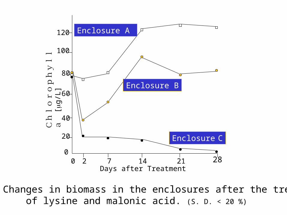
Fig.3 Changes in biomass in the enclosures after the treatment of lysine and malonic acid. (S. D. < 20 %)
28
12
16
20
24
Ce
lls
[
x
10
6/L
]
20 7 14 210
Days after Treatment
4
8
Total phytoplankton
Cyanobacteria
Diatom
Euglena
Enclosure A
12
16
20
24
20 7 14 21 280
Days after Treatment
4
8
Total phytoplankton
Cyanobacteria
Diatom
Euglena
Enclosure B
12
16
20
24
20 7 14 21 280
Days after Treatment
4
8
Total phytoplankton
Cyanobacteria
Diatom
Euglena
Enclosure C
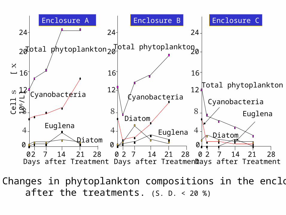
Fig.4 Changes in phytoplankton compositions in the enclosures after the treatments. (S. D. < 20 %)
Cla
do
cera
n
[in
div
idu
als
/L]
20 7 14 21 280
Days after Treatment
Enclosure AEnclosure B
Enclosure C
100
200
300
400
500
600
700
800
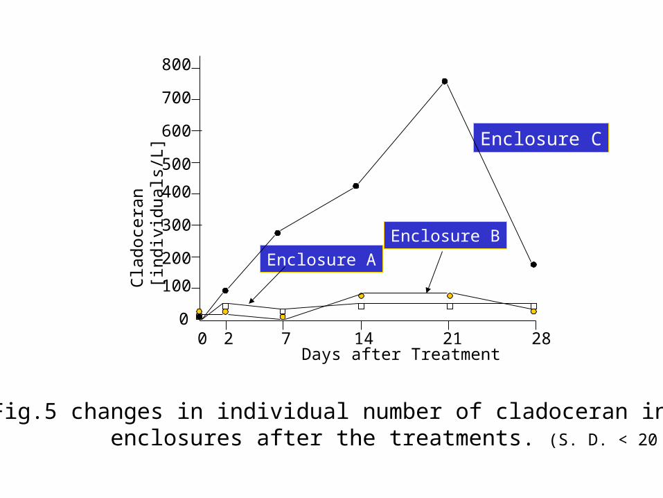
Fig.5 changes in individual number of cladoceran in the enclosures after the treatments. (S. D. < 20 %)
9
12
15
To
tal
mic
rocy
stin
[m
g/L
]
20 7 14 21 280
Days after Treatment
3
6
Enclosure A
Enclosure B
Enclosure C
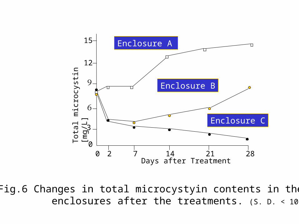
Fig.6 Changes in total microcystyin contents in the enclosures after the treatments. (S. D. < 10 %)
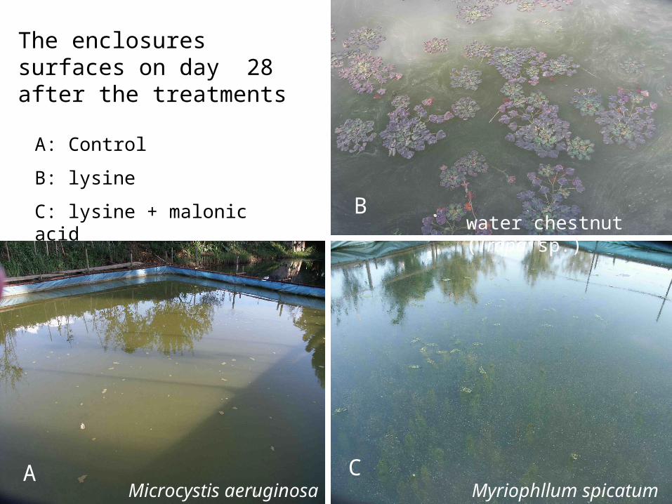
The enclosures surfaces on day 28 after the treatments
A: Control
B: lysine
C: lysine + malonic acid
A
B
CMyriophllum spicatum
water chestnut (Trapa sp.)
Microcystis aeruginosa

Conclusion:
The treatment with lysine plus malonic acid is an effective method for the control of toxic Microcystis blooms. The ecological and water qualitative changes derived from the treatment suggested that the incorporation cycles of nitrogen and phosphorus in eutrophicated water were switched from toxic cyanobacteria (Microcystis) to non-toxic macrophytes.
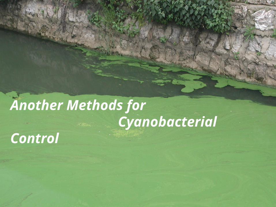
Another Methods for Cyanobacterial Control
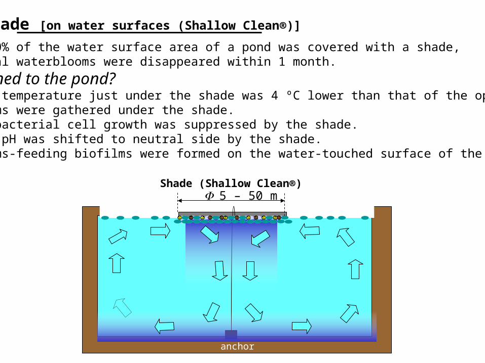
Shade [on water surfaces (Shallow Clean®)]
When about 30% of the water surface area of a pond was covered with a shade, cyanobacterial waterblooms were disappeared within 1 month.
What happened to the pond?1) The water temperature just under the shade was 4 ºC lower than that of the open surface.2) Waterblooms were gathered under the shade.3) The cyanobacterial cell growth was suppressed by the shade.4) The water pH was shifted to neutral side by the shade.5) Waterblooms-feeding biofilms were formed on the water-touched surface of the shade.
5 – 50 m
anchor
Shade (Shallow Clean®)
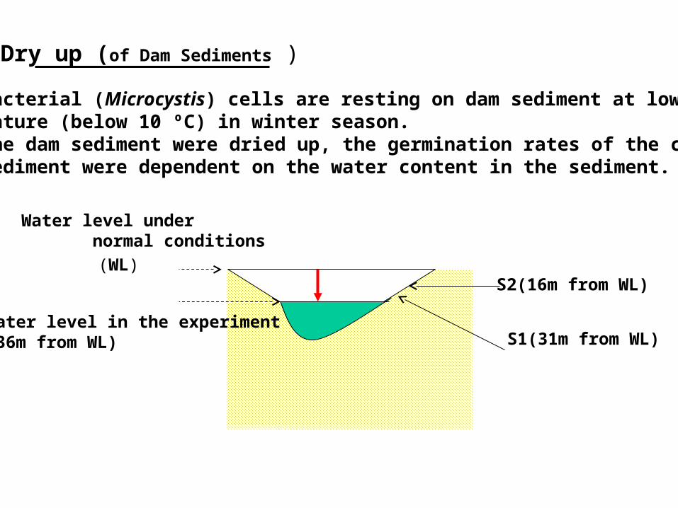
Dry up (of Dam Sediments )
Cyanobacterial (Microcystis) cells are resting on dam sediment at low watertemperature (below 10 ºC) in winter season.When the dam sediment were dried up, the germination rates of the cells on the sediment were dependent on the water content in the sediment.
Water level under normal conditions
S1(31m from WL)
S2(16m from WL)
Water level in the experiment(36m from WL)
(WL)

Water content in the sediment (WCS) and germination rate (GR)
100
0 7 14 21 30Day after dry up
% o
f W
CS
(bar
s)100
75
50
25
0 0
25
50
75
100
% o
f G
R (
clos
ed c
ircl
es)S1
S2
S1
S2
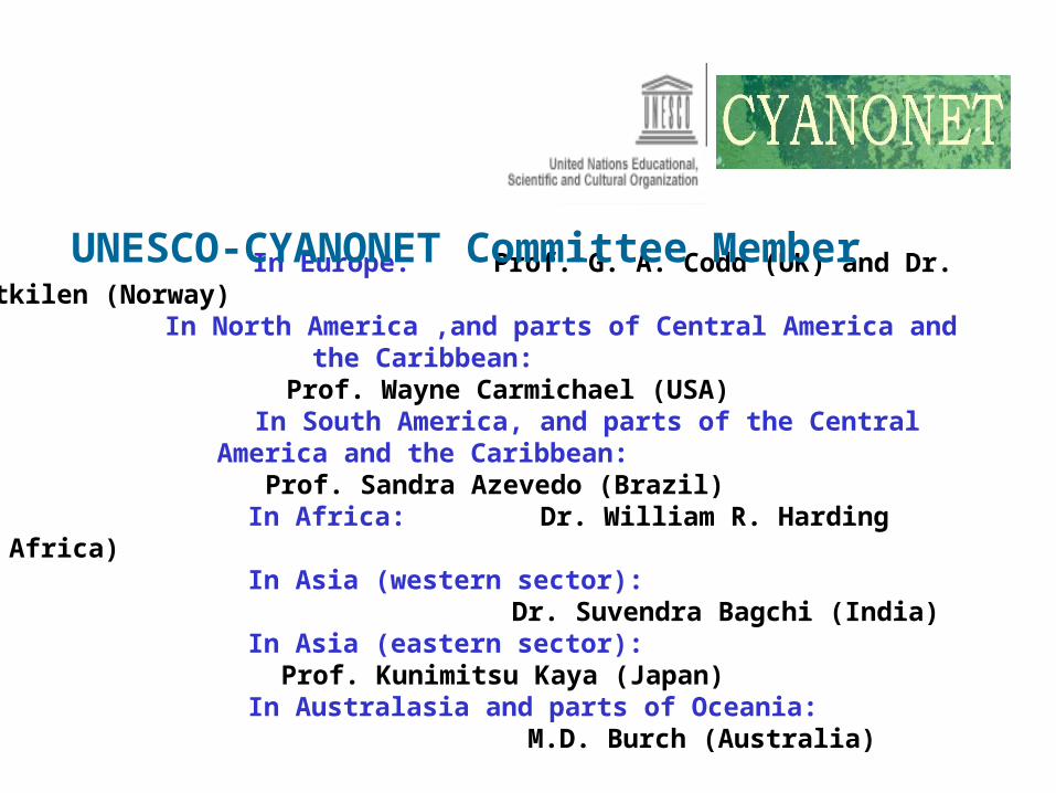
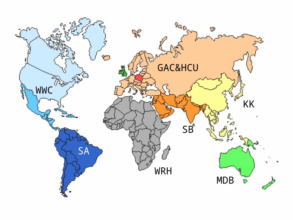
In Europe: Prof. G. A. Codd (UK) and Dr. H.C. Utkilen (Norway)
In North America ,and parts of Central America and the Caribbean: Prof. Wayne Carmichael (USA)
In South America, and parts of the Central America and the Caribbean:
Prof. Sandra Azevedo (Brazil) In Africa: Dr. William R. Harding (South Africa) In Asia (western sector): Dr. Suvendra Bagchi (India) In Asia (eastern sector):
Prof. Kunimitsu Kaya (Japan) In Australasia and parts of Oceania: M.D. Burch (Australia)
UNESCO-CYANONET Committee Member
WWC
KK
GAC&HCU
MDB
SB
WRH
SA

Thank youfor your attention!