keratinocyte differentiation is stimulated by activators of the nuclear hormone receptor pparα
TRANSCRIPT

Keratinocyte Differentiation is Stimulated by Activators of theNuclear Hormone Receptor PPARα
Karen Hanley,*§ Yan Jiang,*§ Shan Shan He,*§ Mark Friedman,*§ Peter M. Elias,*§ Daniel D. Bikle,*†§¶Mary L. Williams,*‡§ and Kenneth R. Feingold*†§¶Departments of *Dermatology, †Medicine, and ‡Pediatrics, University of California, San Francisco, California, U.S.A.; and the §Dermatology and¶Medical Services Department of Veterans Affairs Medical Center, San Francisco, California, U.S.A.
Peroxisome proliferator activated receptors (PPAR)belong to the superfamily of nuclear hormone receptorsthat heterodimerize with the retinoid X receptor andregulate transcription of several genes involved in lipidmetabolism and adipocyte differentiation. Because ofthe role of 1,25-dihydroxyvitamin D3 and retinoic acidworking through similar receptors (the vitamin D recep-tor and retinoic acid receptor, respectively) on keratino-cyte differentiation, we have examined the effects ofactivators of PPARα on keratinocyte differentiation. Therate of cornified envelope formation was increased 3-foldin keratinocytes maintained in low calcium (0.03 mM)and incubated in the presence of clofibric acid, a potentPPARα activator. Involucrin, a cornified envelope pre-cursor, and the cross-linking enzyme transglutaminase,were increased at both the message level (2–7-fold) and theprotein level (4–12-fold) by clofibric acid. Furthermore,
TThe epidermis is comprised of morphologically distinctcell layers, with each strata representative of a progress-ively more advanced stage of differentiation. As cellsmigrate upward and leave the proliferative basal layer,they lose their capacity to divide and begin to sequentially
express specific proteins (reviewed in Fuchs, 1990): first, keratins K1and K10, and then proteins such as filaggrin, involucrin (INV), loricrin,and transglutaminase (TG’ase) 1, specific to later stages of differentiation(Dale et al, 1985; Eichner et al, 1986; Mehrel et al, 1990; Robinsonet al, 1996). During the final stages of differentiation, TG’ase generatesε- (γ-glutamyl) lysine bonds that cross-link substrates such as loricrinand INV to form the cornified cell envelope, a chemically andmechanically resistant layer of insoluble protein located under theplasma membrane (Rice and Green, 1979; Thacher, 1989; Fuchs,1990). The ultimate consequence of epidermal differentiation is theformation of the stratum corneum, layers of terminally differentiatedcornified cells in the outermost epidermis, responsible for the barrierproperties of the skin (Downing, 1992).
Manuscript received June 23, 1997; revised October 25, 1997; accepted forpublication December 1, 1997.
Reprint requests to: Dr. Kenneth R. Feingold, Metabolism Service (111F),Department of Veterans Affairs Medical Center, 4150 Clement Street, SanFrancisco, CA 94121.
Abbreviations: CE, cornified envelope; INV, involucrin; PPAR, peroxisomeproliferator-activated receptor; RXR, retinoid X-activated receptor; TG’ase,transglutaminase.
0022-202X/98/$10.50 · Copyright © 1998 by The Society for Investigative Dermatology, Inc.
368
physiologic doses of the fatty acids oleic acid, linoleicacid, and eicosatetraynoic acid, which are also activatorsof PPARα, also induced involucrin and transglutaminaseprotein and mRNA. In contrast, the PPARγ ligand prosta-glandin J2 had no effect on protein or mRNA levels ofinvolucrin or transglutaminase. Levels of involucrin andtransglutaminase mRNA and protein were induced byclofibric acid in keratinocytes incubated in 1.2 mMcalcium, a concentration which by itself induces keratino-cyte differentiation. Finally, PPARα activators inhibitDNA synthesis. This study demonstrates that PPARαactivators, including putative endogenous ligands suchas fatty acids, induce differentiation and inhibit prolifera-tion in keratinocytes, and suggests a regulatory role forthe PPARα in epidermal homeostasis. Key word: peroxisomeproliferator-activated receptor. J Invest Dermatol 110:368–375,1998
Although the specific cellular signals that initiate the changes ingene expression regulating the different steps of differentiation remainunknown, calcium has been identified as a key regulator of terminaldifferentiation. Keratinocytes incubated in medium containing lowlevels of calcium (0.03 mM) remain in a proliferative, relativelyundifferentiated state, whereas keratinocytes maintained in high extra-cellular calcium concentrations (1.2 mM) express increased levels ofdifferentiation specific proteins, such as INV and TG’ase (Henningset al, 1989; Fuchs, 1990; Dlugosz and Yuspa, 1994). These proteinsare regulated both at the transcriptional and at the translational levels,and their expression leads to increased rates of cornified envelope (CE)formation (Hennings et al, 1989; Pillai and Bikle, 1991; Dlugosz andYuspa, 1994; Gibson et al, 1996).
Other factors such as 1,25-dihydroxyvitamin D3, the hormonallyactive form of vitamin D, have well known effects on keratinocyteproliferation and differentiation. 1,25-dihydroxyvitamin D3 is a potentinducer of keratinocyte differentiation, and, like calcium, acceleratesthe appearance of several CE precursor proteins, and inhibits cellularproliferation (Pillai and Bikle, 1991; Itin et al, 1994). Retinoids andthyroid hormone also affect keratinocyte differentiation, although, incontrast to vitamin D, they inhibit in vitro differentiation andcornification and suppress the expression of differentiation-associatedkeratins (Isseroff et al, 1989; Blumberg et al, 1992; Fisher and Voorhees,1996). Though their effects are diverse, vitamin D3, retinoids, andthyroid hormone are all ligands for nuclear hormone receptors in thesame superfamily, transcription factors that are heterodimeric partnersof the retinoid X-activated receptor (RXR) (Mangelsdorf et al, 1995;Kastner et al, 1995). Recent studies in utero suggest that one or more

VOL. 110, NO. 4 APRIL 1998 PPARα ACTIVATORS INDUCE KERATINOCYTE DIFFERENTIATION 369
members of this receptor superfamily may be key regulators of epidermaldevelopment. Overexpression of a truncated inactive RARα in fetalmurine epidermis results in aberrant cutaneous development, mostlikely due to subversion of the RXR and its partners (Saitou et al,1995; Imakado et al, 1995).
The peroxisome proliferator-activated receptors (PPAR) are alsomembers of the nuclear hormone receptor superfamily. The PPARsubtypes α, δ, and γ are differentially activated by several fatty acids,arachidonic acid metabolites, and hypolipidemic drugs such as clofibrate(Keller et al, 1993; Yu et al, 1995; Forman et al, 1995). Whereas PPARγis involved in the regulation of adipocyte differentiation, the α isoformplays a role in lipid metabolism and homeostasis in the liver and kidney(reviewed in Schoonjans et al, 1996), tissues that, like epidermis, exhibithigh rates of fatty acid synthesis. We have recently demonstrated thatPPARα activators accelerate the development of the stratum corneumand the epidermal permeability barrier in full thickness fetal rat skinexplants incubated in serum- and hormone-free media (Hanley et al,1997a). In this study, we sought to determine whether activators ofPPARα might also alter the rate of human keratinocyte differentiationin vitro. We report here that activators of PPARα are potent stimulatorsof keratinocyte differentiation and inhibitors of proliferation.
MATERIALS AND METHODS
Cell culture Human epidermis was isolated from newborn foreskins byincubation in dispase, and a suspension of keratinocytes was obtained byincubation in ethylenediaminotetraacetic acid and subsequent trypsinization, asdescribed by Gibson et al (1996). The cells were plated in serum-free keratinocytegrowth medium (KGM; Clonetics, San Diego, CA), containing 0.07 mMcalcium, and grown to 60–80% confluence. Cells were then passaged twiceand plated in KGM containing 0.07 mM calcium for 48 h, switched to KGMcontaining 0.03 mM calcium for 48 h, then treated with either clofibric acid,fatty acids, or vehicle [, 0.1% dimethylsulfoxide (DMSO) or , 0.1% ethanolor , 0.1% ethanol with 0.5% bovine serum albumin], or 1.2 mM calciumwith or without clofibric acid, for 0–48 h. Fatty acids and clofibric acid (Sigma,St. Louis, MO) were solubilized in ethanol or DMSO. 15-Deoxy-∆12,14-prostaglandin J2 (Prostaglandin J2) (Cayman, Ann Arbor, MI) was solubilizedin DMSO. Fatty acids were complexed with fatty acid-free bovine serumalbumin (Sigma) immediately prior to the initiation of each experiment, filteredthrough a 0.2 µm filter, and added to the culture medium giving a final bovineserum albumin concentration of 100 µM.
RNA isolation Total RNA was isolated according to the method ofChomczynski et al (1987). Briefly, cells were lyzed in 4 M guanidine isothiocyan-ate/25 mM sodium citrate/0.5% sarcosyl/0.1 M β-mercaptoethanol, extractedwith phenol-chloroform, and precipitated with ethanol. RNA pellets weresuspended in sterile, diethyl pyrocarbonate-treated water, and RNA wasquantitated by absorbance at 260 nm using the 260/280 nm ratio as an indexof purity.
Northern blotting and cDNA probes Total RNA (15 µg per sample) wassize fractionated through a 1% agarose gel containing 2.2 M formaldehyde, asdescribed in Gibson et al (1996). RNA integrity was visualized followingacridine orange staining of the electrophoresed gel. The RNA was transferredto a nylon membrane that was subsequently baked at 80°C for 2 h. Blots werethen hybridized with the appropriate 32P-labeled probe [P1–2 for INV, a giftfrom Dr. Howard Greene (Harvard, Cambridge, MA), and hTG for keratinocytetransglutaminase 1, a gift from Dr. Robert Rice (UCD, Davis)] overnight at65°C. Washes were then performed in a solution containing 0.1% sodiumcitrate/chloride buffer and 0.1% sodium dodecyl sulfate for 20 min at roomtemperature, followed by a 20 min wash at 65°C. Autoradiography wasperformed at –70°C. Blots were probed with β-actin to confirm equal loading.
Involucrin and transglutaminase protein levels Protein concentrationwas assessed by protein electrophoresis and western blotting, as described inGibson et al (1996). Briefly, cells were lyzed in 2% sodium dodecyl sulfate andthe lysate sonicated. Following protein determination [Bicinchoninic acidprotein assay (Pierce, Rockford, IL)], equal amounts of protein were electro-phoresed on 7.5% polyacrylamide gels and electroblotted onto membranes.INV protein was detected by incubation overnight at 4°C with a polyclonalrabbit anti-human INV antibody (a gift from Dr. Robert Rice) (1:1000 inTween 20, 0.5%, and nonfat milk, 5%). Densitometry was used to quantitateINV specific bands on the autoradiograms.
TG’ase protein expression was measured as for INV expression, withmodifications as described in Gibson et al (1996). Briefly, stacking, sample, andrunning buffers all contained 4 M urea, and following electrophoresis, gels were
washed in 4 M urea, 25 mM Tris-HCl, pH 7.4, 75 mM NaCl, 0.1 mMdithiothreitol, and 2 mM ethylenediamine tetraacetic acid for 90 min prior toelectroblotting, allowing TG’ase to be detected by BC.1 primary antibody(Thacher, 1989).
CE formation The rate of CE formation was determined as described inGibson et al (1996). Briefly, cells were incubated with 35S-methionine/cysteinefor 48 h, washed with phosphate-buffered saline, and harvested into 1.1 ml of2% sodium dodecyl sulfate. Aliquots were reserved for protein determinations.The remaining cell lysate (1 ml) was sonicated briefly (10 s). One milliliter of4% sodium dodecyl sulfate/4 mM dithiothreitol was then added, and themixture was heated to .95oC for 30 min. Following cooling, sodium dodecylsulfate/dithiothreitol-insoluble material was collected on filter discs, washedwith 0.5% sodium dodecyl sulfate/0.5% dithiothreitol, and quantitated byscintillation spectrophotometry. To determine total protein synthesized duringthe 48 h of 35S labeling, a reserved aliquot of the cell lysate (taken prior toheating) was precipitated with an equal volume of 2% bovine serum albuminand 1 ml of 10% (wt/vol) trichloroacetic acid on ice for 30 min, and 35S-labeled precipitated protein was collected onto filter disks, washed with 5%trichloroacetic acid, and counted in a scintillation counter. Total protein wasdetermined by the method of Bradford (BioRad, Hercules, CA).
DNA content and synthesis DNA content was measured by the methodof Labarca et al (1980). Briefly, cells were incubated for 2 h with bisbenzimidazole(Hoechst 33258, 1 mg per ml) and fluorescence measured with a Perkin-Elmerfluorimeter, excitation 356, emission 458. A standard curve was made usingdilutions of calf thymus DNA.
The rate of DNA synthesis was determined by measuring [3H]thymidineincorporation into cellular DNA after 4 h of incubation with 2 µCi [3H]thymid-ine (110 Ci methyl per mmol, 1929-[3H]thymidine, Amersham, ArlingtonHeights, IL) per ml media. The radioactivity in the washed trichloroacetic acidprecipitate was quantitated by scintillation spectroscopy.
Statistics Statistical analysis was performed using Student’s t test.
RESULTS
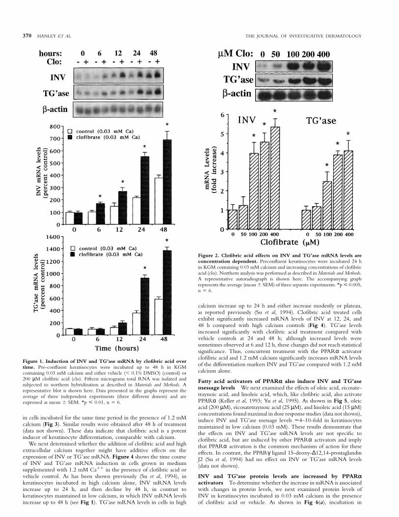
The PPARα activator clofibric acid induces INV and TG’asemRNA levels Keratinocytes maintained in low levels of extracellularcalcium remain in a proliferative state, expressing relatively low messagelevels of differentiation-specific proteins such as INV and TG’ase (Pillaiand Bikle, 1991; Dlugosz and Yuspa, 1994; Gibson et al, 1996). Todetermine whether activators of PPARα induce differentiation incultured keratinocytes, we first measured INV and TG’ase mRNAlevels by northern analysis in cultures maintained in low calcium(0.03 mM), incubated either in the presence of the PPARα activatorclofibric acid or in the vehicle, over a 48 h time period. As shown inFig 1, levels of INV and TG’ase mRNA in both clofibric acid treatedand vehicle treated cells progressively increase throughout the 48 hperiod; however, clofibric acid treated cells exhibit significantlyincreased levels of INV compared with vehicle treated cells at all timepoints examined (6, 12, 24, 48 h), and significantly increased TG’asemRNA levels at 24 and 48 h (Fig 1), with a maximal effect on INVat 12–24 h and on TG’ase at 24–48 h (2–7-fold increase over controlsin three separate experiments). β-actin mRNA levels remained constantover time, and were unaffected by clofibric acid treatment. Theseresults indicate that the PPARα activator clofibric acid induces twodifferent markers of keratinocyte differentiation.
To determine whether the effect by clofibric acid on INV andTG’ase message levels was concentration dependent, we next measuredINV and TG’ase message levels in keratinocytes maintained in 0.03 mMcalcium and incubated in the presence of increasing concentrations ofclofibric acid. As shown in Fig 2, levels of INV and TG’ase mRNAwere significantly increased by 100 µM clofibric acid. Effects ofclofibric acid on mRNA levels of both INV and TG’ase were maximalwith 200–400 µM. Incubation in 500 µM clofibric acid resulted intoxic effects.
Increased calcium is well known to stimulate keratinocyte differenti-ation (Hennings et al, 1989; Fuchs, 1990; Pillai and Bikle, 1991;Dlugosz and Yuspa, 1994; Itin et al, 1994; Gibson et al, 1996).Therefore, we next compared clofibric acid induced message levels ofINV and TG’ase with levels induced by 1.2 mM extracellular calcium.mRNA levels of both INV and TG’ase in clofibric acid treated cellsmaintained in 0.03 mM calcium for 24 h are similar to levels observed
370 HANLEY ET AL THE JOURNAL OF INVESTIGATIVE DERMATOLOGY
Figure 1. Induction of INV and TG’ase mRNA by clofibric acid overtime. Pre-confluent keratinocytes were incubated up to 48 h in KGMcontaining 0.03 mM calcium and either vehicle (, 0.1% DMSO) (control) or200 µM clofibric acid (clo). Fifteen micrograms total RNA was isolated andsubjected to northern hybridization as described in Materials and Methods. Arepresentative blot is shown here. Data presented in the graphs represent theaverage of three independent experiments (three different donors) and areexpressed as mean 6 SEM. *p ø 0.01, n 5 6.
in cells incubated for the same time period in the presence of 1.2 mMcalcium (Fig 3). Similar results were obtained after 48 h of treatment(data not shown). These data indicate that clofibric acid is a potentinducer of keratinocyte differentiation, comparable with calcium.
We next determined whether the addition of clofibric acid and highextracellular calcium together might have additive effects on theexpression of INV or TG’ase mRNA. Figure 4 shows the time courseof INV and TG’ase mRNA induction in cells grown in mediumsupplemented with 1.2 mM Ca11 in the presence of clofibric acid orvehicle control. As has been shown previously (Su et al, 1994), inkeratinocytes incubated in high calcium alone, INV mRNA levelsincrease up to 24 h, and then decline by 48 h, in contrast tokeratinocytes maintained in low calcium, in which INV mRNA levelsincrease up to 48 h (see Fig 1). TG’ase mRNA levels in cells in high
Figure 2. Clofibric acid effects on INV and TG’ase mRNA levels areconcentration dependent. Preconfluent keratinocytes were incubated 24 hin KGM containing 0.03 mM calcium and increasing concentrations of clofibricacid (clo). Northern analysis was performed as described in Materials and Methods.A representative autoradiograph is shown here. The accompanying graphrepresents the average (mean 6 SEM) of three separate experiments. *p ø 0.005,n 5 6.
calcium increase up to 24 h and either increase modestly or plateau,as reported previously (Su et al, 1994). Clofibric acid treated cellsexhibit significantly increased mRNA levels of INV at 12, 24, and48 h compared with high calcium controls (Fig 4). TG’ase levelsincreased significantly with clofibric acid treatment compared withvehicle controls at 24 and 48 h; although increased levels weresometimes observed at 6 and 12 h, these changes did not reach statisticalsignificance. Thus, concurrent treatment with the PPARα activatorclofibric acid and 1.2 mM calcium significantly increases mRNA levelsof the differentiation markers INV and TG’ase compared with 1.2 mMcalcium alone.
Fatty acid activators of PPARα also induce INV and TG’asemessage levels We next examined the effects of oleic acid, eicosate-traynoic acid, and linoleic acid, which, like clofibric acid, also activatePPARα (Keller et al, 1993; Yu et al, 1995). As shown in Fig 5, oleicacid (200 µM), eicosatetraynoic acid (25 µM), and linoleic acid (15 µM)concentrations found maximal in dose response studies (data not shown),induce INV and TG’ase message levels µ4–10-fold in keratinocytesmaintained in low calcium (0.03 mM). These results demonstrate thatthe effects on INV and TG’ase mRNA levels are not specific toclofibric acid, but are induced by other PPARα activators and implythat PPARα activation is the common mechanism of action for theseeffects. In contrast, the PPARγ ligand 15-deoxy-∆12,14-prostaglandinJ2 (Su et al, 1994) had no effect on INV or TG’ase mRNA levels(data not shown).
INV and TG’ase protein levels are increased by PPARαactivators To determine whether the increase in mRNA is associatedwith changes in protein levels, we next examined protein levels ofINV in keratinocytes incubated in 0.03 mM calcium in the presenceof clofibric acid or vehicle. As shown in Fig 6(a), incubation in

VOL. 110, NO. 4 APRIL 1998 PPARα ACTIVATORS INDUCE KERATINOCYTE DIFFERENTIATION 371
Figure 3. Clofibric acid induced levels of INV and TG’ase mRNA aresimilar to calcium induced levels. Preconfluent keratinocytes were incubatedin the presence of 1.2 mM calcium, 0.03 mM calcium, or 0.03 mM calciumplus 200 µM clofibric acid (clo) for 24 h and northern analysis performed asdescribed in Fig. 1. Graphs represent the average of data from two independentexperiments (two different donors). A representative autoradiograph is shownhere. *p ø 0.01, n 5 4.
increasing concentrations of clofibric acid for 24 h resulted in aprogressive increase in INV protein levels (8–12-fold induction by200–400 µM clofibric acid versus vehicle controls in three separateexperiments). The magnitude of the increase was similar to thatproduced by incubation in high (1.2 mM) extracellular calcium(Fig 6a). Similar results were observed after 48 h of treatment (datanot shown). Clofibric acid also induced a further increase in INVprotein in keratinocytes incubated in high calcium (Fig 6b), i.e., levelsof INV protein in keratinocytes incubated in 1.2 mM calcium plusclofibric acid were 2-fold higher than in keratinocytes incubated in1.2 mM calcium plus vehicle only.
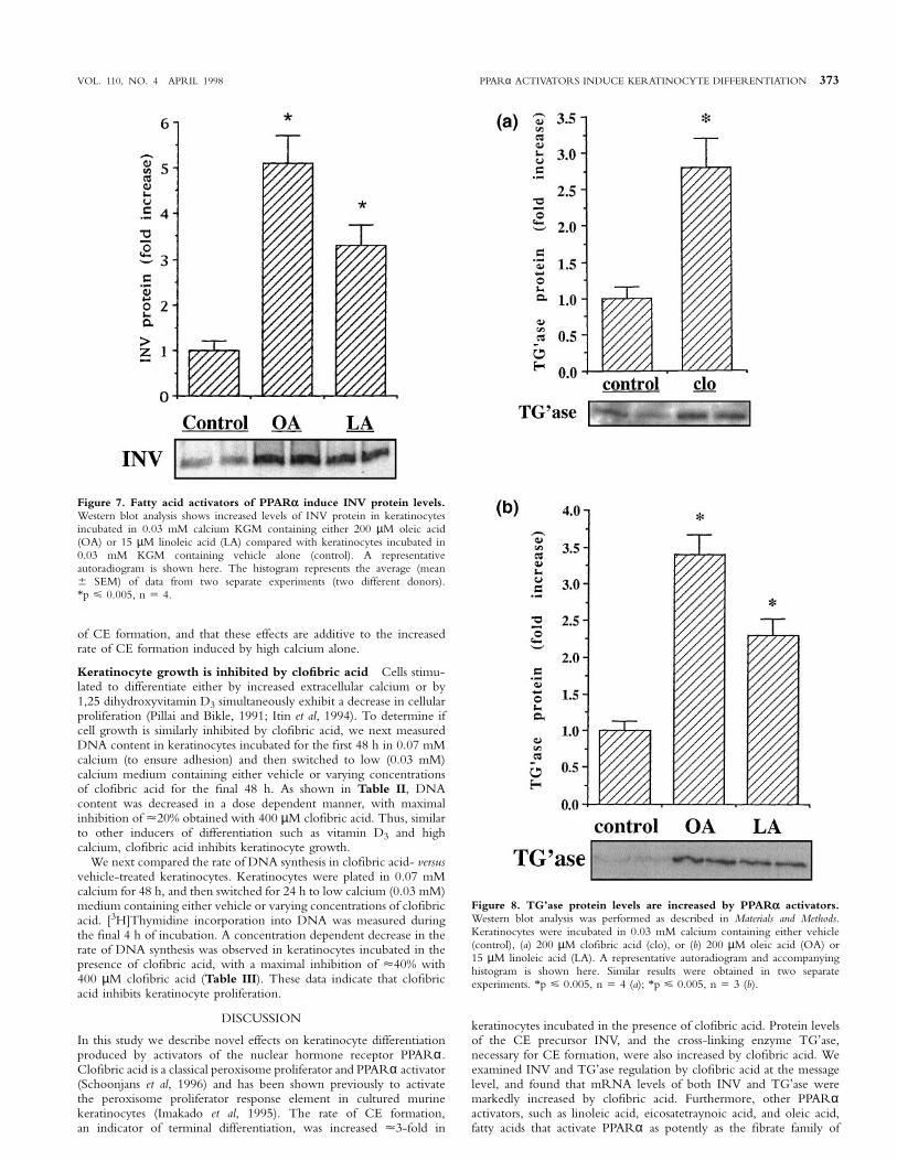
Finally, to determine whether other PPARα activators also increasedINV protein levels, we incubated keratinocytes in 0.03 mM calciumin the absence or presence of oleic or linoleic acid, fatty acid activatorsof the PPARα. As shown in Fig 7, both oleic acid (200 µM) and
Figure 4. Time course of INV and TG’ase mRNA induction by clofibricacid in keratinocytes incubated in 1.2 mM calcium. Pre-confluentkeratinocytes were incubated up to 48 h in KGM containing 1.2 mM calciumand either vehicle (, 0.1% DMSO) (control) or 200 µM clofibric acid. Fifteenmicrograms total RNA was isolated and subjected to northern hybridization asdescribed in Materials and Methods. A representative autoradiograph is shownhere. Data presented in the graphs represent the average of three independentexperiments (three different donors) and are expressed as mean 6 SEM.*p ø 0.005, n 5 6.
linoleic acid (15 µM) also induced INV protein expression. In contrast,the PPARγ ligand 15-deoxy-∆12,14-prostaglandin J2 (Su et al, 1994)had no effect on INV protein expression (data not shown).
As has been shown previously by others, TG’ase protein levels werevery low in keratinocytes maintained in 0.03 mM calcium (Gibsonet al, 1996). TG’ase protein levels were increased µ3-fold in cellsincubated in the presence of clofibric acid for 48 h (Fig 8a), andincreased µ3-fold and 2-fold by oleic acid and linoleic acid, respectively(Fig 8b). These results indicate that PPARα activators induce proteinlevels of both INV and TG’ase.
Clofibric acid increases the rate of CE formation An additional,late marker of epidermal differentiation, CE formation, was assessed incells treated for 48 h with clofibric acid in low (0.03 mM) and in high(1.2 mM) calcium. The results of these studies are shown in Table Iand are expressed as the percentage of the vehicle control value in

372 HANLEY ET AL THE JOURNAL OF INVESTIGATIVE DERMATOLOGY
Figure 5. mRNA levels of INV and TG’ase are induced by fatty acidactivators of PPARα. Preconfluent keratinocytes were incubated for 24 h inKGM containing 0.03 mM calcium and either vehicle alone (control) or200 µM oleic acid (OA), 25 µM eicosatetraynoic acid (ETYA), or 15 µMlinoleic acid (LA). For comparison cells incubated in 1.2 mM calcium aloneare included. Total RNA was isolated and subjected to northern analysis asdescribed in Materials and Methods. The average (mean 6 SEM) of data fromtwo separate experiments (two different donors) is shown here. *p ø 0.005,n 5 4.
0.03 calcium (equals 100%). A concentration dependent increase inCE formation is seen with increasing concentrations of clofibric acid,approaching a 4-fold increase (p , 0.01) with 400 mM clofibric acidin 0.03 mM calcium. Incubation in high calcium alone resulted in analmost 5-fold increase in the rate of CE formation. Yet clofibric acidtogether with high calcium produced a modest but significant increaseover high calcium alone (59% increase, Table I). Compared with0.03 mM calcium vehicle, clofibric acid and 1.2 mM calcium resultedin an approximate 6-fold increase in CE synthesis (Table I). Thesedata indicate that the PPARα activator clofibric acid increases the rate
Figure 6. Protein levels of INV are increased by clofibric acid.Preconfluent keratinocytes were incubated for 24 h in KGM containing (a)0.03 mM calcium and either vehicle alone or 100, 200, or 400 µM clofibricacid (clo), or 1.2 mM calcium and vehicle alone; or (b) 1.2 mM calcium andeither vehicle or 200 µM clofibric acid (Clo). Western analysis was performedand autoradiograms were quantitated as described in Materials and Methods.Representative autoradiograms are shown here. The histogram represents theaverage (mean 6 SEM) of data from two separate experiments (two differentdonors). *p ø 0.01, n 5 4 (a); *p ø 0.01, n 5 6 (b).
VOL. 110, NO. 4 APRIL 1998 PPARα ACTIVATORS INDUCE KERATINOCYTE DIFFERENTIATION 373
Figure 7. Fatty acid activators of PPARα induce INV protein levels.Western blot analysis shows increased levels of INV protein in keratinocytesincubated in 0.03 mM calcium KGM containing either 200 µM oleic acid(OA) or 15 µM linoleic acid (LA) compared with keratinocytes incubated in0.03 mM KGM containing vehicle alone (control). A representativeautoradiogram is shown here. The histogram represents the average (mean6 SEM) of data from two separate experiments (two different donors).*p ø 0.005, n 5 4.
of CE formation, and that these effects are additive to the increasedrate of CE formation induced by high calcium alone.
Keratinocyte growth is inhibited by clofibric acid Cells stimu-lated to differentiate either by increased extracellular calcium or by1,25 dihydroxyvitamin D3 simultaneously exhibit a decrease in cellularproliferation (Pillai and Bikle, 1991; Itin et al, 1994). To determine ifcell growth is similarly inhibited by clofibric acid, we next measuredDNA content in keratinocytes incubated for the first 48 h in 0.07 mMcalcium (to ensure adhesion) and then switched to low (0.03 mM)calcium medium containing either vehicle or varying concentrationsof clofibric acid for the final 48 h. As shown in Table II, DNAcontent was decreased in a dose dependent manner, with maximalinhibition of µ20% obtained with 400 µM clofibric acid. Thus, similarto other inducers of differentiation such as vitamin D3 and highcalcium, clofibric acid inhibits keratinocyte growth.
We next compared the rate of DNA synthesis in clofibric acid- versusvehicle-treated keratinocytes. Keratinocytes were plated in 0.07 mMcalcium for 48 h, and then switched for 24 h to low calcium (0.03 mM)medium containing either vehicle or varying concentrations of clofibricacid. [3H]Thymidine incorporation into DNA was measured duringthe final 4 h of incubation. A concentration dependent decrease in therate of DNA synthesis was observed in keratinocytes incubated in thepresence of clofibric acid, with a maximal inhibition of µ40% with400 µM clofibric acid (Table III). These data indicate that clofibricacid inhibits keratinocyte proliferation.
DISCUSSION
In this study we describe novel effects on keratinocyte differentiationproduced by activators of the nuclear hormone receptor PPARα.Clofibric acid is a classical peroxisome proliferator and PPARα activator(Schoonjans et al, 1996) and has been shown previously to activatethe peroxisome proliferator response element in cultured murinekeratinocytes (Imakado et al, 1995). The rate of CE formation,an indicator of terminal differentiation, was increased µ3-fold in
Figure 8. TG’ase protein levels are increased by PPARα activators.Western blot analysis was performed as described in Materials and Methods.Keratinocytes were incubated in 0.03 mM calcium containing either vehicle(control), (a) 200 µM clofibric acid (clo), or (b) 200 µM oleic acid (OA) or15 µM linoleic acid (LA). A representative autoradiogram and accompanyinghistogram is shown here. Similar results were obtained in two separateexperiments. *p ø 0.005, n 5 4 (a); *p ø 0.005, n 5 3 (b).
keratinocytes incubated in the presence of clofibric acid. Protein levelsof the CE precursor INV, and the cross-linking enzyme TG’ase,necessary for CE formation, were also increased by clofibric acid. Weexamined INV and TG’ase regulation by clofibric acid at the messagelevel, and found that mRNA levels of both INV and TG’ase weremarkedly increased by clofibric acid. Furthermore, other PPARαactivators, such as linoleic acid, eicosatetraynoic acid, and oleic acid,fatty acids that activate PPARα as potently as the fibrate family of

374 HANLEY ET AL THE JOURNAL OF INVESTIGATIVE DERMATOLOGY
Table I. The rate of CE formation is increased by clofibratea
Clofibrate concentration Rate of CE formation(µM) (% of control)
0.03 mM calcium 0 100.0 6 12.550 110.5 6 14.9
200 338.3 6 26.8*400 386.8 6 28.7*
1.2 mM calcium 0 100.0 6 15.850 94.9 6 10.3
200 131.3 6 12.2**400 158.9 6 15.5**
aPreconfluent keratinocytes were treated with vehicle or with varying concentrations ofclofibrate for 48 h and 35S-methionine incorporation into detergent and reducing agentinsoluble protein was measured as described in Materials and Methods (average value forcontrols, 2192 cpms per mg protein; 400 µM clofibrate-treated, 8549). Results presentedhere are expressed as the mean 6 SEM of two independent experiments. *p , 0.01compared with 0.03 calcium controls; ** p , 0.01 compared with 1.2 calcium controls;n 5 6.
Table II. Clofibrate inhibits keratinocyte growtha
Clofibrate DNA content after 48 h(µM) (µg per dish) Significance
0 15.5 6 0.9 –50 14.9 6 1.5 not significant
200 13.8 6 1.1 p , 0.05400 12.1 6 1.3 p , 0.01
aPreconfluent keratinocytes were incubated in 0.03 mM calcium for 48 h in the presenceof vehicle or varying concentrations of clofibrate. DNA content was determined asdescribed in Materials and Methods. Results are presented as mean 6 SEM, n 5 3.
Table III. The rate of DNA synthesis is inhibited byclofibratea
Clofibrate cpms per µg DNA(µM) (per cent control) Significance
0 100.0 6 11.3 –50 98.2 6 8.5 not significant
200 79.3 6 10.1 p , 0.01400 61.3 6 8.4 p , 0.005
aPreconfluent keratinocytes were incubated in 0.03 mM calcium for 24 h in the presenceof vehicle or varying concentrations of clofibrate and [3H]thymidine incorporation wasmeasured over the final 4 h of incubation as described in Materials and Methods (100%control 5 5390 cpms per µg DNA). Results are presented as mean 6 SEM, n 5 4.
peroxisomal proliferators (Keller et al, 1993; Yu et al, 1995; Schoonjanset al, 1996), also significantly increased INV and TG’ase proteinand message levels, whereas the PPARγ ligand 15-Deoxy-∆12,14-prostaglandin J2 had no effect. In addition, stimulatory effects on INVand TG’ase protein and message levels by clofibric acid were alsoobserved in keratinocytes incubated in the presence of physiologiccalcium levels (1.2 mM). Together, these results strongly suggest thatPPARα plays a regulatory role in keratinocyte differentiation.
The nuclear hormone receptor PPARα is a member of the thyroid/vitamin D/retinoid superfamily of transcription factors that requireheterodimerization with the RXR for optimal DNA binding(Mangelsdorf and Evans, 1995; Kastner et al, 1995; Mangelsdorfet al, 1995). Whereas effects by PPARα activators on keratinocytedifferentiation have not been previously reported, it is well knownthat activators/ligands of other nuclear hormone receptors in thissuperfamily regulate keratinocyte growth and differentiation. Forexample, 1,25-dihydroxyvitamin D3 increases intracellular calcium,promotes terminal differentiation, and inhibits growth of keratinocytes,evidenced by increased levels of INV and TG’ase mRNA, and increasedCE formation (Smith et al, 1986; Pillai and Bikle, 1991; Su et al, 1994).Retinoic acid and its analogs, as well as thyroid hormone, also havewell-known effects on the terminal differentiation of keratinocytes,but in contrast to PPARα activators and 1,25-dihydroxyvitamin
D3, they inhibit cornification in cultured keratinocytes and suppressexpression of differentiation specific keratins via their respective nuclearreceptors (Isseroff et al, 1989; Blumberg et al, 1992; Fisher and Voorhees,1996). Thus, although their effects are diverse, activators/ligands forthis nuclear hormone receptor superfamily are important modulatorsof keratinocyte growth and differentiation. The mechanisms by whichthe PPARα activators affect keratinocyte differentiation remain unclear.For example, it remains to be determined whether PPARα activatorsregulate INV and TG’ase gene expression directly.
Studies in vivo also suggest an important role for this superfamily ofnuclear receptors in epidermal formation. Overexpression of an abnor-mal RARα in fetal murine basal epidermal cells results in thin, fragileskin (Saitou et al, 1995), whereas overexpression in suprabasal cellsresults in a severe skin phenotype characterized by the absence ofmature lamellar membrane structures in the stratum corneum interstices,leading to impaired barrier function (Imakado et al, 1995). Because themutated RARα retains its ability to dimerize and bind DNA, butthe resultant complex is transcriptionally inactive, signaling pathwaysdependent on RXR heterodimeric complexes are perturbed. Thus,the superfamily of nuclear hormone receptors may play an importantrole early in the initial differentiation of keratinocytes and later in theregulation of lipid metabolism crucial to the formation of a functionalepidermal barrier.
Our laboratory has been studying fetal epidermal barrier and stratumcorneum development. Several hormones that interact with receptorsin the nuclear hormone receptor superfamily, such as glucocorticoids,estrogen, and testosterone, affect epidermal ontogenesis when given inpharmacologic doses (Aszterbaum et al, 1993; Hanley et al, 1996a, b).Furthermore, physiologic concentrations of thyroid hormone, as wellas activators of the PPARα and farnesoid X-activated receptor, acceler-ate the development of the stratum corneum and the epidermal barrierin fetal skin explants (Hanley et al, 1996a, 1997a); however, it is notclear which one or more of these factors are required for the finalsteps of stratum corneum formation, because development proceedsnormally in fetal rat skin explants incubated in hormone- and serum-free medium (Hanley et al, 1996a). Moreover, fetal mice madehypothyroid due to a mutation in the TSH receptor, though exhibitinga temporary delay in epidermal development prenatally, are born withapparently normal epidermal barriers (Hanley et al, 1997b). Similarly,PPARα may not be essential in embryonic development, as micelacking PPARα are born viable and fertile, with no obvious cutaneousdefects (Lee et al, 1995). These data suggest functional redundancyamong RXR partners, with a single defect resulting in a mildphenotype. In contrast, overexpression of a mutated RARα that canperturb effective dimerization of this family of hormone receptorsresults in a dramatic phenotype and demonstrates the central role ofRXR in epidermal development.
PPARα is abundant in liver and kidney where it plays a role in theregulation of fatty acid metabolism (reviewed in Schoonjans et al,1996). It is activated by molecules that induce peroxisomal proliferation,such as the synthetic agents clofibric acid and WY14,643, used for thetherapy of hyperlipidemia, as well as by several free fatty acids (Kelleret al, 1993; Yu et al, 1995; Schoonjans et al, 1996). Because all layersof the epidermis actively synthesize fatty acids (Feingold, 1991),significant endogenous levels of these fatty acids are likely to be presentthroughout the epidermis. Indeed, the fatty acid concentrations thatinduced keratinocyte differentiation in this study (15 µM linoleic acidor 200 µM oleic acid) are consistent with physiologic concentrationranges for fatty acids in the epidermis (0.3–2 mM) and with fatty acidconcentrations that activate PPARα in other cell types (Long, 1970;Lampe et al, 1983; Keller et al, 1993; Yu et al, 1995). Thus, endogenousfatty acids are potential candidates as key local regulators of keratinocytedifferentiation. It is of interest that glucocorticoids, which accelerateepidermal barrier development in vivo and in vitro in fetal rats, increasethe expression of PPARα mRNA in other cell types (Lemberger et al,1994; Steineger et al, 1994). This raises the possibility that theeffect of glucocorticoids on epidermal formation may be mediatedvia PPARα.
In summary, we demonstrate here that activators of the nuclearhormone receptor PPARα, a member of the vitamin D receptor/

VOL. 110, NO. 4 APRIL 1998 PPARα ACTIVATORS INDUCE KERATINOCYTE DIFFERENTIATION 375
RAR/RXR superfamily of transcription factors, induce keratinocytedifferentiation. These studies, taken together with evidence that PPARαactivators accelerate fetal epidermal maturation, suggest a physiologicrole for PPARα in epidermal homeostasis.
The authors appreciate the technical assistance provided by Sally Pennypacker (U.C.Cell Culture Facility, V.A. Medical Center), and the technical advice provided by Dr.David Gibson. This study was supported by NIH grants HD 29706, AR 39639,AR29706, and P039448 and the Medical Research Service, Department of VeteransAffairs Medical Center.
REFERENCES
Aszterbaum M, Feingold KR, Menon GM, Williams ML: Glucocorticoids accelerate fetalmaturation of the epidermal permeability barrier in the rat. J Clin Invest 91:2703–2708, 1993
Blumberg M, Connolly DM, Freedberg IM: Regulation of keratin gene expression: therole of nuclear receptors for retinoic acid, thyroid hormone, and vitamin D3. JInvest Dermatol 98:42S–49S, 1992
Chomczynski P, Sacchi N: Single-step method of RNA isolation by acid guanidiniumthiocyanate-phenol-chloroform extraction. Anal Biochem 162:156–159, 1987
Dale BA, Resing KA, Landsdale-Eccles JD: A keratin filament associated protein. Ann NYAcad Sci 455:330–342, 1985
Dlugosz AA, Yuspa SH: Protein kinase C regulates keratinocyte transglutaminase (TGK)gene expression in cultured primary mouse epidermal keratinocytes induced toterminally differentiate by calcium. J Invest Dermatol 102:409–414, 1994
Downing DT: Lipid and protein structures in the permeability barrier of mammalianepidermis. J Lipid Res 33:301–313, 1992
Eichner R, Sun TT, Aebi U: The role of keratin subfamilies and keratin pairs in theformation of human epidermal intermediate filaments. J Cell Biol 102:1767–1777, 1986
Feingold KR: The regulation and role of epidermal lipid synthesis. Adv Lipid Res 24:57–82, 1991
Fisher GJ, Voorhees JJ: Molecular mechanisms of retinoid actions in skin. FASEB J10:1002–1013, 1996
Forman BM, Tontonoz P, Chen J, Brun RP, Spiegelman BM, Evans RM: 15-Deoxy-e12,14-prostaglandin J2 is a ligand for the adipocyte determination factor PPARγ.Cell 83:803–812, 1995
Fuchs E: Epidermal differentiation: the bare essentials. J Cell Biol 111:2807–2814, 1990Gibson D, Ratnam AV, Bikle DD: Evidence for separate control mechanisms at the
message, protein, and enzyme activation levels for transglutaminase during calcium-induced differentiation of normal and transformed human keratinocytes. J InvestDermatol 106:154–161, 1996
Hanley K, Rassner U, Elias PM, Williams ML, Feingold KR: Epidermal barrier ontogenesis:maturation in serum-free media and acceleration by glucocorticoids and thyroidhormone but not selected growth factors. J Invest Dermatol 106:404–411, 1996a
Hanley K, Rassner U, Jiang Y, et al: Hormonal basis for the gender difference in epidermalbarrier formation in the fetal rat. Acceleration by estrogen and delay by androgen.J Clin Invest 97:2576–2584, 1996b
Hanley K, Jiang Y, Crumrine D, et al: Activators of the nuclear hormone receptors PPARαand FXR accelerate the development of the fetal epidermal permeability barrier. JClin Invest 100:705–712, 1997a
Hanley K, Devaskar UP, Hicks SJ, et al: Hypothyroidism delays fetal stratum corneumdevelopment in mice. Pediatr Res 42:610–614, 1997b
Hennings H, Kruszewski FH, Yuspa SH, Tucker RW: Intracellular calcium alterations in
response to increased external calcium in normal and neoplastic keratinocytes.Carcinogenesis 4:777–780, 1989
Imakado S, Bickenbach JR, Bundman D, et al: Targeting expression of a dominant-negative retinoic acid receptor mutant in the epidermis of transgenic mice results inloss of barrier function. Genes Dev 9:317–329, 1995
Isseroff RR, Chun KT, Rosenberg RM: Triiodothyronine alters the cornification ofcultured human keratinocytes. Br J Dermatol 120:503–510, 1989
Itin PH, Pittelkow MR, Kumar R: Effects of vitamin D metabolites on proliferation anddifferentiation of cultured human epidermal keratinocytes grown in serum-free ordefined culture medium. Endocrinology 135:1793–1798, 1994
Kastner P, Mark M, Chambon P: Nonsteroid nuclear receptors: what are genetic studiestelling us about their role in real life? Cell 83:841–850, 1995
Keller H, Dreyer C, Medin J, Mahfoudi A, Ozato K, Wahli W: Fatty acids and retinoidscontrol lipid metabolism through activation of peroxisome proliferator-activatedreceptor-retinoid X receptor heterodimers. Proc Natl Acad Sci 90:2160–2164, 1993
Labarca C, Paigen K: A simple, rapid, and sensitive DNA assay procedure. Anal Biochem102:344–352, 1980
Lampe MA, Williams ML, Elias PM: Human epidermal lipids: characterization andmodulations during differentiation. J Lipid Res 24:131–140, 1983
Lee SS-T, Pineau T, Drago J, et al: Targeted disruption of the alpha isoform of theperoxisome proliferator-activated receptor gene in mice results in abolishment ofthe pleiotropic effects of peroxisome proliferators. Molec Cell Biol 15:3012–3022, 1995
Lemberger T, Staels B, Saladin R, Desvergne B, Auwerx J, Wahli W: Regulation of theperoxisome proliferator-activated receptor α gene by glucocorticoids. J Biol Chem269:24527–24530, 1994
Long VJW: Variations in lipid composition at different depths in the cow snout epidermis.J Invest Dermatol 55:269–273, 1970
Mangelsdorf DJ, Evans RM: The RXR heterodimers and orphan receptors. Cell 83:841–850, 1995
Mangelsdorf DJ, Thummel C, Beato M, et al: The nuclear receptor superfamily: thesecond decade. Cell 83:835–839, 1995
Mehrel T, Hohl D, Rothnagel JA, et al: Identification of a major keratinocyte cell envelopeprotein, loricrin. Cell 61:1103–1112, 1990
Pillai S, Bikle DD: Role of intracellular-free calcium in the cornified envelope formationof keratinocytes: differences in the mode of action of extracellular calcium and 1,25dihydroxyvitamin D3. J Cell Physiol 146:94–100, 1991
Rice RH, Green H: Presence in human epidermal cells of a soluble protein precursor ofthe cross-linked envelope: activation of the cross-linking by calcium ions. Cell18:681–694, 1979
Robinson NA, LaCelle PT, Eckert RL: Involucrin is a covalently cross-linked constituentof highly purified epidermal corneocytes: evidence for a common pattern ofinvolucrin cross-linking in vivo and in vitro. J Invest Dermatol 107:101–107, 1996
Saitou M, Sugal S, Tanaka T, Shimouchi K, Fuchs E, Narumiya S, Kakizuka A: Inhibitionof skin development by targeted expression of a dominant-negative retinoic acidreceptor. Nature 374:159–162, 1995
Schoonjans K, Staels B, Auwerx J: Role of the peroxisome proliferator-activated receptor(PPAR) in mediating the effects of fibrates and fatty acids on gene expression. JLipid Res 37:907–925, 1996
Smith EL, Walworth NC, Holick MF: Effect of 1,25-dihydroxyvitamin D3 on themorphologic and biochemical differentiation of cultured human epidermalkeratinocytes grown in serum-free conditions. J Invest Dermatol 86:709–714, 1986
Steineger HH, Sorensen HN, Tugwood JD, Skevede S, Spydevold O, Gautvik KM:Dexamethasone and insulin demonstrate marked and opposite regulation of thesteady-state mRNA level of the peroxisomal proliferator-activated receptor (PPAR)in hepatic cells. Euro J Biochem 225:967–974, 1994
Su M-J, Bikle DD, Mancianti ML, Pillai S: 1,25-dihydroxyvitamin D3 potentiates thekeratinocyte response to calcium. J Biol Chem 269:14723–14729, 1994
Thacher SM: Purification of keratinocyte transglutaminase and its expression duringsquamous differentiation. J Invest Dermatol 92:578–584, 1989
Yu K, Bayona W, Kallen CB, et al: Differential activation of peroxisome proliferator-activated receptors by eicosanoids. J Biol Chem 270:23975–23983, 1995