kaido siimon layout-1 - dspace.ut.eedspace.ut.ee/bitstream/handle/10062/52394/siimon_kaido.pdf ·...
TRANSCRIPT
Tartu 2016
ISSN 2228-0928ISBN 978-9949-77-177-6
1
DISSERTATIONES SCIENTIAE
MATERIALIS TARTUENSIS
16
KAIDO SIIMON
Electrospun gelatincross-linked by glucose

DISSERTATIONES SCIENTIAE MATERIALIS UNIVERSITATIS TARTUENSIS
16
DISSERTATIONES SCIENTIAE MATERIALIS UNIVERSITATIS TARTUENSIS
16
KAIDO SIIMON
Electrospun gelatin cross-linked by glucose
The study was carried out at the Institute of Physics, Faculty of Science and Technology, University of Tartu. The dissertation was admitted on 10th of June, 2016 in partial fulfilment of the requirements for the degree of Doctor of Philosophy in Materials Science, and was allowed for defence by the Scientific Council on Materials Science of the Faculty of Science and Technology of the University of Tartu. Supervisors: Martin Järvekülg, Institute of Physics, University of Tartu,
Estonia
Uno Mäeorg, Institute of Chemistry, University of Tartu, Estonia
Ivo Laidmäe, Institute of Pharmacy, University of Tartu, Estonia
Opponent: Dr. Tekla Tammelin, VTT Technical Research Centre of
Finland, Finland Defence: 30th of August 2016, University of Tartu (Tartu, Estonia) The study was financially supported by the European Union through the Euro-pean Regional Development Fund via projects “Carbon Nanotube Reinforced Electrospun Nano-fibres and Yarns“ (3.2.1101.12-0018), “SmaCell“ (3.2.1101.12-0017), Centre of Excellence “Mesosystems: Theory and Applica-tions” (3.2.0101.11-0029), Estonian Science Foundation grant IUT2-25, Esto-nian Nanotechnology Competence Centre (EU29996).
ISSN 2228-0928 ISBN 978-9949-77-177-6 (print) ISBN 978-9949-77-178-3 (pdf) Copyright: Kaido Siimon, 2016 University of Tartu Press www.tyk.ee

5
CONTENTS PUBLICATIONS ........................................................................................ 6 1. PREFACE .............................................................................................. 7 2. INTRODUCTION .................................................................................. 9
2.1 Electrospun gelatin ......................................................................... 9 2.2 Cross-linking of electrospun gelatin ............................................... 9 2.3 Mechanical properties of electrospun gelatin ................................. 10
3. MATERIALS AND METHODS ........................................................... 13 3.1 Preparation of fibrous gelatin ......................................................... 13 3.2 Scanning electron microscopy (SEM) ............................................ 14 3.3 Fourier transform infrared microscopy (FTIR) .............................. 14 3.4 Characterization of mechanical properties of electrospun
gelatin ............................................................................................. 15 3.4.1 Tensile test ........................................................................... 15 3.4.2 Modelling ............................................................................. 15
4. RESULTS .............................................................................................. 19 4.1 Preparation of electrospun gelatin .................................................. 19 4.2 Effect of concentration of the electrospinning solution on
fibre diameter .................................................................................. 20 4.3 Effect of aging of the electrospinning solution on fibre
diameter .......................................................................................... 22 4.4 The extent of cross-linking ............................................................. 22
4.4.1 FTIR analysis ....................................................................... 22 4.4.2 Monitoring the cross-linking process ................................... 24 4.4.3 Determination of the extent of cross-linking ........................ 25
4.5 Mechanical properties of electrospun gelatin ................................. 27 4.5.1 Tensile test and simulation ................................................... 27 4.5.2 Effect of glucose content ...................................................... 28 4.5.3 Effect of aging of the electrospinning solution .................... 29
4.6 Effect of alum on electrospun gelatin cross-linked by glucose ............................................................................................ 29 4.6.1 Preparation of alum-containing fabrics ................................ 29 4.6.2 Mechanical properties of alum-containing fabrics ............... 30
5. DISCUSSION ........................................................................................ 33 6. APPLICATIONS – TISSUE ENGINEERING ...................................... 35 7. CONCLUSIONS .................................................................................... 36 SUMMARY IN ESTONIAN ...................................................................... 37 ACKNOWLEDGEMENTS ........................................................................ 39 REFERENCES ............................................................................................ 40 PUBLICATIONS ........................................................................................ 47 CURRICULUM VITAE ............................................................................. 79 ELULOOKIRJELDUS ................................................................................ 81

6
PUBLICATIONS The current dissertation is based on the following publications:
I. Kaido Siimon, Paula Reemann, Annika Põder, Martin Pook, Triin
Kangur, Külli Kingo, Viljar Jaks, Uno Mäeorg, Martin Järvekülg. Effect of glucose content on thermally cross-linked fibrous gelatin scaf-folds for tissue engineering. Materials Science & Engineering C 2014; 42: 538–545. doi: 10.1016/j.msec.2014.05.075
II. Kaido Siimon, Hele Siimon, Martin Järvekülg. Mechanical character-
ization of electrospun gelatin scaffolds cross-linked by glucose. Journal of Materials Science: Materials in Medicine 2015; 26:37. doi: 10.1007/s10856-014-5375-1
III. Kaido Siimon, Karol Mõisavald, Hele Siimon, Martin Järvekülg.
Increasing mechanical strength of electrospun gelatin nanofibres by the addition of aluminium potassium sulphate. Journal of Applied Polymer Science 2015; 42431. doi: 10.1002/app.42431
The author of the current dissertation, closely cooperating with co-authors of the above-mentioned articles, was responsible for designing the study, con-ducting experiments, analysing results, writing the manuscripts (except for sec-tions 2.5–2.9 and 3.4 in publication I), and serves as the corresponding author for all the above-mentioned publications. The articles are referred to using Roman numerals in the text.
7
1. PREFACE The initial purpose of our work concerning electrospun gelatin was developing transplantable tissue engineering scaffolds for skin regeneration, focusing on using natural, non-toxic substances and simple, cost-effective methods. Such a scaffold is basically a thin mesh, which is used to cover a wound to promote healing. The first step of the scaffold preparation process is preparing an artificial extracellular matrix on which the cells can be cultured. Gelatin was chosen to be the main component of the artificial extracellular matrix, because it is a protein derived by hydrolysis from collagen, a naturally occurring protein most abundant in connective tissue such as skin [1, 2]. However, gelatin is cheaper than collagen and easier to process. Both collagen and gelatin have been considered as materials for tissue engineering scaffolds and share many advantages (biocompatibility, biodegradability, non-toxicity) as well as disadvantages (nonhomogeneous composition and inconsistent physicochemical properties) [3].
Natural extracellular matrix consists mainly of bundled fibres. In order to mimic this structure to a certain degree, fibrous gelatin scaffolds were prepared using the electrospinning technique. However, electrospun gelatin is water-soluble and must be cross-linked to make it insoluble, since cells are cultured in an aqueous environment. Previously, mostly expensive and more or less toxic chemical cross-linking agents have been used to treat fibrous gelatin. The current dissertation is based on the discovery that electrospun gelatin fibres can be thermally cross-linked by glucose. The discovery brought up several questions – how much glucose should be added in order to achieve desired extent of cross-linking, how does cross-linking by glucose affect the properties of the material, can the fabrics be used for cell culture applications, etc. The abovementioned articles provide answers to many of such questions. The results are summarised in the current dissertation, providing additional information where necessary.
A large part of the dissertation is dedicated to mechanical properties of elec-trospun gelatin cross-linked by glucose because of the following reasons. Firstly, mechanical behaviour plays a key role in many applications, including biomedicine, filtering, protective clothing and fabrics. However, low mechani-cal strength has been limiting the use of fibrous gelatin in these applications, and it is vital to find ways to increase the strength of the material. Secondly, mechanical properties of biopolymer fibres are influenced by numerous factors including the quality of the used gelatin, fabrication process parameters, solvent used to prepare the electrospinning solution, humidity, fibre size etc. As a result of all these different factors influencing the properties of electrospun materials, the reported elastic moduli of electrospun gelatin treated with miscellaneous cross-linkers vary to a very large degree and it is hard to reproduce the results presented in literature and compare it to one’s own results, which creates a lot of confusion. Therefore, it is vital to study mechanical behaviour of the material

8
in great detail in order to be able to repeatedly prepare fibres with similar mechanical properties.
The main purpose of the study was to prepare and characterize electrospun gelatin cross-linked thermally by glucose, and to develop strategies for gaining control over its mechanical properties.

9
2. INTRODUCTION 2.1 Electrospun gelatin
Interest in natural fibrous materials is continuously increasing. The fibrous structure that can be achieved by electrospinning is important in various appli-cations in the biomedical field, cosmetics, energy storage, filtration, protective clothing, sensors, space applications, optoelectronics, and in composite materi-als [4, 5, 6]. Electrospun gelatin has been studied mainly for biomedical applications, including wound healing, tissue engineering blood vessels, heart valves, bone, cornea, regeneration of periodontal tissue, nerve tissue, controlled release [7, 8, 9, 10, 11, 12, 13, 14].
Several methods can be used to fabricate fibrous materials, including phase separation, self-assembly, surface patterning, interfacial complexation, wet-, bio-, melt- and microfluidic spinning and electrospinning, the latter being a well-established, simple, cost-effective and flexible technique that takes advantage of electrostatic forces to produce polymer fibres [15, 16, 17, 18]. Gelatin can be electrospun from solvents like 1,1,1,3,3,3-hexafluoro-2-propa-nol, 2,2,2-trifluoroethanol and solvent systems like acetic acid/ethyl ace-tate/water or aqueous formic acid [19, 20, 21, 22]. It has been reported that gelatin can be electrospun from water above 36°C [23, 24]. At room tempera-ture, gelatin can be electrospun from acetic acid or its water solutions, as it is done in the current work, but fibres are produced only when the concentration of acetic acid is sufficiently high [25].
Gelatin has been co-electrospun with several other polymers including elas-tin, chitosan, silk, hyaluronic acid, fibrinogen, poly(ε-caprolactone), poly-urethane, poly(vinyl alcohol) [26, 27, 28, 29, 30, 31, 32, 33]. Several additives can be used to alter the properties of electrospun gelatin, including nano-particles, herbal extracts, salts, and so forth [III, 34, 35, 36].
In conclusion, electrospun gelatin can easily be prepared using only natural substances and easy, cost-effective methods. Such fibrous gelatin is also rela-tively cheap, extremely biocompatible and biodegradable material. However, electrospun gelatin is water-soluble and mechanically weak. Overcoming these disadvantages is discussed in the following chapters.
2.2 Cross-linking of electrospun gelatin
Cross-linking is a widely used strategy for making electrospun gelatin insoluble and to increase its strength. Cross-linking decreases solubility by creating chemical bonds between polymer chains and has been shown to increase mechanical strength of electrospun gelatin [37, 38]. Cross-linking of gelatin fibres can be carried out using chemical, physical, enzymatic [39] or combined methods. Physical cross-linking methods include thermal, plasma and ultra-violet treatment [40, 41, 42, 43, 44]. However, physical cross-linking methods

10
generally result in a low degree of cross-linking, which can be sufficient to make gelatin insoluble in water, but the use of chemical cross-linking agents is necessary to achieve considerable changes in mechanical properties of the mate-rial. Additionally, chemically cross-linked electrospun gelatin has been reported to be better suited for tissue engineering applications compared to thermally cross-linked electrospun gelatin [45]. Numerous cross-linking agents have been used to treat electrospun gelatin, including glutaraldehyde, terephthalaldehyde, dextran aldehyde, genipin, oxidized sucrose, caffeic acid, tannic acid, procyani-dine, proanthocyanidin [46, 47, 48, 49, 50, 51, 52, 53]. However, most chemical cross-linkers are costly and potentially toxic. Cytotoxicity of a cross-linking agent depends on the type and amount of cross-linking agent and the cross-linking process [54]. The latter is often quite hard to control, which makes the use of potentially toxic cross-linking agents more or less dangerous in bio-medical applications.
Glucose has many advantages (biocompatibility, non-toxicity, cost-effec-tiveness) compared to widely used chemical cross-linking agents such as dial-dehydes. However, glucose alone has been reported to be a relatively ineffective cross-linking agent [55], mostly because the formation of glucose cross-links requires glucose molecules to be in their linear form [56], but at normal condi-tions glucose molecules are in the closed ring form [57]. Nevertheless, it is known that amino acids and reducing sugars react at high temperatures through the Maillard reaction pathway and that the presence of sugars can alter the con-formation and interactions of proteins, having an effect on the cross-linking process [58, 59, 60]. As described in the current work [I–III], electrospun gela-tin can be thermally cross-linked by glucose simply by placing the glucose-containing as-electrospun fabric into a preheated oven at about 170°C, ensuring proper cross-linking and preserving the nanofibrous structure of the fabric.
2.3 Mechanical properties of electrospun gelatin
Mechanical behaviour of materials plays a key role in several applications, including biomedicine. For example, stiffness of artificial substrate is known to influence cell growth, proliferation and differentiation in tissue engineering applications [61, 62]. Low mechanical strength has been limiting the use of fibrous gelatin.
Mechanical strength of electrospun gelatin depends on several factors. Prop-erties of gelatin depend on the source of collagen, age of animal, type of colla-gen, type of conversion of collagen to gelatin (acidic or basic hydrolysis) [63]. The properties of electrospun materials are affected by fabrication process parameters, fibre orientation, orientation of macromolecules inside the fibres, mechanical anisotropy, humidity, existence of beads on the fibre surface [64, 65, 66, 67]. Furthermore, the stress-strain behaviour of electrospun fibres has been found to be different from bulk material from which it was prepared [68],
11
making evaluation of fibre properties even harder. Mechanical strength of cross-linked polymers is determined by the type and amount of the cross-linking agent, the chosen cross-linking method, and the extent of cross-linking. Exact characterization of mechanical properties of electrospun bio-polymeric fibres remains challenging, mainly because it is difficult to take all these factors into account at the same time.
A variety of approaches have been applied towards mechanical characteri-zation of nanofibres and fabrics by employing nanoindentation, bending tests, resonance frequency measurements and tensile tests [69, 70, 71]. Mechanical properties of fibrous materials have been modelled at micro- and macro-scale in various ways mostly based on bending and tensile tests [72, 73, 74, 75].
The effect of fibre size on mechanical strength of fibrous bio-polymeric materials is rarely considered. Some authors have reported that the strength of electrospun polymers increases with decreasing fibre diameter [76, 77]. Additionally, it is known that various surface effects can cause the strength of inorganic nanofibres to increase with decreasing fibre diameter [78, 79, 80]. The strength of cross-linked gelatin fibres is greatly affected by the presence of intra-fibrous hydrogen bonds and cross-links, which’s concentration is propor-tional to fibre diameter, which contributes more to the strength of larger fibres.
In the current study, the efforts made to increase mechanical strength of electrospun gelatin are based on the following speculations. Firstly, it is known that physical cross-linking techniques such as thermal treatment result in a very low extent of cross-linking. In order to significantly increase mechanical strength of electrospun gelatin, it is necessary to achieve a higher extent of cross-linking. This can be achieved by using glucose as a cross-linking agent. In order to do that, the fabrics were heated to about 170°C. As mentioned above, amino acids like gelatin and reducing sugars like glucose react at high temper-atures through the Maillard reaction pathway. The Maillard reaction involves mainly certain amino groups of gelatin (ε-amino groups of lysine and α-amino groups of terminal amino acids) [81]. However, lysine content in gelatin is quite low (4–5% of dry ash-free protein) [82, 83]. This is one of the aspects limiting the maximum extent of cross-linking that can be achieved by using glucose as a cross-linking agent. In order to further increase mechanical strength of the fibres, other functional groups must be involved in the cross-linking process.
The type A gelatin used in the current work contains 78–80 mmol of free carboxyl groups per 100 g of protein (according to the product information sheet). It is known that aluminium forms cationic basic ions, capable of reacting with collagen and the reaction sites for Al(III) are the carboxyl groups of colla-gen [84]. Aluminium potassium sulphate, a natively occurring salt, has long been used in leather tanning to enhance its mechanical properties and make it more durable [85]. This suggests that the addition of AlK(SO4)2 could also lead to an increase in mechanical strength of gelatin-based materials by initiating additional cross-linking mechanism involving carboxyl groups of gelatin. How-ever, in case of leather tanning the additive is used to treat existing bundled

12
fibres, whereas in case of artificially prepared fibres the additive can be placed directly inside the fibres. Therefore, the effect of the additive can be expected to be stronger and last longer than in tanning applications.
In the current dissertation, an increase in mechanical strength of electrospun gelatin is achieved by combining thermal treatment with glucose- and alum-initiated cross-linking. In addition to the effects of cross-linking, other aspects influencing the strength of the fabrics are analysed, including the effect of aging of the electrospinning solution.

13
3. MATERIALS AND METHODS 3.1 Preparation of fibrous gelatin
Gelatin type A from porcine skin, glucose, and glacial acetic acid were pur-chased from Sigma-Aldrich. Aluminium potassium sulphate dodecahydrate was purchased from Lach-Ner. All the process parameters described below were obtained as a result of process optimization and preliminary experiments.
Gelatin was mixed with glucose so that the mixtures contained up to 30% glucose. In order to study the effect of aluminium potassium sulphate on mechanical strength of electrospun gelatin cross-linked by glucose, aluminium potassium sulphate dodecahydrate was added to a mixture containing 10% glu-cose so that the mixtures contained up to 17% alum. The mixtures were dis-solved in 10 M aqueous acetic acid to obtain 19–25% solutions.
Gelatin fabrics were prepared from these solutions by electrospinning (Figure 1). A syringe containing the solution was mounted on a New Era Pump Systems NE-511 pump operating at 6–10 μl/min. High voltage (17.5 kV) was applied to a blunted metallic syringe needle using Heinzinger LNC 30000 high voltage power supply. A grounded target (plate collector) was placed at 12–15 cm distance from the needle tip. Fibrous meshes were collected from the target after electrospinning for 15–30 min and stored for further treatment. Fibre diameter was manipulated by varying the concentration of the electrospinning solution – the higher the concentration, the larger the fibres.
Figure 1 Schematic representation of the used electrospinning setup comprising a syringe pump filled with polymer solution, a high voltage power supply and a grounded target. A droplet forms on the needle tip as the solution is pushed out of the syringe. The droplet becomes charged when high voltage is applied to a metallic syringe needle and a polymer jet is pulled towards the grounded target by electrostatic forces, resulting in the formation of polymer fibres, which deposit on the grounded target one by one and layer by layer, forming a fabric.

14
In order to avoid thermal degradation of gelatin while ensuring proper cross-linking and to operate above melting point and caramelization temperature of glucose, the fabrics were cross-linked by placing them in a preheated oven for 3 h at 170–175°C (Figure 2). In order to ensure proper cross-linking while maintaining structural integrity of the material, the oven must be preheated to this temperature before inserting the fabrics. Pieces of the material were removed from the oven after various times from 5 min to 3 h and analysed to monitor the cross-linking process.
3.2 Scanning electron microscopy (SEM)
Samples were dried using Leica EM CPD300 critical point drier, covered with 5–7 nm layer of gold using Polaron SC7640 sputter coater and analysed by SEM (Tescan Vega). Brightness and contrast of the images was adjusted and the colours of some images were inverted to make them easier to analyse using Adobe Photoshop CS2 software. Fibre diameters were measured from 21000 times magnified images. Fibre diameter distribution graphs were constructed by measuring the number of fibres with diameters ±30 nm from a certain fibre diameter. Such technique allows obtaining smooth histograms and facilitates visual appraisal of the graphs.
Porosity of the meshes was determined in the following way. Firstly, SEM images were divided into layers using colour codes of pixels at the centre of the fibres. Secondly, the diameters and length of the fibres present in the layer were measured. Next, porosity of a layer P was calculated from the equation = 100% ∗ ∑ ( )
, where di is the diameter of the i-th fibre, Li is the length
of the i-th fibre, and V is the volume of the layer under consideration (depth of the layers was evaluated using the measured fibre diameters). Lastly, porosity of the mesh was determined by averaging the porosities of the layers determined from several images.
3.3 Fourier transform infrared microscopy (FTIR)
After drying in Leica EM CPD300 critical point drier, FTIR spectra between 400 cm-1 and 4000 cm-1 were recorded using Bruker Vertex 70 spectrometer equipped with an attenuated total reflection (ATR) accessory. The ATR spectra were converted to absorbance spectra after baseline correction and normalized to a constant penetration depth using OPUS software.
Absolute values of recorded absorbances were not reproducible due to the fibrous structure of the material and small fibre diameter, which did not allow achieving perfect contact between the fabric and ATR crystal. In order to be able to reliably compare infrared spectra of samples with different composition, relative absorbances were calculated by dividing absorbance of the peak in question by the sum of absorbances of all relevant peaks detected in this particular spectrum.

15
3.4 Characterization of mechanical properties of electrospun gelatin
3.4.1 Tensile test
Electrospun fabrics were cut into rectangular segments and their dimensions and weight were measured. Smaller, approximately 3 cm long and 1.5 cm wide samples were cut out from these fabrics and their thickness was measured using a calliper. Tensile test was carried out using a self-built tensile testing station (10 μm distance measurement accuracy) equipped with Sauter FH100 force sensor. Samples were fixed between clamps and pulled at 5–10 mm/h, elonga-tion and force were recorded. The fabrics were photographed during tensile test and the changes in their dimensions at given elongation were evaluated using Adobe Photoshop CS2 software.
3.4.2 Modelling
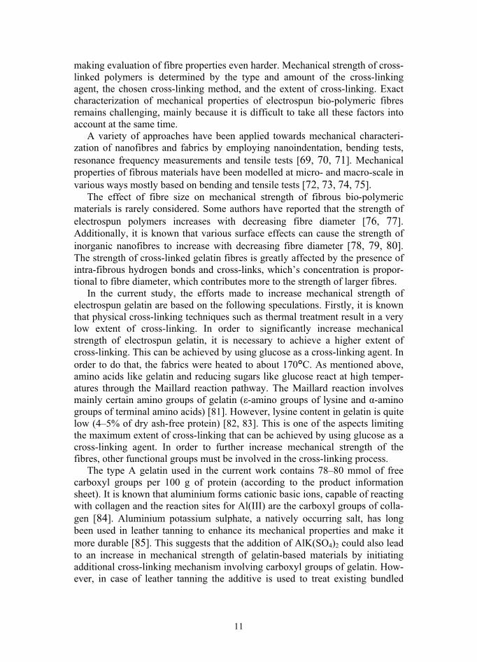
Modelling of fibre deposition The first step of tensile test simulation was creating a virtual fibrous scaffold. This was done by simulating fibre deposition during electrospinning. The total length of fibres per volume in the scaffold was calculated using the dimensions, weight, porosity and fibre diameter measured beforehand. Fibres were placed randomly one by one and layer by layer to form a virtual scaffold until the cal-culated total fibre length was reached. Based on SEM data, the fibres were assumed to be straight and remain straight during tensile test. It was observed from SEM images taken after failure that fibres in the scaffolds tested before cross-linking were pulled straight next to the ripping point (Figure 2a). The fibres that were further away from the ripping point also became noticeably oriented in the direction of the applied force. Scaffolds tested after cross-linking revealed very little fibre orientation right next to the ripping point. Furthermore, no increase in fibre orientation was observed by SEM when the meshes were pulled only in the elastic region of the strain and removed from the tensile test-ing machine before ripping (Figure 2b). This confirms the assumption that cross-links form between individual fibres as well as between gelatin molecules in each fibre and suggests that in the elastic region where deformation is small the fibres do not slide on each other and friction does not have to be taken into account when describing tensile testing of cross-linked scaffolds.
The behaviour of the scaffolds during tensile test depends on the interactions between the fibres. Non-cross-linked fibres are relatively independent of each other, the interactions between fibre segments that are in contact are weak and all the fibres tend to change their orientation in the direction of the applied force during tensile test. Friction alone causes some resistance to fibre reorientation. In case of cross-linked fibres chemical bonds have formed not only between polymer molecules inside each fibre, but also at the interceptions of fibres. These chemical bonds connecting individual fibres make the mesh structure
16
rigid and reorientation of a fibre caused by the applied force becomes dependent on reorientation of other fibres.
Tensile test was simulated for cross-linked scaffolds only, because non-cross-linked scaffolds are water-soluble and thus not usable in practical appli-cations. Comparing SEM images (Figure 2c) with virtual meshes (Figure 2d) constructed in the manner described above confirmed the necessary similarity between the virtual and the real scaffold.
Figure 2 SEM images of (a) a non-cross-linked gelatin fabric after tensile test 100 μm from the ripping point and (b) a cross-linked gelatin fabric after tensile test. Both in case of cross-linked and non-crosslinked samples, the fibres were randomly oriented before tensile test. During tensile test, the non-cross-linked fibres were reoriented in the direction of the applied force, whereas cross-linked fibres were not. (c) 21000 times magnified SEM image of a gelatin scaffold and (d) the virtual 3D scaffold constructed by the model; brightness of the fibres expresses the depth of the scaffold – deeper layers are darker.
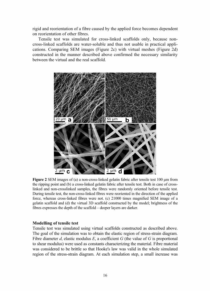
Modelling of tensile test Tensile test was simulated using virtual scaffolds constructed as described above. The goal of the simulation was to obtain the elastic region of stress-strain diagram. Fibre diameter d, elastic modulus E, a coefficient G (the value of G is proportional to shear modulus) were used as constants characterizing the material. Fibre material was considered to be brittle so that Hooke's law was valid in the whole simulated region of the stress-strain diagram. At each simulation step, a small increase was

17
given to the length of the sample. This causes stress in the fibres. The total tensile force is equal to the sum of the forces resulting from all fibres. Decrease in mesh width (neck formation) at larger elongations is related to the strength of interactions between the fibres. It was observed experimentally that the change in shape (forming of neck due to decreased mesh width) is related to glucose concentration in the fibres. The more glucose the scaffolds contained (and therefore the higher the extent of cross-linking) the smaller was the decrease of sample width. Scaffolds containing 15% glucose did not form any neck during tensile testing. During simulation the shape change of a piece of scaffold was determined at each step. In this work we assumed that the studied piece of scaffold remains rectangular during the test. This assumption is strictly valid for central parts of the mesh. However, if the dimensions of the scaffold piece are chosen to be small and tensile test is simulated only in the elastic region where deformation is also small, then the approximation that the scaffold is rectangular describes tensile testing accurately enough even in case of neck-formation.
Figure 3 describes the scheme of the simulation. y-axis is chosen so that it coincides with the direction of the applied force (and scaffold elongation). Each step of the simulation gives one point to the force-elongation diagram and consists of the following parts. Firstly, a small increase is given to the scaffold length, causing relative elongation of the scaffold y, while the width of the scaffold is kept unchanged. This induces elongation and stress in each fibre depending on the angle αi between the fibre and x-axis, which in return causes normal stress i in i-th fibre. Normal stress was calculated using the equation = , where E is the expected elastic modulus, εi is the relative elongation of the i-th fibre and αi is the angle between the i-th fibre and x-axis. Increase in length of the scaffold causes a change i in the angle i. The fibres tend to be oriented towards the y-axis during tensile testing. It was taken into account that the fibres are cross-linked and relatively strong chemical bonds exist at the interceptions of individual fibres. Thus the fibres do not slide on one another and the structure is relatively rigid. Therefore torsion arises around these interceptions. Generally, the shear stress (tangential stress) τi in a fibre at torsion is proportional to the torsion angle. We calculated tangential stress using the equation = , where G is a coefficient characteristic to the fibre material and proportional to its shear modulus.
Figure 3 The conceptual scheme of the tensile test simulation. Applied force causes elongation of a sample, which in returns causes normal stress (σi) and tangential stress (τi) in the fibres.
18
Evaluation of the coefficient G is based on the following speculations. The ratio of the elastic modulus and the shear modulus depends on the Poisson coeffi-cient, i.e. the ratio of the relative decrease of the specimen width to the relative increase of its length. The Poisson coefficient determines the change in volume of a material during tension, and the relative change of the surface area in our tensile test. Therefore we expected the E/G ratio to be connected to the relative change in the dimensions and shape of the scaffold during testing. Using the sample dimensions evaluated from the images taken during experimental tensile testing and comparing them to the dimensions calculated from the simulations at various E/G ratios, it was found that the values of E/G varied between 1.0 (for samples containing 15% glucose) and 1.2 (for samples containing no glucose).
The second part of this simulation step takes advantage of the calculated stresses i and τi to determine the change in mesh width. We calculated x- and y-components of normal and tangential stresses by = cos( ), = sin( ), = sin( ), and = cos( ). The y-components of i and τi add up and their sum is balanced by the applied force. The direction of the x-components of i and τi is determined by the angle i. The x-components ix and τix of each fibre are directed at opposite directions. The difference between their values is related to the change in fibre orientation and the decrease in scaffold width. Additionally, it gives rise to neck formation during testing. It must be mentioned that the sum of forces resulting from the x-components of the stresses in all fibres is equal to zero, whereas the difference between ix and τix in each individual fibre contributes to neck formation. The sum of y-compo-nents of the stresses in all the fibres is proportional to the applied force, = ( /4)∑ + . The force causing the narrowing of the scaffold is caused by the differences in stress components, = ( /4)∑ | − |. Relative decrease of the mesh width, x, follows from the equation
= ,
which gives
= ∙ ∑ | |∑ ( ) . Finally, the values of y and x were used to recalculate fibre lengths, stress in the fibres, and the total force Fy stretching the scaffold. The force directly causing elongation of the sample was calculated as the sum of forces deter-mined for all fibres. In this way, tensile test was simulated by firstly calculating new dimensions of the sample, and secondly the force applied in the y-direction. So, step by step, the force-strain diagram was generated. Tensile test was simu-lated, adjusting the hypothetic elastic modulus, until the elastic region of experimental tensile test graph and the simulated tensile test graph overlapped. The elastic modulus of the fibre material was found for all the samples in this manner.

19
4. RESULTS 4.1 Preparation of electrospun gelatin
Mixtures of gelatin and glucose were found to be electrospinnable from 5 M and more concentrated aqueous acetic acid solutions, but the suitable range of electrospinning parameters became narrower at lower acetic acid concentra-tions. 10 M aqueous acetic acid was found to be suitable for electrospinning all the mixtures used in this work, allowing problem-free production of fibres in a wide range of process parameters. Glucose content had little effect on the suit-able range of electrospinning parameters, which seemed to be determined mostly by the concentration of gelatin.
20–150 μm thick gelatin fabrics were obtained as a result of electrospinning. Smooth and uniformly structured fibres were produced at up to 30% glucose content. However, easy to handle fabric-like material was produced at up to 15% glucose content. At higher glucose concentrations the fabrics became stiff, brittle and broke easily after thermal treatment.
Density of the fabrics varied between 150 and 220 mg/cm3, mostly depend-ing on composition and porosity of a fabric. Porosity of the fabrics determined using SEM images varied from 78% to 90% (average 82%). No correlation was found between fibre diameter and porosity of the fabrics. Porosity of electro-spun gelatin is greatly influenced by randomness of the electrospinning process (Figure 4), which causes variabilities.
Figure 4 SEM images of samples prepared using the same preparation process param-eters indicate that porosity of a fabric is greatly influenced by randomness of the elec-trospinning process.
Porosity of an electrospun mesh also has a strong impact on mechanical strength of the material. Mechanical strength of a fabric is inversely propor-tional to its porosity, leading to the conclusion that porosity must be taken into account in studies concerning the strength of electrospun materials. However, there’s a lot of confusion concerning porosity of electrospun materials. Porosity is affected by the electrospinning process parameters [86]. It has been found

20
that porosity of an electrospun mesh is not dependent on fibre diameter, although porosities determined using various techniques vary greatly [87]. Additionally, porosity has been reported to both increase [88] and decrease [89] with increasing fibre diameter. In the current work, porosity was not found to be in correlation with fibre diameter.
4.2 Effect of concentration of the electrospinning solution on fibre diameter
Varying the concentration of the electrospinning solution (the amount of 10M acetic acid added to the gelatin-glucose mixture) between 19–25% allowed fabrication of gelatin meshes with average fibre diameters between 200–700 nm (Figure 5). A few samples had an average fibre diameter higher than 700 nm or lower than 200 nm. This is caused by hard to control instabilities of the electro-spinning process like the size of the droplet that forms on the tip of the needle used in electrospinning.
Figure 5 21000 times magnified SEM images of electrospun gelatin fabrics containing 10% glucose as a cross-linking agent prepared by varying the concentrations of the electrospinning solution; average fibre diameters: 784 nm (a), 594 nm (b), 472 nm (c), 444 nm (d), 352 nm (e), 288 nm (f), 259 nm (g), 188 nm (h). The colours of the original SEM images were inverted to improve visibility of the deeper layers.
The correlation between the concentration of the electrospinning solution and average fibre diameters is demonstrated in Figure 6. It is important to keep in mind that average fibre diameter is affected by several other factors in addition to the concentration of the electrospinning solution. For example, aging of the electrospinning solution has a strong effect on average fibre diameter, which is

21
considered in a following section of the current work. Therefore, in order to repeatedly prepare fabrics with similar average fibre diameters, all the parameters of the electrospinning process must be kept as constant as possible.
Figure 6 Dependence between the concentration of the electrospinning solution and average fibre diameter. Fibre diameter is determined largely by concentration of the electrospinning solution, although other factors like aging of the solution also have an effect on it.
Fibre diameters vary to some extent in case of all samples. However, the shape of the fibre diameter distribution graphs of samples with various average fibre diameters was largely similar (Figure 7), although the range of fibre diameters increased in case of samples consisting of larger fibres. Additionally, the number of abnormally small or large fibres increased as the average fibre diameters increased. This was probably caused by the used electrospinning setup – higher viscosity of more concentrated solutions caused the formation of a larger droplet on the needle tip, which in turn makes the electrospinning process less stable.
Figure 7 Fibre diameter distributions of samples with the following average fibre diameters: 197 nm (a), 332 nm (b), 551 nm (c)
22
4.3 Effect of aging of the electrospinning solution on fibre diameter
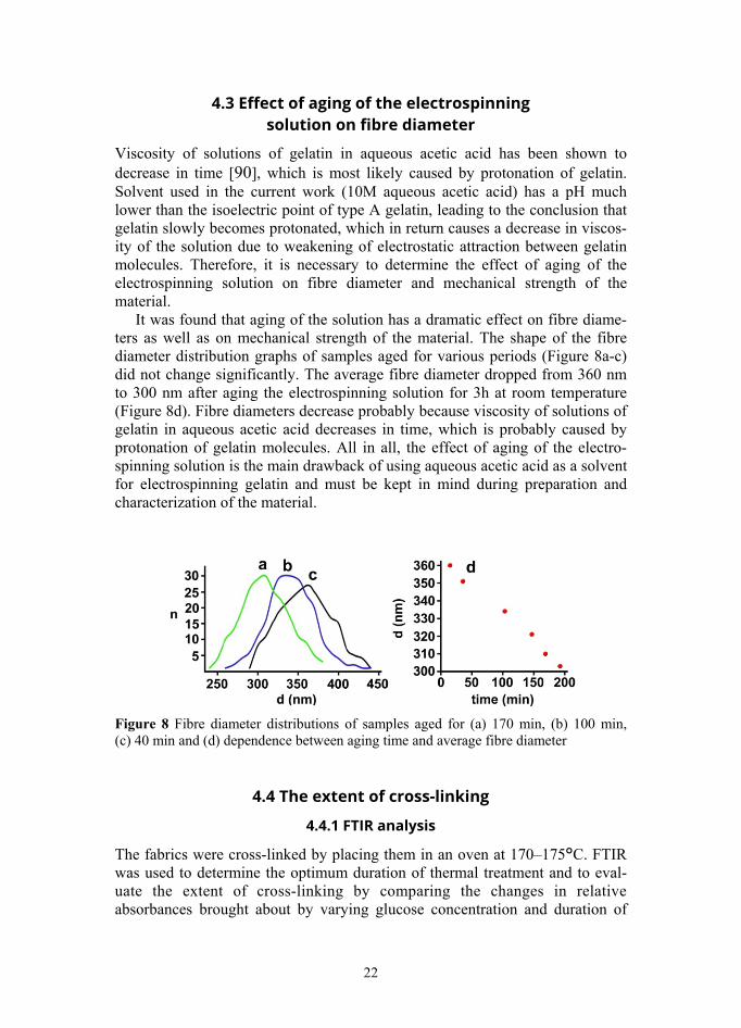
Viscosity of solutions of gelatin in aqueous acetic acid has been shown to decrease in time [90], which is most likely caused by protonation of gelatin. Solvent used in the current work (10M aqueous acetic acid) has a pH much lower than the isoelectric point of type A gelatin, leading to the conclusion that gelatin slowly becomes protonated, which in return causes a decrease in viscos-ity of the solution due to weakening of electrostatic attraction between gelatin molecules. Therefore, it is necessary to determine the effect of aging of the electrospinning solution on fibre diameter and mechanical strength of the material.
It was found that aging of the solution has a dramatic effect on fibre diame-ters as well as on mechanical strength of the material. The shape of the fibre diameter distribution graphs of samples aged for various periods (Figure 8a-c) did not change significantly. The average fibre diameter dropped from 360 nm to 300 nm after aging the electrospinning solution for 3h at room temperature (Figure 8d). Fibre diameters decrease probably because viscosity of solutions of gelatin in aqueous acetic acid decreases in time, which is probably caused by protonation of gelatin molecules. All in all, the effect of aging of the electro-spinning solution is the main drawback of using aqueous acetic acid as a solvent for electrospinning gelatin and must be kept in mind during preparation and characterization of the material.
Figure 8 Fibre diameter distributions of samples aged for (a) 170 min, (b) 100 min, (c) 40 min and (d) dependence between aging time and average fibre diameter
4.4 The extent of cross-linking
4.4.1 FTIR analysis
The fabrics were cross-linked by placing them in an oven at 170–175°C. FTIR was used to determine the optimum duration of thermal treatment and to eval-uate the extent of cross-linking by comparing the changes in relative absorbances brought about by varying glucose concentration and duration of
23
thermal treatment. Analysing the changes brought about by thermal treatment using FTIR is a challenging task complicated by the following factors. Firstly, absolute values of recorded absorbances were not reproducible due to the fibrous structure of the material and small fibre diameter, which did not allow achieving perfect contact between the fabric and ATR crystal. In order to be able to reliably compare infrared spectra of samples with different composition and analyse the changes brought about by thermal treatment, it was necessary to calculate relative absorbances as described above. Secondly, infrared absorption is greatly affected by water content in the sample. It was found that analysing spectra by calculating relative absorbances produces meaningful results only if the fabrics are properly dried prior to recording spectra. In the current work, the samples were dried in a critical point drier before analysing them. Thirdly, FTIR spectra are affected by the non-homogeneous composition of gelatin. The effect is small, but noticeable, and can be overcome by recording several spectra of every sample and averaging the absorbances.
It was concluded that FTIR can be used to analyse the cross-linking process and to determine the extent of cross-linking, given that these problems are properly addressed during experimental work. The described method cannot be used to determine the extent of cross-linking of a particular sample – it is only suitable to study the process, because it is necessary to prepare and analyse large patches of samples in order to get meaningful results.
FTIR spectra of gelatin, glucose and the fabrics before and after thermal treatment are presented in Figure 9. Peaks were detected at 3285, 3077 cm-1 (mainly OH and NH vibrations), 2939 and 2879 cm-1 (mainly CH2 asymmetric and symmetric vibrations respectively), 1657, 1640 and 1631 cm-1 (the amide I band, mainly C=O vibrations), 1562, 1547 and 1535 cm-1 (the amide II band, mainly NH bending), 1452, 1405, 1336, 1203 and 1163 cm-1 (different in plane vibrations) and 1240 cm-1 (the amide III band, in plane NH bending and CN stretching), 1081, 1035 and 921 cm-1 (mainly CO vibrations overlapping with over vibrations in glucose [I, 91, 92, 93, 94]. It was found that the changes in gelatin brought about by thermal treatment of the fabrics are best represented by changes in relative absorbance of the amide I peak, whereas changes in glucose are best represented by changes in relative absorbance of a peak at 1035 cm-1.

24
Figure 9 FTIR spectra of glucose, type A gelatin and electrospun fabrics containing 15.1% glucose before and after thermal treatment.
4.4.2 Monitoring the cross-linking process
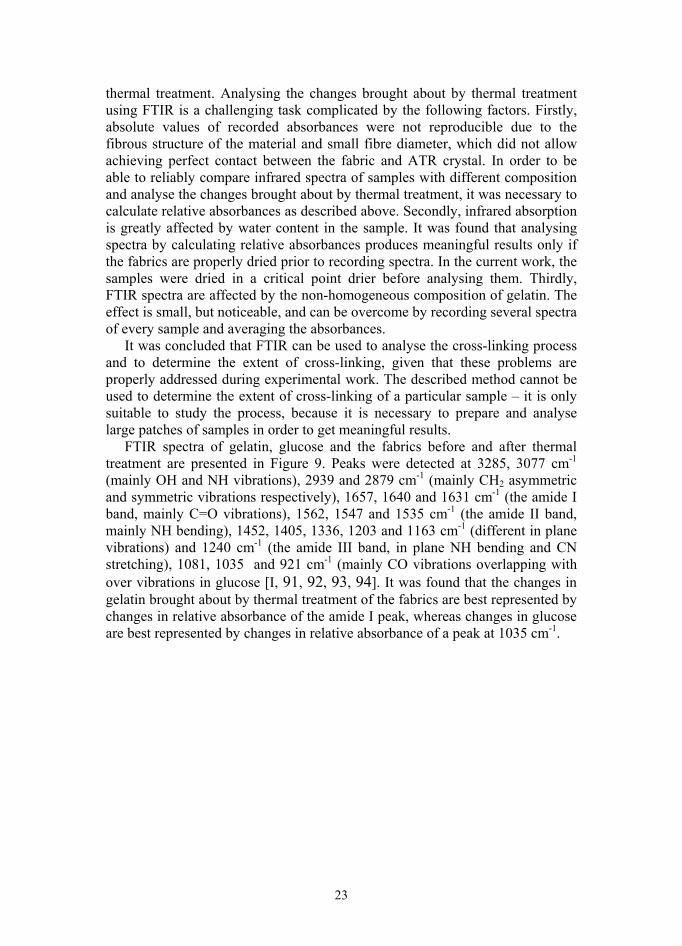
Increasing glucose content in the fibres from 0% to 30% led to considerable increase in relative absorbance of peaks at 1081 cm-1, 1035 cm-1 and 921 cm-1. At the same time, relative absorbance of amide I and amide II bands decreased. These are the peaks by which we can best distinguish between changes in gela-tin and changes in glucose brought about by thermal treatment. In order to find the optimal duration of thermal treatment, the cross-linking process was moni-tored by removing pieces of scaffolds from the oven after various times, ana-lysing these by FTIR and comparing the resulting spectra. It was found that changes in relative absorbance during thermal cross-linking are exponential (Figure 10). Considerable changes in relative absorbance were detected during the first hour of cross-linking. After 2 hours of thermal treatment, very little further changes in relative absorbance were detected with an average of less than 0.1% change between 2 and 3 hours as opposed to an average of about 5% change in relative absorbance during the first 5 minutes of thermal treatment. It was concluded that maximum extent of cross-linking is reached after nearly 3 h of thermal treatment.
25
Figure 10 Changes in relative absorbance during thermal treatment of the fabrics (19.9% glucose content)
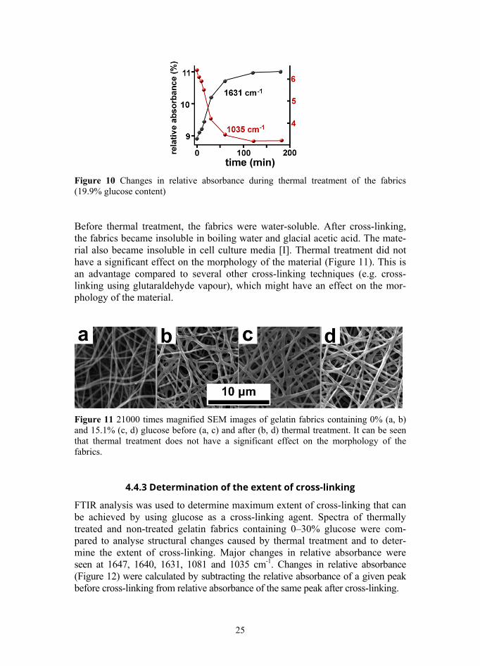
Before thermal treatment, the fabrics were water-soluble. After cross-linking, the fabrics became insoluble in boiling water and glacial acetic acid. The mate-rial also became insoluble in cell culture media [I]. Thermal treatment did not have a significant effect on the morphology of the material (Figure 11). This is an advantage compared to several other cross-linking techniques (e.g. cross-linking using glutaraldehyde vapour), which might have an effect on the mor-phology of the material.
Figure 11 21000 times magnified SEM images of gelatin fabrics containing 0% (a, b) and 15.1% (c, d) glucose before (a, c) and after (b, d) thermal treatment. It can be seen that thermal treatment does not have a significant effect on the morphology of the fabrics.
4.4.3 Determination of the extent of cross-linking
FTIR analysis was used to determine maximum extent of cross-linking that can be achieved by using glucose as a cross-linking agent. Spectra of thermally treated and non-treated gelatin fabrics containing 0–30% glucose were com-pared to analyse structural changes caused by thermal treatment and to deter-mine the extent of cross-linking. Major changes in relative absorbance were seen at 1647, 1640, 1631, 1081 and 1035 cm-1. Changes in relative absorbance (Figure 12) were calculated by subtracting the relative absorbance of a given peak before cross-linking from relative absorbance of the same peak after cross-linking.

26
The interpretation of spectral changes is made harder by the number and complexity of reactions going on during cross-linking. Whereas spectral changes in pure gelatin fabrics are limited, it was observed that samples with higher glucose content undergo rapid changes during thermal treatment, which indicates that the majority of reactions going on during cross-linking are directly caused by the presence of glucose. By far the biggest change detected was an increase in relative absorbance of the amide I band, coupled with the decrease of peaks at 1081 and 1035 cm-1 associated with C-O vibrations mainly in glucose.
This brings up the question of the amount of glucose at which gelatin is cross-linked to maximum extent that can be achieved using glucose as cross-linking agent. The extent of gelatin cross-linking is often evaluated by changes in free ε-amino group concentration [95], although it has been suggested that at high temperatures a cross-linking mechanism without an amino group involve-ment occurs [96]. The increase in relative absorbance at 1657, 1640 and 1631 cm-1 was strongest for fabrics containing about 20% glucose, while relative absorbance at 1081 and 1035 cm-1 decreased further still when scaffolds con-tained more glucose. This suggests that some other reaction, perhaps carameli-zation, becomes more dominant at over 20% glucose content.
FTIR analysis alone does not prove that the reactions caused by thermal treatment actually lead to formation of cross-links between gelatin molecules. However, taking into account that mechanical strength of the fibre material increases with increasing glucose content [II] and that the material becomes more resistant to enzymatic degradation [I], it can be concluded that glucoses does indeed act as a cross-linking agent for gelatin, and that the maximum extent of cross-linking is achieved at about 20% glucose content. However, fabric-like, easy to handle material can be obtained from gelatin-glucose blends containing up to 15% glucose. At higher glucose concentrations the fabrics become impractically brittle after thermal treatment.
Figure 12 Changes in relative absorbance (ΔR) at 1640 cm-1 caused by thermal treatment indicate that maximum extent of cross-linking is achieved at about 20% glucose content. Changes in relative absorbance were calculated by subtracting relative absorbance of a given peak before cross-linking from relative absorbance of the same peak after cross-linking.
27
4.5 Mechanical properties of electrospun gelatin
4.5.1 Tensile test and simulation
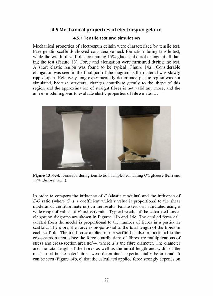
Mechanical properties of electrospun gelatin were characterized by tensile test. Pure gelatin scaffolds showed considerable neck formation during tensile test, while the width of scaffolds containing 15% glucose did not change at all dur-ing the test (Figure 13). Force and elongation were measured during the test. A short elastic region was found to be typical (Figure 14a). Considerable elongation was seen in the final part of the diagram as the material was slowly ripped apart. Relatively long experimentally determined plastic region was not simulated, because structural changes contribute greatly to the shape of this region and the approximation of straight fibres is not valid any more, and the aim of modelling was to evaluate elastic properties of fibre material.
Figure 13 Neck formation during tensile test: samples containing 0% glucose (left) and 15% glucose (right).
In order to compare the influence of E (elastic modulus) and the influence of E/G ratio (where G is a coefficient which’s value is proportional to the shear modulus of the fibre material) on the results, tensile test was simulated using a wide range of values of E and E/G ratio. Typical results of the calculated force-elongation diagrams are shown in Figures 14b and 14c. The applied force cal-culated from the model is proportional to the number of fibres in a particular scaffold. Therefore, the force is proportional to the total length of the fibres in each scaffold. The total force applied to the scaffold is also proportional to the cross-section area, since the force contributions of fibres are multiplications of stress and cross-section area πd2/4, where d is the fibre diameter. The diameter and the total length of the fibres as well as the initial length and width of the mesh used in the calculations were determined experimentally beforehand. It can be seen (Figure 14b, c) that the calculated applied force strongly depends on

28
the elastic modulus of the fibre material whereas the dependence on the E/G ratio is rather weak.
Figure 14 Experimental tensile test (a) and simulation results demonstrating the effect of elastic modulus (b) and E/G ratio (c) on force-elongation curve in the elastic region
4.5.2 Effect of glucose content
The elastic modulus of the fibre material was determined for samples prepared using 0–15% glucose. It was found (Figure 15a) that cross-linking by glucose considerably increases the elastic modulus of gelatin fibres from about 0.3 GPa (0% glucose) to 1.1 GPa (15% glucose). Simulation results were used to draw connections between the elastic modulus of the scaffold and the elastic modulus of the fibre material (Figure 15b) at different E/G ratios calculated for a wider range of values of E. The analysis revealed that the elastic modulus of the fibre material exceeds the elastic modulus of the scaffold around 8 times. These results seem to be reliable, taking into account the porosity and fibrous structure of the mesh.
Figure 15 The effect of glucose concentration on elastic modulus of gelatin fibres (A); Dependence between elastic modulus of the fibre material and elastic modulus of the mesh (B)
29
4.5.3 Effect of aging of the electrospinning solution
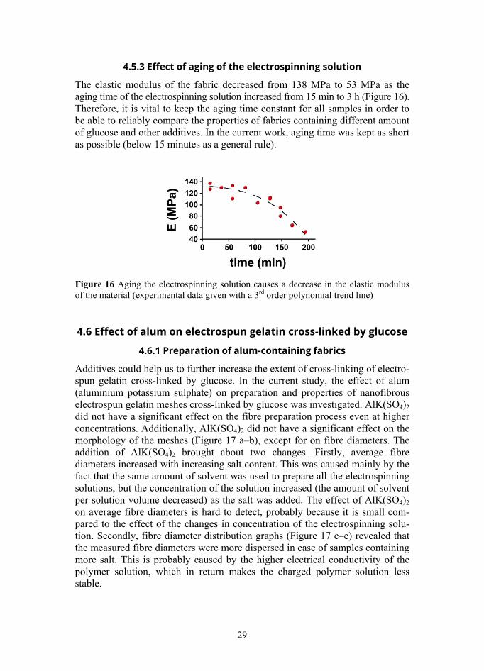
The elastic modulus of the fabric decreased from 138 MPa to 53 MPa as the aging time of the electrospinning solution increased from 15 min to 3 h (Figure 16). Therefore, it is vital to keep the aging time constant for all samples in order to be able to reliably compare the properties of fabrics containing different amount of glucose and other additives. In the current work, aging time was kept as short as possible (below 15 minutes as a general rule).
Figure 16 Aging the electrospinning solution causes a decrease in the elastic modulus of the material (experimental data given with a 3rd order polynomial trend line)
4.6 Effect of alum on electrospun gelatin cross-linked by glucose
4.6.1 Preparation of alum-containing fabrics
Additives could help us to further increase the extent of cross-linking of electro-spun gelatin cross-linked by glucose. In the current study, the effect of alum (aluminium potassium sulphate) on preparation and properties of nanofibrous electrospun gelatin meshes cross-linked by glucose was investigated. AlK(SO4)2 did not have a significant effect on the fibre preparation process even at higher concentrations. Additionally, AlK(SO4)2 did not have a significant effect on the morphology of the meshes (Figure 17 a–b), except for on fibre diameters. The addition of AlK(SO4)2 brought about two changes. Firstly, average fibre diameters increased with increasing salt content. This was caused mainly by the fact that the same amount of solvent was used to prepare all the electrospinning solutions, but the concentration of the solution increased (the amount of solvent per solution volume decreased) as the salt was added. The effect of AlK(SO4)2 on average fibre diameters is hard to detect, probably because it is small com-pared to the effect of the changes in concentration of the electrospinning solu-tion. Secondly, fibre diameter distribution graphs (Figure 17 c–e) revealed that the measured fibre diameters were more dispersed in case of samples containing more salt. This is probably caused by the higher electrical conductivity of the polymer solution, which in return makes the charged polymer solution less stable.
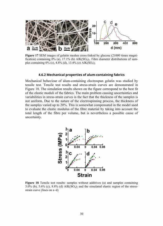
30
Figure 17 SEM images of gelatin meshes cross-linked by glucose (21000 times magni-fication) containing 0% (a), 17.1% (b) AlK(SO4)2. Fibre diameter distributions of sam-ples containing 0% (c), 4.8% (d), 13.4% (e) AlK(SO4)2
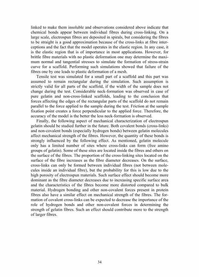
4.6.2 Mechanical properties of alum-containing fabrics
Mechanical behaviour of alum-containing electrospun gelatin was studied by tensile test. Tensile test results and stress-strain curves are demonstrated in Figure 18. The simulation results shown on the figure correspond to the best fit of the elastic moduli of the fabrics. The main problem causing uncertainties and variabilities in stress-strain curves is the fact that the thickness of the samples is not uniform. Due to the nature of the electrospinning process, the thickness of the samples varied up to 20%. This is somewhat compensated in the model used to evaluate the elastic modulus of the fibre material by taking into account the total length of the fibre per volume, but is nevertheless a possible cause of uncertainty.
Figure 18 Tensile test results: samples without additives (a) and samples containing 3.0% (b), 5.6% (c), 8.8% (d) AlK(SO4)2 and the simulated elastic region of the stress-strain curve (lines on a–d)

31
As a general rule, the most important part of the stress-strain curve is the linear region, because in most applications the material only works in the elastic region of the stress-strain diagram. Therefore, plastic region and failure of the meshes were not studied. The elastic moduli of the meshes with different com-position were calculated using the linear region of the stress-strain curve. Next, tensile test was simulated by varying the hypothetic elastic modulus of the fibre material, until the elastic region of experimental stress-strain curves and simu-lated stress-strain curves overlapped. In this case the elastic modulus of a fibrous material calculated from the simulation is equal to the experimentally determined elastic modulus of the mesh. The elastic modulus of the fibre mate-rial was found in this way for all samples.
AlK(SO4)2 increased the elastic modulus of glucose-cross-linked gelatin fibres up to about 10% salt content (Figure 19). However, it should be kept in mind that the absolute values of the determined elastic moduli are dependent on numerous factors mentioned in the introduction, including molecular mass of gelatin, extent of cross-linking determined by the cross-linking method (the amount of cross-linking agent, duration of thermal treatment, temperature), possible degradation of gelatin, surface effects, etc. In the current study, these parameters were kept as constant as possible in order to distinguish the effect of the additive from other parameters. Thereby, the effect of AlK(SO4)2 was clearly evident. In the current work, the elastic modulus of electrospun gelatin fabrics increased from 20 MPa to 70 MPa and the elastic modulus of the fibre material increased from 150 MPa to 620 MPa as the salt content increased from 0% to 9.6%. However, varying the abovementioned parameters can cause fibrous gelatin-based materials to be either mechanically stronger or weaker than hereby presented. During preparation of the fabrics, obtaining homo-geneous electrospinning solutions took longer in case of gelatin mixtures con-taining alum (around 15 min), compared to mixtures containing only gelatin and glucose (in which case 5 min of stirring was sufficient). This is a drawback of the addition of alum, since the strength of the fibres decreases with increasing solution aging time.
Figure 19 Dependence of the elastic modulus of the fibre material on AlK(SO4)2 concentration.

32
The elastic modulus of the fibre material is roughly 8 times higher than the elastic modulus of the mesh. This difference is caused firstly by the high poros-ity of electrospun materials and secondly by random fibre orientation. The fact that the addition of AlK(SO4)2 increased the elastic modulus of the material indicates that the presence of the salt induces cross-linking, which in return determines mechanical strength of the material. At about 10% AlK(SO4)2 con-tent maximum extent of cross-linking that can be reached by the addition of AlK(SO4)2 is achieved and the elastic modulus of the material drops at higher salt concentrations. It is not certain whether the addition of AlK(SO4)2 brings about covalent or ionic cross-linking, but significant changes in FTIR spectra between 950–1300 cm-1 brought about by the presence of the salt, in addition to considerable increase in strength of the material indicate that thermal treatment initiates covalent cross-linking (probably involving carboxyl groups of gelatin), although the exact chemistry of it requires further studies.

33
5. DISCUSSION Cross-linking of electrospun gelatin Evaluating the extent of cross-linking by analysing FTIR spectra proved to be effective. However, it must be noted that the determined extent of cross-linking is dependent on the method used to evaluate the extent of cross-linking. There-fore, the results are hard to compare to those obtained by other methods, for example near-infrared spectrophotometry [97] and use of chemical assays [98]. FTIR analysis indicated that gelatin reacts chemically with glucose at up to 20% glucose content. At higher glucose concentrations some other kind of reaction, probably caramelization, becomes prevalent. The following additional infor-mation must be considered in order to prove that the chemical reaction between gelatin and glucose leads to the formation of cross-links between gelatin mole-cules. Firstly, mechanical strength of electrospun gelatin increases with increasing glucose content. This indicates that cross-links between gelatin mol-ecules do form. However, only meshes containing up to 15% glucose were tested since the fabrics became very brittle at over 15% glucose content, and the attempts to test samples containing more glucose were not successful. Secondly, it was discovered that meshes containing up to 15% glucose were enzymatically digestible, meshes containing 20% were partially enzymatically digestible, and meshes containing over 20% glucose were totally resistant to enzymatic degra-dation [I]. Taking all the results into account, it can be concluded that electro-spun gelatin can be thermally cross-linked by glucose and that the extent of cross-linking increases up to 20% glucose content. Compared to other widely used cross-linking agents, glucose is not only biocompatible and non-toxic, but also relatively cheap and allows producing the fabrics using only natural sub-stances and easy, cost-effective methods.
Glucose-initiated cross-linking reaction involves mainly certain amino groups of gelatin. Non-cross-linked gelatin contains around 23*10-5 mol/g free amino groups [99]. Samples containing 20% glucose contain around 110*10-5 mol/g glucose molecules. Therefore, certain amount of caramelization probably occurs in glucose-containing gelatin meshes, which is supported by the fact that thermal treatment was carried out at over melting point and caramelization tem-perature of glucose. This in return indicates that the cross-links between gelatin molecules might actually be caramel molecules of varying size, which further supports the formation of cross-links.
Mechanical properties of electrospun gelatin The model used to evaluate the elastic modulus of the fibre material in the cur-rent study describes mechanical behaviour of fibrous cross-linked or otherwise interconnected materials and allows simulating tensile test in the elastic region. Remaining in the elastic region is related to the approximation that the fibres in the concerned part of a scaffold are straight during tensile test and do not slide on each other. This assumption is only valid if there are chemical bonds present at fibre interceptions. In case of gelatin scaffolds, the fibres must be cross-
34
linked to make them insoluble and observations considered above indicate that chemical bonds appear between individual fibres during cross-linking. On a large scale, electrospun fibres are deposited in spirals, but considering the fibres to be straight is a good approximation because of the cross-links at fibre inter-ceptions and the fact that the model operates in the elastic region. In any case, it is the elastic region that is of importance in most applications. However, for brittle fibre materials with no plastic deformation one may determine the maxi-mum normal and tangential stresses to simulate the formation of stress-strain curve for a scaffold. Performing such simulations showed that failure of the fibres one by one leads to plastic deformation of a mesh.
Tensile test was simulated for a small part of a scaffold and this part was assumed to remain rectangular during the simulation. Such assumption is strictly valid for all parts of the scaffold, if the width of the sample does not change during the test. Considerable neck-formation was observed in case of pure gelatin and non-cross-linked scaffolds, leading to the conclusion that forces affecting the edges of the rectangular parts of the scaffold do not remain parallel to the force applied to the sample during the test. Friction at the sample fixation point creates a force perpendicular to the applied force. Therefore, the accuracy of the model is the better the less neck-formation is observed.
Finally, the following aspect of mechanical characterization of electrospun gelatin should be studied further in the future. Both covalent bonds (cross-links) and non-covalent bonds (especially hydrogen bonds) between gelatin molecules affect mechanical strength of the fibres. However, the quantity of these bonds is strongly influenced by the following effect. As mentioned, gelatin molecule only has a limited number of sites where cross-links can form (free amino groups of gelatin). Some of these sites are located inside the fibres and others on the surface of the fibres. The proportion of the cross-linking sites located on the surface of the fibre increases as the fibre diameter decreases. On the surface, cross-links can only be formed between individual fibres (not between mole-cules inside an individual fibre), but the probability for this is low due to the high porosity of electrospun materials. Such surface effect should become more dominant as the fibre diameter decreases due to increasing specific surface area and the characteristics of the fibres become more distorted compared to bulk material. Hydrogen bonding and other non-covalent forces present in protein fibres also have a similar effect on mechanical strength of the fibres. The for-mation of covalent cross-links can be expected to decrease the importance of the role of hydrogen bonds and other non-covalent forces in determining the strength of gelatin fibres. Such an effect should contribute more to the strength of larger fibres.

35
6. APPLICATIONS – TISSUE ENGINEERING Tissue engineering is an interdisciplinary field of research, which unites mate-rials science, engineering, chemistry, physics, medicine and cell biology. In the current study [I], electrospun gelatin meshes thermally cross-linked by glucose were used as cell culture scaffolds to evaluate their suitability for tissue engi-neering applications. In order to do that, fibroblasts were seeded onto glass cover slips covered with electrospun gelatin scaffolds [I]. Cells grown on elec-trospun gelatin are compared to cells grown on glass in Figure 20.
Figure 20. SEM images of cells grown on electrospun gelatin (left) and glass (right [I]).
Morphological differences between cells grown on glass and cells grown on the scaffolds can be explained in the following way. Natural extracellular matrix consists of bundled nanofibres. In order to mimic this structure, artificial extra-cellular matrix should also consist of fibres with sub-micron diameters. Electro-spinning has the advantage of producing structural features somewhat similar to native extracellular matrix, which has been shown to result in different cell morphology compared to cells grown on smooth substrate [100].
Cell culture experiments, biological stability test and observations made during scaffold preparation [I] indicate that scaffolds containing up to 15% glucose can be considered for tissue engineering applications, most importantly because fabric-like easy to handle scaffolds were produced at 0–15% glucose content, secondly because the number of cells on scaffolds started to drop at higher glucose concentrations, and thirdly because at over 15% glucose content the scaffolds become resistant to enzymatic degradation.
36
7. CONCLUSIONS The following conclusions were made based on the current study.
Gelatin was electrospun from 10M aqueous acetic acid. Average fibre diameter varies from 200 nm to 700 nm depending on the concentration of the electrospinning solution. FTIR analysis, results of mechanical characterization of the material, biological degradation tests, and observations made during the experiments indicate that electrospun gelatin can be thermally cross-linked by glucose by placing the fabrics in an oven at about 170°C.
Maximum extent of cross-linking is achieved at nearly 20% glucose content. However, easy to handle fabrics can be obtained from gelatin-glucose blends containing up to 15% glucose. At higher glucose concentrations the material becomes impractically brittle after thermal treatment. The extent of cross-link-ing depends on glucose concentration and duration of thermal treatment. Maxi-mum extent of cross-linking is reached after nearly 3 h of thermal treatment.
Mechanical properties of the material were evaluated by tensile test and modelling. The model simulates deposition of fibres during electrospinning and tensile test of fibrous meshes in order to evaluate the elastic modulus of electro-spun materials and is effective when applied to cross-linked fabrics with bonds between both polymer molecules and at fibre interceptions. Cross-linking elec-trospun gelatin by glucose increases mechanical strength of the material. The addition of aluminium potassium sulphate further increases mechanical strength of electrospun gelatin cross-linked by glucose up to about 10% alum content. Aging of the electrospinning solution decreases mechanical strength and fibre diameter of gelatin electrospun from 10M aqueous acetic acid.
Preliminary short-term cell culture experiments indicate that electrospun gelatin cross-linked thermally by glucose is suitable for tissue engineering applications [I].
All in all, the goals of the current work were fully achieved and a promising new material, electrospun gelatin cross-linked by glucose, was developed as a result of the work.
37
SUMMARY IN ESTONIAN Glükoosiga ristsidestatud elektrospinnitud želatiin
Käesolevas doktoritöö algseks eesmärgiks oli siirdamiseks sobivate želatiini-põhiste naharakkude kasvualuste väljatöötamine, kasutades võimalikult loodus-sõbralikke lähteaineid ja meetodeid. Želatiin valiti peamiseks lähteaineks, kuna see on äärmiselt biosobiv, mitte-toksiline, kergesti töödeldav ja odav. Želatiini saadakse kollageeni, ühe sidekoe peamise koostisosa, hüdrolüüsi tulemusel. Kollageeni ja želatiini keemiline koostis on seetõttu peaaegu identne, mis teeb želatiini eriti sobivaks lähteaineks naharakkude kasvualuste valmistamisel. Želatiini peamiseks puuduseks võrreldes sünteetiliste lähteainetega on mitte-homogeenne koostis ja sellest tulenev füüsikaliste ja keemiliste omaduste varieeruvus, kuid antud juhul on eeliseid peetud tunduvalt kaalukamaks puu-dustest.
Elusorganismis koosneb normaalne rakuväline keskkond põhimõtteliselt kokkukeerdunud nanokiudude pundardest. Sellise struktuuri teatud ulatuses jäljendamiseks valmistati kasvualused elektrospinnimise meetodil. Elektro-spinnitud želatiin on aga vees lahustuv ja mehaaniliselt nõrk. Need puudused on võimalik ületada želatiinimolekulide ristsidestamise teel. Varasemalt on selleks otstarbeks kasutatud mitmeid kalleid ja vähem või rohkem toksilisi keemilisi ristsidestajaid. Käesolev dissertatsioon põhineb avastusel, et elektrospinnitud želatiini on võimalik ristsidestada glükoosi abil. See avastus tekitas mitmeid küsimusi – kui palju glükoosi peaks želatiinile lisama soovitud tulemuse saamiseks, kui kaua peaks želatiinikangast kuumutama, kuidas ristsidestamine mõjutab materjali omadusi, kas glükoosiga ristsidestatud želatiinikangas on sobiv regeneratiivses meditsiinis kasutamiseks jne. Käesolev dissertatsioon koos selle aluseks olevate artiklitega [I–III] annab vastuse mitmetele prakti-listele küsimustele.
Suur osa dissertatsioonist on pühendatud glükoosiga ristsidestatud elektro-spinnitud želatiinikanga mehaanilistele omadustele, kuna just need mängivad võtmerolli paljudes rakendustes, sealhulgas biomeditsiinis. Samas on just žela-tiini mehaaniline nõrkus olnud tema laiema kasutamise üheks peamiseks takistuseks ja on väga oluline keskenduda nende meetodite arendamisele, mis aitaksid želatiinikiude tugevamaks muuta. Lisaks on biopolümeersete kiudude mehaanilised omadused küllaltki komplekssed ja mõjutatud muuhulgas lähte-aineks oleva želatiini kvaliteedi, valmistamisprotsessi parameetrite, lahusti omaduste, niiskuse, kiu läbimõõdu jms poolt. Kõigi nende mõjurite tõttu varieerub želatiinikiudude kirjanduses toodud elastsusmoodul äärmiselt suures ulatuses, kirjanduses leiduvaid tulemusi on omavahel väga raske võrrelda ja katsete korratavus ei ole hea, mis tekitab palju segadust. Seega on äärmiselt tähtis uurida elektrospinnitud želatiini mehaanilisi omadusi piisavalt suure detailsuse astmega, et oleks võimalik korratavalt valmistada kindlate mehaani-liste omadustega kangaid. Eeltoodust tulenevalt on käesoleva uurimuse

38
peamiseks eesmärgiks valmistada ja iseloomustada elektrospinnitud glükoosiga termiliselt ristsidestatud želatiinikangaid.
Töö tulemusena leiti, et želatiini ning želatiini, glükoosi ja teiste lisandite segusid on võimalik elektrospinnida 10 M äädikhappe vesilahusest. Keskmised kiudude läbimõõdud jäävad vahemikku 200–700 nm olenevalt želatiini ja lisandite kontsentratsioonist elektrospinnimise lahuses. Kangataolist, kergesti käsitletavat materjali on võimalik saada kuni 15% glükoosi sisaldavatest segu-dest. Suurema glükoosisisalduse juures muutub materjal pärast kuumutamist väga hapraks.
FTIR analüüs, želatiinikanga mehaaniliste omaduste määramise katsete tulemused, lagundamiskatsed ja katsete käigus tehtud tähelepanekud kinnitasid seda, et glükoos tõepoolest ristsidestab kiude moodustavaid želatiinimolekule, kui ta paigutada ahju umbes 170 °C juures. Seeläbi muutub želatiinikangas lahustumatuks ja mehaaniliselt palju tugevamaks. Ristsidestumise määr sõltub glükoosikontsentratsioonist ja kuumutamise kestvusest. Maksimaalne rist-sidestumise ulatus saavutatakse umbes 3 tunni kuumas ahjus hoidmise järel.
Želatiinikanga mehaanilisi omadusi iseloomustati tõmbekatse ja modelleeri-mise teel. Mudel simuleerib esiteks kiudude kogunemist elektrospinnimise ajal, teiseks kiududest koosneva kanga tõmbekatset eesmärgiga määrata kiumaterjali elastsusmoodul. Mudel on eriti sobiv rakendamiseks ristsidestatud kiudmater-jalidele, mille puhul on keemilised sidemed mitte üksnes kiude moodustavate želatiinimolekulide vahel, vaid seotud on ka kiud omavahel kiudude ristumis-kohtades. Leiti, et glükoosiga ristsidestamine suurendab oluliselt elektro-spinnitud želatiini elastsusmoodulit. AlK(SO4)2 lisamine muudab glükoosiga ristsidestatud elektrospinnitud želatiini mehaaniliselt veelgi tugevamaks. Samas elektrospinnimise lahuse vanandamine vähendab valmistatava kanga mehaa-nilist tugevust, kui lahustiks on äädikhappe vesilahus.
Rakendusuuringute tulemusena leiti, et glükoosiga ristsidestatud elektro-spinnitud želatiin on sobiv regeneratiivse meditsiini rakendusteks.

39
ACKNOWLEDGEMENTS I am grateful to my supervisors. I would like to thank all the co-authors of the publications on which the dis-sertation is based on – Martin Järvekülg, Uno Mäeorg, Karol Mõisavald, Triin Kangur, Paula Reemann, Martin Pook, Annika Põder, Külli Kingo, Viljar Jaks. I would like to express my gratitude to colleagues and friends at the Faculty of Science and Technology and Institute of Physics. The study was financially supported by the European Union through the European Regional Development Fund via projects “Carbon Nanotube Re-inforced Electrospun Nano-fibres and Yarns“ (3.2.1101.12-0018), “SmaCell“ (3.2.1101.12-0017), Centre of Excellence “Mesosystems: Theory and Applica-tions” (3.2.0101.11-0029), Estonian Science Foundation grant IUT2-25, Esto-nian Nanotechnology Competence Centre (EU29996).
40
REFERENCES
1 Gorgieva S, Kokol V. Collagen- vs. Gelatin-based Biomaterials and Their Bio-compatibility: Review and Perspectives. In: R. Pignatello (Ed.). Biomaterials Applications for Nanomedicine 2011, Chapter 2.
2 Gomez-Guillen MC, Perez-Mateos M, Gomez-Estaca J, Lopez-Caballero E, Gimenez B, Montero P. Fish gelatin: a renewable material for developing active biodegradable films. Trends in Food Science & Technology 2009; 20:3–16.
3 Zhong SP, Zhang YZ, Lim CT. Tissue scaffolds for skin wound healing and der-mal reconstruction. Wiley Interdisciplinary Reviews – Nanomedicine and Nano-biotechnology 2010; 2(5) 510–25.
4 Raghavan P, Lim D-H, Ahn J-H, Nah C, Sherrington DC, Ryu H-S, Ahn H-J. Electrospun polymer nanofibers: The booming cutting edge technology. Reactive & Functional Polymers 2012; 72: 915–930.
5 Huang Z-M, Zhang Y-Z, Kotaki M, Ramakrishna S. A review on polymer nano-fibers by electrospinning and their applications in nanocomposites. Composites Science and Technology 2003; 63: 2223–2253.
6 Gao C, Gao Q, Li Y. In vitro evaluation of electrospun gelatin-bioactive glass hybrid scaffolds for bone regeneration. J. Appl. Polym. Sci. 2013; 127(4): 2588–2599.
7 Chong EJ, Phan TT, Lim IF, Zhang YZ, Bay, Ramakrishna S, Lim CT. Evaluation of electrospun PCL/gelatin nanofibrous scaffold for wound healing and dermal reconstruction. Acta Biomaterialia 2007; 3: 321–330.
8 Wang S, Zhang Y, Yin G, Wang H, Dong Z. Electrospun polylactide/silk fibroin composite tubular scaffolds for small-diameter tissue engineering blood vessels. Journal of Applied Polymer Science 2009; 113(4): 2675–2682.
9 Wong C, Shital P, Chen R, Owida A, Morsi Y. Biomimetic electrospun gelatin-chitosan polyurethane for heart valve leaflets. Journal of Mechanics in Medicine and Biology 2010; 10(4): 563–576.
10 Ren L, Wang J, Yang F-Y, Wang L, Wang D, Wang T-X, Tian M-M. Fabrication of gelatin-siloxane fibrous mats via sol-gel and electrospinning procedure and its application for bone tissue engineering. Materials Science and Engineering C 2010; 30(3): 437–444.
11 Tonsomboon K, Oyen ML. Composite electrospun gelatin fiber-alginate gel scaf-folds for mechanically robust tissue engineered cornea. Journal of the Mechanical Behavior of Biomedical Materials 2013; 21: 185–194.
12 Zhang S, Huang Y, Yang X, Mei F, Ma Q, Chen G, Ryu S, Deng X. Gelatin nano-fibrous membrane fabricated by electrospinning of aqueous gelatin solution for guided tissue regeneration. Journal of Biomedical Materials Research Part A 2009; 90A(3): 671–679.
13 Alvarez-Perez MA, Guarino V, Cirillo V, Ambrosio L. Influence of gelatin Cues in PCL Electrospun Membranes on Nerve Outgrowth. Biomacromolecules 2010; 11: 2238–2246.
14 Okhawilai M, Rangkupan R, Kanokpanont S, Damrongsakkul S. Preparation of Thai silk fibroin/gelatin electrospun fiber mats for controlled release applications. International Journal of Biological Macromolecules 2010; 46(5): 544–550.
15 Wang X, Ding B, Li B. Biomimetic electrospun nanofibrous structures for tissue engineering. Materials Today 2013; 16(6): 229–241.

41
16 Tamayol A, Akbari M, Annabi N, Paul A, Khademhosseini A, Juncker D. Fiber-based tissue engineering: Progress, challenges, and opportunities, Biotechnology Advances 2013; 31: 669–687.
17 Feng C, Khulbe K.C, Matsuura T. Recent Progress the Preparation, Characteriza-tion, and Applications of Nanofibers and Nanofiber Membranes via Electro-spinning/Interfacial Polymerization. J. Appl. Polym. Sci. 2010; 115(2): 756–776.
18 Pham QP, Sharma U, Mikos AG. Electrospinning of Polymeric Nanofibers for Tissue Engineering Applications: A Review. Tissue Engineering 2006; 12 (5): 1197–1211.
19 Li M, Guo Y, Wei Y, MacDiarmid AG, Lelkes PI. Electrospinning polyaniline-contained gelatin nanofibers for tissue engineering applications. Biomaterials 2006; 27: 2705–2715.
20 Skotak M, Noriega S, Larsen G, Subramanian A. Electrospun cross-linked gelatin fibers with controlled diameter: The effect of matrix stiffness on proliferative and biosynthetic activity of chondrocytes cultured in vitro. Journal of Biomedical Materials Research Part A 2010; 95A(3): 828-836.
21 Sisson K, Zhang C, Farach-Carson MC, Chase DB, Rabolt JF. Evaluation of Cross-Linking Methods for Electrospun Gelatin on Cell Growth and Viability. Biomacromolecules 2009; 10(7): 1675-1680.
22 Ki CS, Baek DH, Gang KD, Lee KH, Um IC, Park YH. Characterization of gelatin nanofiber prepared from gelatin-formic acid solution. Polymer 2005; 46: 5094–5102.
23 Elliott DE, Davis FJ, Mitchell GR, Olley RH. Structure development in electro-spun fibres of gelatin. Journal of Physics: Conference Series 2009; 183: 012021.
24 Moon S, Farris RJ. Electrospinning of heated gelatin-sodium alginate-water solu-tions. Polymer Engineering & Science 2009; 49(8): 1616–1620.
25 Song J-H, Kim H-E, Kim H-W. Production of electrospun gelatin nanofiber by water-based co-solvent approach. Journal of Materials Science – Materials in Medicine 2008; 19(1): 95–102.
26 Li M, Mondrinos MJ, Chen X, Gandhi MR, Ko FK, Lelkes PI. Co-electrospun poly(lactic-co-glycolide), gelatin, and elastin blends for tissue engineering scaf-folds. Journal of Biomedical Materials Research Part A 2006; 79A(4): 963–973.
27 Wang S, Zhang Y, Wang H, Yin G, Dong Z. Fabrication and Properties of the Electrospun Polylactide/Silk Fibroin-Gelatin Composite Tubular Scaffold. Bio-macromolecules 2009; 10(8): 2240–2244.
28 Zhuang X, Cheng B, Kang W, Xu X. Electrospun chitosan/gelatin nanofibers containing silver nanoparticles. Carbohydrate Polymers 2010; 82(2): 524–527.
29 Li J, He A, Zheng J, Han CC. Gelatin and Gelatin-Hyaluronic Acid Nanofibrous Membranes Produced by Electrospinning of Their Aqueous Solutions. Biomacro-molecules 2006; 7: 2243–2247.
30 Balasubramanian P, Prabhakaran MP, Kai D, Ramakrishna S. Human cardiomyo-cyte interaction with electrospun fibrinogen/gelatin nanofibers for myocardial regeneration. Journal of Biomaterials Science, Polymer Edition 2013; 24(14): 1660–1675.
31 Zhang Y, Ouyang H, Lim CT, Ramakrishna S. Electrospinning of Gelatin Fibers and Gelatin/PCL Composite Fibrous Scaffolds. Journal of Biomedical Materials Research Part B – Applied Biomaterials 2005; 72B(1): 156–165.

42
32 Kim SE, Heo DN, Lee JB, Kim JR, Park SH, Jeon SH, Kwon IK. Electrospun gelatin/polyurethane blended nanofibers for wound healing. Biomedical Materials 2009; 4: 044106.
33 Yang C, Wu X, Zhao Y, Xu L, Wei S. Nanofibrous Scaffold Prepared by Electro-spinning of Poly(vinyl alcohol)/Gelatin Aqueous Solutions. Journal of Applied Polymer Science 2011; 121(5): 3047–3055.
34 Rujitanaroj P, Pimpha N, Supaphol P. Wound-dressing materials with antibacterial activity from electrospun gelatin fiber mats containing silver nanoparticles. Poly-mer 2008; 49: 4723–4732.
35 Xu X, Zhou M. Antimicrobial Gelatin Nanofibers Containing Silver Nano-particles. Fibers and Polymers 2008; 9(6): 685–690.
36 Sikareepaisan P, Suksamrarn A, Supaphol P. Electrospun gelatin fiber mats containing a herbal-Centella asiatica-extract and release characteristics of asiati-coside. Nanotechnology 2008; 19: 015102.
37 Nguyen T-H, Lee B-T. Fabrication and characterization of cross-linked gelatin electro-spun nano-fibers. J. Biomedical Science and Engineering 2010; 3: 1117–1124.
38 Zhang YZ, Venugopal J, Huang Z-M, Lim CT, Ramakrishna S. Crosslinking of the electrospun gelatin nanofibers. Polymer 2006; 47: 2911–2917.
39 Bertoni F, Barbani N, Giusti P, Ciardelli G. Transglutaminase reactivity with gela-tine: perspective applications in tissue engineering. Biotechnology letters 2006; 28(10): 697–702.
40 Birshtein VY, Tul'chinskii VM. A study of gelatin by IR spectroscopy. Chemistry of Natural Compounds 1982; 18(6): 697–700.
41 Kozlov PV, Burdygina GI. The structure and properties of solid gelatin and the principles of their modification. Polymer 1983; 24(6): 651–666.
42 Zhan J, Lan P. The Review on Electrospun Gelatin Fiber Scaffold. Journal of Research Updates in Polymer Science 2012; 1: 59–71.
43 Lin W-H, Tsai W-B. In situ UV-crosslinking gelatin electrospun fibers for tissue engineering applications. Biofabrication 2013; 5(3): 035008.
44 Ratanavaraporn J, Rangkupan R, Jeeratawatchai H, Kanokpanont S, Damrongsakkul S. Influences of physical and chemical crosslinking techniques on electrospun type A and B gelatin fiber mats. International Journal of Biological Macromolecules 2010; 47: 431–438.
45 Gomes SR, Rodrigues G, Martins GG, Henriques CMR, Silva JC. In vitro evalua-tion of crosslinked electrospun fish gelatin scaffolds. Materials Science and Engi-neering C 2013; 33: 1219–1227.
46 Yang L-J, Ou Y-C. The micro patterning of glutaraldehyde (GA)-crosslinked gela-tin and its application to cell-culture. Lab on a chip 2005; 5(9): 979–984.
47 Biscarat J, Galea B, Sanchez J, Pochat-Bohatier C. Effect of chemical cross-link-ing on gelatin membrane solubility with a non-toxic and non-volatile agent: Terephthalaldehyde. International Journal of Biological Macromolecules 2015; 74: 5–11.
48 Jalaja K, Kumar PRA, Dey T, Kundu SC, James NR. Modified dextran cross-linked electrospun gelatin nanofibres for biomedical applications. Carbohydrate Polymers 2014; 114: 467–475.
49 Panzavolta S, Gioffre M, Focarete ML, Gualandi C, Foroni L. Electrospun gelatin nanofibers: Optimization of genipin cross-linking to preserve fiber morphology after exposure to water. Acta Biomaterialia 2011; 7: 1702–1709.
43
50 Jalaja K, James NR. Electrospun gelatin nanofibers: A facile cross-linking approach using oxidized sucrose. International Journal of Biological Macro-molecules 2015; 73: 270-278.
51 Zhang X, Do MD, Casey P, Sulistio A, Qiao GG, Lundin L, Lillford P, Kosaraju S. Chemical Cross-Linking Gelatin with Natural Phenolic Compounds as Studied by High-Resolution NMR Spectroscopy. Biomacromolecules 2010; 11(4): 1125–1132.
52 Chen Z, Wang L, Jiang H. The effect of procyanidine crosslinking on the proper-ties of the electrospun gelatin membranes. Biofabrication 2012; 4: 035007.
53 Huang C-H, Chi C-Y, Chen Y-S, Chen K-Y, Chen P-L, Yao C-H. Evaluation of proanthocyanidin-crosslinked electrospun gelatin nanofibers for drug delivering system. Materials Science and Engineering C 2012; 32: 2476–2483.
54 Ulubayram K, Aksu E, Gurhan SID, Serbetci K, Hasirci N. Cytotoxicity evalua-tion of gelatin sponges prepared with different cross-linking agents. Journal of Biomaterials Science, Polymer Edition 2002; 13(11): 1203–1219.
55 Ohan MP, Weadock KS, Dunn MG. Synergistic effects of glucose and ultraviolet irradiation on the physical properties of collagen. Journal of Biomedical Materials Research 2002; 60(3): 384–391.
56 Bunn HF, Higgins PJ. Reaction of monosaccharides with proteins: possible evolu-tionary significance. Science 1981; 213(4504): 222–224.
57 Angyal SJ. The Composition of Reducing Sugars in Solution. Advances in Carbo-hydrate Chemistry and Biochemistry 1984; 42: 15–68.
58 Gu X, Campbell LJ, Euston SR. Influence of sugars on the characteristics of glu-cono-δ-lactone-induced soy protein isolate gels. Food Hydrocolloids 2009; 23: 314–326.
59 Rich LM, Foegeding EA. Effects of Sugars on Whey Protein Isolate Gelation. Journal of Agricultural and Food Chemistry 2000; 48(10): 5046–5052.
60 Cortesi R, Nastruzzi C, Davis SS. Sugar cross-linked gelatin for controlled release: microspheres and disks. Biomaterials 1998; 19(18): 1641–1649.
61 Engler AJ, Sen S, Sweeney HL, Discher DE. Matrix elasticity directs stem cell lineage specification. Cell 2006; 126: 677–689.
62 Rao N, Grover GN, Vincent LG, Evans SC, Choi YS, Spencer KH, Hui EE, Englerac AJ, Christman KL. A co-culture device with a tunable stiffness to under-stand combinatorial cell–cell and cell–matrix interactions, Integrative Biology 2013; 5(11): 1344–1354.
63 Sajkiewicz P, Kołbuk D. Electrospinning of gelatin for tissue engineering – molec-ular conformation as one of the overlooked problems. Journal of Biomaterials Sci-ence, Polymer Edition 2014; 25(18): 2009–2022.
64 Baji A, Mai Y-W, Wong S-C, Abtahi M, Chen P. Electrospinning of polymer nanofibers: Effects on oriented morphology, structures and tensile properties. Composites Science and Technology 2010; 70: 703–718.
65 Tan EPS, Ng SY, Lim CT. Tensile testing of a single ultrafine polymeric fiber. Biomaterials 2005; 26: 1453–1456.
66 McManus MC, Boland ED, Koo HP, Barnes CP, Pawlowski KJ, Wnek GE, Simpson DG, Bowlin GL. Mechanical properties of electrospun fibrinogen structures. Acta Biomaterialia 2006; 2: 19–28.
67 Huang Z-M, Zhang YZ, Ramakrishna S, Lim CT. Electrospinning and mechanical characterization of gelatin nanofibers. Polymer 2004; 45(15): 5361–5368.
44
68 Pedicini A, Farris RJ. Mechanical behavior of electrospun polyurethane. Polymer 2003; 44: 6857–6862.
69 Strange DGT, Tonsomboon K, Oyen ML. Mechanical behaviour of electrospun fibre-reinforced hydrogels. Journal of Materials Science – Materials in Medicine 2014; 25(3): 681–690.
70 Bhardwaj N, Kundu SC. Electrospinning: A fascinating fiber fabrication tech-nique. Biotechnology Advances 2010; 28: 325–347.
71 Tan EPS, Lim CT. Mechanical characterization of nanofibers – A review. Compo-sites Science and Technology 2006; 66: 1102–1111.
72 Molnar K, Vas LM, Czigany T. Determination of tensile strength of electrospun single nanofibers through modeling tensile behavior of the nanofibrous mat. Com-posites Part B: Engineering 2012; 43(1): 15–21.
73 Syerko E, Comas-Cardona S, Binetruy C. Models of mechanical proper-ties/behavior of dry fibrous materials at various scales in bending and tension: A review. Composites: Part A 2012; 43: 1365–1388.
74 Rizvi MS, Kumar P, Katti DS, Pal A. Mathematical model of mechanical behav-iour of micro/nanofibrous materials designed for extracellular matrix substitutes. Acta Biomaterialia 2012; 8: 4111–4122.
75 Pai C-L, Boyce MC, Rutledge GC. On the importance of fiber curvature to the elastic moduli of electrospun nonwoven fiber meshes. Polymer 2011; 52(26): 6126–6133.
76 Papkov D, Zou Y, Andalib MN, Goponenko A, Cheng SZD, Dzenis YA. Simultaneously Strong and Tough Ultrafine Continuous Nanofibers. ACS NANO 2013; 7(4): 3324–3331.
77 Baji A, Mai Y-W, Wong S-C. Effect of fiber size on structural and tensile proper-ties of electrospun polyvinylidene fluoride fibers. Polymer Engineering & Science 2015; 55(8): 1812-1817.
78 Cuenot S, Frétigny C, Demoustier-Champagne S, Nysten B. Surface tension effect on the mechanical properties of nanomaterials measured by atomic force microscopy. Physical Review B 2004; 69(16): 165410.
79 Tang S, Vongehr S, Wan N, Meng X. Rapid synthesis of pentagonal silver nanowires with diameter-dependent tensile yield strength. Materials Chemistry and Physics 2013; 142: 17–26.
80 Jing GY, Duan HL, Sun XM, Zhang ZS, Xu J, Li YD, Wang JX, Yu DP. Surface effects on elastic properties of silver nanowires: Contact atomic-force microscopy. Physical review B 2006; 73: 235409.
81 Martins SIFS, Jongen WMF, van Boekel MAJS. A review of Maillard reaction in food and implications to kinetic modelling. Trends Food Sci. Technol. 2001; 11: 364–373.
82 Hafidz RNRM, Yaakob CM, Amin I, Noorfaizan A. Chemical and functional properties of bovine and porcine skin gelatin. Int. Food Res. J. 2011; 18: 813–817.
83 Eastoe JE. The Amino Acid Composition of Mammalian Collagen and Gelatin. Biochem. J. 1955; 61(4): 589–600.
84 Covington AD. Tanning Chemistry: The Science of Leather. Royal Society of Chemistry, Cambridge 2011; Chapter 12: 259–262.
85 Pequignot A, Tumosa CS, von Endt DW. The effects of tanning and fixing processes on the properties of taxidermy skins. In Collection Forum 2006; 21(1–2): 133–142.

45
86 Rnjak-Kovacina J, Wise SG, Li Z, Maitz PKM, Young CJ, Wang Y, Weiss AS. Tailoring the porosity and pore size of electrospun synthetic human elastin scaf-folds for dermal tissue engineering. Biomaterials 2011; 32: 6729–6736.
87 Tornello PRC, Caracciolo PC, Cuadrado TR, Abraham GA. Structural characterization of electrospun micro/nanofibrous scaffolds by liquid extrusion porosimetry: A comparison with other techniques. Materials Science and Engi-neering C 2014; 41: 335–342.
88 Meng ZX, Wang YS, Ma C, Zheng W, Li L, Zheng YF. Electrospinning of PLGA/gelatin randomly-oriented and aligned nanofibers as potential scaffold in tissue engineering. Materials Science and Engineering C 2010; 30: 1204–1210.
89 Powell HM, Boyce ST. Fiber density of electrospun gelatin scaffolds regulates morphogenesis of dermal–epidermal skin substitutes. Journal of Biomedical Mate-rials Research A 2008; 84A(4): 1078–1086.
90 Oraby MA, Waley AI, El-Dewany AI, Saad EA. Electrospinning of Gelatin Func-tionalized with Silver Nanoparticles for Nanofiber Fabrication. Modeling and Numerical Simulation of Material Science 2013; 3: 95–105.
91 Ibrahim M, Alaam M, El-Haes H, Jalbout AF, de Leon A. Analysis of the structure and vibrational spectra of glucose and fructose. Ecletica Quimica 2006; 31(3): 15–21.
92 Muyonga JH, Cole CGB, Duodu KG. Fourier transform infrared (FTIR) spectro-scopic study of acid soluble collagen and gelatin from skins and bones of young and adult Nile perch (Lates niloticus). Food Chemistry 2004; 86: 325–332.
93 Lin L-H, Chen K-M, Liu H-J, Chu H-C, Kuo T-C, Hwang M-C, Wang C-F. Preparation and surface activities of modified gelatin-glucose conjugates. Colloids and Surfaces A: Physicochem. Eng. Aspects 2012; 408: 97–103.
94 Garidel P, Schott H. Fourier-Transform Midinfrared Spectroscopy for Analysis and Screening of Liquid Protein Formulations. BioProcess International 2006; 4(6): 48–55.
95 Bigi A, Cojazzi G, Panzavolta S, Roveri N, Rubini K. Stabilization of gelatin films by crosslinking with genipin. Biomaterials 2002; 23: 4827–4832.
96 Ofner III CM, Zhang Y-E, Jobeck VC, Bowman BJ. Crosslinking Studies in Gela-tin Capsules Treated with Formaldehyde and in Capsules Exposed to Elevated Temperature and Humidity. Journal of Pharmaceutical Sciences 2001; 90(1): 79–88.
97 Gold TB, Buice RG, Lodder RA, Digenis GA. Determination of Extent of Formal-dehyde-Induced Crosslinking in Hard Gelatin Capsules by Near-Infrared Spectro-photometry. Pharmaceutical Research 1997; 14 (8): 1046–1050.
98 Ofner CM, Bubnis WA. Chemical and Swelling Evaluations of Amino Group Crosslinking in Gelatin and Modified Gelatin Matrices. Pharmaceutical Research 1996; 13(12): 1821–1827.
99 Grover CN, Farndale RW, Best SM, Cameron RE. The interplay between physical and chemical properties of protein films affects their bioactivity. Accessed 30.06.2016: https://www.repository.cam.ac.uk/bitstream/handle/1810/243440/ 2012%20Grover.pdf
100 Bolin MH, Svennersten K, Wang X, Chronakis IS, Richter-Dahlfors A, Jager EWH, Berggren M. Nano-fiber scaffold electrodes based on PEDOT for cell stimulation, Sensors and Actuators B 2009; 142: 451–456.
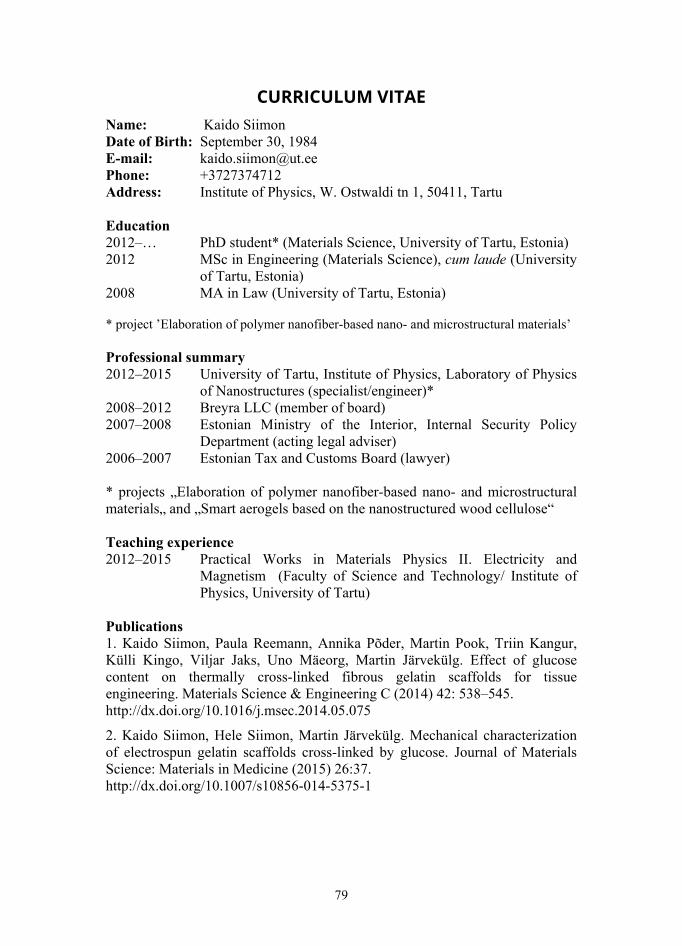
79
CURRICULUM VITAE Name: Kaido Siimon Date of Birth: September 30, 1984 E-mail: [email protected] Phone: +3727374712 Address: Institute of Physics, W. Ostwaldi tn 1, 50411, Tartu Education 2012–… PhD student* (Materials Science, University of Tartu, Estonia) 2012 MSc in Engineering (Materials Science), cum laude (University
of Tartu, Estonia) 2008 MA in Law (University of Tartu, Estonia) * project ’Elaboration of polymer nanofiber-based nano- and microstructural materials’ Professional summary 2012–2015 University of Tartu, Institute of Physics, Laboratory of Physics
of Nanostructures (specialist/engineer)* 2008–2012 Breyra LLC (member of board) 2007–2008 Estonian Ministry of the Interior, Internal Security Policy
Department (acting legal adviser) 2006–2007 Estonian Tax and Customs Board (lawyer) * projects „Elaboration of polymer nanofiber-based nano- and microstructural materials„ and „Smart aerogels based on the nanostructured wood cellulose“ Teaching experience 2012–2015 Practical Works in Materials Physics II. Electricity and
Magnetism (Faculty of Science and Technology/ Institute of Physics, University of Tartu)
Publications 1. Kaido Siimon, Paula Reemann, Annika Põder, Martin Pook, Triin Kangur, Külli Kingo, Viljar Jaks, Uno Mäeorg, Martin Järvekülg. Effect of glucose content on thermally cross-linked fibrous gelatin scaffolds for tissue engineering. Materials Science & Engineering C (2014) 42: 538–545. http://dx.doi.org/10.1016/j.msec.2014.05.075
2. Kaido Siimon, Hele Siimon, Martin Järvekülg. Mechanical characterization of electrospun gelatin scaffolds cross-linked by glucose. Journal of Materials Science: Materials in Medicine (2015) 26:37. http://dx.doi.org/10.1007/s10856-014-5375-1
80
3. Kaido Siimon, Karol Mõisavald, Hele Siimon, Martin Järvekülg. Increasing mechanical strength of electrospun gelatin nanofibers by the addition of aluminum potassium sulfate. Journal of Applied Polymer Science (2015), 132 (35): 42431. http://dx.doi.org/10.1002/app.42431 Awards 2015 Memorial Stipend of Valda and Bernard Õun 2016 Outstanding Reviewer (Materials Science & Engineering C)
81
ELULOOKIRJELDUS Nimi: Kaido Siimon Sünniaeg: 30.09.1984 E-mail: [email protected] Telefon: +3727374712 Aadress: Füüsika instituut, W. Ostwaldi tn 1, 50411, Tartu Haridus 2012–… doktorant* (materjaliteadus, Tartu Ülikool, Eesti) 2012 MSc (materjaliteadus), cum laude (Tartu Ülikool, Eesti) 2008 MA (Tartu Ülikool, Eesti) * projekt ’Polümeersetel nanofiibritel põhinevate nano- ja mikrostruktuursete materjalide arendamine’ Töö 2012–2015 Tartu ülikool, füüsika instituut, nanostruktuuride füüsika labor
(spetsialist/insener)* 2008–2012 Breyra OÜ (juhatuse liige) 2007–2008 Siseministeerium, sisejulgeolekupoliitika osakond (õigus-
nõuniku kt) 2006–2007 Maksu- ja Tolliamet (jurist) * projektid „Elaboration of polymer nanofiber-based nano- and microstructural materials„ and „Smart aerogels based on the nanostructured wood cellulose“ Õpetamiskogemus 2012–2015 Materjalifüüsika praktikum II. Elekter ja magnetism (Tartu Ülikool, Füüsika Instituut) Publikatsioonid 1. Kaido Siimon, Paula Reemann, Annika Põder, Martin Pook, Triin Kangur, Külli Kingo, Viljar Jaks, Uno Mäeorg, Martin Järvekülg. Effect of glucose content on thermally cross-linked fibrous gelatin scaffolds for tissue engineering. Materials Science & Engineering C (2014) 42: 538–545. http://dx.doi.org/10.1016/j.msec.2014.05.075
2. Kaido Siimon, Hele Siimon, Martin Järvekülg. Mechanical characterization of electrospun gelatin scaffolds cross-linked by glucose. Journal of Materials Science: Materials in Medicine (2015) 26:37. http://dx.doi.org/10.1007/s10856-014-5375-1
82
3. Kaido Siimon, Karol Mõisavald, Hele Siimon, Martin Järvekülg. Increasing mechanical strength of electrospun gelatin nanofibers by the addition of aluminum potassium sulfate. Journal of Applied Polymer Science (2015), 132 (35): 42431. http://dx.doi.org/10.1002/app.42431 Auhinnad 2015 Valda ja Bernard Õuna mälestusstipendium (biomeditsiinialase
teadustöö eest) 2016 Outstanding Reviewer (Materials Science & Engineering C)
DISSERTATIONES SCIENTIAE MATERIALIS UNIVERSITATIS TARTUENSIS
1. Martin Järvekülg. Tubular microstructures by Hf-, Zr- and Ti-butoxide gel sheet rolling. Tartu, 2011, 112 p.
2. Sergei Vlassov. Investigation of nanoscale interactions and mechanical properties of nanostructures using quartz tuning fork based real-time mea-surements. Tartu, 2011, 171 p.
3. Margus Kodu. Pulsed Laser Deposition of Magnesium Oxide and Barium Ternary Oxides for Plasma Display Protective Layers. Tartu, 2011, 89 p.
4. Rainer Pärna. Surface studies of some oxide films. Tartu, 2011, 129 p. 5. Jevgeni Šulga. Self-assembly and interaction of nanostructures. Tartu,
2011, 114 p. 6. Wojciech Kuznik. Quantum-chemical computer simulations of the linear
and non-linear optical properties of pyrazoloquinoline and dicyanopyrazine derivatives. Tartu, 2012, 89 p.
7. Leonid Dorogin. Structural and tribological properties of zero- and one-dimensional nanocrystals. Tartu, 2012, 193 p.
8. Viljar Palmre. Fabrication and characterization of microporous carbon-based electroactive polymer actuators. Tartu, 2012, 99 p.
9. Madis Paalo. Synthesis of CNT-metal oxide nanocomposites: sol-gel process, rheology, structural and functional properties. Tartu, 2014, 175 p.
10. Raul Välbe. Developement of ionic liquid composites by sol-gel method for elaboration of industrial nano- and microstructures. Tartu, 2014, 97 p.
11. Urmas Joost. Impurity and preparation dependent properties of titania thin films. Tartu, 2014, 155 p.
12. Madis Umalas. Application of sol-gel technology for production of ceramic nanocomposites and functional coatings. Tartu, 2015, 136 p.
13. Elena Samsonova. Energy transfer probe as a tool to study morphological and structural origins of fluorescence quenching in rare-earth doped nano-phosphors. Tartu, 2015, 114 p.
14. Jay Mondal. Novel Corrosion Protective Nanostructured Composite Coatings. Tartu, 2016, 104 p.
15. Kathriin Utt. Metal oxide mesostructures for optical applications. Tartu, 2016, 88 p.