jumping plant lice of the family carsidaridae (hemiptera .... bio. & env. sci. 2015 1 ......
TRANSCRIPT
J. Bio. & Env. Sci. 2015
1 | Yana et al.
RESEARCH PAPER OPEN ACCESS
Jumping plant lice of the family Carsidaridae (Hemiptera :
Psylloidea) from Cameroon: taxonomic, faunisitic, phenology
and host plants
W. Yana1,2, Y.P. Ndankeu Mveyo1,3, V.J. Dzokou1, J.L. Tamesse1*
1Laboratory of Zoology, Higher Teacher’s Training College, University of Yaounde I, Yaounde,
Cameroon
2Laboratory of Biological Science, Faculty of Sciences, University of Bamenda, Bambili, Cameroon
3Laboratory of Zoology, Faculty of Science, University of Yaounde I, Yaounde, Cameroon
Article published on June 05, 2015
Key words: Psyllids, Carsidaridae, Taxonomy, Faunistic, Cameroon.
Abstract
Cameroonian psyllids members of the Carsidaridae family are revised. Six species are recognized within this
family including three known species, two newly described species; one species remain undescribed because the
material is not sufficient. The newly described species are Carsidara camerunensis sp.n., psyllid of Sterculia
tragacantha and Mesohomotoma njinei sp.n., psyllid of Desplatsia dewevrei. Adults and fifth instars’ larvae of
the five Carsidaridae species are diagnosed and illustrated. The host plants of the five described species belong in
Malvaceae family. The psyllid outbreak period of each species depends on the availability of young leaves on host
plants. Then, the population dynamic of each species depends mainly on the phenology of the host plant which
depends of climatic factors.
*Corresponding Author: J.L. Tamesse [email protected]
Journal of Biodiversity and Environmental Sciences (JBES) ISSN: 2220-6663 (Print) 2222-3045 (Online)
Vol. 6, No. 6, p. 1-20, 2015
http://www.innspub.net
J. Bio. & Env. Sci. 2015
2 | Yana et al.
Introduction
Psylloids are small plant sap-sucking insects of
Hemiptera commonly called jumping plant lice. As
other hemipterans, jumping plant lice have piercing-
sucking mouth-parts. When feeding, the mandibular
and maxillary stylets are inserted into the host tissue,
saliva is injected and then the liquid food is absorbed.
Before feeding, the insects probe more or less
extensively. The probing also involves injection of
saliva, which is particularly relevant in species which
transmit bacterial or viral pathogens (Burckhardt and
De Queiroz, 2012). Currently, 3850 species have been
described world-wide (Li, 2011), which is probably
less than half of the existing number of species.
Recent revised classification of psyllid indicated that
eight families could be defined: Aphalaridae,
Calophyidae, Carsidaridae, Homotomidae, Liviidae,
Phacopteronidae, Psyllidae and Triozidae
(Burckhardt and Ouvrard, 2012). Psyllids are
distributed among all biogeographic regions but they
are most numerous in warmer regions (Burckhardt,
1994). Their distribution in the tropical regions of
Africa is poorly known, therefore they could exist
numerous undescribed psyllid species from this part
of world.
The first biodiversity data of psyllids from Cameroon
was published by (Tamesse et al., 2007); these
authors listed 35 species of Triozidae. Since, Dzokou
et al. (2009a), in the West Region of Cameroon, listed
37 species of Psyllidae family; Yana et al. (2009), in
the Centre Region of Cameroon, listed 11 species of
Phacopteronidae family; Yana et al. (2010) listed, in
the Centre Region of Cameroon, 45 species of
Psyllidae family and Mveyo et al. (2011) listed, in the
South Region of Cameroon, 35 species of Psyllidae.
Several species recorded by those authors are new
records. Until now no record of the biodiversity of
psyllid of Carsidaridae family from Cameroon is not
yet described. Carsidarids psyllid group, according to
Heslop-Harrison (1958), are considers as a tribe of
the subfamily Ciriacreminae. In this subfamily, the
author places 6 genera Carsidara, Mesohomotoma,
Mastigimas, Protyora, Epicarsa and Diceraopsylla.
Vondráček (1957) considers Carsidaridae as a group
close to Triozidae. For his part, Becker-Migdisova
(1973) recognizes seven subfamilies among
Carsidaridae: Calophyinae, Pauropsyllinae,
Leptynopterinae, Phacopterinae, Tenaphalarinae,
Carsidarinae and Homotominae. The family gathers
by Becker-Migdisova (1973), a large number of genera
among Psylloidea. White and Hodkinson (1985)
conducted a comprehensive definition of
Carsidaridae; two subfamilies are retained by the
authors in this family: Mastigimatinae- Becker
Migdisova (1973) and Carsidarinae Crawford with the
following generas Mastigimas Enderlein,
Tenaphalara Kuwayama, Protyora Kieffer,
Mesohomotoma Kuwayama, Paracarsidara Heslop-
Harrison and Carsidara Walker. Hollis (1987)
reconsider the classification of Mastigimas and
transfers it to the Calophyidae family. Burckhardt and
Ouvrard (2012), in the newly psyllid classification
confirmed Carsidaridae as a family within the
Hemiptera Psylloidea.
The psyllids of Carsidaridae family are associated
with host plants of the families Sterculiaceae,
Bombacaceae and Malvaceae, ie the plants of
Malvales group (Hollis (1987),. The carsidarids host
plants are of some economic importance. Theobroma
cacao L. (Sterculiaceae) originated from tropical
America has been introduced in Cameroon. This plant
is cultivated in various regions: South, Centre, East,
West and Littoral (Mbondji, 1984). Cocoa is a major
export crop in several West African countries. The
update of the study of the population dynamic of the
cocao psyllid is indispensable for an integrated pest
management.
Previously in Cameroon, very few scientific notes
included psyllids members of carsidarids group:
Mesohomotoma tessmanni (Aulmann) (Messi,
1986), M. hollisi Messi (Messi and Nguefang, 1993)
and Tenaphalara camerunus (Aulmann) (Hollis,
1987). No other records of Carsidaridae psyllids were
published from this country. This paper described the
biodiversity of psyllids of Carsidaridae family from
J. Bio. & Env. Sci. 2015
3 | Yana et al.
Cameroon, provided keys for their identification and
the taxonomic description of two new species within
his family. Regular field surveys give us useful also
information of the population dynamic of species of
economic importance such as cocoa psyllid.
Materials and methods
Study sites
Psyllids were sampled in four sites for regular
monthly prospection: Kala (3°47’ N, 11°24’E),
Minkoameyos (3°51’N, 11°31’E), Nkomilong (3°47’ N,
11°24’E) and Soa (3°57’N, 11°36’E). The four localities
are in the Center region of Cameroon, central Africa.
Population dynamic study of psyllid
Field surveys, for population dynamic study, took
place for a period of 24 months (January 2006-
December 2007). Psyllids for its various
developmental stages were counted on a selected
branch of five host plant in each locality. Adult
psyllids were captured with a sweep net of 0.5 mm
mesh size and an aspirator. Larvae were sampled
directly from buds and leaves of the host plant
Taxonomic study, terminology and abbreviations
Drawings were made under a microscope with slide-
mounted specimens of insects. Morphological
terminology follows Hollis (1973, 1987); and
Ossiannilsson(1992). The following abbreviations are
used: LZUY= Laboratory of Zoology, University of
Yaounde I; RMCA= Royal Museum of Central Africa;
NHMB= Naturhistorisches Museum Basel,
Switzerland; NHY= National Herbarium of Yaounde,
Cameroon. The following abbreviations are used in
the descriptions and measurement tables. Adult: BL,
body length; BW, body width; HW, head width; AL,
antenna length; F1, length of first antennal
flagellomere; WL, forewing length; WW, forewing
width; wL, hindwing length; wW, hindwing width;
MTL, metatibial length; MFL, metafemur length; MP,
male proctiger length; PL, paramere length; DL,
length of distal segment of aedeagus; FP, female
proctiger length; SL, female subgenital plate length.
Fifth instar larva: BL, body length; BW, body width;
AL, antenna length; FL, forewing-pad length; ML,
metatibial length.
Museum specimen deposit
The specimens are preserved dry and slide-mounted
or in 70% ethanol and are deposited in LZUY, RMCA
and NHMB. The host plants were identified at NHY.
Drawings and measurements were made from slide-
mounted material.
Results and discussion
Carsidaridae Crawford
Carsidaridae synonymies and diagnosis characters are
given by (Hollis, 1987). (Burckhardt and Ouvrard,
2012) recently confirmed the family status of
Carsidarisae and listed the nine genera within this
family along with the type species of each genus. The
taxonomic of Carsidaridae species from Cameroon
follows the classification of (Hollis, 1987) and
(Burckhardt and Ouvrard, 2012).
Table 1. Measurements (in mm) of last instar larvae of Carsidaridae species (N= number of measured
specimens).
Species N BL BW AL FL MTL BL/BW BL/AL AL/FL
Carsidara camerounensis sp.n. 6 3.8 1.6 1.8 1.3 1.1 2.4 2.1 1.4
Mesohomotoma tessmanni 15 2.4 0.5 1.2 0.7 0.4 4.8 2 1.7
Mesohomotoma hibisci 30 2.6 0.9 1.7 0.8 0.7 2.8 1.5 2.1
Mesohomotoma njinei sp.n. 12 3.1 0.8 1.9 0.8 0.7 3.8 1.6 2.4
Tenaphalara camerunus 6 2.9 0.9 1.1 0.7 0.5 3.2 2.6 1.6
Psyllid species belonging in Carsidaridae family are
characterized by: antennal sockets enlarged and
swollen ventromedially and vertex often deeply
divided by median giving the head a cleft appearance
in dorsal view. Antennal flagellum with single
subapical rhinarium presents on flagellomere 3 in
addition to those on flagellomeres 2, 4, 6 and 7. False
rs-m cross-vein present in forewing or rs and M1+2 in
J. Bio. & Env. Sci. 2015
4 | Yana et al.
broad contact, costal break absent. Hind tibia with a
well-developed basal spine; hind basitarsus with a
single apical spur. Male subgenital plate with a pair of
secondary lobes anterior to parameres, these lobes
appear to be sclerotised projections arising from the
membrane lining the inner surface of the subgenital
plate (Hollis, 1987). Final instar larva elongate,
clearly divided into head, thorax and abdomen;
antennae elongate, 10 segmented; legs elongate,
tarsal arolium sessile and fan-shaped or globular;
wing buds small, without humeral lobes; thoracic
sclerites poorly differentiated; caudal region of
abdomen differentiated and bearing convoluted pore
bands, anus terminal or termino dorsal; body setae
mainly simple but scattered, small, lanceolate setae
present on caudal sclerites marginally and
submarginally (Hollis, 1987).
Table 2. Measurements (in mm) of male adult of Carsidaridae species (N= number of measured specimens).
Species N BL BW HW AL F1 WL WW wL ww MTL MFL MP PL DL
Carsidara camerounensis sp.n 20 6.7 1.8 1.1 4.1 0.6 7.5 2.9 4.7 1.8 1.4 1.2 0.5 0.4 0.4
Mesohomotoma tessmanni 5 2.5 0.8 0.6 1.7 0.3 3.6 1.2 2.5 0.8 0.7 0.6 0.4 0.4 0.2
Mesohomotoma hibisci 35 3.8 0.8 0.7 2.6 0.4 4.2 1.4 2.5 0.8 0.8 0.7 0.3 0.4 0.4
Mesohomotoma njinei sp.n. 9 4.5 0.8 0.7 2.5 0.4 4.7 1.6 2.9 0.9 1.0 0.8 0.3 0.4 0.3
Tenaphalara camerunus 3 3.3 0.6 0.6 1.4 0.3 2.9 0.9 1.9 0.6 0.6 0.5 0.3 0.3 0.3
Key to Carsidaridae genera from Cameroon
1. Forewing narrowing to a subacute apex, Rs and
M1+2 not in contact but connected by a false rs-m
cross-vein (Fig. 32, 33), cu1 much wider than high and
with a value of at least 1.7; antennal flagellum
elongate, 1st flagellar segment long and narrow, not
less than nine times longer than its greatest width.
Table 3. Measurements (in mm) of female adult of Carsidaridae species (N= number of measured specimens).
Species N BL BW HW AL F1 WL WW wL ww MTL MFL FP SL
Carsidara camerounensis sp.n. 20 6.8 1.9 1.2 4.0 0.6 8.1 3.2 5.2 1.7 1.4 1.2 1.2 0.5
Mesohomotoma tessmanni 8 3.0 0.9 0.6 1.7 0.4 4.0 1.4 2.6 0.8 0.7 0.6 0.7 0.5
Mesohomotoma hibisci 35 4.3 0.9 0.8 2.7 0.4 4.9 1.6 3.0 0.9 0.9 0.8 1.0 0.9
Mesohomotoma njinei sp.n. 4 4.4 0.9 0.8 2.6 0.4 5.2 2.0 3.2 1.2 1.1 0.9 1.1 0.7
Tenaphalara camerunus 4 3.6 0.7 0.6 1.5 0.3 3.4 0.9 2.3 0.6 0.5 0.4 0.6 0.4
2. Pterostigma absent and M+Cu about half as long as
Cu stem; male proctiger bipartite, with a large, anvil-
shaped median posterior lobe in addition to lateral
lobes (fig. 52, 53, 54)….Mesohomotoma. Pterostigma
present (Fig. 31), male proctiger unipartite, without
median posterior lobe and lateral lobes (Fig. 51).
Table 4. Ratios of male adult of Carsidaridae species.
Species BL/BW AL/HW AL/F1 BL/HW WL/HW WL/WW WL/wL wL/ww MTL/HW MP/HW PL/HW
C. camerounensis 3.7 3.7 6.8 6.1 6.8 2.6 1.6 2.6 1.3 0.5 0.4
M. tessmanni 3.1 2.8 5.6 4.2 6.0 3.0 1.4 3.1 1.2 0.6 0.6
M. hibisci 4.7 3.7 6.5 5.4 6.0 3.0 1.7 3.1 1.1 0.4 0.6
M. njinei 5.6 3.6 6.2 6.4 6.7 2.9 1.6 3.2 1.4 0.4 0.6
T. camerunus 5.5 2.3 4.6 5.5 4.8 3.2 1.5 3.2 1.0 0.5 0.5
3. M+Cu very short, about one third as long as R stem
and less than half as long as Cu stem (Fig. 31); female
terminalia, in profile, rounded dorsally, ventrolateral
margins of proctiger with dense fringes of setae,
lateral palps ridged (Fig. 65)...Carsidara -M+Cu
longer, about as long as or longer than R stem and Cu
stem (Fig. 40); female terminalia, in profile conical,
proctiger sometimes with a median lobe posterior to
anal pore, lateral palps not ridged (Fig.69).
4. False r1-rs crossveing absent (Fig. 40.

J. Bio. & Env. Sci. 2015
5 | Yana et al.
5. Radular area absent in cu1a, claval suture reaching
hind margin of forewing distant from apex of Cu1b
(Fig.40) Tenaphalara.
Carsidara Walker 1869
Carsidara synonymies and diagnosis characters are
given by (Hollis, 1987).
Carsidara camerunensis Tamesse sp.n.
Fifth instar larva.
Colouration
body orange brown dorsally and pale yellow to
ochreous ventrally; compound eyes reddish; claws
dark brown.
Table 5. Ratios of female adult of Carsidaridae species.
Species BL/BW AL/HW AL/F1 BL/HW WL/HW WL/WW WL/wL wL/ww MTL/HW FP/HW FP/SL
Carsidara camerounensis sp.n 3.6 3.3 6.6 5.6 6.7 2.5 1.5 3.0 1.2 1.0 2.4
Mesohomotoma tessmanni 3.3 2.8 4.2 5.0 6.6 2.8 1.5 3.2 1.2 1.2 1.4
Mesohomotoma hibisci 4.7 3.4 6.7 5.4 6.1 3.1 1.6 3.3 1.1 1.2 1.1
Mesohomotoma njinei sp.n. 4.8 3.2 6.5 5.5 6.5 2.6 1.6 2.6 1.4 1.4 1.6
Tenaphalara camerunus 5.1 2.5 5.0 6.0 5.6 3.7 1.5 3.8 0.8 1.0 1.5
Structure
Final instar larva (Fig. 1) elongate, not clearly divided
into head, thorax and abdomen. Antenna elongate, 10
segmented; flagellum distinctly subdivided with two
subapical rhinaria present on flagellomeres 2, 3, 4,
and a single subapical rhinarium on flagellomeres 6,
7, 8 (Fig. 2). Antennae, legs, head, thorax and
abdomen are covered by minute lanceolate setae.
Dorsally, abdomen with three stronger short setae in
its margin. Wing pads, with many marginal lanceolate
setae. Legs elongate, tarsal arolium sessile and
globular (Fig. 3). Caudal region of abdomen
differentiated and bearing a convoluted pore band
and many oval patches of pores (Fig. 4). Anus in
ventral and terminal position. Measurements and
ratios in table 1.
Fig. 1-8. Carsidaridae fifth instar larvae. 1,2,3,4, Carsidara camerunensis ; 5,6,7,8, Tenaphalara camerunus.
Scale lines : 0.3 mm (1) ; 0.06 mm (2) ; 0.15 mm (3) ; 0.08 mm (4, 6) ; 0.10 mm (5) ; 0.04 mm (7, 8).
J. Bio. & Env. Sci. 2015
6 | Yana et al.
Adult
Colouration
Overall body color brown with dark brown markings
dorsally. Antennal flagellomeres 1-5 dark-brown
apically, eyes dark-brown. Forewings yellowish with a
large brown dot in cu2b cell. Hindwings transparent;
spurs and claws dark-brown. Vertex with a dark-
brown transverse band.
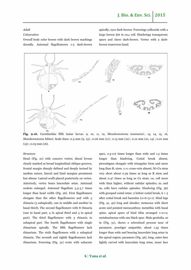
Fig. 9-16. Carsidaridae fifth instar larvae. 9, 10, 11, 12, Mesohomotoma tessmanni ; 13, 14, 15, 16,
Mesohomotoma hibisci. Scale lines: 0.3 mm (9, 13) ; 0.06 mm (11) ; 0.15 mm (12) ; 0.12 mm (10, 14) ; 0.01 mm
(15) ; 0.03 mm (16).
Structure
Head (Fig. 21) with concave vertex; discal foveae
clearly marked as broad longitudinal oblique grooves,
frontal margin sharply defined and deeply incised by
median suture, lateral and hind margins prominent
but obtuse. Lateral ocelli placed posteriorly on vertex.
Anteriorly, vertex bears lanceolate setae. Antennal
sockets enlarged. Antennal flagellum 3.3-3.7 times
longer than head width (Fig. 26). First flagellomere
elongate than the other flagellomeres and with 5
rhinaria (3 subapically, one in middle and another in
basal third). The second flagellomere with 8 rhinaria
(one in basal part, 4 in apical third and 3 in apical
part). The third flagellomere with 3 rhinaria in
subapical part. The fourth flagellomere with single
rhinarium apically. The fifth flagellomere lack
rhinarium. The sixth flagellomere with 2 subapical
rhinaria. The seventh and eighth flagellomeres lack
rhinarium. Forewing (Fig. 31) ovate with subacute
apex, 2.5-2.6 times longer than wide and 1.5 times
longer than hindwing. Costal break absent,
pterostigma elongate with triangular form and more
long than R1 stem. r1-rs cross-vein absent; M+Cu stem
very short about 0.39 times as long as R stem and
about 0.47 times as long as Cu stem; cu1 cell more
wide than higher, without radular spinules; m1 and
m2 cells have radular spinules. Hindwing (Fig. 36)
with grouped costal setae: 2 before costal break, 6 + 5
after costal break and hamulus (2+6+5+1). Hind legs
(Fig. 41, 42) long and slender; metacoxa with short
acute and pointed meracanthus; metatibia with basal
spine, apical spurs of hind tibia arranged 1+2+2;
metabasitarsus with one black spur. Male genitalia, as
in (Fig. 51), shows a sclerotized process near of
paramere; proctiger unipartite, about 1.55 times
longer than wide and bearing lanceolate long setae in
the apical region; paramere (Fig. 56), long, outer face
lightly curved with lanceolate long setae, inner face
J. Bio. & Env. Sci. 2015
7 | Yana et al.
hightly concaved with lanceolate long setae,
posteroapical lobe in addition to posteroapical hook;
apical segment of aedeagus highly modified (Fig. 60),
end tube of ductus ejaculatorius heavily sclerotised.
Female genitalia as in (Fig. 65); proctigere without
posterodorsal lobe, apex strongly sclerotised
upcurved, posterolateral margins with fringes of long
setae, circumanal compressed anteriorly with two
rows of pores, subgenital plate short apex lightly
rounded, external margin with lanceolate long setae,
inner margin with lanceolate short setae, lateral
valvulae swallowed up in ventral valvula.
Measurements and ratios in tables 2, 3, 4 and 5.
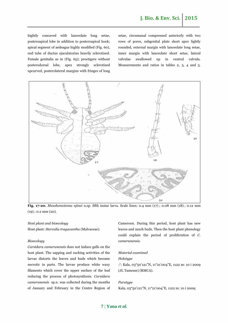
Fig. 17-20. Mesohomotoma njinei n.sp. fifth instar larva. Scale lines: 0.4 mm (17) ; 0.08 mm (18) ; 0.12 mm
(19) ; 0.2 mm (20).
Host plant and bioecology
Host plant: Sterculia tragacantha (Malvaceae).
Bioecology
Carsidara camerunensis does not induce galls on the
host plant. The sapping and sucking activities of the
larvae distorts the leaves and buds which become
necrotic in parts. The larvae produce white waxy
filaments which cover the upper surface of the leaf
reducing the process of photosynthesis. Carsidara
camerunensis sp.n. was collected during the months
of January and February in the Centre Region of
Cameroon. During this period, host plant has new
leaves and much buds. Then the host plant phenology
could explain the period of proliferation of C.
camerunensis.
Material examined
Holotype
♂: Kala, 03°50’121”N, 11°21’004”E, 1122 m: 10 i 2009
(JL Tamesse) (RMCA).
Paratype
Kala, 03°50’121”N, 11°21’004”E, 1122 m: 10 i 2009;
J. Bio. & Env. Sci. 2015
8 | Yana et al.
20 ♂, 21♀, 8 larvae; Minkoameyos, 03°52’290”N,
11°25’420”E, 740 m: 18 ii 2007, 2 larvae; Nkomilong,
03°49’954”E, 1161 m: 29 i 2007, 2♂, 3♀, 4 larvae; 19
ii 2007, 3♂, 2♀, 2 larvae.
Comment
(Hollis, 1987) described, in the Afro-tropical one
species: Carsidara africana Hollis. Carsidara
camerunensis sp.n. described in this work from
Cameroon is different from C. africana by the
following characters: the posterior margin of the
vertex of C. africana is concave and that of C.
camerunensis sp.n. is inverted V-shaped. The
antennal flagellomeres 1-6 of C. africana are dark
brown apically and flagellomeres 7-8 entirely dark
brown but on C. camerunensis sp.n., the
flagellomeres 1-5 are dark brown apically and
flagellomeres 7-8 are yellowish; antennal flagellum of
C. africana 2.7- 3.0 times longer than head width,
and the one ofC. camerunensis sp.n. is 3.3-3.7 times
longer than head width; first flagellomere of C.
africana, with 6-12 rhinaria in apical third, second
flagellomere with 10-20 rhinaria in apical half, third
flagellomere with 5-7 rhinaria in apical quarter,
fourth flagellomere with 2 subapical rhinaria and up
to 5 more in apical third, fifth flagellomere with 0 or 1
subapical rhinarium and up to 3 more in apical third,
seventh flagellomere with a single subapical
rhinarium but first flagellomere of C. camerunensis
sp.n., with 5 rhinaria (3 subapically, one in middle
and another in basal third), second flagellomere with
8 rhinaria (one in basal part, 4 in apical third and 3 in
apical part), third flagellomere with 3 rhinaria in
subapical part, fourth flagellomere with single
rhinarium apically, fifth flagellomere lack rhinarium,
sixth flagellomere with 2 subapical rhinaria, seventh
and eighth flagellomeres lack rhinarium. The
pterostigma is small, about 3 times longer than wide
in C. Africana and greater, about 4.7 times longer
than wide, in C. camerunensis sp.n. On C. africana
forewing, M+Cu stem about 0.33 times as long as R
stem and about 0.5 times as long as Cu stem but on C.
camerunensis sp.n. forewing, M+Cu stem about 0.35
times as long as R stem and about 0.6 times as long
Cu stem. Cu1 cubital cell is about 3 times longer than
wide in C. camerunensis sp.n. and about 2 times as
long as wide in C. africana. The posterior lateral
expansion of the male proctiger is convex in C.
camerunensis sp.n. and relatively more slender than
in C. africana male proctiger; the male proctiger
about 1.4 times longer than wide on C. africana and
about 1.5 times longer than wide on C. camerunensis
sp.n. The apex of the subgenital plate process is
slightly cracked in C. camerunensis sp.n. and sharp
and pointed in C. africana. The proximal portion of
the paramere has a square section in C. camerunensis
sp.n. and rounded in C. africana. The process of
ventral apex of aedeagus in C. Africana is longer than
the one of C. camerunensis sp.n.
Etymology
The species is named after the country, Cameroun
where this work was conducted. This species is the
first psyllid species described within the genus
Casidara from Cameroon.
Tenaphalara camerunus (Aulmann): redescription
Fifth instar larva.
Colouration
Body whitish; eyes reddish; antennal flagellomeres 2,
4 and 6 apically dark-brown, flagellomeres 7 and 8
entirely dark-brown; claws and spurs of hind leg dark
brown.
Structure
Final instar larva (Fig. 5), elongate and oblate
dorsoventrally; antenna 10 segmented; flagellum
distinctly subdivided with one apical rhinarium
present on flagellomeres 2, 4, 6 and 7 (Fig. 6);
antennae, legs, head and abdomen are covered by
minute lanceolate setae; wing pads elongate and well-
developed without setae; metatibia apically with 5
spurs, metabasitarsus and metatarsus with one 1 spur
each, tarsal arolium sessile and globular (Fig. 7);
caudal region of abdomen differentiated and bearing
a convoluted pore bands (Fig. 8); anus in ventral
position. Measurements and ratios in table 1.
J. Bio. & Env. Sci. 2015
9 | Yana et al.
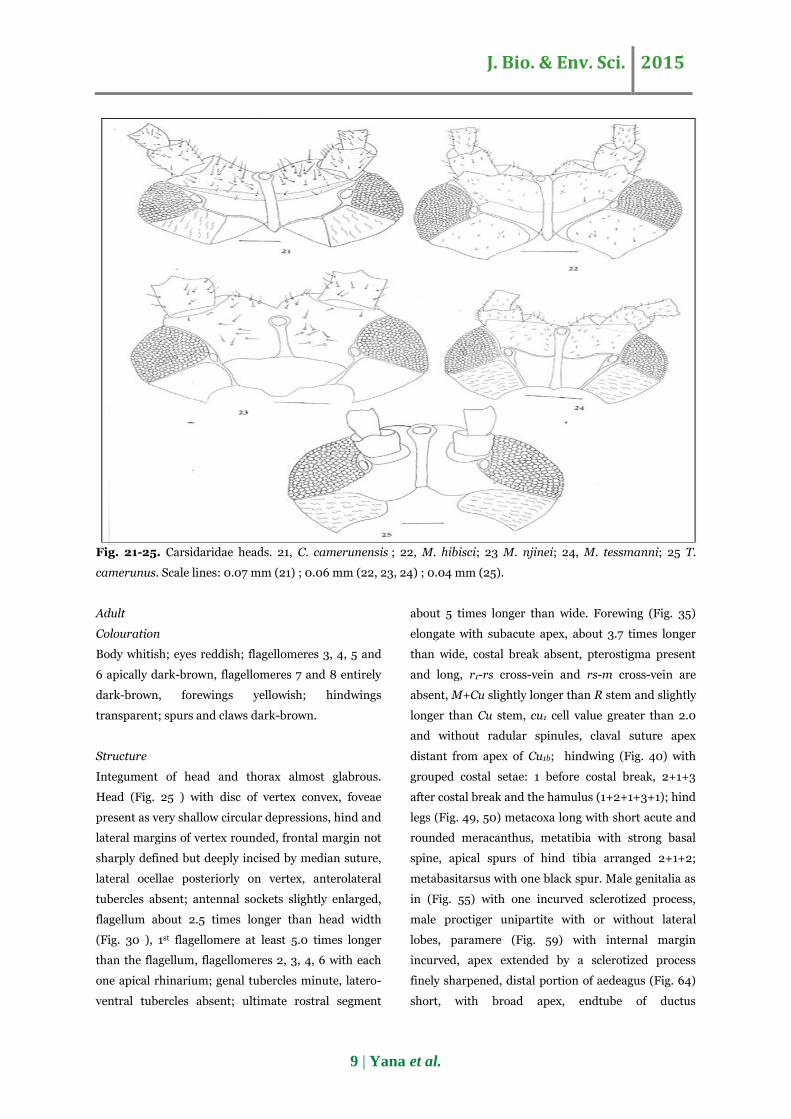
Fig. 21-25. Carsidaridae heads. 21, C. camerunensis ; 22, M. hibisci; 23 M. njinei; 24, M. tessmanni; 25 T.
camerunus. Scale lines: 0.07 mm (21) ; 0.06 mm (22, 23, 24) ; 0.04 mm (25).
Adult
Colouration
Body whitish; eyes reddish; flagellomeres 3, 4, 5 and
6 apically dark-brown, flagellomeres 7 and 8 entirely
dark-brown, forewings yellowish; hindwings
transparent; spurs and claws dark-brown.
Structure
Integument of head and thorax almost glabrous.
Head (Fig. 25 ) with disc of vertex convex, foveae
present as very shallow circular depressions, hind and
lateral margins of vertex rounded, frontal margin not
sharply defined but deeply incised by median suture,
lateral ocellae posteriorly on vertex, anterolateral
tubercles absent; antennal sockets slightly enlarged,
flagellum about 2.5 times longer than head width
(Fig. 30 ), 1st flagellomere at least 5.0 times longer
than the flagellum, flagellomeres 2, 3, 4, 6 with each
one apical rhinarium; genal tubercles minute, latero-
ventral tubercles absent; ultimate rostral segment
about 5 times longer than wide. Forewing (Fig. 35)
elongate with subacute apex, about 3.7 times longer
than wide, costal break absent, pterostigma present
and long, r1-rs cross-vein and rs-m cross-vein are
absent, M+Cu slightly longer than R stem and slightly
longer than Cu stem, cu1 cell value greater than 2.0
and without radular spinules, claval suture apex
distant from apex of Cu1b; hindwing (Fig. 40) with
grouped costal setae: 1 before costal break, 2+1+3
after costal break and the hamulus (1+2+1+3+1); hind
legs (Fig. 49, 50) metacoxa long with short acute and
rounded meracanthus, metatibia with strong basal
spine, apical spurs of hind tibia arranged 2+1+2;
metabasitarsus with one black spur. Male genitalia as
in (Fig. 55) with one incurved sclerotized process,
male proctiger unipartite with or without lateral
lobes, paramere (Fig. 59) with internal margin
incurved, apex extended by a sclerotized process
finely sharpened, distal portion of aedeagus (Fig. 64)
short, with broad apex, endtube of ductus
J. Bio. & Env. Sci. 2015
10 | Yana et al.
ejaculatorius simple. Female genitalia as in (Fig. 69);
proctiger with a well-developed dorsal lobe, anus
terminal, circumanal expanded and cover over the ½
of proctiger length with two rows of pores, subgenital
plate apex short. Measurements and ratios in tables 2,
3, 4 and 5.
Host plant and bioecology
Host plant
Ceiba pentandra (Malvaceae).
Bioecology
Tenaphalara camerunus does not induce damage
visible on the host. The larvae produce white wax
filaments which covers buds of the host plant. T.
camerunus is not frequent on his host plant during
the year. But the highest number of individuals was
noted on February in the Centre Region of Cameroon.
During that period the host plant renews its leaves
and buds. Then the host plant phenology could
explain the period of proliferation of T. camerunus.
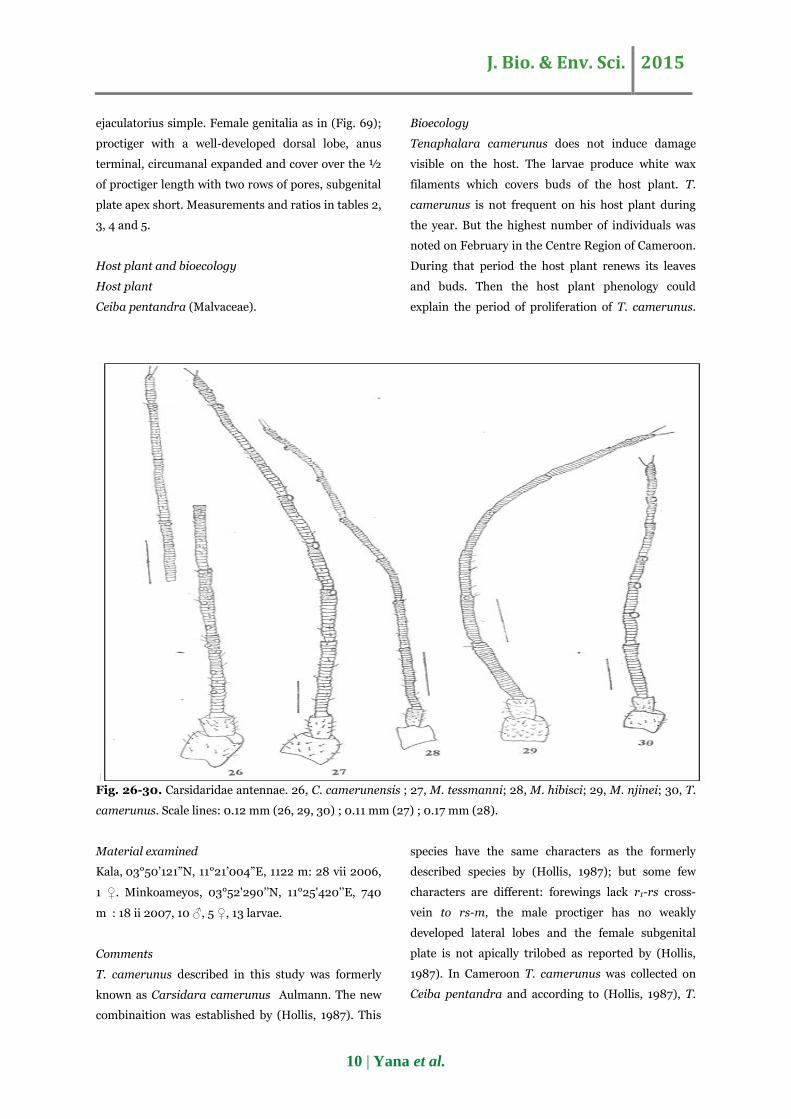
Fig. 26-30. Carsidaridae antennae. 26, C. camerunensis ; 27, M. tessmanni; 28, M. hibisci; 29, M. njinei; 30, T.
camerunus. Scale lines: 0.12 mm (26, 29, 30) ; 0.11 mm (27) ; 0.17 mm (28).
Material examined
Kala, 03°50’121”N, 11°21’004”E, 1122 m: 28 vii 2006,
1 ♀. Minkoameyos, 03°52'290’’N, 11°25'420’’E, 740
m : 18 ii 2007, 10 ♂, 5 ♀, 13 larvae.
Comments
T. camerunus described in this study was formerly
known as Carsidara camerunus Aulmann. The new
combinaition was established by (Hollis, 1987). This
species have the same characters as the formerly
described species by (Hollis, 1987); but some few
characters are different: forewings lack r1-rs cross-
vein to rs-m, the male proctiger has no weakly
developed lateral lobes and the female subgenital
plate is not apically trilobed as reported by (Hollis,
1987). In Cameroon T. camerunus was collected on
Ceiba pentandra and according to (Hollis, 1987), T.
J. Bio. & Env. Sci. 2015
11 | Yana et al.
camerunus feed on Ceiba pentandra, Bombax
buonopozense and B. sessile (Malvaceae).
Genus Mesohomotoma Kuwayama
Mesohomotoma synonymies and diagnosis characters
are given by (Hollis, 1987).
Included species of Mesohomotoma genus
Mesohomotoma tessmanni (Aulmann): redescription
Fifth instar larva.
Colouration
Body color orange brown, eyes reddish, claws and
spurs dark brown, caudal region more dark brown
than the other part of abdomen.
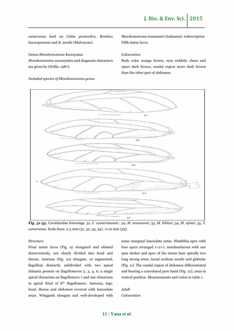
Fig. 31-35. Carsidaridae forewings. 31, C. camerunensis ; 32, M. tessmanni; 33, M. hibisci; 34, M. njinei; 35, T.
camerunus. Scale lines: 0.3 mm (31, 32, 33, 34) ; 0.12 mm (35).
Structure
Final instar larva (Fig. 9) elongated and oblated
dorsoventraly, not clearly divided into head and
thorax. Antenna (Fig. 10) elongate, 10 segmented;
flagellum distinctly subdivided with two apical
rhinaria present on flagellomeres 2, 3, 4, 6; a single
apical rhinarium on flagellomere 7 and one rhinarium
in apical third of 8th flagellomere. Antenna, legs,
head, thorax and abdomen covered with lanceolate
setae. Wingpads elongate and well-developed with
some marginal lanceolate setae. Hindtibia apex with
four spurs arranged 1+2+1; metabasitarsus with one
spur darker and apex of the tarsus bear apically two
long strong setae; tarsal arolium sessile and globular
(Fig. 11). The caudal region of abdomen differentiated
and bearing a convoluted pore band (Fig. 12); anus in
ventral position. Measurements and ratios in table 1.
Adult
Colouration
J. Bio. & Env. Sci. 2015
12 | Yana et al.
Overall body color light brown with dark brown
markings dorsally. The antennal flagellomeres 2-6
dark-brown apically, flagellomeres 7-8 entirely dark-
brown. Compound eyes reddish. Forewings orange
with a dark-brown dot in cu2b cell. Hindwings
transparent; spurs and claws dark-brown.
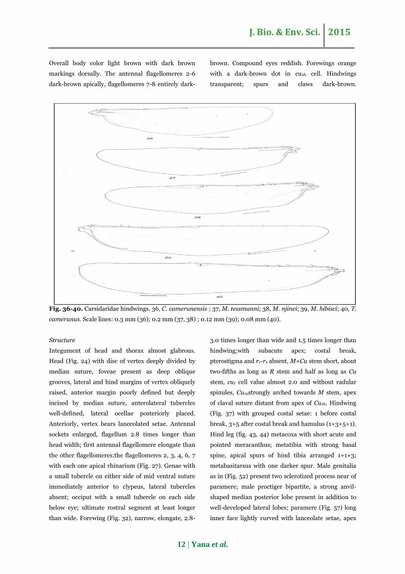
Fig. 36-40. Carsidaridae hindwings. 36, C. camerunensis ; 37, M. tessmanni; 38, M. njinei; 39, M. hibisci; 40, T.
camerunus. Scale lines: 0.3 mm (36); 0.2 mm (37, 38) ; 0.12 mm (39); 0.08 mm (40).
Structure
Integument of head and thorax almost glabrous.
Head (Fig. 24) with disc of vertex deeply divided by
median suture, foveae present as deep oblique
grooves, lateral and hind margins of vertex obliquely
raised, anterior margin poorly defined but deeply
incised by median suture, anterolateral tubercles
well-defined, lateral ocellae posteriorly placed.
Anteriorly, vertex bears lanceolated setae. Antennal
sockets enlarged, flagellum 2.8 times longer than
head width; first antennal flagellomere elongate than
the other flagellomeres;the flagellomeres 2, 3, 4, 6, 7
with each one apical rhinarium (Fig. 27). Genae with
a small tubercle on either side of mid ventral suture
immediately anterior to clypeus, lateral tubercles
absent; occiput with a small tubercle on each side
below eye; ultimate rostral segment at least longer
than wide. Forewing (Fig. 32), narrow, elongate, 2.8-
3.0 times longer than wide and 1.5 times longer than
hindwing;with subacute apex; costal break,
pterostigma and r1-rs absent, M+Cu stem short, about
two-fifths as long as R stem and half as long as Cu
stem, cu1 cell value almost 2.0 and without radular
spinules, Cu1astrongly arched towards M stem, apex
of claval suture distant from apex of Cu1b. Hindwing
(Fig. 37) with grouped costal setae: 1 before costal
break, 3+5 after costal break and hamulus (1+3+5+1).
Hind leg (fig. 43, 44) metacoxa with short acute and
pointed meracanthus; metatibia with strong basal
spine, apical spurs of hind tibia arranged 1+1+3;
metabasitarsus with one darker spur. Male genitalia
as in (Fig. 52) present two sclerotized process near of
paramere; male proctiger bipartite, a strong anvil-
shaped median posterior lobe present in addition to
well-developed lateral lobes; paramere (Fig. 57) long
inner face lightly curved with lanceolate setae, apex
J. Bio. & Env. Sci. 2015
13 | Yana et al.
sclerotised with two strong setae; aedeagus (Fig. 61)
narrow apically, apex of ductus ejaculatorius
prominent, strongly produced from aedeagal apex
and expanded apically. Female genitalia as in (Fig.
66), possess a large circumanal with two rows of
pores;female proctiger in profile, strongly stepped
posteriorly, apical part narrow elongate and bearing
short and thickened setae, apex weakly barbed;
subgenital plate long apex lightly pointed.
Measurements and ratios in tables 2, 3, 4 and 5.
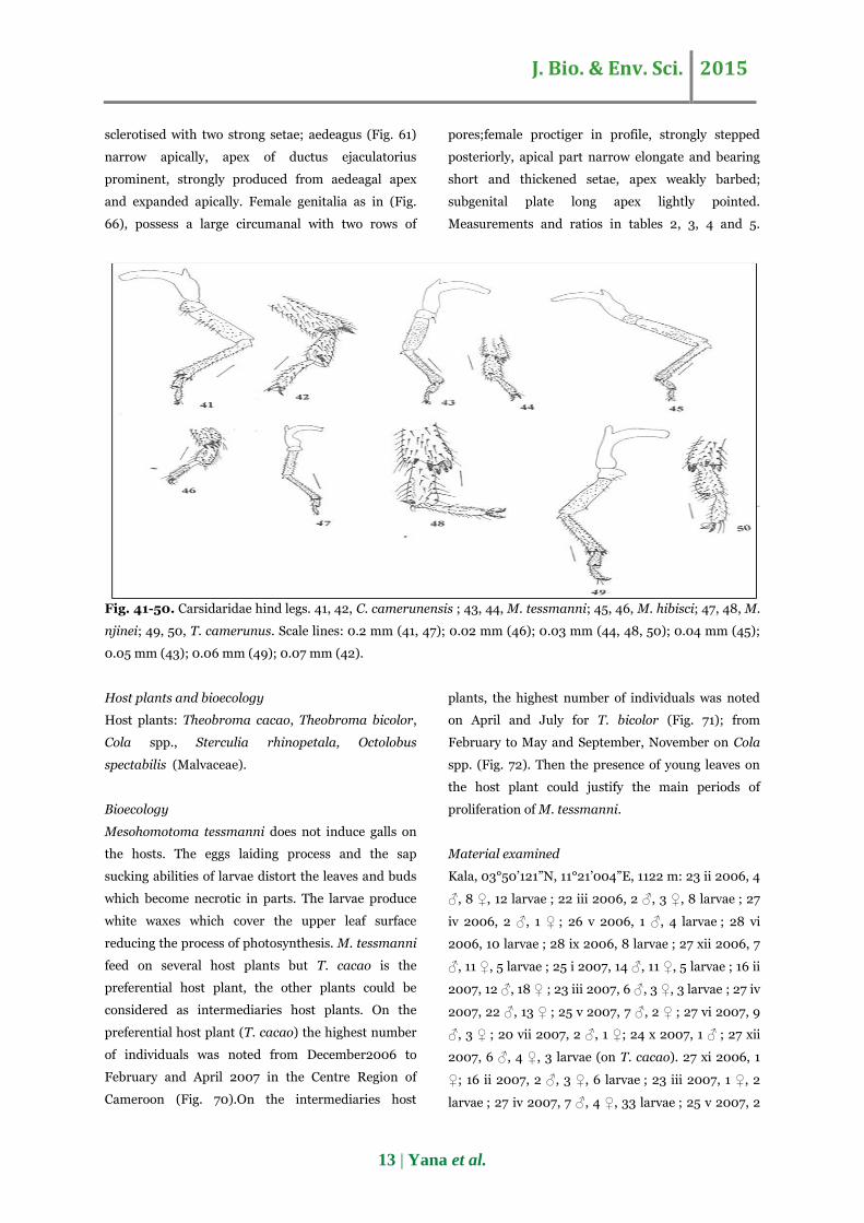
Fig. 41-50. Carsidaridae hind legs. 41, 42, C. camerunensis ; 43, 44, M. tessmanni; 45, 46, M. hibisci; 47, 48, M.
njinei; 49, 50, T. camerunus. Scale lines: 0.2 mm (41, 47); 0.02 mm (46); 0.03 mm (44, 48, 50); 0.04 mm (45);
0.05 mm (43); 0.06 mm (49); 0.07 mm (42).
Host plants and bioecology
Host plants: Theobroma cacao, Theobroma bicolor,
Cola spp., Sterculia rhinopetala, Octolobus
spectabilis (Malvaceae).
Bioecology
Mesohomotoma tessmanni does not induce galls on
the hosts. The eggs laiding process and the sap
sucking abilities of larvae distort the leaves and buds
which become necrotic in parts. The larvae produce
white waxes which cover the upper leaf surface
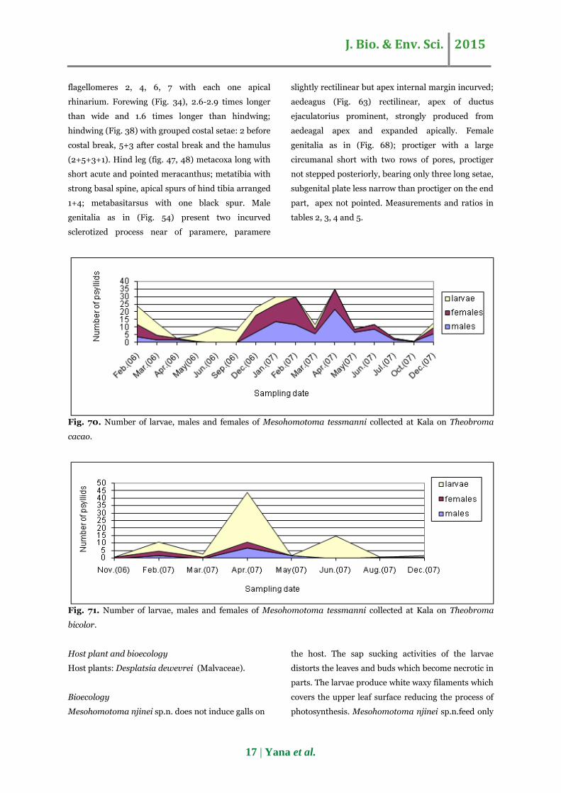
reducing the process of photosynthesis. M. tessmanni
feed on several host plants but T. cacao is the
preferential host plant, the other plants could be
considered as intermediaries host plants. On the
preferential host plant (T. cacao) the highest number
of individuals was noted from December2006 to
February and April 2007 in the Centre Region of
Cameroon (Fig. 70).On the intermediaries host
plants, the highest number of individuals was noted
on April and July for T. bicolor (Fig. 71); from
February to May and September, November on Cola
spp. (Fig. 72). Then the presence of young leaves on
the host plant could justify the main periods of
proliferation of M. tessmanni.
Material examined
Kala, 03°50’121”N, 11°21’004”E, 1122 m: 23 ii 2006, 4
♂, 8 ♀, 12 larvae ; 22 iii 2006, 2 ♂, 3 ♀, 8 larvae ; 27
iv 2006, 2 ♂, 1 ♀ ; 26 v 2006, 1 ♂, 4 larvae ; 28 vi
2006, 10 larvae ; 28 ix 2006, 8 larvae ; 27 xii 2006, 7
♂, 11 ♀, 5 larvae ; 25 i 2007, 14 ♂, 11 ♀, 5 larvae ; 16 ii
2007, 12 ♂, 18 ♀ ; 23 iii 2007, 6 ♂, 3 ♀, 3 larvae ; 27 iv
2007, 22 ♂, 13 ♀ ; 25 v 2007, 7 ♂, 2 ♀ ; 27 vi 2007, 9
♂, 3 ♀ ; 20 vii 2007, 2 ♂, 1 ♀; 24 x 2007, 1 ♂ ; 27 xii
2007, 6 ♂, 4 ♀, 3 larvae (on T. cacao). 27 xi 2006, 1
♀; 16 ii 2007, 2 ♂, 3 ♀, 6 larvae ; 23 iii 2007, 1 ♀, 2
larvae ; 27 iv 2007, 7 ♂, 4 ♀, 33 larvae ; 25 v 2007, 2
J. Bio. & Env. Sci. 2015
14 | Yana et al.
♂ ; 27 vi 2007, 15 larvae ; 23 viii 2007, 1 ♂ ; 27 xii
2007, 1 ♂, 1 larva (on T. bicolor). 27 iv 2006, 1 ♀, 5
larvae ; 28 ix 2006, 2 ♂, 10 larvae, 19 x 2006, 1 ♀ ; 27
xi 2006, 2 ♂, 6 ♀, 12 larvae ; 27 xii 2006, 1 ♂ ; 16 ii
2007, 2 ♂, 8 larvae ; 27 iv 2007, 22 ♂, 8 larvae ; 25 v
2007, 12 larvae (on Cola spp.). 22 ix 2007, 1 ♂, 1 ♀, 1
larva. Minkoameyos,03°52'290’’N, 11°25'420’’E, 740
m : 18 ii 2007, 4 ♂, 5 ♀, 1 larva ; 31 iii 2007, 3 larvae ;
29 iv 2007, 1 ♂. Soa, 03°58'112’’N, 11°35'435’’E, 674
m : 24 iii 2007, 1 ♀ (on Sterculia rhinopetala).
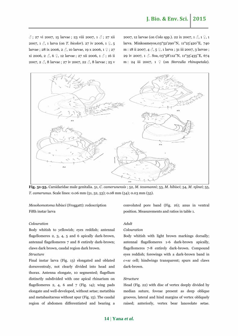
Fig. 51-55. Carsidaridae male genitalia. 51, C. camerunensis ; 52, M. tessmanni; 53, M. hibisci; 54, M. njinei; 55,
T. camerunus. Scale lines: 0.06 mm (51, 52, 53); 0.08 mm (54); 0.03 mm (55).
Mesohomotoma hibisci (Froggatt): redescription
Fifth instar larva
Colouration
Body whitish to yellowish; eyes reddish; antennal
flagellomeres 2, 3, 4, 5 and 6 apically dark-brown,
antennal flagellomeres 7 and 8 entirely dark-brown;
claws dark brown, caudal region dark brown.
Structure
Final instar larva (Fig. 13) elongated and oblated
dorsoventraly, not clearly divided into head and
thorax. Antenna elongate, 10 segmented; flagellum
distinctly subdivided with one apical rhinarium on
flagellomeres 2, 4, 6 and 7 (Fig. 14); wing pads
elongate and well-developed, without setae; metatibia
and metabasitarsus without spur (Fig. 15). The caudal
region of abdomen differentiated and bearing a
convoluted pore band (Fig. 16); anus in ventral
position. Measurements and ratios in table 1.
Adult
Colouration
Body whitish with light brown markings dorsally;
antennal flagellomeres 1-6 dark-brown apically,
flagellomeres 7-8 entirely dark-brown. Compound
eyes reddish; forewings with a dark-brown band in
c+sc cell; hindwings transparent; spurs and claws
dark-brown.
Structure
Head (Fig. 22) with disc of vertex deeply divided by
median suture, foveae present as deep oblique
grooves, lateral and hind margins of vertex obliquely
raised; anteriorly, vertex bear lanceolate setae.
J. Bio. & Env. Sci. 2015
15 | Yana et al.
Antennal (Fig. 28), sockets enlarged, flagellum 3.4-
3.7 times longer than head width; first antennal
flagellomere elongate than the other flagellomeres;
flagellomeres 1, 2, 3, 4, 6, 7 with one apical rhinarium
on each flegellomere. Forewing (Fig. 33), 3.1 times
longer than wide and 1.7 times longer than hindwing;
hindwing (Fig. 39) with grouped costal setae: 1 before
costal break, 3+3 after costal break (1+3+3). Hind leg
(Fig. 45, 46) metacoxa long with short acute and
pointed meracanthus; metatibia with strong basal
spine, apical spurs of hind tibia arranged 1+2+1+1;
metabasitarsus with one black spur. Male genitalia as
in (Fig. 53) with two sclerotized process near the
paramere, inner lobe of proctiger less developed,
paramere (Fig. 58) long, external face curved with
lanceolate setae, apex sclerotised with one long strong
setae and one short strong setae. Aedeagus (Fig. 62)
rectilinear, apex of ductus ejaculatorius prominent,
strongly produced from aedeagal apex and expanded
apically. Female genitalia as in (Fig. 67); circumanal
larger anteriorly and shorter posteriorly with two
rows of pores, proctiger bearing several long setae,
subgenital plate long, shorter than proctiger, apex not
pointed; valves well developed. Measurements and
ratios in tables 2, 3, 4 and 5.
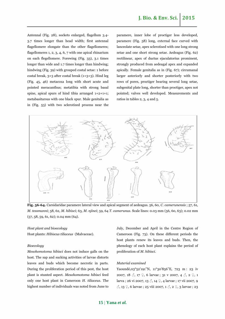
Fig. 56-64. Carsidaridae paramere lateral view and apical segment of aedeagus. 56, 60, C. camerunensis ; 57, 61,
M. tessmanni; 58, 62, M. hibisci; 63, M. njinei; 59, 64 T. camerunus. Scale lines: 0.03 mm (56, 60, 63); 0.02 mm
(57, 58, 59, 61, 62); 0.04 mm (64).
Host plant and bioecology
Host plants: Hibiscus tiliaceus (Malvaceae).
Bioecology
Mesohomotoma hibisci does not induce galls on the
host. The sap and sucking activities of larvae distorts
leaves and buds which become necrotic in parts.
During the proliferation period of this pest, the host
plant is stunted aspect. Mesohomotoma hibisci feed
only one host plant in Cameroon H. tiliaceus. The
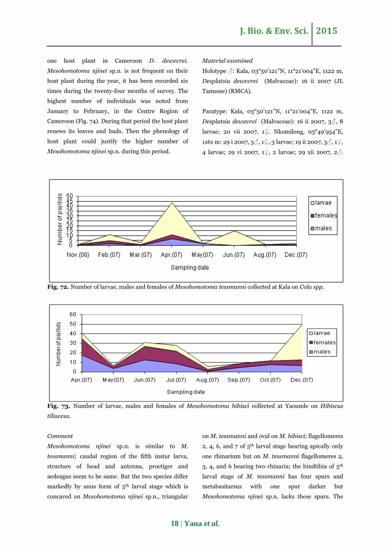
highest number of individuals was noted from June to
July, December and April in the Centre Region of
Cameroon (Fig. 73). On these different periods the
host plants renew its leaves and buds. Then, the
phenology of each host plant explains the period of
proliferation of M. hibisci.
Material examined
Yaoundé,03°52'191’’N, 11°30'856’’E, 723 m : 23 iv
2007, 18 ♂, 17 ♀, 6 larvae ; 31 v 2007, 4 ♂, 2 ♀, 1
larva ; 26 vi 2007, 13 ♂, 14 ♀, 4 larvae ; 17 vii 2007, 9
♂, 13 ♀, 6 larvae ; 25 viii 2007, 1 ♂, 2 ♀, 3 larvae ; 23

J. Bio. & Env. Sci. 2015
16 | Yana et al.
ix 2007, 5 ♂, 4 ♀ ; 25 x 2007, 8 ♂, 4 ♀; 28 xii 2007, 7
♂, 6 ♀, 37 larvae.
Comment
Mesohomotoma hibisci described in Cameroon for
the first time has the same characters as the formerly
described species by (Hollis, 1987). In Cameroon, this
species feed on H. tiliaceus and according to (Hollis,
1987), M. hibisci feed on H. tiliaceus H. rosasinensis
and H. boryanus.
Fig. 65-69. Carsidaridae female genitalia. 65, C. camerunensis ; 66, M. tessmanni; 67, M. hibisci; 68, M. njinei;
69, T. camerunus. Scale lines: 0.06 mm (65); 0.05 mm (66); 0.02 mm (67); 0.07 mm (68); 0.03 mm (69).
Mesohomotoma njinei Tamesse sp.n.
Fifth instar larva
Colouration
Body pale yellow with orange brown bands dorsally;
eyes reddish; antennal flagellomeres 2, 3, 4, 5 and 6
apically dark-brown, flagellomeres 7 and 8 entirely
dark-brown; claws dark brown. The caudal region is
more dark brown than the other part of abdomen.
Structure
Final instar larva (Fig. 17) elongated and oblated
dorsoventraly, not clearly divided into head and
thorax. Antenna elongate, 10 segmented; flagellum
distinctly subdivided with one apical rhinarium on
flagellomeres 2, 4, 6 and 7 (Fig. 18); wing pads
elongate and well-developed with setae; metatibia
and metabasitarsus not clearly separated (Fig. 19).
The caudal region of abdomen differentiated and
bearing a convoluted pore band (Fig. 20); anus
terminal, in ventral position. Measurements and
ratios in table 1.
Adult
Colouration
Body dark brown, dorsal view darker; eyes reddish;
forewings with a large and two small patches dark-
brown in cu2 cell; hindwings transparent; spurs and
claws dark-brown.
Structure
Head (Fig. 23) with disc of vertex deeply divided by
median suture, foveae present as deep oblique
grooves, lateral and hind margins of vertex obliquely
raised; anteriorly, vertex bear lanceolate setae.
Antennal (Fig. 29), sockets enlarged, flagellum 3.2-
3.6 times longer than head width; first antennal
flagellomere elongate than the other flagellomeres;
J. Bio. & Env. Sci. 2015
17 | Yana et al.
flagellomeres 2, 4, 6, 7 with each one apical
rhinarium. Forewing (Fig. 34), 2.6-2.9 times longer
than wide and 1.6 times longer than hindwing;
hindwing (Fig. 38) with grouped costal setae: 2 before
costal break, 5+3 after costal break and the hamulus
(2+5+3+1). Hind leg (fig. 47, 48) metacoxa long with
short acute and pointed meracanthus; metatibia with
strong basal spine, apical spurs of hind tibia arranged
1+4; metabasitarsus with one black spur. Male
genitalia as in (Fig. 54) present two incurved
sclerotized process near of paramere, paramere
slightly rectilinear but apex internal margin incurved;
aedeagus (Fig. 63) rectilinear, apex of ductus
ejaculatorius prominent, strongly produced from
aedeagal apex and expanded apically. Female
genitalia as in (Fig. 68); proctiger with a large
circumanal short with two rows of pores, proctiger
not stepped posteriorly, bearing only three long setae,
subgenital plate less narrow than proctiger on the end
part, apex not pointed. Measurements and ratios in
tables 2, 3, 4 and 5.
Fig. 70. Number of larvae, males and females of Mesohomotoma tessmanni collected at Kala on Theobroma
cacao.
Fig. 71. Number of larvae, males and females of Mesohomotoma tessmanni collected at Kala on Theobroma
bicolor.
Host plant and bioecology
Host plants: Desplatsia dewevrei (Malvaceae).
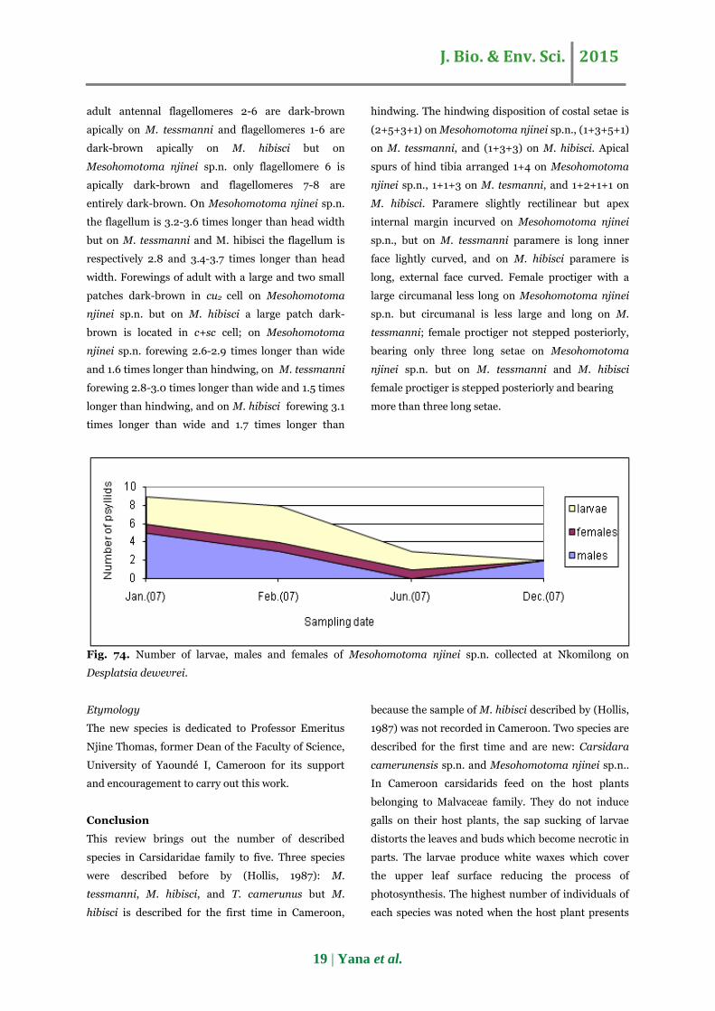
Bioecology
Mesohomotoma njinei sp.n. does not induce galls on
the host. The sap sucking activities of the larvae
distorts the leaves and buds which become necrotic in
parts. The larvae produce white waxy filaments which
covers the upper leaf surface reducing the process of
photosynthesis. Mesohomotoma njinei sp.n.feed only
J. Bio. & Env. Sci. 2015
18 | Yana et al.
one host plant in Cameroon D. dewevrei.
Mesohomotoma njinei sp.n. is not frequent on their
host plant during the year, it has been recorded six
times during the twenty-four months of survey. The
highest number of individuals was noted from
January to February, in the Centre Region of
Cameroon (Fig. 74). During that period the host plant
renews its leaves and buds. Then the phenology of
host plant could justify the higher number of
Mesohomotoma njinei sp.n. during this period.
Material examined
Holotype ♂: Kala, 03°50’121”N, 11°21’004”E, 1122 m,
Desplatsia dewevrei (Malvaceae): 16 ii 2007 (JL
Tamesse) (RMCA).
Paratype: Kala, 03°50’121”N, 11°21’004”E, 1122 m,
Desplatsia dewevrei (Malvaceae): 16 ii 2007, 3♂, 8
larvae; 20 vii 2007, 1♀. Nkomilong, 03°49’954”E,
1161 m: 29 i 2007, 5♂, 1♀, 3 larvae; 19 ii 2007, 3♂, 1♀,
4 larvae; 29 vi 2007, 1♀, 2 larvae; 29 xii 2007, 2♂.
Fig. 72. Number of larvae, males and females of Mesohomotoma tessmanni collected at Kala on Cola spp.
Fig. 73. Number of larvae, males and females of Mesohomotoma hibisci collected at Yaounde on Hibiscus
tiliaceus.
Comment
Mesohomotoma njinei sp.n. is similar to M.
tessmanni; caudal region of the fifth instar larva,
structure of head and antenna, proctiger and
aedeagus seem to be same. But the two species differ
markedly by anus form of 5th larval stage which is
concaved on Mesohomotoma njinei sp.n., triangular
on M. tessmanni and oval on M. hibisci; flagellomeres
2, 4, 6, and 7 of 5th larval stage bearing apically only
one rhinarium but on M. tessmanni flagellomeres 2,
3, 4, and 6 bearing two rhinaria; the hindtibia of 5th
larval stage of M. tessmanni has four spurs and
metabasitarsus with one spur darker but
Mesohomotoma njinei sp.n. lacks these spurs. The
J. Bio. & Env. Sci. 2015
19 | Yana et al.
adult antennal flagellomeres 2-6 are dark-brown
apically on M. tessmanni and flagellomeres 1-6 are
dark-brown apically on M. hibisci but on
Mesohomotoma njinei sp.n. only flagellomere 6 is
apically dark-brown and flagellomeres 7-8 are
entirely dark-brown. On Mesohomotoma njinei sp.n.
the flagellum is 3.2-3.6 times longer than head width
but on M. tessmanni and M. hibisci the flagellum is
respectively 2.8 and 3.4-3.7 times longer than head
width. Forewings of adult with a large and two small
patches dark-brown in cu2 cell on Mesohomotoma
njinei sp.n. but on M. hibisci a large patch dark-
brown is located in c+sc cell; on Mesohomotoma
njinei sp.n. forewing 2.6-2.9 times longer than wide
and 1.6 times longer than hindwing, on M. tessmanni
forewing 2.8-3.0 times longer than wide and 1.5 times
longer than hindwing, and on M. hibisci forewing 3.1
times longer than wide and 1.7 times longer than
hindwing. The hindwing disposition of costal setae is
(2+5+3+1) on Mesohomotoma njinei sp.n., (1+3+5+1)
on M. tessmanni, and (1+3+3) on M. hibisci. Apical
spurs of hind tibia arranged 1+4 on Mesohomotoma
njinei sp.n., 1+1+3 on M. tesmanni, and 1+2+1+1 on
M. hibisci. Paramere slightly rectilinear but apex
internal margin incurved on Mesohomotoma njinei
sp.n., but on M. tessmanni paramere is long inner
face lightly curved, and on M. hibisci paramere is
long, external face curved. Female proctiger with a
large circumanal less long on Mesohomotoma njinei
sp.n. but circumanal is less large and long on M.
tessmanni; female proctiger not stepped posteriorly,
bearing only three long setae on Mesohomotoma
njinei sp.n. but on M. tessmanni and M. hibisci
female proctiger is stepped posteriorly and bearing
more than three long setae.
Fig. 74. Number of larvae, males and females of Mesohomotoma njinei sp.n. collected at Nkomilong on
Desplatsia dewevrei.
Etymology
The new species is dedicated to Professor Emeritus
Njine Thomas, former Dean of the Faculty of Science,
University of Yaoundé I, Cameroon for its support
and encouragement to carry out this work.
Conclusion
This review brings out the number of described
species in Carsidaridae family to five. Three species
were described before by (Hollis, 1987): M.
tessmanni, M. hibisci, and T. camerunus but M.
hibisci is described for the first time in Cameroon,
because the sample of M. hibisci described by (Hollis,
1987) was not recorded in Cameroon. Two species are
described for the first time and are new: Carsidara
camerunensis sp.n. and Mesohomotoma njinei sp.n..
In Cameroon carsidarids feed on the host plants
belonging to Malvaceae family. They do not induce
galls on their host plants, the sap sucking of larvae
distorts the leaves and buds which become necrotic in
parts. The larvae produce white waxes which cover
the upper leaf surface reducing the process of
photosynthesis. The highest number of individuals of
each species was noted when the host plant presents
J. Bio. & Env. Sci. 2015
20 | Yana et al.
young leaves. The dynamic population of each species
depends of the phenology of the host plant which
depends of climatic factors..
References
Becker-Migdisova EE. 1973. Systematic of the
Psyllomorpha and the position of the group within
the order Homoptera. In Narchuk, E.P. (Ed.),
Doklady na dvadzat chetvertom escheghodnom
chtenii pamyati N.A. Kholodovskogo 1971. Leningrad,
1973: 90-117. (In Russian, English translation, British
Library, Boston Spa.).
Burckhardt D. 1994. Psylloid pests of temperate
and subtropical crops and ornamental plants
(Hemiptera: Psyllidea): a review. Trends in
Agricultural Sciences, Entomology 2, 173-186.
Burckhardt D, Ouvrard D. 2012. A revised
classification of the jumping plant-lice (Hemiptera:
Psylloidea). Zootaxa 3509, 1-34.
Burckhardt D, De Queiroz DL. 2012. Checklist
and comments on jumping plant-lice (Hemiptera:
Psylloidea) from Brazil. Zootaxa 3571, 26-48.
Dzokou VJ, Tamesse JL, Burckhardt D.
2009a.Jumping plant lice of the family Psyllidae
(Hemiptera-Psylloidea) from West-Cameroon;
Biodiversity and host plants. Journal of Entomology 6
(1), 1-17.
Heslop-Harrison G. 1958. Subfamily separation in
the homopterous Psyllidae III (a-c). Annals and
Magazine of Natural History 13(1), 561-579.
Hollis D. 1973. African gall bugs of the genus
Phytolyma (Hemiptera, Psylloidea). Bulletin of
Entomological Research 63, 143-154.
Hollis D. 1987. A review of the Malvales-feeding
psyllid family Carsidaridae (Homoptera). Bulletin of
the British Museum (Natural History), Entomology
series 56 (2), 87-127.
Li F. 2011. Psyllidomorpha of China (Insecta:
Hemiptera).Beijing, China: Science Press., 1976 p.
Mbondji PM. 1984. Main insects pests of cocoa and
coffee trees of Cameroon. Bionomic and pest
management. Ed. CEPER, Yaounde (Cameroon), p.
94.
Messi J. 1986.Mise en évidence des dégâts causés
aux cacaoyers par le mode d’insertion de l’œuf de
Mesohomotoma tessmanni (Homoptera-Psyllidae).
Café, Cacao, Thé 30 (1), 51-56.
Messi J, Nguefang M. 1993.Mesohomotoma
hollisi, espèce nouvelle de psylles inféodées à
Scaphopetalum blackii Mast (Homoptera, Psyllidae).
Bulletin de la Société Entomologique de France 98
(2), 127-130.
Mveyo Ndankeu YP, Tamesse JL, Burckhardt
D, Messi J. 2011.Biodiversity of jumping plant-lice
of Psyllidae family (Hemiptera : Psylloidea) from the
South Region of Cameroon : faunistics, phenology
and host plants. Journal of Entomology 8(2), 123-
138.
Ossiannilsson F. 1992. Psylloidea (Homoptera) of
Fennoscandia and Denmark. Fauna Entomologica
Scandinavica 26, 1-347.
Tamesse JL, Burckhardt D, Dzokou VJ, Yana
W, Mveyo Ndankeu YP, Foko Dadji GA, Messi
J. 2007.Jumping plant-lice of the family Triozidae
(Hemiptera: Triozidea) from Cameroon: Biodiversity
and Host Plants. Journal of Entomology 4(3), 181-
193.
Vondráček K. 1957. Mery Psylloidea. Fauna CSR 9,
1-431.
Yana W, Tamesse JL, Burckhardt D. 2009.
Jumping plant-lice of the family Phacopteronidae
(Hemiptera: Psylloidea) from the Center Region of
Cameroon: biodiversity and host plants. Syllabus
Review 1, 1-9.
Yana W, Tamesse JL, Burckhardt D. 2010.
Jumping plant-lice of the family Psyllidae Latreille
(Hemiptera : Psylloidea) from the Center Region of
Cameroon : faunistics, phenology and host plants.
Journal of Entomology 7(1), 1-18.