journal of the association for research in otolaryngology · serial analysis of gene expression in...
TRANSCRIPT

Serial Analysis of Gene Expression in the Chicken Otocyst
SAKU T. SINKKONEN,1,3 VERONIKA STARLINGER,1,4 DEEPA J. GALAIYA,1 ROMAN D. LASKE,1 SAMUELMYLLYKANGAS,2 KAZUO OSHIMA,1 AND STEFAN HELLER
1
1Departments of Otolaryngology–Head & Neck Surgery and Molecular & Cellular Physiology, Stanford University School ofMedicine, 801 Welch Road, Stanford, CA 94305-5739, USA2Division of Oncology, Stanford University School of Medicine, CCSR 3240, 269 Campus Drive, Stanford, CA 94305, USA
Received: 9 February 2011; Accepted: 2 August 2011; Online publication: 19 August 2011
ABSTRACT
The inner ear arises from multipotent placodal precur-sors that are gradually committed to the otic fate andfurther differentiate into all inner ear cell types, with theexception of a few immigrating neural crest-derived cells.The otocyst plays a pivotal role during inner ear develop-ment: otic progenitor cells sub-compartmentalize intonon-sensory and prosensory domains, giving rise toindividual vestibular and auditory organs and theirassociated ganglia. The genes and pathways underlyingthis progressive subdivision and differentiation processare not entirely known. The goal of this study was toidentify a comprehensive set of genes expressed in thechicken otocyst using the serial analysis of gene expres-sion (SAGE) method. Our analysis revealed severalhundred transcriptional regulators, potential signalingproteins, and receptors. We identified a substantialcollection of genes that were previously known in thecontext of inner ear development, but we also foundmany new candidate genes, such as SOX4, SOX5, SOX7,
SOX8, SOX11, and SOX18, which previously were notknown to be expressed in the developing inner ear.Despite its limitation of not being all-inclusive, thegenerated otocyst SAGE library is a practical bioinfor-matics tool to study otocyst gene expression and toidentify candidate genes for developmental studies.
Keywords: gene array, inner ear development, oticvesicle, SAGE, Sox
INTRODUCTION
The otic vesicle, or otocyst, is one of the earliestmorphological manifestations of the vertebrate innerear. It arises by invagination of the otic placode, which isan ectodermal thickening that develops near thedeveloping hindbrain. This process happens in chickenembryos during the second and third days of embryonicdevelopment (E2–E3). In intermediate stages, the oticplacode has folded inward to form a pouch that is alsocalled otic pit. The otic pit subsequently pinches offfrom the surface ectoderm into the underlying mesen-chyme, resulting in the formation of the otocyst.
It has been hypothesized that axis formation of thedeveloping inner ear already happens during otocystformation, where signals from the surrounding tissuesresult in the regionalization of the developing oticplacode, pit, and otocyst (reviewed in Fekete 1996;Fekete and Wu 2002). Despite a complex patterningprocess that is already manifest at the otocyst stage byregionalized expression of specific markers (for areview, see Streit 2007), the otocyst itself is a remarkablestructure because it contains all the necessary progen-itor cells to form the major cell types of the inner ear.This autonomy was revealed by grafting otocysts into
Present address: Saku T. Sinkkonen, Department of Otolaryngology,Helsinki University Central Hospital, POB 220, FIN-00029 HUCH,Helsinki, Finland.Present address: Veronika Starlinger, Department of Otorhinolaryng-ology, Medical University of Vienna, Waehringer Guertel 18-20,1090, Vienna, Austria.
Correspondence to: Stefan Heller & Departments of Otolaryngology–Head & Neck Surgery and Molecular & Cellular Physiology & StanfordUniversity School of Medicine & 801 Welch Road, Stanford, CA 94305-5739, USA. Telephone: +1-650-7248086; fax: +1-650-7258502?; email:[email protected]
Electronic supplementary material The online version of this article(doi: 10.1007/s10162-011-0286-z) contains supplementary material,which is available to authorized users.
Saku T. Sinkkonen and Veronika Starlinger equally contributed tothis work.
JARO 12: 697–710 (2011)DOI: 10.1007/s10162-011-0286-zD 2011 Association for Research in Otolaryngology
697
JAROJournal of the Association for Research in Otolaryngology

other regions of the developing body (Swanson et al.1990) as well as by determining that the otic lineage thatis specified during otic induction has already reached alargely committed state when the otocyst is formed(Groves and Bronner-Fraser 2000).
Inner ear cell regeneration research has beenutilizing the fact that stem cell-derived mammalianotic progenitor cells, defined by the expression ofotocyst markers such as PAX2, PAX8, and DLX5,display a certain degree of commitment toward theotic lineage (Li et al. 2003a, b; Oshima et al. 2007,2010). This was demonstrated by showing the poten-tial of otic progenitors to differentiate into differentcell types that express makers indicative of neurons,hair cells, and supporting cells. Based on thesefindings, it has been hypothesized that stem cell-derived otic progenitors are similar to otocyst cells(Brigande and Heller 2009; Diensthuber et al. 2009).
Here, we present the results of an unbiased inter-rogation of gene expression in the chicken otocyst.This analysis was spurred by the lack of comprehen-sive gene expression information at this importantstage of development. We decided to utilize serialanalysis of gene expression (SAGE), which is aquantitative method that can be used to identifyknown as well as new genes (Saha et al. 2002;Velculescu et al. 1995). Of the 39,326 seventeen-basepair sequence tags that we found, we evaluated 16,008unique sequences that resulted in 4,153 unequivocallyidentified genes. Although our study lacked thesensitivity of more modern high-throughput deepsequencing methods, we consider the results as animportant contribution because they provide a com-prehensive summary of genes that are expressed atmedium and high levels. Our analysis revealedpotential signaling proteins and receptors as well asalmost 300 transcriptional regulators that areexpressed in the chicken otocyst. Some of theseregulators have been previously known to play impor-tant roles during inner ear development, but wefound additional candidate genes, such as severalmembers of the Sox gene family, which have thus far
not been evaluated in the context of the developinginner ear.
METHODS
Tissue dissection, RNA preparation, and SAGElibrary preparation
Fertilized eggs (Gallus gallus) of the white leghornstrain (California Golden Eggs, Dinuba, CA) wereincubated at 38°C for 72 h on a rocking platform in ahumidified egg incubator (Sportsman 1202A, GQFManufacturing, Savannah, GA). The embryos wereremoved from the eggs, washed in Hanks’ balancedsalt solution (HBSS; Invitrogen, Carlsbad, CA), andcollected in HBSS. Staging was conducted accordingto Hamburger and Hamilton’s (HH) guidelines(Hamburger and Hamilton 1992), and only HH stage18–19 embryos with clearly identifiable closed oticvesicles and unpigmented eyes were used. Oticvesicles were microdissected using fine forceps andattention was given to remove as much of thesurrounding periotic mesenchyme as possible(Fig. 1A). The vesicles were individually inspectedand frozen in bulks of 10–20 in liquid nitrogen andstored at −80°C.
Total RNA was isolated using a commercial kit(RNeasy Mini Kit, Qiagen, Hilden, Germany). RNAintegrity and quality was confirmed by gel electro-phoresis and by visual assessment. Five microgramstotal RNA, the combined yield of 200 otocysts, wasused for SAGE library synthesis (I-SAGE Long kit,Invitrogen) starting with attaching polyA+ RNA tooligo(dT)-paramagnetic beads, reverse transcription,and second strand synthesis. The resulting cDNA wascleaved with NlaIII, divided into two fractions, andbound to two different adapters containing a type IISrestriction nuclease recognition site. The adapterswith adjacent 21-bp cDNA pieces were released fromthe oligo(dT)-magnetic beads using the type IISrestriction endonuclease MmeI. The two pools ofreleased adapter-linked tags were ligated to 130-bp
FIG. 1. SAGE library from the chickenotocyst. A HH stage 18–19 chickenembryo. The otocyst is indicated with awhite box. Inset, otocyst after dissection.B SAGE tag frequency (tag count) plot forall tags identified.
698 SINKKONEN ET AL.: Serial Analysis of Gene Expression in the Chicken Otocyst

ditags and amplified with 27 PCR cycles with specificprimers to each adapter. The adapters were thencleaved with NlaIII, 34-bp ditags were purified fromthe adapters by polyacrylamide gel electrophoresis,and concatenated. Concatemers were fractioned bysize, gel-purified, and then cloned into pZErO-1provided with the kit.
Quality control was conducted in two steps. First,20 colonies were picked, plasmid DNA was prepared,and the resulting plasmids were digested with NsiI,which resulted in the release of the individual inserts.We obtained 20 distinct restriction patterns with amean insert length of 671 bp (±425 bp (SD)). Thesmallest fragment was 200 bp and the largest was1,700 bp. In a second step, we sequenced 24 inserts,obtaining 12,025 bp of raw data. These insert lengthsslightly exceeded the expected ≈25 SAGE tags perclone predicted by the kit manufacturer. Threethousand eight hundred forty colonies were roboti-cally picked and the plasmid concatemer inserts weredirectly sequenced (GeneWiz, South Plainfield, NJ).
Modifications from the manufacturer’s protocolincluded that the gel electrophoreses for the 130- and34-bp ditags were performed on 10% polyacrylamidegels (Novex TBE Gels, Invitrogen) and that DNA wasisolated from polyacrylamide gels with QIAEXII beads(Qiagen). Concatemers were separated on a 2%agarose gel and purified before subcloning with acolumn-based gel extraction kit (QIAquick, Qiagen).
Data analysis
At the highest stringency settings for DNA sequencequality and tag extraction, 39,326 individual 17-bp tagswere extracted from the 3,512 sequencing data filesusing SAGE2000 analysis software (version 4.5, Invi-trogen). Only unequivocal sequences were used forlibrary construction, which resulted in a library aboutthree times smaller than predicted by the qualitycontrol samples. A potential reason for this shortfallmight be that the sequence quality achieved withdirect sequencing was lower than the sequencingresults obtained with plasmid DNA, which was usedfor the quality control clones. For computerizedmapping, we appended the NlaIII restriction site (5′-CATG-3′) to the 5′ end of each tag. The resulting 21-bptags were mapped using 22,290 G. gallus cDNA sequen-ces available through the Ensembl 52 database (www.ensembl.org/info/data/ftp/), 19,307 G. gallus cDNAsavailable from the RefSeq database (ftp.ncbi.nih.gov),and 33,383 G. gallus sequences from the Unigenedatabase (www.ncbi.nlm.nih.gov/unigene). Mappingwas conducted using MAQ software (Li et al. 2008;maq.sourceforge.net) with all parameters set to defaultallowing for two mismatches in the sequence align-ments. Ingenuity Pathway Analysis (IPA) software was
accessed via the Stanford University BioinformaticsResource (cmgm.stanford.edu).
Reverse transcriptase PCR
Chicken otocyst RNA was isolated (Absolutely RNAMiniprep Kit, Stratagene/Agilent Technologies, LaJolla, CA) and treated with RNase-free DNase I(Roche Diagnostics, Mannheim, Germany). TheRNA concentration was determined by spectrophoto-metric analysis using a NanoDrop (Thermo FisherScientific, Wilmington, DE). Total RNA extracts werethen used for reverse transcription (RT) into cDNA(first strand) using SuperScript III Reverse Tran-scriptase (Invitrogen) and Oligo(dT)18 primer (Invi-trogen) with 350 ng of total RNA per 20 μl reaction.To prevent RNA degradation, 1 μl RiboLock RNaseInhibitor (Fermentas, Thermo Fisher Scientific, GlenBurnie,MD) was also included in each reaction. Controlreactions were done without reverse transcriptase.
Oligonucleotide primer pairs were designed foreach gene of interest using NCBI Primer-BLAST(www.ncbi.nlm.nih.gov/tools/primer-blast) with NCBIReference Sequences as template. For each genetested, at least two primer pairs covering two non-overlapping 300- to 700-bp regions were used toconfirm mRNA expression. A full list of primers testedcan be found in Electronic supplementary materials(ESM) Table 1. PCR was performed using GoTaqGreen Master Mix (Promega, Madison, WI) with 2 μlcDNA template and 1 μl 400 nM each of forward andreverse gene-specific primers. The following cyclingconditions were employed: initial denaturation at 94°C (3 min); 30 cycles of denaturation at 94°C (30 s),annealing at 55°C (1 min), and elongation at 72°C(1 min); and a hold at 4°C. Aliquots of PCR productswere electrophoresed in a 2.0% agarose gel, stainedwith SYBR Safe (Invitrogen) in 1X TAE buffer at120 V for 35 min, and documented using UV trans-illumination and digital photography (Kodak GelLogic 200 Imaging System).
In situ hybridization
The T7 promoter sequence (5′-TAATACGACTCACTATAGGG-3′) was added to the 5′ end of the forwardor reverse primer for the different Sox2 cDNAs toallow for conversion of the PCR product to sense andantisense cRNA probes for in situ hybridization. Ofthe PCR product, 500 ng was used to synthesizedigoxigenin-labeled antisense probes (DIG RNALabeling Kit, Roche Diagnostics), which were resus-pended in 30 μl RNAse-free water. Embryos weredissected at HH stage 18–19 (E3) and HH stage 26–28(E5), fixed overnight with 4% paraformaldehyde inphosphate-buffered saline (PBS, pH 7.4), transferred
SINKKONEN ET AL.: Serial Analysis of Gene Expression in the Chicken Otocyst 699

into 30% sucrose in PBS for 24–36 h, and embeddedin O.C.T compound (Tissue-Tek). Sections were cutwith a cryomicrotome (CM3050 S, Leica), collectedon ultrastick slides (precleaned Gold Seal, Rite-on,Micro Slides), dried at 37°C for 45 min, and storedfrozen at −70°C. For hybridization, the sections werebrought to room temperature and rehydrated in100 μl diluted probe (1:100) in 50% formamide,10% dextran sulfate, 1 mg/ml yeast RNA, 1x Denhardt’ssolution, 185 mM NaCl, 5.6 mM NaH2PO4, 5 mMNa2HPO4, 5 mM EDTA, and 15 mM Tris at pH 7.5.After coverslipping and overnight incubation at 65°C ina chamber humidified with 50% formamide in 150 mMNaCl, 15 mM trisodium citrate, pH 7 (1× SSC), thecoverslips were removed in 5x SSC and the slides washedtwice for 30 min each in 50% formamide and 0.1%Triton X-100 in 1x SSC at 65°C. Thereafter, the slideswere washed for 15 min in 0.2x SSC and for 15 min in150 mM NaCl and 100 mM Tris at pH 7.5 at roomtemperature. For antibody detection, the sections wereblocked for 30 min in 0.5% blocking powder (RocheDiagnostics), 10% heat-inactivated goat serum, 100 mMNaCl, 0.1% Triton X-100, and 100 mM Tris at pH 7.5.The slides were then incubated for 2 h at room temper-ature in a blocking solution pre-incubated for 1 h withalkaline phosphatase-conjugated anti-digoxigenin Fabfragments (1:500, Roche Diagnostics). Unbound Fabfragments were removed by washing twice for 30 mineach in 150 m NaCl and 100 mM Tris at pH 7.5. Thesections were first incubated in detection buffer(100 mM NaCl, 50 mM MgCl2, 100 mM Tris at pH 9.5)for 10min. For detection, the sections were then coveredwith 200 μl of chromogen solution consisting of 20 μlNBT/BCIP stock solution (Roche Diagnostics) and 50 μl
Levamisol stock solution (20x concentrate, Invitrogen)in 1 ml detection buffer, coverslipped, and incubatedovernight at room temperature in a humidified cham-ber. Coverslips were removed and color reaction wasstopped in 1 mMEDTA and 10mMTris at pH 8.1. Slideswere embedded in 50% glycerol in PBS and cover-slipped. Analysis and photography was conducted on anAxiovert 25 microscope with an AxioCam MRC camera,using AxioVision software (V 4.6.3.0, Zeiss).
RESULTS
SAGE library of the chicken otocyst
Otocysts were dissected from HH stage 18–19 chickenembryos (Fig. 1A), total RNA was extracted, andsubjected to a commercial long-SAGE protocol, result-ing in a library of concatemerized tags. Individual clonesof SAGE concatemers were sequenced, resulting in39,326 seventeen-base pair tags with tag counts up to 718for the most abundant tag; 3,292 tags were representedbetween two and five times, whereas the majority of tags(11,717) were only found once (Fig. 1B). Overall, weidentified 16,008 unique sequence tags (ESM Table 2;NCBI Geo DataSet accession no. GSM651351).
Although the chicken genome has been fullysequenced (The Chicken Genome Consortium 2004),the annotation of chicken genes is far from complete, andconsequently, gene annotation needed to be conductedby combining several database resources (Fig. 2). Westarted by mapping all 16,008 tags consisting of the 4-bpNlaIII restriction site and each individual 17-bp SAGE tagusing the MAQ software (Li et al. 2008). Threereference databases were used—Ensembl 52, RefSeq,
FIG. 2. Flowchart of SAGE tag analysis resulting in a library of 4,153 annotated genes expressed in the chicken otocyst.
700 SINKKONEN ET AL.: Serial Analysis of Gene Expression in the Chicken Otocyst

and Unigene—resulting in 7,026, 8,682, and 13,405matches, respectively. To compare thematches from thethree different databases that were composed of assort-ments of unofficial and official gene names, weimported the results into the IPA software, whichrevealed official gene symbols as common identifiers.IPA software recognized 5,632, 7,326, and 7,529 geneidentifiers from Ensembl, RefSeq, and Unigene data-base matches, respectively. In a next step, official genesymbols created in IPA were used to directly comparematches of different databases using a relational data-base. Only when all three different databases suggesteda match for a given gene was the gene accepted forfurther analysis. In case of discrepancies in matchesbetween different databases or in case of missingmatches in some of the databases, we performedmanual BLAST searches (blast.ncbi.nlm.nih.gov/Blast.cgi) with each tag. If a match was found, the resultinggene was added for further analysis. In 52 cases, a singletag corresponded to two different genes, and in onecase, a single tag corresponded to five different genes.These 53 ambiguous tags (ESM Table 3) were removedfrom further analysis. After this step, the libraryconsisted of 7,912 gene matches with unique tags.However, many genes were represented in the librarywith two or more different tags, which could be due toalternative poly-adenylation sites, internal priming, oralternative splicing near the 3′ end of the mRNA. Theseduplicates were combined, resulting in 4,180 genes thatwere re-imported into IPA for final analysis. At this finalstep, IPA recognized 4,153 genes, which in the finalannotated SAGE library were associated with theaggregate count number of all the tags of every givengene (ESM Table 4).
Gene annotation reveals abundanceof transcriptional regulators
Analysis of the relation between tag count and anno-tated gene number revealed that 50% of all tags in the
otocyst were encoded by only 180 genes, whereas themajority of genes were represented by fewer than 10 tags(Fig. 3A, B). Not unexpectedly, the genes with thehighest tag count were housekeeping genes involved inthe maintenance of basal cellular functions (Table 1).The IPA database provided unequivocal gene familyinformation for about half of the identified genes.Analysis of all unambiguous gene family annotationidentifiers revealed that the majority of otocyst genesencoded unclassified enzymes as well as kinases, phos-phatases, and peptidases. The second largest family ofgenes identified encoded transcriptional regulators,followed by transporters, transmembrane domain-con-taining receptors, and ion channels (Fig. 3C). Transla-tional regulators, growth factors, cytokines, and otherprotein families comprised the rest.
Overall, we identified 299 genes that encode tran-scriptional regulators (ESM Table 5) which can becategorized into transcription factors containing zinc-coordinating DNA-binding domains (11%), helix-loop-helix domains (13%), basic domains (15%), ß-scaffoldfactors with minor groove contacts (16%), and others(45%). Fifty-one transcriptional regulators were previ-ously known to be expressed in the developing innerear. Known examples for each respective category areGATA2 and GATA3 (Lillevali et al. 2007) for zinc-coordinating DNA-binding domains, PAX2 and FOXG1(Herbrand et al. 1998; Li et al. 2004; Pauley et al. 2006)for helix-loop-helix domains, NEUROG1 and NEUROD1(Liu et al. 2000; Ma et al. 2000) for basic domains, andSOX10 andNOTCH1 (Lewis et al. 1998; Stone and Rubel1999; Watanabe et al. 2000) for ß-scaffold factors withminor groove contacts. Two hundred forty-eight tran-scriptional regulators were previously unknown in thecontext of early inner ear development.
Secreted proteins and transmembrane proteins
Genes that encode growth factors, cytokines, andother secreted proteins are the second group of
FIG. 3. SAGE library after annotation. A Tag count frequency plot for all 4,153 annotated genes. B Cumulative tag count analysis reveals that180 of the most abundantly expressed annotated genes represent 50% of all identified tags. C Family representation of all unambiguouslyidentified annotated genes.
SINKKONEN ET AL.: Serial Analysis of Gene Expression in the Chicken Otocyst 701

developmentally interesting otocyst genes (ESMTable 6). Of the 172 genes that we identified in thisgroup, several were previously known in inner eardevelopment and include BMP7, FGF10, FGF19, FRZB,TGFß2, NETRIN1, SLIT1, WNT3, and WNT5A (Abrairaet al. 2008; Alsina et al. 2004; Battisti and Fekete 2008;Hollyday et al. 1995; Liu et al. 2008; Oh et al. 1996;Okano et al. 2005; Sanchez-Calderon et al. 2007;Sienknecht and Fekete 2009). Transcripts encodingthe secreted signaling protein midkine (MDK) wereby far the most abundantly expressed mRNA that wedetected. Midkine has been previously reported in thepostnatal mouse cochlea, and it has been shown thatthe protein is involved in regulating the expression ofthe tectorial membrane component ß-tectorin (Jia etal. 2001; Zou et al. 2006), but early developmentalroles in the inner ear have not been reported. Otherproteins, such as opticin (OPTC), have previouslybeen shown in the otic vesicle, but their function ininner ear development remains unknown (Frolova etal. 2004). Several genes emerged in our screen asnovel candidates for roles in inner ear development,such as olfactomedin-like 2A, 2B, 3 (OLFML2A,OLFML2B, OOLFML3), which belong to a class of
proteins implicated in a variety of developmentalprocesses (reviewed by Tomarev and Nakaya 2009), orneudesin (NENF), which may play roles in neuronaldifferentiation and development (Kimura et al. 2006).We identified various TGFß antagonists such as twistedgastrulation protein homolog 1 (TWSG1) or follistatin-like 3 (FSTL3). Lastly, we identified various secretedproteins of unknown function during development, butwith previous implications in cancer or other cellgrowth- and death-related processes; examples for theseproteins are AGR3, CLU, EGFL7, and HDGF.
Our analysis of transmembrane-spanning proteinsrevealed high transcript expression levels of many tightjunction and cell adhesion proteins such as claudin 1(CLDN1), CLDN3, and CLDN17; integrins α3 (ITGA3)and α6 (ITGA6); integrins ß1, ß2, ß3, and ß5 (ITGB1,ITGB2, ITGB3, ITGB5); neurexin 1 (NRXN1); as well ascell adhesion molecule 1 (CADM1) and epithelial celladhesion molecule TACSTD1, among others. One ofthe most abundant genes identified in this categoryencodes protein tyrosine kinase 7 (PTK7), a proteinimplicated in the regulation of planar cell polarity,convergent extension, andWnt signaling (Lu et al. 2004;Puppo et al. 2011; Yen et al. 2009). Another interesting
TABLE 1
Genes with the highest expression based on SAGE tag count
Gene symbol Count Family Gene name
COX1 840 Enzyme Cytochrome c oxidase IARIH1 560 Enzyme Ariadne homolog, ubiquitin-conjugating enzyme E2 binding protein, 1COX2 526 Enzyme Cytochrome c oxidase IICOX3 307 Enzyme Cytochrome c oxidase IIIATP6 298 Transporter ATP synthase 6, ATPase subunit 6NPM1 273 Transcription regulator Nucleophosmin (nucleolar phosphoprotein B23, numatrin)ND4 257 Enzyme NADH dehydrogenase, subunit 4 (complex I)GAPDH 254 Enzyme Glyceraldehyde-3-phosphate dehydrogenaseRPL13 230 Ribosomal structure Ribosomal protein L13RPL10A 218 Ribosomal structure Ribosomal protein L10aRPL4 187 Ribosomal structure Ribosomal protein L4TUBA4A 162 Cytoskeletal structure Tubulin, α4aRPL23 161 Ribosomal structure Ribosomal protein L23RPS27A 156 Ribosomal structure Ribosomal protein S27aMDK 154 Growth factor Midkine (neurite growth-promoting factor 2)ND5 151 Enzyme NADH dehydrogenase, subunit 5 (complex I)RPS3 146 Ribosomal structure Ribosomal protein S3EEF1A1 142 Translation regulator Eukaryotic translation elongation factor 1 α1ATP5B 139 Transporter ATP synthase, H+ transporting, mitochondrial F1 complex, β polypeptideRPS29 136 Ribosomal structure Ribosomal protein S29RPS27L 131 Ribosomal structure Ribosomal protein S27-likeACTB 124 Cytoskeletal structure Actin, βRPS15 120 Ribosomal structure Ribosomal protein S15TUBB2A 118 Cytoskeletal structure Tubulin, β 2ARPLP1 116 Ribosomal structure Ribosomal protein, large, P1RPS3A 112 Ribosomal structure Ribosomal protein S3ARPL21 107 Ribosomal structure Ribosomal protein L21RPL36 102 Ribosomal structure Ribosomal protein L36RPL35 96 Ribosomal structure Ribosomal protein L35HNRNPA3 94 Nucleic acid binding Heterogeneous nuclear ribonucleoprotein A3
702 SINKKONEN ET AL.: Serial Analysis of Gene Expression in the Chicken Otocyst

gene in this regard encodes the Ig superfamily proteinprotogenin (PRTG), which has been shown to play arole in suppressing premature neural differentiationand whose roles in other tissues might similarly be incontrolling the timing of transitions between earlyprogenitor state and differentiation (Ito et al. 2011;Wong et al. 2010). Probably the most interesting groupof genes that we identified encodes receptors forsignaling proteins because they might reveal informa-tion about the developmental processes happening inthe otocyst. These include genes that encode receptorsfor ligands that are already known for playing roles inotic development such as FGFR1, FZD1, FZD2, FZD3,FZD4, FZD7,NGFR, andNOTCH1, which have previouslybeen shown to be expressed in the vertebrate otocyst(Adam et al. 1998; Pirvola et al. 2002; Sienknecht andFekete 2009; Stevens et al. 2003; von Bartheld et al. 1991;Wright and Mansour 2003). BMPR1, BMPR2, LGFR1,SMO1, PTCH1, DISP1, and TGFBR2 are genes that werepresumed to be expressed in the otocyst because theirligands, such as BMPs and other TGFß family members,IGF, as well as hedgehog signaling proteins, have beenshown to be expressed and active during inner eardevelopment (Bok et al. 2005; Frenz et al. 1991, 1992;Liu et al. 2002; Oh et al. 1996; Riccomagno et al. 2002;Yamashita and Oesterle 1995). Other identified genesinclude receptors for somatostatin (SSTR1), interleukin11 (IL11RA), endothelin (EDNRB), and tumor necrosisfactors (TNFRSF1A, TNFRSF6B, TNFRSF19) and orphanreceptors such as lathrophilin 3 (LPNHN3; Sudhof2001).
Other potentially interesting transcripts encodedtransmembrane proteins involved in cell recognitionand adhesion that play roles in axonal guidance and cellmigration such as the semaphorins SEMA4B, SEMA5B,SEMA6D, SEMA7A and some components of theirreceptor complex such as Plexins A1 and B2 (PLXNA1,PLXNB2; Perrot et al. 2002). Additional genes withsimilar roles include ephrin B1 (EFNB1) and ephrinreceptors (EPHA4, EPHA5, EPHB3), netrin G1 and thenetrin receptor UNC5B, the Slit receptors ROBO1 andROBO2, as well as the Slit-like transmembrane proteinSLITRK6. The possible roles of some of these genes inaxon guidance and cell migration has been discussed inthe context of the inner ear (Fekete and Campero 2007;Webber and Raz 2006), and their expression patternsand potential function are the focus of intensiveresearch (Battisti and Fekete 2008; Katayama et al.2009; Matilainen et al. 2007).
As initial validation of mRNA expression, weconducted RT-PCR on otic vesicle RNA template witholigonucleotide primer pairs specific for selectedgenes representing transcriptional regulators, signal-ing proteins, receptors, and other genes of interest(Fig. 4 and Table 2). Transcripts for all selected geneswere detectable, and although the RT-PCR was not
conducted in quantitative fashion, we observed ageneral trend where strong amplification productscorresponded to genes with higher SAGE tag counts.
Known and novel Sox genes expressedin the otocyst
One of the most strongly represented groups oftranscription factors in the chicken otocyst SAGElibrary were the Sox genes. Previous reports showthe expression of SOX1, SOX2, SOX3, SOX6, SOX9,SOX10, and SOX21 in the chicken otocyst, or in theotic vesicle of various species including African clawedfrog, zebrafish, and mouse (Barrionuevo et al. 2008;Liu et al. 2003; Neves et al. 2007; Uchikawa et al. 1999;Watanabe et al. 2000). Clearly highlighting thelimitation of SAGE, showing that about 40,000 tagsare far from exhaustive, is the fact that tags for SOX2,SOX3, and SOX6 were not represented in our SAGElibrary. Nevertheless, we found six Sox genes thatpreviously were not known to be expressed in thedeveloping inner ear, which include SOX4, SOX5,SOX7, SOX8, SOX11, and SOX18.
We verified the expression of the Sox genes by RT-PCR (Fig. 4) and by in situ hybridization at the otocyststage (Fig. 5). SOX8 and SOX11 were abundantlyexpressed throughout the otocyst and also in theadjacent hindbrain. SOX4 mRNA appeared to be moreregionalized with stronger expression in the ventrome-dial regions of the otocyst, indicative of a potentialsubsequent expression in the prosensory domains.SOX5 transcripts were weakly detectable throughoutthe otocyst, with a possible stronger expression in thedorsolateral part of the otocyst. SOX7 and SOX18mRNAwas detectable throughout the otocyst, perhaps with aslightly stronger expression in the ventral portion.
At E5, when the basilar papilla and vestibularcompartments of the chicken inner ear are clearlydefined and prosensory domains have been formed,only SOX11 appeared to be specifically associated withthe prosensory domains of the basilar papilla andvestibular maculae (Fig. 6). SOX7 was no longerdetectable, whereas SOX4 and SOX5 transcripts weredetectable in the roof of the basilar papilla, presum-ably in the premordial tegmentum vasculosum. SOX8expression was strong throughout the inner ear aswell as in the adjacent hindbrain (not shown).
DISCUSSION
The chicken embryo is one of the major animalmodels used to study inner ear induction and devel-opment. In the past decades, many genes have beenfound that are expressed by cells of the otocyst, andthe specific roles of some of these genes have been
SINKKONEN ET AL.: Serial Analysis of Gene Expression in the Chicken Otocyst 703

elucidated. Nevertheless, no comprehensive study hasbeen conducted on gene expression in the chickenotocyst. We hypothesized that the existing collectionof otocyst markers and genes is just the tip of aniceberg, and we consequently decided to investigate,using a high-throughput method, gene expression inthis clearly defined transient structure. Unlike themouse and human genomes, the chicken genome iscomparably poorly annotated, which complicated theanalysis strategy. We refrained from using gene arrayswhose preselected genes are constrained by theseshortcomings. Additionally, at the onset of this study,no comprehensive chicken gene arrays were commer-cially available and next-generation sequencing tech-niques, likewise, were not yet developed. We decidedto employ SAGE, which is a relatively unbiasedmethod, based on sequencing of short tags that aredirectly adjacent to a NlaIII restriction site in the 3′region of any given mRNA (Velculescu et al. 1995).The NlaIII recognition sequence is 4-bp long (5′-CATG-3′) and theoretically occurs once in every256 bp. Using long-SAGE (Saha et al. 2002), which
employs 17-mer tags instead of 10-mer tags, whichwere used in initial SAGE protocols, we were able toutilize a specificity of 421. Indeed, we only found 53ambiguous tags, which either occur more than oncein the transcriptome or were associated with morethan one gene as a result of annotation ambiguities.
Our analysis is not based on comparative or sub-tractive studies, and consequently, many genes identi-fied are widely expressed. Nevertheless, the results ofour study do not preclude the use of bioinformatic toolsto extract subtractive or otherwise user-defined datasets,and the reader is encouraged to use our dataset asneeded. A recent very elegant gene array study focusingon FGF-based otic induction in mouse embryos is anexample of the powerful specificity that can be achievedby selecting proper tissues for comparison (Urness et al.2010). In this specific case, wild-type mouse otic placodetissue was compared with tissue from the prospectiveotic placode of Fgf3−/−;Fgf10−/− mice in which oticdevelopment fails to be initiated. This study revealedseveral transcriptional regulator genes that depend onFGF-based otic induction, includingHmx2,Hmx3, Foxg1,
FIG. 4. Shown are agarose gels stained with ethidium bromide to visualize RT-PCR fragments for genes expressed in the chicken otocyst(indicated with “+”). Control reactions without reverse transcription are labeled with “−”. Faint bands in the “−” control lanes are likely the resultof residual genomic DNA contamination. The corresponding gene names are listed in Table 2. Predicted PCR product sizes are listed in ESMTable 1. SAGE tag count numbers are indicated in parentheses.
704 SINKKONEN ET AL.: Serial Analysis of Gene Expression in the Chicken Otocyst
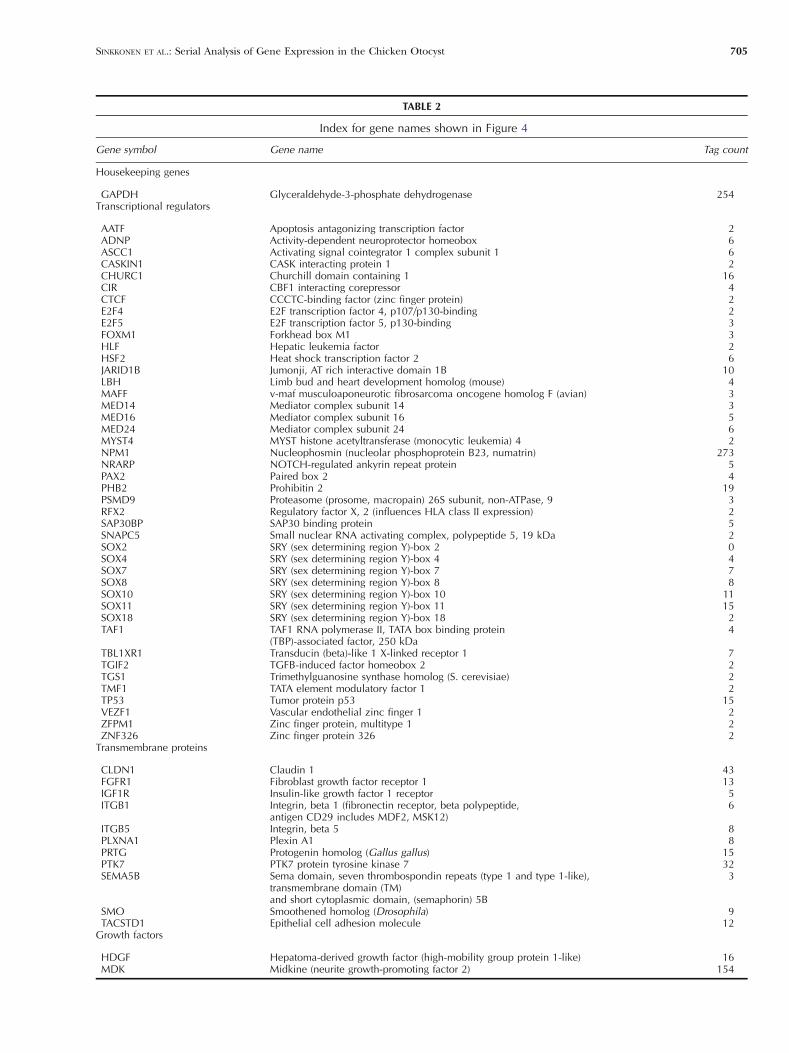
TABLE 2
Index for gene names shown in Figure 4
Gene symbol Gene name Tag count
Housekeeping genes
GAPDH Glyceraldehyde-3-phosphate dehydrogenase 254Transcriptional regulators
AATF Apoptosis antagonizing transcription factor 2ADNP Activity-dependent neuroprotector homeobox 6ASCC1 Activating signal cointegrator 1 complex subunit 1 6CASKIN1 CASK interacting protein 1 2CHURC1 Churchill domain containing 1 16CIR CBF1 interacting corepressor 4CTCF CCCTC-binding factor (zinc finger protein) 2E2F4 E2F transcription factor 4, p107/p130-binding 2E2F5 E2F transcription factor 5, p130-binding 3FOXM1 Forkhead box M1 3HLF Hepatic leukemia factor 2HSF2 Heat shock transcription factor 2 6JARID1B Jumonji, AT rich interactive domain 1B 10LBH Limb bud and heart development homolog (mouse) 4MAFF v-maf musculoaponeurotic fibrosarcoma oncogene homolog F (avian) 3MED14 Mediator complex subunit 14 3MED16 Mediator complex subunit 16 5MED24 Mediator complex subunit 24 6MYST4 MYST histone acetyltransferase (monocytic leukemia) 4 2NPM1 Nucleophosmin (nucleolar phosphoprotein B23, numatrin) 273NRARP NOTCH-regulated ankyrin repeat protein 5PAX2 Paired box 2 4PHB2 Prohibitin 2 19PSMD9 Proteasome (prosome, macropain) 26S subunit, non-ATPase, 9 3RFX2 Regulatory factor X, 2 (influences HLA class II expression) 2SAP30BP SAP30 binding protein 5SNAPC5 Small nuclear RNA activating complex, polypeptide 5, 19 kDa 2SOX2 SRY (sex determining region Y)-box 2 0SOX4 SRY (sex determining region Y)-box 4 4SOX7 SRY (sex determining region Y)-box 7 7SOX8 SRY (sex determining region Y)-box 8 8SOX10 SRY (sex determining region Y)-box 10 11SOX11 SRY (sex determining region Y)-box 11 15SOX18 SRY (sex determining region Y)-box 18 2TAF1 TAF1 RNA polymerase II, TATA box binding protein
(TBP)-associated factor, 250 kDa4
TBL1XR1 Transducin (beta)-like 1 X-linked receptor 1 7TGIF2 TGFB-induced factor homeobox 2 2TGS1 Trimethylguanosine synthase homolog (S. cerevisiae) 2TMF1 TATA element modulatory factor 1 2TP53 Tumor protein p53 15VEZF1 Vascular endothelial zinc finger 1 2ZFPM1 Zinc finger protein, multitype 1 2ZNF326 Zinc finger protein 326 2
Transmembrane proteins
CLDN1 Claudin 1 43FGFR1 Fibroblast growth factor receptor 1 13IGF1R Insulin-like growth factor 1 receptor 5ITGB1 Integrin, beta 1 (fibronectin receptor, beta polypeptide,
antigen CD29 includes MDF2, MSK12)6
ITGB5 Integrin, beta 5 8PLXNA1 Plexin A1 8PRTG Protogenin homolog (Gallus gallus) 15PTK7 PTK7 protein tyrosine kinase 7 32SEMA5B Sema domain, seven thrombospondin repeats (type 1 and type 1-like),
transmembrane domain (TM)and short cytoplasmic domain, (semaphorin) 5B
3
SMO Smoothened homolog (Drosophila) 9TACSTD1 Epithelial cell adhesion molecule 12
Growth factors
HDGF Hepatoma-derived growth factor (high-mobility group protein 1-like) 16MDK Midkine (neurite growth-promoting factor 2) 154
SINKKONEN ET AL.: Serial Analysis of Gene Expression in the Chicken Otocyst 705

and Sox9, which we also found in our dataset. Otherstudies that focused on the identification of otocyst
genes used differential display of chicken otocyst RNAagainst RNA from surrounding tissues (Gong et al.
FIG. 5. In situ hybridization analysis of Sox gene expression in sections of the chicken otocyst. Numbers in parentheses indicate the SAGE tagcount for each individual gene. Sense controls were negative for all probes used; a representative control is shown.
FIG. 6. In situ hybridization analysis ofSox gene expression in cross-sections ofthe E5 chicken inner ear. Sense controlswere negative for all probes used; arepresentative control is shown.
706 SINKKONEN ET AL.: Serial Analysis of Gene Expression in the Chicken Otocyst
1997) and on cDNA subtraction of mouse otocyst minusliver cDNA (Powles et al. 2004). The differential displaystudy identified only a small number of unknown genes,and the collection of 280 specific transcripts found inthemouse otocyst cannot be directly compared with ourdata because the dataset was only partially annotatedand has not been deposited in a format usable for insilicio comparison, for example via the NCBI GeneExpression Omnibus (GEO) database (http://www.ncbi.nlm.nih.gov/geo/).
An obvious limitation of the SAGE method is thenumber of tags which results in libraries that arereasonable large, but that are far from exhaustive,particularly when dealing with complex tissues con-sisting of different cell types. Analysis of our chickenotocyst dataset clearly revealed this limitation. Forexample, known and easily detectable otocyst genessuch as SOX2, PAX8, and FOXI3 (Groves and Bronner-Fraser 2000; Ohyama and Groves 2004; Uchikawa etal. 1999; Wood and Episkopou 1999) were notrepresented in our library, and 45% of all annotatedtags were only represented once. The consequence ofthis limitation is probably a major reason why theSAGE method appears to be a transient technologythat is in the process of being replaced with much morecomprehensive and massive parallel next-generationsequencing methods capable of generating datasets of
tens of millions of tags with a single run. Likewise,microarray and cross-species comparison methods arebecoming increasingly more accessible to study geneexpression in avian species and have already beensuccessfully used in recent years (Hawkins et al. 2007).
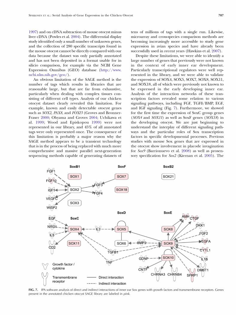
Despite these limitations, we were able to identify alarge number of genes that previously were not knownin the context of early inner ear development.Particularly transcriptional regulators were well rep-resented in the library, and we were able to validatethe expression of SOX4, SOX5, SOX7, SOX8, SOX11,and SOX18, all of which were previously not known tobe expressed in the early developing inner ear.Analysis of the interaction networks of these tran-scription factors revealed some relation to varioussignaling pathways, including FGF, TGFß/BMP, EGF,and IGF signaling (Fig. 7). Furthermore, we showedfor the first time the expression of SoxC group genes(SOX4 and SOX11) as well as SoxF genes (SOX18) inthe developing otocyst. We are just beginning tounderstand the interplay of different signaling path-ways and the particular roles of Sox transcriptionfactors in specific developmental processes. Previousstudies with mouse Sox genes that are expressed inthe otocyst show involvement in placode invaginationfor Sox9 (Barrionuevo et al. 2008) as well as prosen-sory specification for Sox2 (Kiernan et al. 2005). The
FIG. 7. IPA software analysis of direct and indirect interactions of inner ear Sox genes with growth factors and transmembrane receptors. Genespresent in the annotated chicken otocyst SAGE library are labeled in pink.
SINKKONEN ET AL.: Serial Analysis of Gene Expression in the Chicken Otocyst 707

overlapping expression of many Sox genes combinedwith a potential functional redundancy will make itdifficult to study individual Sox gene function in theotocyst because single knockouts might lack a pheno-type. Phylogeny, neofunctionalization, and redundan-cies within the Sox gene family have recently beencomprehensively reviewed, highlighting the impor-tance of this group of genes in many developmentalprocesses (Guth and Wegner 2008).
Our analysis also revealed many other potentiallyimportant genes that have not previously been consid-ered in the context of inner ear development, and somehave just recently been investigated.We found a numberof secreted proteins that are novel candidates for signal-ing functions in the developing otocyst. Transmembraneproteins consisted of members of previously knownfamilies of proteins that are essentially involved in innerear development such as receptors for FGFs, BMPs, andWNTs, as well as NOTCH1, among others. Interestingly,we found a relatively large group of proteins belongingto families that have been implicated in axonal guidanceand cell migration; some of these proteins have pre-viously been shown to be expressed in the otocyst andother developmental stages of the inner ear (Battisti andFekete 2008; Matilainen et al. 2007). The expression andfunction of Slit-like transmembrane protein SLITRK6,for example, has recently been analyzed in mouse innerear development (Katayama et al. 2009). Slitrk6 isstrongly expressed in the prosensory and sensory patchesof the auditory and vestibular organs; the innervationdensity of these organs was reduced or abolished inSlitrk6−/− mice.
In summary, we have used the SAGE method toassemble a list of sequence tags that can be associatedwith gene expression in the chicken otocyst. Althoughnot all-inclusive, this SAGE library is a practical bio-informatics tool to study otocyst gene expression. Foruser-defined analyses, the library is available in elec-tronic formats that can be directly queried online suchas NCBI GEO, or it can be imported into commercial orpublic domain bioinformatic software packages such asIPA. We used the Sox gene family as an example tohighlight the depth as well as the limitations of thelibrary and to demonstrate that the collection of otocystSAGE tags is a useful tool for molecular and devel-opmental studies of early inner ear development.
ACKNOWLEDGMENTS
This project was supported by the Sigrid Jusélius Foundationand Instrumentarium Science Foundation (to S.T.S.), aStanford Dean’s Fellowship, and by fellowship D/06/41764from the German Academic Exchange Service (to V.S.), as wellas grants DC006167, DC010042, and P30 DC010363 from theNational Institutes of Health (to S.H.).
REFERENCES
ABRAIRAVE, DEL RIO T, TUCKER AF, SLONIMSKY J, KEIRNES HL, GOODRICH
LV (2008) Cross-repressive interactions between Lrig3 andnetrin 1 shape the architecture of the inner ear. Development135:4091–4099
ADAM J, MYAT A, LE ROUX I, EDDISON M, HENRIQUE D, ISH-HOROWICZ D,LEWIS J (1998) Cell fate choices and the expression of Notch, Deltaand Serrate homologues in the chick inner ear: parallels withDrosophila sense-organ development. Development 125:4645–4654
ALSINA B, ABELLO G, ULLOA E, HENRIQUE D, PUJADES C, GIRALDEZ F(2004) FGF signaling is required for determination of oticneuroblasts in the chick embryo. Dev Biol 267:119–134
BARRIONUEVO F, NAUMANN A, BAGHERI-FAM S, SPETH V, TAKETO MM,SCHERER G, NEUBUSER A (2008) Sox9 is required for invaginationof the otic placode in mice. Dev Biol 317:213–224
BATTISTI AC, FEKETE DM (2008) Slits and Robos in the developingchicken inner ear. Dev Dyn 237:476–484
BOK J, BRONNER-FRASER M, WU DK (2005) Role of the hindbrain indorsoventral but not anteroposterior axial specification of theinner ear. Development 132:2115–2124
BRIGANDE JV, HELLER S (2009) Quo vadis hair cell regeneration? NatNeurosci 12:679–685
DIENSTHUBER M, OSHIMA K, HELLER S (2009) Stem/progenitor cellsderived from the cochlear sensory epithelium give rise tospheres with distinct morphologies and features. J Assoc ResOtolaryngol 10:173–190
FEKETE DM (1996) Cell fate specification in the inner ear. Curr OpinNeurobiol 6:533–541
FEKETE DM, CAMPERO AM (2007) Axon guidance in the inner ear. IntJ Dev Biol 51:549–556
FEKETE DM, WU DK (2002) Revisiting cell fate specification in theinner ear. Curr Opin Neurobiol 12:35–42
FRENZ DA, VAN DE WATER TR, GALINOVIC-SCHWARTZ V (1991) Trans-forming growth factor beta: does it direct otic capsule forma-tion? Ann Otol Rhinol Laryngol 100:301–307
FRENZ DA, GALINOVIC-SCHWARTZ V, LIU W, FLANDERS KC, VAN DE WATER
TR (1992) Transforming growth factor beta 1 is an epithelial-derived signal peptide that influences otic capsule formation.Dev Biol 153:324–336
FROLOVA EI, FOKINA VM, BEEBE DC (2004) The expression pattern ofopticin during chicken embryogenesis. Gene Expr Patterns4:335–338
GONG TW, HEGEMAN AD, SHIN JJ, LINDBERG KH, BARALD KF, LOMAX MI(1997) Novel genes expressed in the chick otocyst duringdevelopment: identification using differential display of RNA.Int J Dev Neurosci 15:585–594
GROVES AK, BRONNER-FRASER M (2000) Competence, specificationand commitment in otic placode induction. Development127:3489–3499
GUTH SI, WEGNER M (2008) Having it both ways: Sox proteinfunction between conservation and innovation. Cell Mol Life Sci65:3000–3018
HAMBURGER V, HAMILTON HL (1992) A series of normal stages in thedevelopment of the chick embryo. 1951. Dev Dyn 195:231–272
HAWKINS RD, BASHIARDES S, POWDER KE, SAJAN SA, BHONAGIRI V,ALVARADO DM, SPECK J, WARCHOL ME, LOVETT M (2007) Largescale gene expression profiles of regenerating inner ear sensoryepithelia. PLoS ONE 2:e525
HERBRAND H, GUTHRIE S, HADRYS T, HOFFMANN S, ARNOLD HH,RINKWITZ-BRANDT S, BOBER E (1998) Two regulatory genes,cNkx5-1 and cPax2, show different responses to local signalsduring otic placode and vesicle formation in the chick embryo.Development 125:645–654
HOLLYDAY M, MCMAHON JA, MCMAHON AP (1995) Wnt expressionpatterns in chick embryo nervous system. Mech Dev 52:9–25
708 SINKKONEN ET AL.: Serial Analysis of Gene Expression in the Chicken Otocyst

ITO K, NAKAMURA H, WATANABE Y (2011) Protogenin mediates celladhesion for ingression and re-epithelialization of paraxialmesodermal cells. Dev Biol 351:13–24
JIA XQ, NAKASHIMA T, KADOMATSU K, MURAMATSU T (2001) Expressionof midkine in the cochlea. Hear Res 160:10–14
KATAYAMA K, ZINE A, OTA M, MATSUMOTO Y, INOUE T, FRITZSCH B, ARUGA
J (2009) Disorganized innervation and neuronal loss in theinner ear of Slitrk6-deficient mice. PLoS ONE 4:e7786
KIERNAN AE, PELLING AL, LEUNG KK, TANG AS, BELL DM, TEASE C,LOVELL-BADGE R, STEEL KP, CHEAH KS (2005) Sox2 is required forsensory organ development in the mammalian inner ear. Nature434:1031–1035
KIMURA I, KONISHI M, MIYAKE A, FUJIMOTO M, ITOH N (2006) Neudesin,a secreted factor, promotes neural cell proliferation and neuro-nal differentiation in mouse neural precursor cells. J NeurosciRes 83:1415–1424
LEWIS AK, FRANTZ GD, CARPENTER DA, DE SAUVAGE FJ, GAO WQ (1998)Distinct expression patterns of notch family receptors andligands during development of the mammalian inner ear. MechDev 78:159–163
LI H, LIU H, HELLER S (2003A) Pluripotent stem cells from the adultmouse inner ear. Nat Med 9:1293–1299
LI H, ROBLIN G, LIU H, HELLER S (2003B) Generation of hair cells bystepwise differentiation of embryonic stem cells. Proc Natl AcadSci U S A 100:13495–13500
LI H, LIU H, CORRALES CE, MUTAI H, HELLER S (2004) Correlation ofPax-2 expression with cell proliferation in the developingchicken inner ear. J Neurobiol 60:61–70
LI H, RUAN J, DURBIN R (2008) Mapping short DNA sequencingreads and calling variants using mapping quality scores. GenomeRes 18:1851–1858
LILLEVALI K, HAUGAS M, PITUELLO F, SALMINEN M (2007) Comparativeanalysis of Gata3 and Gata2 expression during chicken inner eardevelopment. Dev Dyn 236:306–313
LIU M, PEREIRA FA, PRICE SD, CHU MJ, SHOPE C, HIMES D, EATOCK RA,BROWNELL WE, LYSAKOWSKI A, TSAI MJ (2000) Essential role ofBETA2/NeuroD1 in development of the vestibular and auditorysystems. Genes Dev 14:2839–2854
LIU W, LI G, CHIEN JS, RAFT S, ZHANG H, CHIANG C, FRENZ DA(2002) Sonic hedgehog regulates otic capsule chondrogene-sis and inner ear development in the mouse embryo. DevBiol 248:240–250
LIU D, CHU H, MAVES L, YAN YL, MORCOS PA, POSTLETHWAIT JH,WESTERFIELD M (2003) Fgf3 and Fgf8 dependent and independ-ent transcription factors are required for otic placode specifica-tion. Development 130:2213–2224
LIU W, LI L, LI G, GARRITANO F, SHANSKE A, FRENZ DA (2008)Coordinated molecular control of otic capsule differentiation:functional role of Wnt5a signaling and opposition by sfrp3activity. Growth Factors 26:343–354
LU X, BORCHERS AG, JOLICOEUR C, RAYBURN H, BAKER JC, TESSIER-LAVIGNE M (2004) PTK7/CCK-4 IS A NOVEL REGULATOR OF PLANAR
CELL POLARITY IN VERTEBRATES. Nature 430:93–98MA Q, ANDERSON DJ, FRITZSCH B (2000) Neurogenin 1 null mutant
ears develop fewer, morphologically normal hair cells in smallersensory epithelia devoid of innervation. J Assoc Res Otolaryngol1:129–143
MATILAINEN T, HAUGAS M, KREIDBERG JA, SALMINEN M (2007) Analysisof Netrin 1 receptors during inner ear development. Int J DevBiol 51:409–413
NEVES J, KAMAID A, ALSINA B, GIRALDEZ F (2007) Differentialexpression of Sox2 and Sox3 in neuronal and sensory progen-itors of the developing inner ear of the chick. J Comp Neurol503:487–500
OH SH, JOHNSON R, WU DK (1996) Differential expression of bonemorphogenetic proteins in the developing vestibular andauditory sensory organs. J Neurosci 16:6463–6475
OHYAMA T, GROVES AK (2004) Expression of mouse Foxi classgenes in early craniofacial development. Dev Dyn 231:640–646
OKANO J, TAKIGAWA T, SEKI K, SUZUKI S, SHIOTA K, ISHIBASHI M (2005)Transforming growth factor beta 2 promotes the formation ofthe mouse cochleovestibular ganglion in organ culture. Int J DevBiol 49:23–31
OSHIMA K, GRIMM CM, CORRALES CE, SENN P, MARTINEZ MONEDERO R,GELEOC GS, EDGE A, HOLT JR, HELLER S (2007) Differentialdistribution of stem cells in the auditory and vestibular organs ofthe inner ear. J Assoc Res Otolaryngol 8:18–31
OSHIMA K, SHIN K, DIENSTHUBER M, PENG AW, RICCI AJ, HELLER S(2010) Mechanosensitive hair cell-like cells from embryonic andinduced pluripotent stem cells. Cell 141:704–716
PAULEY S, LAI E, FRITZSCH B (2006) Foxg1 is required for morpho-genesis and histogenesis of the mammalian inner ear. Dev Dyn235:2470–2482
PERROT V, VAZQUEZ-PRADO J, GUTKIND JS (2002) Plexin B regulates Rhothrough the guanine nucleotide exchange factors leukemia-associated Rho GEF (LARG) and PDZ-RhoGEF. J Biol Chem277:43115–43120
PIRVOLA U, YLIKOSKI J, TROKOVIC R, HEBERT JM, MCCONNELL SK,PARTANEN J (2002) FGFR1 is required for the development of theauditory sensory epithelium. Neuron 35:671–680
POWLES N, BABBS C, FICKER M, SCHIMMANG T, MACONOCHIE M (2004)Identification and analysis of genes from the mouse otic vesicleand their association with developmental subprocesses throughin situ hybridization. Dev Biol 268:24–38
PUPPO F, THOME V, LHOUMEAU AC ET AL (2011) Protein tyrosine kinase7 has a conserved role in Wnt/beta-catenin canonical signalling.EMBO Rep 12:43–49
RICCOMAGNO MM, MARTINU L, MULHEISEN M, WU DK, EPSTEIN DJ(2002) Specification of the mammalian cochlea is dependent onSonic hedgehog. Genes Dev 16:2365–2378
SAHA S, SPARKS AB, RAGO C, AKMAEV V, WANG CJ, VOGELSTEIN B, KINZLER
KW, VELCULESCU VE (2002) Using the transcriptome to annotatethe genome. Nat Biotechnol 20:508–512
SANCHEZ-CALDERON H, FRANCISCO-MORCILLO J, MARTIN-PARTIDO G,HIDALGO-SANCHEZ M (2007) Fgf19 expression patterns in thedeveloping chick inner ear. Gene Expr Patterns 7:30–38
SIENKNECHT UJ, FEKETE DM (2009) Mapping of Wnt, frizzled, andWnt inhibitor gene expression domains in the avian oticprimordium. J Comp Neurol 517:751–764
STEVENS CB, DAVIES AL, BATTISTA S, LEWIS JH, FEKETE DM (2003)Forced activation of Wnt signaling alters morphogenesis andsensory organ identity in the chicken inner ear. Dev Biol261:149–164
STONE JS, RUBEL EW (1999) Delta1 expression during avian hair cellregeneration. Development 126:961–973
STREIT A (2007) The preplacodal region: an ectodermal domainwith multipotential progenitors that contribute to sense organsand cranial sensory ganglia. Int J Dev Biol 51:447–461
SUDHOF TC (2001) alpha-Latrotoxin and its receptors: neurexinsand CIRL/latrophilins. Annu Rev Neurosci 24:933–962
SWANSON GJ, HOWARD M, LEWIS J (1990) Epithelial autonomy in thedevelopment of the inner ear of a bird embryo. Dev Biol137:243–257
TOMAREV SI, NAKAYA N (2009) Olfactomedin domain-containingproteins: possible mechanisms of action and functions innormal development and pathology. Mol Neurobiol 40:122–138
UCHIKAWA M, KAMACHI Y, KONDOH H (1999) Two distinct subgroups ofGroup B Sox genes for transcriptional activators and repressors:their expression during embryonic organogenesis of thechicken. Mech Dev 84:103–120
URNESS LD, PAXTON CN, WANG X, SCHOENWOLF GC, MANSOUR SL(2010) FGF signaling regulates otic placode induction and
SINKKONEN ET AL.: Serial Analysis of Gene Expression in the Chicken Otocyst 709

refinement by controlling both ectodermal target genes andhindbrain Wnt8a. Dev Biol 340:595–604
VELCULESCU VE, ZHANG L, VOGELSTEIN B, KINZLER KW (1995) Serialanalysis of gene expression. Science 270:484–487
VON BARTHELD CS, PATTERSON SL, HEUER JG, WHEELER EF, BOTHWELL M,RUBEL EW (1991) Expression of nerve growth factor (NGF)receptors in the developing inner ear of chick and rat. Development113:455–470
WATANABE K, TAKEDA K, KATORI Y, IKEDA K, OSHIMAT, YASUMOTO K, SAITO H,TAKASAKAT, SHIBAHARA S (2000) Expression of the Sox10 gene duringmouse inner ear development. Brain Res Mol Brain Res 84:141–145
WEBBER A, RAZ Y (2006) Axon guidance cues in auditory develop-ment. Anat Rec A Discov Mol Cell Evol Biol 288:390–396
WONG YH, LU AC, WANG YC, CHENG HC, CHANG C, CHEN PH, YU JY, FANNMJ (2010) Protogenin defines a transition stage during embryonicneurogenesis and prevents precocious neuronal differentiation. JNeurosci 30:4428–4439
WOOD HB, EPISKOPOU V (1999) Comparative expression of themouse Sox1, Sox2 and Sox3 genes from pre-gastrulation to earlysomite stages. Mech Dev 86:197–201
WRIGHT TJ, MANSOUR SL (2003) Fgf3 and Fgf10 are required formouse otic placode induction. Development 130:3379–3390
YAMASHITA H, OESTERLE EC (1995) Induction of cell proliferation inmammalian inner-ear sensory epithelia by transforming growthfactor alpha and epidermal growth factor. Proc Natl Acad Sci US A 92:3152–3155
YEN WW, WILLIAMS M, PERIASAMY A, CONAWAY M, BURDSAL C, KELLER R,LU X, SUTHERLAND A (2009) PTK7 is essential for polarized cellmotility and convergent extension during mouse gastrulation.Development 136:2039–2048
ZOU P, MURAMATSU H, SONE M, HAYASHI H, NAKASHIMA T, MURAMATSU T(2006) Mice doubly deficient in the midkine and pleiotrophingenes exhibit deficits in the expression of beta-tectorin gene andin auditory response. Lab Invest 86:645–653
710 SINKKONEN ET AL.: Serial Analysis of Gene Expression in the Chicken Otocyst