journal of renin-angiotensin-aldosterone system · 2 . journal of the renin-angiotensin-aldosterone...
TRANSCRIPT
http://jra.sagepub.com/System
Journal of Renin-Angiotensin-Aldosterone
http://jra.sagepub.com/content/early/2013/02/22/1470320313477174The online version of this article can be found at:
DOI: 10.1177/1470320313477174
published online 25 February 2013Journal of Renin-Angiotensin-Aldosterone Systemthe SCOPE Consortium
Shane D Sykes, Kirsty G Pringle, Ang Zhou, Gustaaf A Dekker, Claire T Roberts, Eugenie R Lumbers and on behalf ofgestation pregnancy is influenced by fetal sex
The balance between human maternal plasma angiotensin II and angiotensin 1-7 levels in early
Published by:
http://www.sagepublications.com
can be found at:Journal of Renin-Angiotensin-Aldosterone SystemAdditional services and information for
http://jra.sagepub.com/cgi/alertsEmail Alerts:
http://jra.sagepub.com/subscriptionsSubscriptions:
http://www.sagepub.com/journalsReprints.navReprints:
http://www.sagepub.com/journalsPermissions.navPermissions:
What is This?
- Feb 25, 2013OnlineFirst Version of Record >>
at UNIV OF AUCKLAND LIB on February 28, 2013jra.sagepub.comDownloaded from
Journal of the Renin-Angiotensin-Aldosterone System0(0) 1 –9© The Author(s) 2013Reprints and permission: sagepub.co.uk/journalsPermissions.navDOI: 10.1177/1470320313477174jra.sagepub.com
Introduction
Pregnancy places increased stress on the maternal cardio-vascular system. The renin–angiotensin aldosterone system (RAAS) is a hormone cascade responsible for blood pres-sure homeostasis and is upregulated during pregnancy.1–3 A significant increase in plasma volume is essential for a suc-cessful pregnancy; these changes in plasma volume are mediated in part by the actions of angiotensin (Ang) II on the adrenal cortex, which releases aldosterone and stimu-lates fluid retention.4
Ang II has vasopressor effects, increasing systemic vas-cular resistance through central and peripheral actions. The RAAS is activated in early pregnancy to maintain blood pressure as maternal peripheral blood flow increases, par-ticularly in the skin,5 kidneys6 and uterus.7 A rise in glo-merular filtration rate (GFR)8 and an increase in progesterone levels9 occur early in pregnancy and would promote salt excretion if not offset by an activated RAAS. As well, sodium retention has to occur to allow for the
increase in maternal blood volume and to meet the demands of the growing fetus.
In pregnant women, plasma levels of active renin, and its precursor prorenin, are increased. Prorenin levels increase to 100 times those of active renin, while in non-pregnant individuals they are only 10 times greater.10–12 Under physiological conditions, 2% of this prorenin will spontaneously activate,13 thereby adding to the circulating renin enzyme activity. Recently, we showed that decidual
The balance between human maternal plasma angiotensin II and angiotensin 1-7 levels in early gestation pregnancy is influenced by fetal sex
Shane D Sykes1,2, Kirsty G Pringle1, Ang Zhou2, Gustaaf A Dekker2,3, Claire T Roberts2 and Eugenie R Lumbers1, on behalf of the SCOPE Consortium
AbstractHypothesis: There are fetal sex-associated differences in the circulating maternal renin–angiotensin system (RAS) in early pregnancy.Methods: Plasma prorenin, angiotensin (Ang) II, Ang 1-7 and angiotensin-converting enzyme (ACE) concentrations were measured at 15 weeks’ gestation in 131 women with uncomplicated pregnancies from the Adelaide SCOPE cohort. Uterine and umbilical artery Doppler sonography was performed at 20 weeks’ gestation.Results: At 15 weeks, women bearing female fetuses had higher maternal Ang II concentrations (p = 0.017) and lower Ang 1-7 to Ang II ratios (p = 0.016) than women bearing males. In women with male fetuses, Ang II positively correlated with birth weight (p = 0.028) and prorenin negatively correlated with placental weight (p = 0.014). Female fetuses had higher umbilical artery resistance indices (p = 0.019) that were related to maternal prorenin concentrations (p = 0.007).Conclusions: In early human pregnancy, the maternal RAS is influenced by fetal sex. The lower Ang 1-7 to Ang II ratios in women with female fetuses may contribute to the lower maternal peripheral microvascular flow as described previously and the lack of any positive effect of Ang II on fetal growth, as seen in women with male fetuses.
KeywordsPregnancy, sexual dimorphism, prorenin, angiotensin-converting enzyme, angiotensin peptides
1Hunter Medical Research Institute, University of Newcastle, Australia2Robinson Institute, University of Adelaide, Australia3Women’s and Children’s Division, Lyell McEwin Hospital, Australia
Corresponding author:Eugenie R. Lumbers, Mothers & Babies Research Centre, Hunter Medical Research Institute, John Hunter Hospital, Locked Bag 1, Hunter Region Mail Centre, 2310, NSW Australia. Email: [email protected]
477174 JRA0010.1177/1470320313477174Journal of the Renin-Angiotensin-Aldosterone SystemSykes et al.2013
Article
at UNIV OF AUCKLAND LIB on February 28, 2013jra.sagepub.comDownloaded from
2 Journal of the Renin-Angiotensin-Aldosterone System 0(0)
explants collected at term from women carrying a female fetus secrete more prorenin ex vivo than explants collected at term from women with a male fetus.14 Furthermore, these sex-specific effects on decidual prorenin secretion were still apparent when decidual explants were incubated for 48 hours ex vivo.
The activity of the RAAS is strongly influenced by the sex steroids, oestrogen and progesterone.15–17 Most signifi-cant is the effect of oestrogen on hepatic production of angiotensinogen (AGT).18 Renin cleaves AGT to form Ang I; a reaction rate limited by AGT levels. Ang I is cleaved by angiotensin-converting enzyme (ACE) to produce Ang II. Angiotensin-converting enzyme 2 (ACE2) is the predomi-nant enzyme for producing the heptapeptide Ang 1-7, with this reaction 500 times faster when using Ang II as the sub-strate rather than being generated from Ang I.19 Renin and prorenin are also influenced by sex, being lower in the cir-culation in women than in men, possibly because the influ-ence of oestrogens on AGT generates more Ang II leading to negative feedback on renin release.20,21 Conversely, Broughton-Pipkin et al.,22 who examined the renin and angiotensin levels in children less than 8 years of age, have found that plasma renin activity and Ang II levels are lower in boys than girls.
Ang peptides have been measured in pregnancy and both plasma Ang II and Ang (1-7) are increased.23,24 Urinary Ang 1-7 also increases throughout gestation.25 Ang II has not been measured in early gestation at the same time as Ang 1-7. Ang 1-7 has been shown to be a vasodilator by inducing endothelial nitric oxide (NO) production,26 kinins27 and prostaglandins28 so the balance between the levels of Ang II and Ang 1-7 may be important in regulating maternal vascular tone. Interestingly, in preeclampsia and gestational diabetes Ang (1-7) levels are low in late gesta-tion.29,30 In order to fully elucidate the functional role of these peptides in pregnancy and the use of Ang 1-7 and/or Ang II as potential biomarkers for pregnancy pathologies such as preeclampsia and gestational hypertension in which the RAAS has been implicated,31 we first needed to estab-lish the normal levels of these peptides and other compo-nents of the RAAS in early gestation in women with uncomplicated pregnancies.
We therefore tested the following hypotheses: first, that a factor associated with fetal sex alters the balance between the Ang II and Ang 1-7 axes of the maternal RAS; second, these sex-based differences in angiotensin peptides are due to differences in the upstream components of the RAS, i.e. prorenin and ACE; third, since the circulating RAAS is a key regulator of cardiovascular and renal function and Ang II is a pro-inflammatory peptide,32,33 these differences in circulating RAAS components would affect other measures of maternal cardiovascular and renal health as well as a marker of inflammation (C-reactive protein; CRP). Finally, we postulated that differences in Ang peptides, and there-fore differences in the vasodilator/vasoconstrictor balance,
would alter uterine artery and umbilical artery resistance indices as well as fetal growth.
Materials and methods
Study design
The current study is a nested case-control study within the Adelaide Screening for Pregnancy Endpoints (SCOPE) cohort. Women attending the Lyell McEwin Hospital (South Australia, Australia) were recruited, after giving informed written consent, if they were nulliparous with a singleton pregnancy, less than 15 weeks’ gestation and had fewer than three previous terminations of pregnancy or miscarriages. Samples collected at 15 weeks’ gestation were selected if women had no pregnancy-associated com-plications (n = 131), that is, if they remained normotensive (<140 and/or <90 mmHg prior to labour), showed no pro-teinuria, delivered a live born baby who was normally grown after 37 weeks’ gestation and had no other sign of pregnancy complications. All the women included in the current study self-reported as being Caucasian, except for one who was listed as a Maori/Cook Islander. Ethics approval for this work was given by the Central Northern Adelaide Health Service Ethics of Human Research Committee (study number: REC 1714/5/2008).
Sample collection
Non-fasting blood was collected into ethylenediaminetet-raacetic acid (EDTA) vacutainers at 15 weeks’ gestation by venepuncture from patients who had been either sitting or supine for 10 minutes (min). Blood pressure and other clin-ical measurements were also recorded at this time. Mid-stream urine was collected into 50 ml pots. All samples were placed on ice before processing and storage at –80°C within 30 min of collection. Doppler sonography was con-ducted at 20 weeks’ gestation on the umbilical and uterine arteries to measure blood flow resistance. Birth and placen-tal weights were measured following delivery. Maternal daily cigarette consumption from before and during preg-nancy was also recorded.
Laboratory measurements
Plasma Ang II and Ang 1-7 were measured at ProSearch International Australia Pty. Ltd. (Malvern, Victoria, Australia) using a direct radioimmunoassay employing delayed tracer addition. Ang II assay sensitivity was 4 pmol/l; with intra- and inter-assay coefficients of variation of 6.4% and 12%, respectively. Cross-reactivity to Ang I, Ang 1-7 and all other pertinent hormones is 0.52%, 0.01% and <0.1%, respectively. Cross-reactivity to Ang III and Ang IV are 98% and 100%, respectively, as these peptides have the same c-terminal as Ang II. Quality controls for
at UNIV OF AUCKLAND LIB on February 28, 2013jra.sagepub.comDownloaded from
Sykes et al. 3
Ang II measured 35.9, 37.5 and 33.8 pmol/l. Ang 1-7 assay sensitivity was 13 pmol/l; with intra- and inter-assay coef-ficients of variation of 4.5% and 10%, respectively. Cross reactivity to Ang I, Ang II, Ang III and Ang IV were 0.11%, 0.04%, 0.53% and 0.25%, respectively. Quality controls for Ang 1-7 measured 135.8, 158.2 and 149.2 pmol/l. The Ang 1-7 to Ang II ratios were derived from the plasma concen-trations of these peptides.
Maternal plasma concentrations of ACE (Duoset, R&D Systems, MN, USA) and prorenin (Molecular Innovations, MI, USA) were measured using commercially available enzyme-linked immunosorbent assay kits and conducted according to manufacturer’s instructions.
Biological analytes were measured by SA Pathology at the Institute of Medical and Veterinary Science (South Australia, Australia). Plasma and urinary electrolytes were measured using ion-selective electrodes, while cre-atinine was measured using the Jaffé method and CRP determined with an immuno-turbidimetric assay. All assays were read on an Olympus AU5400 Chemistry-Immuno Analyzer.
Data analysis
The rate pressure product, which is an indirect measure of cardiac work, was calculated by multiplying heart rate and systolic blood pressure. Plasma creatinine was used as a surrogate measure of GFR because equations that derive GFR from the plasma creatinine cannot be used in preg-nancy because of the physiological changes in GFR and the progressive changes in body weight.34 Urinary protein to creatinine, urinary albumin to creatinine and urinary sodium to potassium ratios were determined and the frac-tional excretion of sodium calculated from the formula:
FENa (%) = (UNa × (PCr/1000))/(PNa × UCr)*100
Where UNa and PNa are urinary and plasma sodium levels (mmol/l), respectively, and UCr and PCr are urinary (mmol/l) and plasma creatinine levels (µmol/l), respectively.
Values are expressed as medians and interquartile ranges, unless otherwise stated. Statistical significance was deemed as p < 0.05. Mann-Whitney U tests were used to determine fetal sex-dependent differences (male fetuses: n = 68, female
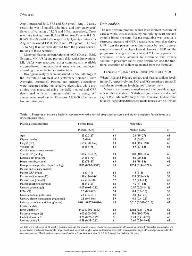
Table 1. Measures of maternal health in women who had a normal pregnancy outcome and either a singleton female fetus or a singleton male fetus.
Maternal characteristics Female fetus Male fetus
Median (IQR) n Median (IQR) n
Age 23 (20–27) 63 23 (19–27) 68Cigarettes/day 0 (0–6) 63 0 (0–15) 68Height (cm) 165 (158–169) 63 164 (159–168) 68Weight (kg) 69 (59–96) 63 69 (57–88) 68Cardiovascular measurementsSystolic BP (mmHg) 108 (102–116) 63 108 (100–115) 68Diastolic BP (mmHg) 64 (58–70) 63 60 (60–68) 68Heart rate (beats/min) 82 (75–87) 63 84 (78–88) 68Rate pressure product (bpm*mmHg) 8820 (8000–9800) 63 8754 (8144–9723) 68Plasma and urinary analytesPlasma CRP (mg/l) 6 (3–11) 62 4 (2–8) 67Plasma sodium (mmol/l) 138 (136–144) 56 138 (136–143) 58Plasma urea (mmol/l) 2.7 (2.4–3.0) 57 2.7 (2.1–3.1) 60Plasma creatinine (µmol/l) 46 (42–51) 63 46 (41–52) 67Urinary protein (g/l) 0.07 (0.05–0.12) 63 0.07 (0.05–0.16) 67FENa (%) 0.5 (0.3–0.7) 54 0.4 (0.3–0.6) 57Urinary sodium:potassium 2.5 (1.5–3.1) 60 2.0 (1.2–2.8) 67Urinary albumin:creatinine (mg/mmol) 0.5 (0.4–0.6) 59 0.5 (0.4–0.8) 64Urinary protein:creatinine (g/mmol) 0.011 (0.009–0.014) 63 0.010 (0.008–0.015) 67Obstetric dataBirth weight (g) 3460 (3290–3830) 63 3,483 (3271–3765) 68Placental weight (g) 600 (500–705) 60 596 (500–700) 63Umbilical artery RI 0.76 (0.73–0.79)a 61 0.74 (0.71–0.78) 68Uterine artery RI 0.58 (0.53–0.62) 61 0.60 (0.54–0.63) 68
All data were collected at 15 weeks’ gestation, except the obstetric data, which were measured at 20 weeks’ gestation by Doppler sonography, and presented as median (interquartile range); birth and placental weights were collected at term. IQR: interquartile range; BP: blood pressure; CRP: C-reactive protein; FENa: fractional excretion of sodium; RI: resistance index; ap = 0.019 using Mann Whitney U tests.
at UNIV OF AUCKLAND LIB on February 28, 2013jra.sagepub.comDownloaded from
4 Journal of the Renin-Angiotensin-Aldosterone System 0(0)
fetuses: n = 63). Pairwise Spearman correlations were used to identify significant relationships between RAS variables thought to interact on a physiological basis. Data were ana-lysed using Stata/IC 11.0 (StataCorp LP, TX, USA) and scatter plots generated with this software. GraphPad Prism 5.0 was used to generate box and whisker plots.
Results
The only difference in maternal physiological variables, measured at 15 weeks’ gestation, between women who
subsequently delivered a female baby compared with those who had a male baby was higher umbilical artery resistance indices at 20 weeks’ gestation in women who carried female fetuses (Table 1).
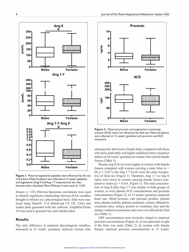
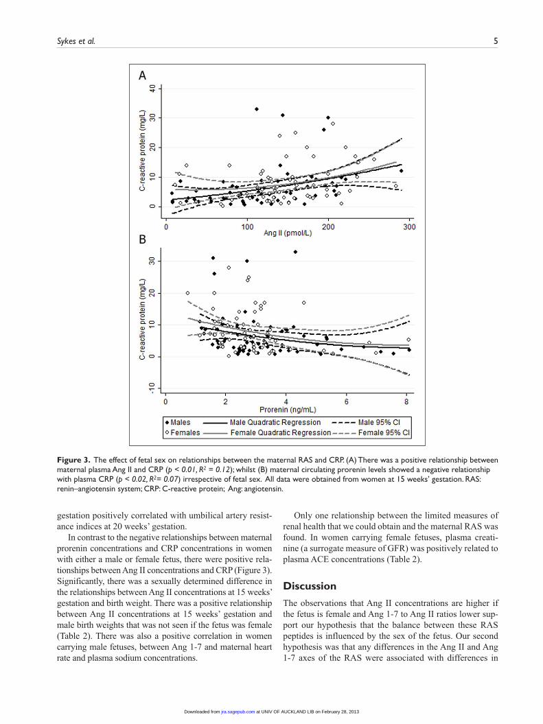
Plasma Ang II levels were higher in women with female fetuses compared with women carrying a male fetus (n = 68, p = 0.017), but Ang 1-7 levels were the same irrespec-tive of fetal sex (Figure 1). Therefore, Ang 1-7 to Ang II ratios were lower in women carrying female fetuses com-pared to males (p = 0.016, Figure 1). The total concentra-tion of Ang II plus Ang 1-7 was similar in both groups of women, as were plasma ACE concentrations and prorenin concentrations (Figure 2). At 15 weeks’ gestation, maternal heart rate, blood pressure, rate pressure product, plasma urea, plasma sodium, plasma creatinine, urinary albumin to creatinine ratio, urinary protein to creatinine ratio and the urinary sodium to potassium ratio were not affected by fetal sex (Table 1).
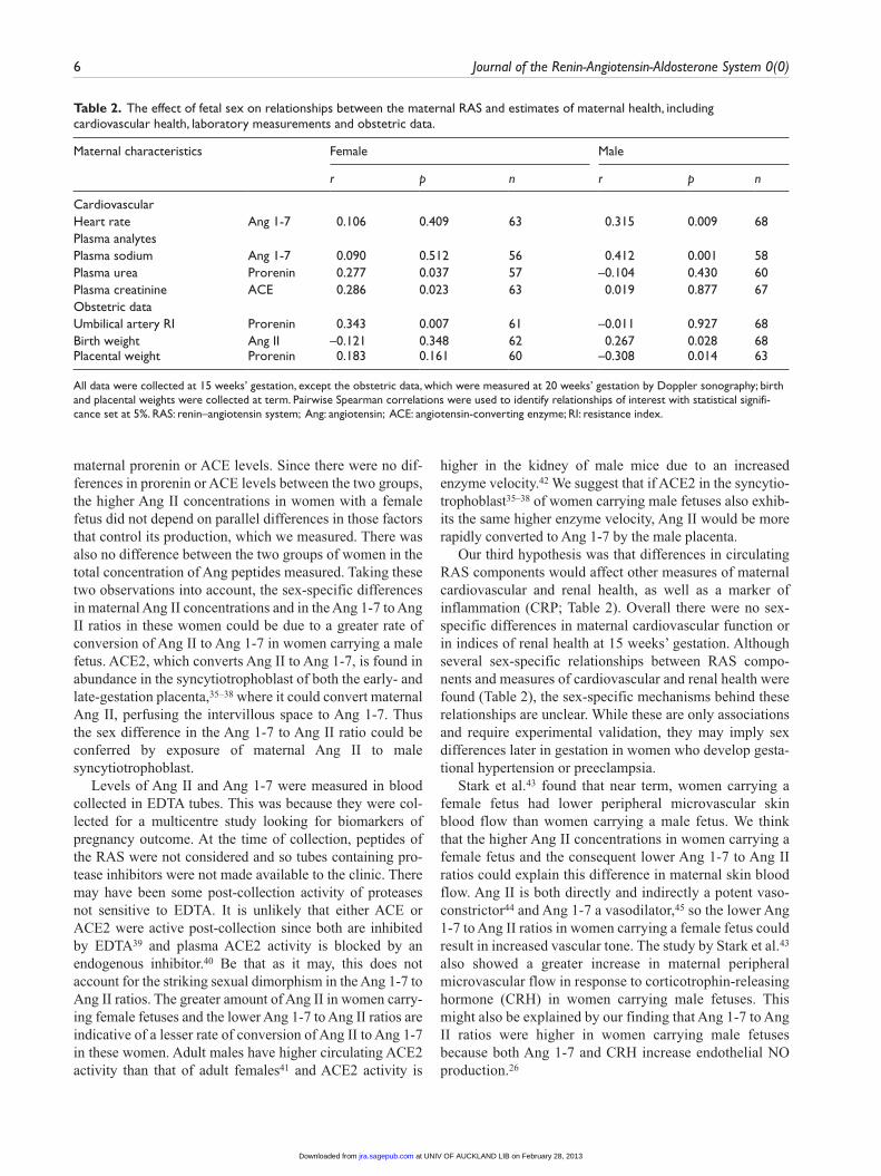
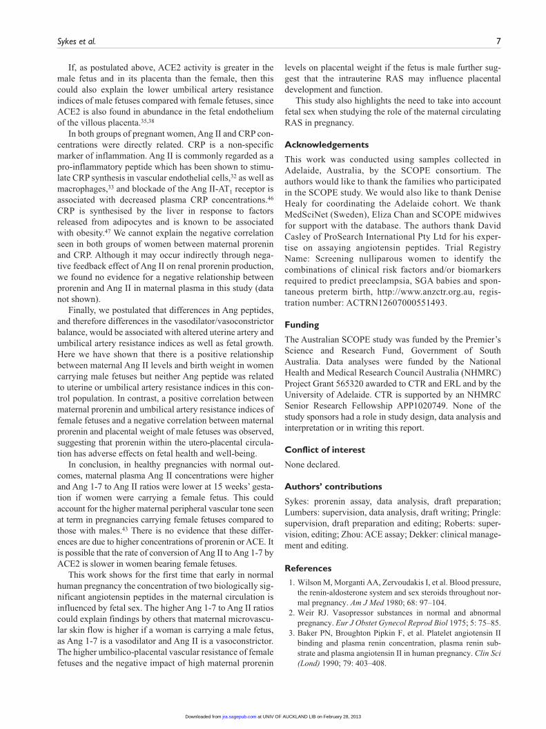
CRP concentrations were inversely related to maternal prorenin concentrations (Figure 3), as was placental weight if the fetus was male (Table 2). In women with female fetuses, maternal prorenin concentrations at 15 weeks’
Figure 1. Maternal angiotensin peptides were influenced by the sex of the fetus. Maternal plasma was collected at 15 weeks’ gestation and angiotensin (Ang) II and Ang 1-7 measured and the ratio between them calculated. Mann Whitney U tests used; *p < 0.02.
Figure 2. Maternal prorenin and angiotensin-converting enzyme (ACE) were not influenced by fetal sex. Maternal plasma was collected at 15 weeks’ gestation and prorenin and ACE measured.
at UNIV OF AUCKLAND LIB on February 28, 2013jra.sagepub.comDownloaded from
Sykes et al. 5
gestation positively correlated with umbilical artery resist-ance indices at 20 weeks’ gestation.
In contrast to the negative relationships between maternal prorenin concentrations and CRP concentrations in women with either a male or female fetus, there were positive rela-tionships between Ang II concentrations and CRP (Figure 3). Significantly, there was a sexually determined difference in the relationships between Ang II concentrations at 15 weeks’ gestation and birth weight. There was a positive relationship between Ang II concentrations at 15 weeks’ gestation and male birth weights that was not seen if the fetus was female (Table 2). There was also a positive correlation in women carrying male fetuses, between Ang 1-7 and maternal heart rate and plasma sodium concentrations.
Only one relationship between the limited measures of renal health that we could obtain and the maternal RAS was found. In women carrying female fetuses, plasma creati-nine (a surrogate measure of GFR) was positively related to plasma ACE concentrations (Table 2).
Discussion
The observations that Ang II concentrations are higher if the fetus is female and Ang 1-7 to Ang II ratios lower sup-port our hypothesis that the balance between these RAS peptides is influenced by the sex of the fetus. Our second hypothesis was that any differences in the Ang II and Ang 1-7 axes of the RAS were associated with differences in
Figure 3. The effect of fetal sex on relationships between the maternal RAS and CRP. (A) There was a positive relationship between maternal plasma Ang II and CRP (p < 0.01, R2 = 0.12); whilst (B) maternal circulating prorenin levels showed a negative relationship with plasma CRP (p < 0.02, R2= 0.07) irrespective of fetal sex. All data were obtained from women at 15 weeks’ gestation. RAS: renin–angiotensin system; CRP: C-reactive protein; Ang: angiotensin.
at UNIV OF AUCKLAND LIB on February 28, 2013jra.sagepub.comDownloaded from
6 Journal of the Renin-Angiotensin-Aldosterone System 0(0)
maternal prorenin or ACE levels. Since there were no dif-ferences in prorenin or ACE levels between the two groups, the higher Ang II concentrations in women with a female fetus did not depend on parallel differences in those factors that control its production, which we measured. There was also no difference between the two groups of women in the total concentration of Ang peptides measured. Taking these two observations into account, the sex-specific differences in maternal Ang II concentrations and in the Ang 1-7 to Ang II ratios in these women could be due to a greater rate of conversion of Ang II to Ang 1-7 in women carrying a male fetus. ACE2, which converts Ang II to Ang 1-7, is found in abundance in the syncytiotrophoblast of both the early- and late-gestation placenta,35–38 where it could convert maternal Ang II, perfusing the intervillous space to Ang 1-7. Thus the sex difference in the Ang 1-7 to Ang II ratio could be conferred by exposure of maternal Ang II to male syncytiotrophoblast.
Levels of Ang II and Ang 1-7 were measured in blood collected in EDTA tubes. This was because they were col-lected for a multicentre study looking for biomarkers of pregnancy outcome. At the time of collection, peptides of the RAS were not considered and so tubes containing pro-tease inhibitors were not made available to the clinic. There may have been some post-collection activity of proteases not sensitive to EDTA. It is unlikely that either ACE or ACE2 were active post-collection since both are inhibited by EDTA39 and plasma ACE2 activity is blocked by an endogenous inhibitor.40 Be that as it may, this does not account for the striking sexual dimorphism in the Ang 1-7 to Ang II ratios. The greater amount of Ang II in women carry-ing female fetuses and the lower Ang 1-7 to Ang II ratios are indicative of a lesser rate of conversion of Ang II to Ang 1-7 in these women. Adult males have higher circulating ACE2 activity than that of adult females41 and ACE2 activity is
higher in the kidney of male mice due to an increased enzyme velocity.42 We suggest that if ACE2 in the syncytio-trophoblast35–38 of women carrying male fetuses also exhib-its the same higher enzyme velocity, Ang II would be more rapidly converted to Ang 1-7 by the male placenta.
Our third hypothesis was that differences in circulating RAS components would affect other measures of maternal cardiovascular and renal health, as well as a marker of inflammation (CRP; Table 2). Overall there were no sex-specific differences in maternal cardiovascular function or in indices of renal health at 15 weeks’ gestation. Although several sex-specific relationships between RAS compo-nents and measures of cardiovascular and renal health were found (Table 2), the sex-specific mechanisms behind these relationships are unclear. While these are only associations and require experimental validation, they may imply sex differences later in gestation in women who develop gesta-tional hypertension or preeclampsia.
Stark et al.43 found that near term, women carrying a female fetus had lower peripheral microvascular skin blood flow than women carrying a male fetus. We think that the higher Ang II concentrations in women carrying a female fetus and the consequent lower Ang 1-7 to Ang II ratios could explain this difference in maternal skin blood flow. Ang II is both directly and indirectly a potent vaso-constrictor44 and Ang 1-7 a vasodilator,45 so the lower Ang 1-7 to Ang II ratios in women carrying a female fetus could result in increased vascular tone. The study by Stark et al.43 also showed a greater increase in maternal peripheral microvascular flow in response to corticotrophin-releasing hormone (CRH) in women carrying male fetuses. This might also be explained by our finding that Ang 1-7 to Ang II ratios were higher in women carrying male fetuses because both Ang 1-7 and CRH increase endothelial NO production.26
Table 2. The effect of fetal sex on relationships between the maternal RAS and estimates of maternal health, including cardiovascular health, laboratory measurements and obstetric data.
Maternal characteristics Female Male
r p n r p n
CardiovascularHeart rate Ang 1-7 0.106 0.409 63 0.315 0.009 68Plasma analytes Plasma sodium Ang 1-7 0.090 0.512 56 0.412 0.001 58Plasma urea Prorenin 0.277 0.037 57 –0.104 0.430 60Plasma creatinine ACE 0.286 0.023 63 0.019 0.877 67Obstetric dataUmbilical artery RI Prorenin 0.343 0.007 61 –0.011 0.927 68Birth weight Ang II –0.121 0.348 62 0.267 0.028 68Placental weight Prorenin 0.183 0.161 60 –0.308 0.014 63
All data were collected at 15 weeks’ gestation, except the obstetric data, which were measured at 20 weeks’ gestation by Doppler sonography; birth and placental weights were collected at term. Pairwise Spearman correlations were used to identify relationships of interest with statistical signifi-cance set at 5%. RAS: renin–angiotensin system; Ang: angiotensin; ACE: angiotensin-converting enzyme; RI: resistance index.
at UNIV OF AUCKLAND LIB on February 28, 2013jra.sagepub.comDownloaded from
Sykes et al. 7
If, as postulated above, ACE2 activity is greater in the male fetus and in its placenta than the female, then this could also explain the lower umbilical artery resistance indices of male fetuses compared with female fetuses, since ACE2 is also found in abundance in the fetal endothelium of the villous placenta.35,38
In both groups of pregnant women, Ang II and CRP con-centrations were directly related. CRP is a non-specific marker of inflammation. Ang II is commonly regarded as a pro-inflammatory peptide which has been shown to stimu-late CRP synthesis in vascular endothelial cells,32 as well as macrophages,33 and blockade of the Ang II-AT1 receptor is associated with decreased plasma CRP concentrations.46 CRP is synthesised by the liver in response to factors released from adipocytes and is known to be associated with obesity.47 We cannot explain the negative correlation seen in both groups of women between maternal prorenin and CRP. Although it may occur indirectly through nega-tive feedback effect of Ang II on renal prorenin production, we found no evidence for a negative relationship between prorenin and Ang II in maternal plasma in this study (data not shown).
Finally, we postulated that differences in Ang peptides, and therefore differences in the vasodilator/vasoconstrictor balance, would be associated with altered uterine artery and umbilical artery resistance indices as well as fetal growth. Here we have shown that there is a positive relationship between maternal Ang II levels and birth weight in women carrying male fetuses but neither Ang peptide was related to uterine or umbilical artery resistance indices in this con-trol population. In contrast, a positive correlation between maternal prorenin and umbilical artery resistance indices of female fetuses and a negative correlation between maternal prorenin and placental weight of male fetuses was observed, suggesting that prorenin within the utero-placental circula-tion has adverse effects on fetal health and well-being.
In conclusion, in healthy pregnancies with normal out-comes, maternal plasma Ang II concentrations were higher and Ang 1-7 to Ang II ratios were lower at 15 weeks’ gesta-tion if women were carrying a female fetus. This could account for the higher maternal peripheral vascular tone seen at term in pregnancies carrying female fetuses compared to those with males.43 There is no evidence that these differ-ences are due to higher concentrations of prorenin or ACE. It is possible that the rate of conversion of Ang II to Ang 1-7 by ACE2 is slower in women bearing female fetuses.
This work shows for the first time that early in normal human pregnancy the concentration of two biologically sig-nificant angiotensin peptides in the maternal circulation is influenced by fetal sex. The higher Ang 1-7 to Ang II ratios could explain findings by others that maternal microvascu-lar skin flow is higher if a woman is carrying a male fetus, as Ang 1-7 is a vasodilator and Ang II is a vasoconstrictor. The higher umbilico-placental vascular resistance of female fetuses and the negative impact of high maternal prorenin
levels on placental weight if the fetus is male further sug-gest that the intrauterine RAS may influence placental development and function.
This study also highlights the need to take into account fetal sex when studying the role of the maternal circulating RAS in pregnancy.
Acknowledgements
This work was conducted using samples collected in Adelaide, Australia, by the SCOPE consortium. The authors would like to thank the families who participated in the SCOPE study. We would also like to thank Denise Healy for coordinating the Adelaide cohort. We thank MedSciNet (Sweden), Eliza Chan and SCOPE midwives for support with the database. The authors thank David Casley of ProSearch International Pty Ltd for his exper-tise on assaying angiotensin peptides. Trial Registry Name: Screening nulliparous women to identify the combinations of clinical risk factors and/or biomarkers required to predict preeclampsia, SGA babies and spon-taneous preterm birth, http://www.anzctr.org.au, regis-tration number: ACTRN12607000551493.
Funding
The Australian SCOPE study was funded by the Premier’s Science and Research Fund, Government of South Australia. Data analyses were funded by the National Health and Medical Research Council Australia (NHMRC) Project Grant 565320 awarded to CTR and ERL and by the University of Adelaide. CTR is supported by an NHMRC Senior Research Fellowship APP1020749. None of the study sponsors had a role in study design, data analysis and interpretation or in writing this report.
Conflict of interest
None declared.
Authors’ contributions
Sykes: prorenin assay, data analysis, draft preparation; Lumbers: supervision, data analysis, draft writing; Pringle: supervision, draft preparation and editing; Roberts: super-vision, editing; Zhou: ACE assay; Dekker: clinical manage-ment and editing.
References
1. Wilson M, Morganti AA, Zervoudakis I, et al. Blood pressure, the renin-aldosterone system and sex steroids throughout nor-mal pregnancy. Am J Med 1980; 68: 97–104.
2. Weir RJ. Vasopressor substances in normal and abnormal pregnancy. Eur J Obstet Gynecol Reprod Biol 1975; 5: 75–85.
3. Baker PN, Broughton Pipkin F, et al. Platelet angiotensin II binding and plasma renin concentration, plasma renin sub-strate and plasma angiotensin II in human pregnancy. Clin Sci (Lond) 1990; 79: 403–408.
at UNIV OF AUCKLAND LIB on February 28, 2013jra.sagepub.comDownloaded from
8 Journal of the Renin-Angiotensin-Aldosterone System 0(0)
4. Laragh JH and Sealey JE. The plasma renin test reveals the contribution of body sodium-volume content (V) and renin-angiotensin (R) vasoconstriction to long-term blood pressure. Am J Hypertens 2011; 24: 1164–1180.
5. Myhrman P, Jansson I and Lundgren Y. Skin blood flow in normal pregnancy measured by venous occlusion plethys-mography of the hand. Acta Obstet Gynecol Scand 1980; 59: 107–110.
6. Conrad KP, Gandley RE, Ogawa T, et al. Endothelin medi-ates renal vasodilation and hyperfiltration during pregnancy in chronically instrumented conscious rats. Am J Physiol 1999; 276: F767–F776.
7. Elkayam U and Gleicher N (eds). Cardiac problems in preg-nancy: Diagnosis and management of maternal and fetal heart disease. 3rd ed. New York: Wiley-Liss, 1998.
8. Krutzen E, Olofsson P, Back SE, et al. Glomerular filtration rate in pregnancy: A study in normal subjects and in patients with hypertension, preeclampsia and diabetes. Scand J Clin Lab Invest 1992; 52: 387–392.
9. Ledoux F, Genest J, Nowaczynski W, et al. Plasma proges-terone and aldosterone in pregnancy. Can Med Assoc J 1975; 112: 943–947.
10. Hsueh WA, Luetscher JA, Carlson EJ, et al. Changes in active and inactive renin throughout pregnancy. J Clin Endocrinol Metab 1982; 54: 1010–1016.
11. Sealey JE, Wilson M, Morganti AA, et al. Changes in active and inactive renin throughout normal pregnancy. Clin Exp Hypertens A 1982; 4: 2373–2384.
12. Skinner SL, Cran EJ, Gibson R, et al. Angiotensins I and II, active and inactive renin, renin substrate, renin activity, and angiotensinase in human liquor amnii and plasma. Am J Obstet Gynecol 1975; 121: 626–630.
13. Danser AH and Deinum J. Renin, prorenin and the putative (pro)renin receptor. Hypertension 2005; 46: 1069–1076.
14. Wang Y, Pringle KG, Sykes SD, et al. Fetal sex affects expres-sion of renin-angiotensin system components in term human decidua. Endocrinology 2012; 153: 462–468.
15. Skinner SL, Lumbers ER and Symonds EM. Alteration by oral contraceptives of normal menstrual changes in plasma renin activity, concentration and substrate. Clin Sci 1969; 36: 67–76.
16. Walters WA and Lim YL. Haemodynamic changes in women taking oral contraceptives. J Obstet Gynaecol Br Commonw 1970; 77: 1007–1012.
17. Oelkers W, Schöneshofer M and Blümel A. Effects of proges-terone and four synthetic progestagens on sodium balance and the renin-aldosterone system in man. J Clin Endocrinol Metab 1974; 39: 882–890.
18. Clauser E, Gaillard I, Wei L, et al. Regulation of angioten-sinogen gene. Am J Hypertens 1989; 2: 403–410.
19. Ferrario CM. ACE2: More of Ang-(1–7) or less Ang II? Curr Opin Nephrol Hypertens 2011; 20: 1–6.
20. Blair-West JR, Coghlan JP, Denton DA, et al. Inhibition of renin secretion by systemic and intrarenal angiotensin infu-sion. Am J Physiol 1971; 220: 1309–1315.
21. Danser AH, Derkx FH, Schalekamp MA, et al. Determinants of interindividual variation of renin and prorenin concentra-tions: Evidence for a sexual dimorphism of (pro)renin levels in humans. J Hypertens 1998; 16: 853–862.
22. Broughton Pipkin F, Smales OR and O’Callaghan M. Renin and angiotensin levels in children. Arch Dis Child 1981; 56: 298–302.
23. Baker PN, Broughton Pipkin F and Symonds EM. Longitu-dinal study of platelet angiotensin II binding in human preg-nancy. Clin Sci (Lond) 1992; 82: 377–381.
24. Merrill DC, Karoly M, Chen K, et al. Angiotensin-(1–7) in normal and preeclamptic pregnancy. Endocrine 2002; 18: 239–245.
25. Valdes G, Germain AM, Corthorn J, et al. Urinary vasodila-tor and vasoconstrictor angiotensins during menstrual cycle, pregnancy, and lactation. Endocrine 2001; 16: 117–122.
26. Heitsch H, Brovkovych S, Malinski T, et al. Angioten-sin-(1–7)-stimulated nitric oxide and superoxide release from endothelial cells. Hypertension 2001; 37: 72–76.
27. Brosnihan KB, Li P and Ferrario CM. Angiotensin-(1–7) dilates canine coronary arteries through kinins and nitric oxide. Hypertension 1996; 27: 523–528.
28. Oliveira MA, Fortes ZB, Santos RA, et al. Synergistic effect of angiotensin-(1–7) on bradykinin arteriolar dilation in vivo. Peptides 1999; 20: 1195–1201.
29. Brosnihan KB, Neves LA, Anton L, et al. Enhanced expres-sion of Ang-(1–7) during pregnancy. Braz J Med Biol Res 2004; 37: 1255–1262.
30. Nogueira AI, Souza Santos RA, Simões ESAC, et al. The pregnancy-induced increase of plasma angiotensin-(1–7) is blunted in gestational diabetes. Regul Pept 2007; 141: 55–60.
31. Anton L and Brosnihan KB. Systemic and uteroplacental renin–angiotensin system in normal and pre-eclamptic preg-nancies. Ther Adv Cardiovasc Dis. 2008; 2: 349–362.
32. Han C, Liu J, Liu X, et al. Angiotensin II induces C-reactive pro-tein expression through ERK1/2 and JNK signaling in human aortic endothelial cells. Atherosclerosis 2010; 212: 206–212.
33. Li M, Liu J, Han C, et al. Angiotensin II induces the expression of c-reactive protein via MAPK-dependent signal pathway in U937 macrophages. Cell Physiol Biochem 2011; 27: 63–70.
34. Maynard SE and Thadhani R. Pregnancy and the kidney. J Am Soc Nephrol 2009; 20: 14–22.
35. Marques FZ, Pringle KG, Conquest A, et al. Molecular characterization of renin-angiotensin system components in human intrauterine tissues and fetal membranes from vaginal delivery and cesarean section. Placenta 2011; 32: 214–221.
36. Pringle KG, Tadros MA, Callister RJ, et al. The expres-sion and localization of the human placental prorenin/renin- angiotensin system throughout pregnancy: Roles in tropho-blast invasion and angiogenesis? Placenta 2011; 32: 956–962.
37. Valdes G, Kaufmann P, Corthorn J, et al. Vasodilator factors in the systemic and local adaptations to pregnancy. Reprod Biol Endocrinol 2009; 7: 79.
38. Valdes G, Neves LA, Anton L, et al. Distribution of angioten-sin-(1–7) and ACE2 in human placentas of normal and patho-logical pregnancies. Placenta 2006; 27: 200–207.
39. Tipnis SR, Hooper NM, Hyde R, et al. A human homolog of angiotensin-converting enzyme. Cloning and functional expression as a captopril-insensitive carboxypeptidase. J Biol Chem 2000; 275: 33238–33243.
40. Lew RA, Warner FJ, Hanchapola I, et al. Angiotensin- converting enzyme 2 catalytic activity in human plasma is masked by an endogenous inhibitor. Exp Physiol 2008; 93: 685–693.
at UNIV OF AUCKLAND LIB on February 28, 2013jra.sagepub.comDownloaded from
Sykes et al. 9
41. Soro-Paavonen A, Gordin D, Forsblom C, et al. Circulat-ing ACE2 activity is increased in patients with type 1 dia-betes and vascular complications. J Hypertens 2012; 30: 375–383.
42. Liu J, Ji H, Zheng W, et al. Sex differences in renal angio-tensin converting enzyme 2 (ACE2) activity are 17beta-oes-tradiol-dependent and sex chromosome-independent. Biol Sex Differ 2010; 1: 6.
43. Stark MJ, Dierkx L, Clifton VL, et al. Alterations in the maternal peripheral microvascular response in pregnancies complicated by preeclampsia and the impact of fetal sex. J Soc Gynecol Investig 2006; 13: 573–578.
44. Fujii AM and Vatner SF. Direct versus indirect pressor and vasoconstrictor actions of angiotensin in conscious dogs. Hypertension 1985; 7: 253–261.
45. Brosnihan KB, Li P, Tallant EA, et al. Angiotensin-(1-7): A novel vasodilator of the coronary circulation. Biol Res 1998; 31: 227–234.
46. Wang CH, Li SH, Weisel RD, et al. C-reactive protein upreg-ulates angiotensin type 1 receptors in vascular smooth muscle. Circulation 2003; 107: 1783–1790.
47. Visser M, Bouter LM, McQuillan GM, et al. Elevated C-reac-tive protein levels in overweight and obese adults. JAMA 1999; 282: 2131–2135.
at UNIV OF AUCKLAND LIB on February 28, 2013jra.sagepub.comDownloaded from