journal of molecular structure - adatbankadatbank.transindex.ro/vendeg/htmlk/pdf9512.pdf ·...
TRANSCRIPT

Journal of Molecular Structure 997 (2011) 78–86
Contents lists available at ScienceDirect
Journal of Molecular Structure
journal homepage: www.elsevier .com/locate /molstruc
Structural investigation of chitosan-based microspheres with someanti-inflammatory drugs
Simina Dreve a, Iren Kacso a, Adriana Popa a, Oana Raita a, Felicia Dragan b, A. Bende a, Gh. Borodi a, I. Bratu a,⇑a National Institute for R&D of Isotopic and Molecular Technologies, 65-103 Donáth st., RO-400293 Cluj-Napoca, Romaniab University of Oradea, Faculty of Medicine and Pharmacy, 54 N. Jiga st., Oradea, Romania
a r t i c l e i n f o a b s t r a c t
Article history:Received 5 April 2011Received in revised form 3 May 2011Accepted 3 May 2011Available online 14 May 2011
Keywords:ChitosanMicrospheresAnti-inflammatory drugsMolecular spectroscopy
0022-2860/$ - see front matter � 2011 Elsevier B.V. Adoi:10.1016/j.molstruc.2011.05.001
⇑ Corresponding author. Tel.: +40 264 584037; fax:E-mail address: [email protected] (I. Bratu).
The use of chitosan as an excipient in oral formulations, as a drug delivery vehicle for ulcerogenic anti-inflammatory drugs and as base in polyelectrolyte complex systems, to prepare solid release systems assponges was investigated. The preparation by double emulsification of chitosan hydrogels carrying dic-lofenac, acetyl-salycilic acid and hydrocortisone acetate as anti-inflammatory drugs is reported. The con-centration of anti-inflammatory drug in the chitosan hydrogel generating the sponges was 0.08 mmol.Chitosan-drug loaded sponges with anti-inflammatory drugs were prepared by freeze–drying at�60 �C and 0.009 atm. Structural investigations of the solid formulations were done by Fourier-transformed infrared and ultraviolet-visible spectroscopy, spectrofluorimetry, differential scanning calo-rimetry and X-ray diffractometry. The results indicated that the drug molecules are forming temporarychelates in chitosan hydrogels and sponges. Electron paramagnetic resonance demonstrates the presenceof free radicals in a wide range and the antioxidant activity for chitosan-drug supramolecular cross-linked assemblies.
� 2011 Elsevier B.V. All rights reserved.
1. Introduction
The concept of designing a specified delivery system has beenoriginated from the perception of Paul Ehrlich, who proposed drugdelivery to be as a ‘magic bullet’ [1], where a drug-carrier complex/conjugate, delivers drug(s) exclusively to the preselected targetcells in a specific manner. The objective of drug targeting is toachieve desired pharmacological response at a selected site with-out undesirable interactions. It is well known that most of anti-inflammatory drugs administered in therapeutic doses, increasethe sensitivity of the gastric mucosa against the aggressive factor,and the acid can produce gastro-intestinal inflammations or ulcers[2]. Between natural polymers generating novel biomaterials, chi-tin and chitosan with their copolymers, are intensively studieddue to their many potential applications. Chitosan [poly(b-(10/4)-2-amino-2-deoxy-Dglucose)] (CTS) is a natural cationic polysac-charide derived from chitin, which is copolymer, a glucosamineand an N-acetyl glucosamine units, combined together [3]. Chito-san is being widely used as a pharmaceutical excipient, comprisinga series of polymers varying in their degree of deacylation, molec-ular weight, viscosity, pKa, etc. The presence of a number of aminogroups permits CTS to chemically react with anionic systems,thereby resulting in alteration of physicochemical characteristics
ll rights reserved.
+40 264 420042.
of reactants and developing new properties of such combinations[4]. Moreover, chitosan has antiacid and antiulcer characteristics,which prevents or weakens drug irritation in the stomach [5].Modern biocompatible systems target not only infectious diseases,but also autoimmune disorders, allergies, chronic inflammatorydiseases and cancer [6]. Small molecular weight anti-inflammatorydrugs that contain chemical groups able to establish temporaryphysical or chemical bonding with the amino group of the CTS link-age are suitable for the targeted and controlled drug CTS-baseddelivery systems. In support of this presumption the chemical for-mulae of CTS, additives and anti-inflammatory drugs used in theexperiment are illustrated in Fig. 1. The present study was aimedto develop and characterize a CTS novel solid complex withTween-80 and oleic acid as drug carrier for controlled drug deliv-ery, with possible use in controlled (slow) drug delivery, avoidinggastrointestinal painful injuries. The hydrogel poly-electrolytecomplexes of chitosan (PEC CTS) were prepared by double emulsi-fication and coacervation method using the 0.02% Tween-80, 0.02%oleic acid in 3% CTS acidic solution [7]. Diclofenac sodium (DCF-Na)and acetyl-salycilic acid (ASA) are among the most useful non-steroidal anti-inflammatory drugs (NSAIDs). Both, and the steroi-dal anti-inflammatory drug hydrocortisone acetate (HcAc) too, dis-solve in gastro-intestinal fluid [8,9] causing gastric mucosaldamage. The pH of the gastrointestinal tract (GIT) varies from pH1 to 3 in the stomach and increases to approximately 7–8 in the co-lon. Another factor influencing the activity of drugs are the struc-
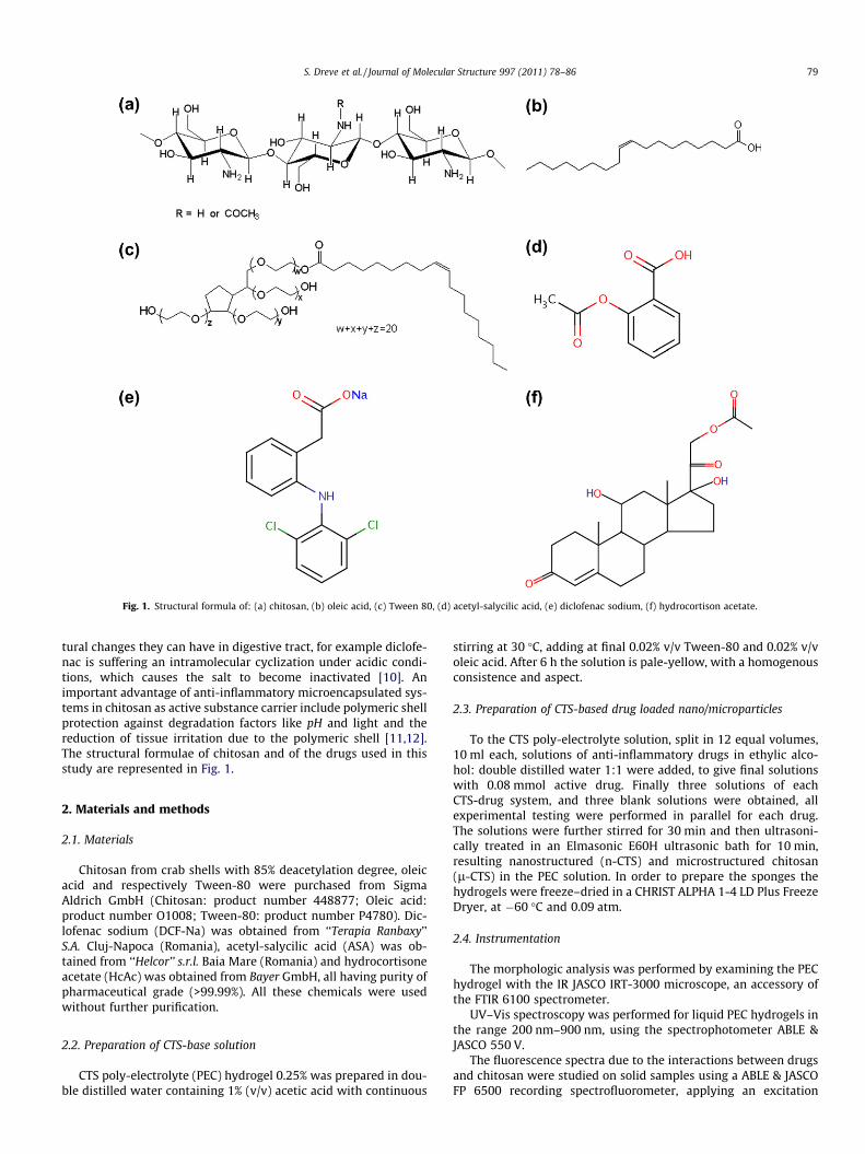
Fig. 1. Structural formula of: (a) chitosan, (b) oleic acid, (c) Tween 80, (d) acetyl-salycilic acid, (e) diclofenac sodium, (f) hydrocortison acetate.
S. Dreve et al. / Journal of Molecular Structure 997 (2011) 78–86 79
tural changes they can have in digestive tract, for example diclofe-nac is suffering an intramolecular cyclization under acidic condi-tions, which causes the salt to become inactivated [10]. Animportant advantage of anti-inflammatory microencapsulated sys-tems in chitosan as active substance carrier include polymeric shellprotection against degradation factors like pH and light and thereduction of tissue irritation due to the polymeric shell [11,12].The structural formulae of chitosan and of the drugs used in thisstudy are represented in Fig. 1.
2. Materials and methods
2.1. Materials
Chitosan from crab shells with 85% deacetylation degree, oleicacid and respectively Tween-80 were purchased from SigmaAldrich GmbH (Chitosan: product number 448877; Oleic acid:product number O1008; Tween-80: product number P4780). Dic-lofenac sodium (DCF-Na) was obtained from ‘‘Terapia Ranbaxy’’S.A. Cluj-Napoca (Romania), acetyl-salycilic acid (ASA) was ob-tained from ‘‘Helcor’’ s.r.l. Baia Mare (Romania) and hydrocortisoneacetate (HcAc) was obtained from Bayer GmbH, all having purity ofpharmaceutical grade (>99.99%). All these chemicals were usedwithout further purification.
2.2. Preparation of CTS-base solution
CTS poly-electrolyte (PEC) hydrogel 0.25% was prepared in dou-ble distilled water containing 1% (v/v) acetic acid with continuous
stirring at 30 �C, adding at final 0.02% v/v Tween-80 and 0.02% v/voleic acid. After 6 h the solution is pale-yellow, with a homogenousconsistence and aspect.
2.3. Preparation of CTS-based drug loaded nano/microparticles
To the CTS poly-electrolyte solution, split in 12 equal volumes,10 ml each, solutions of anti-inflammatory drugs in ethylic alco-hol: double distilled water 1:1 were added, to give final solutionswith 0.08 mmol active drug. Finally three solutions of eachCTS-drug system, and three blank solutions were obtained, allexperimental testing were performed in parallel for each drug.The solutions were further stirred for 30 min and then ultrasoni-cally treated in an Elmasonic E60H ultrasonic bath for 10 min,resulting nanostructured (n-CTS) and microstructured chitosan(l-CTS) in the PEC solution. In order to prepare the sponges thehydrogels were freeze–dried in a CHRIST ALPHA 1-4 LD Plus FreezeDryer, at �60 �C and 0.09 atm.
2.4. Instrumentation
The morphologic analysis was performed by examining the PEChydrogel with the IR JASCO IRT-3000 microscope, an accessory ofthe FTIR 6100 spectrometer.
UV–Vis spectroscopy was performed for liquid PEC hydrogels inthe range 200 nm–900 nm, using the spectrophotometer ABLE &JASCO 550 V.
The fluorescence spectra due to the interactions between drugsand chitosan were studied on solid samples using a ABLE & JASCOFP 6500 recording spectrofluorometer, applying an excitation
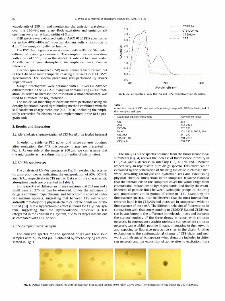
Fig. 3. UV–Vis spectra of ASA, DCF-Na and HcAc, respectively in CTS matrix.
Table 1Absorption peaks of CTS, and anti-inflammatory drugs ASA, DCF-Na, HcAc, and oftheir complex hydrogels.
Examined substance/assembly Wavelenght (nm)
CTS 234ASA 202, 232.6DCF-Na 202, 233HcAc 202, 232.6, 260.7, 299CTS/ASA 241, 277CTS/DCF-Na 240, 279.5CTS/HcAc 238, 275
80 S. Dreve et al. / Journal of Molecular Structure 997 (2011) 78–86
wavelength of 230 nm and monitoring the emission wavelengthover the 250–440 nm range. Both excitation and emission slitopenings were set at bandwidths of 3 lm.
FTIR spectra were obtained with a JASCO 6100 FTIR spectrome-ter in the 4000–400 cm�1 spectral domain with a resolution of4 cm�1 by using KBr pellet technique.
The DSC thermograms were obtained with a DSC-60 Shimadzudifferential scanning calorimeter. The samples’ heating was donewith a rate of 10 �C/min in the 20–500 �C interval by using sealedAl cells, in nitrogen atmosphere. An empty cell was taken asreference.
Electron spin resonance (ESR) measurements were carried outin the X-band at room temperature using a Bruker E-500 ELEXSYSspectrometer. The spectra processing was performed by BrukerXepr software.
X-ray diffractograms were obtained with a Bruker D8 Advancediffractometer in the 2h = 2–50� angular domain using Cu Ka1 radi-ation. In order to increase the resolution a monochromator wasused to eliminate the Ka2 radiation.
The molecular modeling calculations were performed using thedensity functional-based tight binding method combined with theself-consistent charge technique (SCC-DFTB), including the empir-ically correction for dispersion and implemented in the DFTB pro-gram code.
3. Results and discussion
3.1. Morphologic characterization of CTS-based drug loaded hydrogel
In order to evidence PEC nano- and micro-spheres obtainedafter sonication, the FTIR microscopy images are presented inFig. 2. As one side of the image is 200 lm, we can assume thatthe microparticles have dimensions of tenths of micrometers.
3.2. UV–Vis spectroscopy
The analysis of UV–Vis spectra, see Fig. 3, revealed characteris-tic absorption peaks, indicating the encapsulation of ASA, DCF-Naand HcAc, respectively in CTS matrix. Data with the characteristicabsorption bands are presented in Table 1.
In the spectra of chitosan an intense maximum at 234 nm and asmall peak at 275 nm can be observed. Under the influence ofdrugs a combined hyperchromic and batochromic effect of chito-san maxima appears, suggesting that between CTS matrix andanti-inflammatory drug physical–chemical stable bonds are estab-lished [13]. A low hyperchromic effect is found for CTS/HcAc sys-tem, suggesting that the hydrocortisone molecule is lessintegrated in the chitosan PEC system, due to its larger dimensionsas compared with DCF or ASA.
3.3. Spectrofluorimetric analysis
The emission spectra for the specified drugs and their solidsamples with n-CTS and l-CTS obtained by freeze–drying are pre-sented in Fig. 4.
Fig. 2. Optical microscopy images for chitosan hydrogel drug loaded vesicles
The analysis of the spectra obtained from the fluorescence mea-surements (Fig. 4) reveals the increase of fluorescence intensity inCTS/ASA, and a decrease in intensity CTS/DCF-Na and CTS/HcAcrespectively, in report with pure drugs spectra. This effect can beexplained by the penetration of the drug molecule in chitosan net-work, activating carboxylic and hydroxilic sites and establishingphysical–chemical interactions in the composite. It can be assumedthat the interactions in the composite cover the whole range fromelectrostatic interactions to hydrogen bonds, and finally the estab-lishment of peptide links between carboxylic groups of the drugand unprotected amino-groups of chitosan [14]. Examining thefluorescence spectra, it can be observed that the most intense fluo-rescence band is for CTS/ASA and increased in comparison with thefluorescence of pure ASA. The different behavior of fluorescence incomparison with that corresponding to CTS/DCF-Na and CTS/HcAc,can be attributed to the difference in molecular mass and betweenthe stereochemistry of the three drugs, in report with chitosannetwork. In consequence aspirin molecule can penetrate chitosannetwork, can establish peptide linkage, integrating in the network,and exposing to fluoresce new active sites in the chain. Anotherexplanation is the conformational change of CTS chain and net-work, as in drugs, which appears when drugs are included in chito-san network and the exposition of active sites to excitation more
(0.08 mmol active drug). The dimensions of the image are 200 � 200 lm.
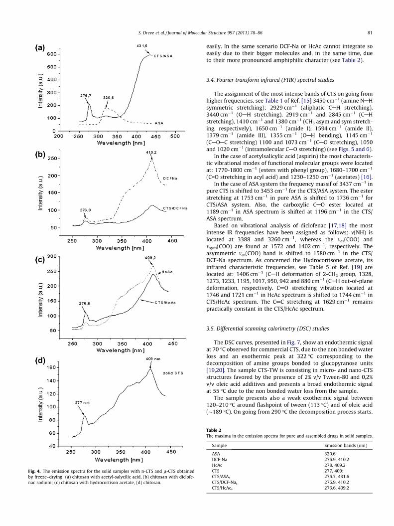
Fig. 4. The emission spectra for the solid samples with n-CTS and l-CTS obtainedby freeze–drying: (a) chitosan with acetyl-salycilic acid, (b) chitosan with diclofe-nac sodium; (c) chitosan with hydrocortison acetate, (d) chitosan.
S. Dreve et al. / Journal of Molecular Structure 997 (2011) 78–86 81
easily. In the same scenario DCF-Na or HcAc cannot integrate soeasily due to their bigger molecules and, in the same time, dueto their more pronounced amphiphilic character (see Table 2).
3.4. Fourier transform infrared (FTIR) spectral studies
The assignment of the most intense bands of CTS on going fromhigher frequencies, see Table 1 of Ref. [15] 3450 cm�1 (amine NAHsymmetric stretching); 2929 cm�1 (aliphatic CAH stretching),3440 cm�1 (OAH stretching), 2919 cm�1 and 2845 cm�1 (CAHstretching), 1410 cm�1 and 1380 cm�1 (CH3 asym and sym stretch-ing, respectively), 1650 cm�1 (amide I), 1594 cm�1 (amide II),1379 cm�1 (amide III), 1355 cm�1 (OAH bending), 1145 cm�1
(CAOAC stretching) 1100 and 1073 cm�1 (CAO stretching), 1050and 1020 cm�1 (intramolecular CAO stretching) (see Figs. 5 and 6).
In the case of acetylsalicylic acid (aspirin) the most characteris-tic vibrational modes of functional molecular groups were locatedat: 1770-1800 cm�1 (esters with phenyl group), 1680–1700 cm�1
(C=O stretching in acyl acid) and 1230–1250 cm�1 (acetates) [16].In the case of ASA system the frequency massif of 3437 cm�1 in
pure CTS is shifted to 3453 cm�1 for the CTS/ASA system. The esterstretching at 1753 cm�1 in pure ASA is shifted to 1736 cm�1 forCTS/ASA system. Also, the carboxylic CAO ester located at1189 cm�1 in ASA spectrum is shifted at 1196 cm�1 in the CTS/ASA spectrum.
Based on vibrational analysis of diclofenac [17,18] the mostintense IR frequencies have been assigned as follows: m(NH) islocated at 3388 and 3260 cm�1, whereas the mas(COO) andmsym(COO) are found at 1572 and 1402 cm�1, respectively. Theasymmetric mas(COO) band is shifted to 1580 cm�1 in the CTS/DCF-Na spectrum. As concerned the Hydrocortisone acetate, itsinfrared characteristic frequencies, see Table 5 of Ref. [19] arelocated at: 1406 cm�1 (CAH deformation of 2-CH2 group, 1328,1273, 1233, 1195, 1017, 950, 942 and 880 cm�1 (CAH out-of-planedeformation, respectively. C@O stretching vibration located at1746 and 1721 cm�1 in HcAc spectrum is shifted to 1744 cm�1 inCTS/HcAc spectrum. The C@C stretching at 1629 cm�1 remainspractically constant in the CTS/HcAc spectrum.
3.5. Differential scanning calorimetry (DSC) studies
The DSC curves, presented in Fig. 7, show an endothermic signalat 70 �C observed for commercial CTS, due to the non bonded waterloss and an exothermic peak at 322 �C corresponding to thedecomposition of amine groups bonded to glucopyranose units[19,20]. The sample CTS-TW is consisting in micro- and nano-CTSstructures favored by the presence of 2% v/v Tween-80 and 0,2%v/v oleic acid additives and presents a broad endothermic signalat 55 �C due to the non bonded water loss from the sample.
The sample presents also a weak exothermic signal between120–210 �C around flashpoint of tween (113 �C) and of oleic acid(�189 �C). On going from 290 �C the decomposition process starts.
Table 2The maxima in the emission spectra for pure and assembled drugs in solid samples.
Sample Emission bands (nm)
ASA 320.6DCF-Na 276.9, 410.2HcAc 278, 409.2CTS 277, 409;CTS/ASAs 276.7, 431.6CTS/DCF-Nas 276.9, 410.2CTS/HcAcs 276.6, 409.2

Fig. 5. FTIR spectra of CTS/drug, 4000–2500 cm�1 spectral domain.
Fig. 6. FTIR spectra of CTS/drug, 1800–400 cm�1 spectral domain.
Fig. 7. DSC thermograms of chitosan-anti-inflammatory drug formulations.
82 S. Dreve et al. / Journal of Molecular Structure 997 (2011) 78–86
The pure drugs melting points by literature data: DCF-Na melt-ing at 287 �C followed by decomposition [21], ASA melting withdecomposition between 133–135 �C [22,23] and HcAc meltingwith decomposition at 223 �C [24].
The sample with HcAc presents a broad endothermic signal at200 �C. On going from 285 �C the decomposition process starts.On the thermogram of CTS/DCF-Na system a weak broad endother-
mic signal with maximum at 60 �C is observed, due to the waterexpulsion and from 230 �C the decomposition process starts. DCFis melted at 284 �C with the decomposition. The CTS/ASA thermo-gram presents a broad endothermic signal at 55 �C, followed by abroad exothermic plateau centered at 148 �C (possible due to theASA decomposition); around 330 �C the decomposition processstarts. All four micro- and nanostructured CTS samples with andwithout drug included decompose around 300 �C excepting CTS/DCF-Na system for which the decomposition starts at a lower tem-perature. All thermograms do not show the drug melting temper-atures that demonstrates the inclusion of drug inside CTSmicrospheres.
3.6. ESR studies
All investigated systems were analyzed by ESR spectroscopy inorder to determine the existence of free radicals present in thematerials. The same quantity of all spectra was analyzed and thecorresponding ESR spectra are presented in the Fig. 8. For all inves-tigated samples an ESR signal (g = 1.99) formed by a narrow linewith 16G line width typically for CTS is obtained.
A possible origin of this line could be attributed to an initial oxi-dation stage. There are several ESR studies on CTS with differentdeacylation degrees [25], where an increase of the signal intensityat g � 1.99 is due to the oxygen presence in the CTS extraction,purifying and deacylation processes.
For each investigated samples we can observe also different gvalues determined as follows:

Fig. 8. ESR spectra for analyzed samples.
Fig. 10. ESR spectra for DCF-Na and CTS/DCF-Na samples before and afterirradiation.
Fig. 11. ESR spectra for HcAc and CTS/HcAc samples before and after irradiation.
S. Dreve et al. / Journal of Molecular Structure 997 (2011) 78–86 83
– CTS/ASA: g = 2.25– CTS/DCF-Na: g = 2.88– CTS/HcAc: g = 2.35
Due to the fact that before validation of in vivo experiments thebiocompatible materials suffer formulation and conditioning pro-cesses, it is important to determine structural changes that appearafter these procedures.
Consequently, these spongy materials were UV irradiated(k = 320 nm) for 4 h; after that ESR experiments were repeated.ESR spectra after irradiation are presented in Figs. 9–11, respec-tively. As a function of radiation type and its duration, the samplescould offer chemical structural changes by chain breaking and theopening of the glycosidic cycles. The drugs present a weak ESR sig-nal at different g values as compared to the starting materials. CTS/DCF-Na material presents a strong signal at g = 2.14 that is ob-served also in DCF-Na spectrum but with a reduced intensity.The irradiation does not produce changes. All investigated materi-als present the same absorption line at g � 1.99 but with a higherintensity. This behavior is in good agreement with the literature[20] and could be related to the increase of the free radical concen-tration of CTS itself.
The analysis of ESR spectra of the active substances before andafter irradiation revealed that UV irradiation does not generate freeradicals or these radicals are of short life time, irrelevant for even-tually structural changes.
Fig. 9. ESR spectra for ASA and CTS/ASA samples before and after irradiation.
The monomer radicals and of CTS polymeric chain are rapidlyannihilated due to the concurrent chemical reactions (esterifica-tion or amidolyse) which are produced during the supramolecularcoupling between CTS (as polymeric chain) and DCF-Na, ASA, orHcAc, respectively, that are more probably in the reaction mediumby applying coupling techniques.
3.7. X-ray diffraction
X-ray diffractograms (see Figs. 12–14) show that CTS presentsan amorphous structure and in the case of CTS/ASA andCTS/DCF-Na systems the inclusion of the drug does not preservetheir crystalline structure (see Figs. 12 and 13). In the case ofCTS/HcAc system, due to the higher dimension of this moleculeas compared to ASA or DCF ones, the inclusion of HcAc in CTS doesnot destroy its crystallinity (see Fig. 14).
3.8. Molecular modeling
In order to study the polymer (oligomer) properties of the CTS/ASA and CTS/DCF structures, at least 3–4 CTS-drug units must beincluded in our molecular geometry structure. These structuresusually contain a large number of atoms (over 180 atoms) which
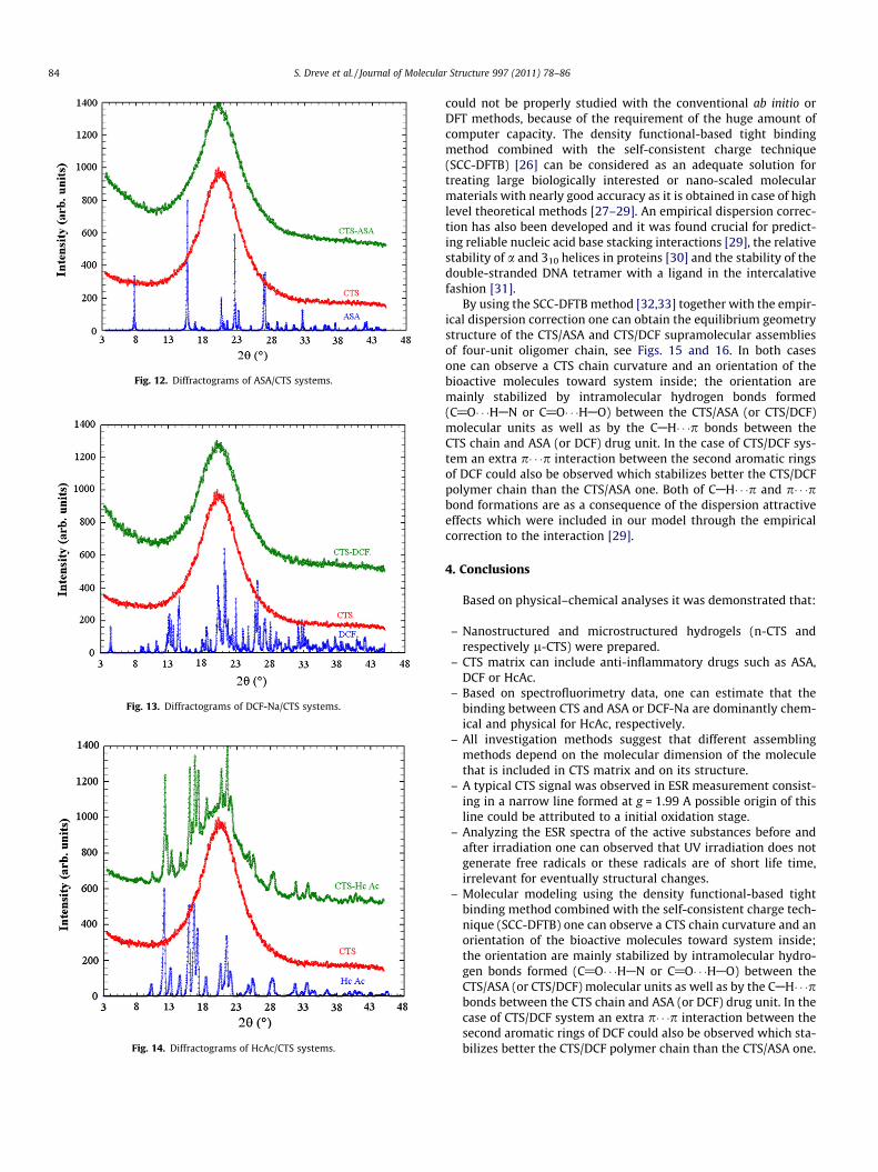
Fig. 12. Diffractograms of ASA/CTS systems.
Fig. 13. Diffractograms of DCF-Na/CTS systems.
Fig. 14. Diffractograms of HcAc/CTS systems.
84 S. Dreve et al. / Journal of Molecular Structure 997 (2011) 78–86
could not be properly studied with the conventional ab initio orDFT methods, because of the requirement of the huge amount ofcomputer capacity. The density functional-based tight bindingmethod combined with the self-consistent charge technique(SCC-DFTB) [26] can be considered as an adequate solution fortreating large biologically interested or nano-scaled molecularmaterials with nearly good accuracy as it is obtained in case of highlevel theoretical methods [27–29]. An empirical dispersion correc-tion has also been developed and it was found crucial for predict-ing reliable nucleic acid base stacking interactions [29], the relativestability of a and 310 helices in proteins [30] and the stability of thedouble-stranded DNA tetramer with a ligand in the intercalativefashion [31].
By using the SCC-DFTB method [32,33] together with the empir-ical dispersion correction one can obtain the equilibrium geometrystructure of the CTS/ASA and CTS/DCF supramolecular assembliesof four-unit oligomer chain, see Figs. 15 and 16. In both casesone can observe a CTS chain curvature and an orientation of thebioactive molecules toward system inside; the orientation aremainly stabilized by intramolecular hydrogen bonds formed(C@O� � �HAN or C@O� � �HAO) between the CTS/ASA (or CTS/DCF)molecular units as well as by the CAH� � �p bonds between theCTS chain and ASA (or DCF) drug unit. In the case of CTS/DCF sys-tem an extra p� � �p interaction between the second aromatic ringsof DCF could also be observed which stabilizes better the CTS/DCFpolymer chain than the CTS/ASA one. Both of CAH� � �p and p� � �pbond formations are as a consequence of the dispersion attractiveeffects which were included in our model through the empiricalcorrection to the interaction [29].
4. Conclusions
Based on physical–chemical analyses it was demonstrated that:
– Nanostructured and microstructured hydrogels (n-CTS andrespectively l-CTS) were prepared.
– CTS matrix can include anti-inflammatory drugs such as ASA,DCF or HcAc.
– Based on spectrofluorimetry data, one can estimate that thebinding between CTS and ASA or DCF-Na are dominantly chem-ical and physical for HcAc, respectively.
– All investigation methods suggest that different assemblingmethods depend on the molecular dimension of the moleculethat is included in CTS matrix and on its structure.
– A typical CTS signal was observed in ESR measurement consist-ing in a narrow line formed at g = 1.99 A possible origin of thisline could be attributed to a initial oxidation stage.
– Analyzing the ESR spectra of the active substances before andafter irradiation one can observed that UV irradiation does notgenerate free radicals or these radicals are of short life time,irrelevant for eventually structural changes.
– Molecular modeling using the density functional-based tightbinding method combined with the self-consistent charge tech-nique (SCC-DFTB) one can observe a CTS chain curvature and anorientation of the bioactive molecules toward system inside;the orientation are mainly stabilized by intramolecular hydro-gen bonds formed (C@O� � �HAN or C@O� � �HAO) between theCTS/ASA (or CTS/DCF) molecular units as well as by the CAH� � �pbonds between the CTS chain and ASA (or DCF) drug unit. In thecase of CTS/DCF system an extra p� � �p interaction between thesecond aromatic rings of DCF could also be observed which sta-bilizes better the CTS/DCF polymer chain than the CTS/ASA one.

Fig. 15. Model of CTS/ASA assembly.
Fig. 16. Model of CTS/DCF-Na assembly.
S. Dreve et al. / Journal of Molecular Structure 997 (2011) 78–86 85
– All analysis demonstrates that aspirin and sodium diclofenacare better included in chitosan matrix than hydrocortisone ace-tate, and all the analytical methods used suggested that the nat-ure of the bonds between chitosan matrix and guest moleculeare depending on the molecular weight of guest molecule andon the reciprocal stereochemistry of the partners in the system.
Acknowledgments
This work was possible due to the financial support of theRomanian Research and Education Ministry under PN-09-44 0201/2009, PN-09-44 02 05/2009 and PN II 72-190/2008 projects.We gratefully acknowledge to Data Center of National Institutefor Research and Development of Isotopic and Molecular Technol-
ogies Cluj-Napoca for providing computer facilities for the molec-ular modeling calculations.
References
[1] P. Ehrlich, Collected Papers of Paul Ehrlich: Immunology and Cancer Research,Pergamon Press, London, 1902.
[2] I. Szelenyi, K. Thiemer, Arch. Toxicol. 41 (1978) 99.[3] J.A. Ko, H.J. Park, S.J. Hwang, J.B. Park, J.S. Lee, Int. J. Pharm. 249 (2002) 165.[4] E.V. Svirshchevskaya, L.G. Alekseeva, P.D. Reshetov, N.N. Phomicheva, S.A.
Parphenyuk, A.V. Ilyina, V.S. Zueva, S.A. Lopatin, A.N. Levov, V.P. Varlamov, Eur.J. Med. Chem. 44 (2009) 2030.
[5] K.C. Gupta, M.N. Ravi Kumar, Biomaterials 21 (2000) 1115.[6] C.S. Sweetman (Ed.), Martindale: The Complete Drug Reference, 33rd ed.,
Pharmaceutical Press, London, 2002 (Chapter 3).[7] V.R. Sinha, A.K. Singla, S. Wadhawan, R. Kaushik, R. Kumria, K. Bansal, S.
Dhawan, Int. J. Pharm. 274 (2004) 1.

86 S. Dreve et al. / Journal of Molecular Structure 997 (2011) 78–86
[8] T. Peng, K.D. Yao, Y. Chen, M.F. Goosen, J. Polym. Sci. Polym. Chem. Ed. 32(1994) 591.
[9] J. Kevin, M.D. Ivey, Am J. Med. 84 (1988) 41.[10] M.E. Palomo, M.P. Ballesteros, P. Frutos, J. Pharm. Biomed. Anal. 21 (1999) 83.[11] C. Pinto, R. Neufeld, A. Ribeiro, F. Veiga, Nanomedicine 2 (2006) 53.[12] N. Anton, J.P. Benoit, P. Saulnier, J. Control Release 128 (2008) 185.[13] Alina Sionkowska, J. Photochem Photobiol., B: Biology 82 (1) (2006) 9.[14] J.L. Atwood, J.W. Steed (Eds.), Encyclopedia of Supramolecular Chemistry,
Marcel Dekker, New York, 2004 (Chapter 3).[15] M. Montalti, A. Credi, L. Prodi, M. Teresa Gandolfi, Handbook of
Photochemistry, 3rd ed., Taylor &Francis Group, Boca Raton, 2006 (Chapter 5).[16] N.B. Colthup, L.H. Daly, S.E. Wiberley, Introduction to Infrared and Raman
Spectroscopy, Academic Press, New York, 1975.[17] N. Kourkoumelis, M.A. Demertzis, D. Kovala-Demertzi, A. Koutsodimou, A.
Moukarika, Spectr. Acta. A 60 (2004) 2253.[18] S. Dreve, I. Kacsó, I. Bratu, E. Indrea, J. Phys. Conf. Series 182 (2009),
doi:10.1088/1742-6596/182/1/012065.[19] R.J. Mesley, Spectr. Acta 22 (1966) 889.[20] G.L. Simionatto, C.É.T. Gomes, Thermochim. Acta 444 (2) (2006) 128.[21] I. Pasquali, R. Bettini, F. Giordan, J. Therm. Anal. Calorim. 90 (3) (2007) 903.[22] George D. Beal, Chester R. Szalkowski, J. Am. Pharm. Assoc. 22 (1) (1933) 36.
[23] G.L. Perlovich, A. Bauer-Brandl, J. Therm. Anal. Calorim. 63 (2001) 653.[24] http://www.caslab.com/Hydrocortisone_acetate_CAS_50-03-3/ http://www.
sigmaaldrich.com/catalog/ProductDetail.do?lang=en&N4=H4126|SIGMA&N5=SEARCH_CONCAT_PNO|BRAND_KEY&F=SPEC.
[25] J. Estrela dos Santos, E.R. Dockal, É.T.G. Cavalheiro, J. Thermal. Anal. Calorim. 79(2005) 243.
[26] M. Elstner, D. Porezag, G. Jungnickel, J. Elsner, M. Haugk, T. Frauenheim, S.Suhai, G. Seifert, Phys. Rev. B 58 (1998) 7260.
[27] M. Elstner, K.J. Jalkanen, M. Knapp-Mohammady, T. Frauenheim, S. Suhai,Chem. Phys. 256 (2000) 15.
[28] M. Elstner, K.J. Jalkanen, M. Knapp-Mohammady, T. Frauenheim, S. Suhai,Chem. Phys. 263 (2001) 203.
[29] M. Elstner, P. Hobza, T. Frauenheim, S. Suhai, E. Kaxiras, J. Chem. Phys. 114(2001) 5149.
[30] H.Y. Liu, M. Elstner, E. Kaxiras, T. Frauenheim, J. Hermans, W.T. Yang, Proteins:Struct. Funct. Genet. 44 (2001) 484–489.
[31] T. Kubar, P. Jurecka, J. Cerny, J. Rezác, M. Otyepka, H. Valéds, P. Hobza, J. Phys.Chem. A 111 (2007) 5642.
[32] DFTB+ 1.0.1 is a DFTB implementation, which is free for noncommercial use.For details, see <http://www.dftb-plus.info>.
[33] B. Aradi, B. Hourahine, T. Frauenheim, J. Phys. Chem. A 111 (26) (2007) 5678.