journal of clinical & experimental cardiology · above the antecubital fossa, however, the wire...
TRANSCRIPT
Volume 3 • Issue 2 • 1000178J Clinic Experiment CardiolISSN:2155-9880 JCEC, an open access journal
Case Report Open Access
Chitsaz et al. J Clinic Experiment Cardiol 2012, 3:2 DOI: 10.4172/2155-9880.1000178
Keywords: Radial artery; Heart catheterization; Radial loop;Arteriovenous fistula; Transradial access
IntroductionTransradial access (TRA) for percutaneous transluminal cardiac
angiography (PTCA) has gained more popularity during the last decade owing largely to its lower rate of access site complications than the transfemoral approach (TFA), patient comfort, and earlier ambulation [1,2]. TRA is also considered as an alternative approach in patients with difficulties in their ilio-femoral anatomy [3], as well as those who are aggressively anticoagulated and at high risk for post-procedural bleeding [4,5]. However, anatomical variations of radial artery are not rare; roughly 10-15% of patient undergoing PTCA via TRA have been shown to have some sort of anomaly including excessive tortuosity, stenosis, hypoplasia, loop, accessory radial arteries etc. [6,7]. These variations can be associated with access failure or complications such as perforation or thrombosis [7]. Radial artery loop is one of the more challenging variations encountered during TRA, and the commonest anomaly that leads to access failure [8,9].
Case ReportA 90-year-old male with critical aortic stenosis (AS) was referred to
the catheterization laboratory for preoperative coronary angiography prior to planned surgical aortic valve replacement (AVR). His past medical history included hypothyroidism, osteoporosis, and hyperlipidemia well-controlled by statins. His renal function was impaired with a calculated creatinine clearance of 35 ml/min by the Cockroft-Gault equation. He had a very active life-style, and previously biked and walked several hours daily, until he developed mild heart failure 3 months before referral. He did not have any history of hypertension, diabetes, cigarette smoking, and peripheral or cerebral vascular disease. His last echocardiogram had shown valvular aortic stenosis and a moderate post-stenosis dilation of the ascending aorta. Surprisingly, in light of his degree of physical activity, there was a mean transvalvular gradient of 136 mmHg and a calculated aortic valve area of 0.4 cm2. Recent Duplex study of the carotid arteries did not show any significant abnormalities. His pre-catheterization physical exam was completely normal in terms of peripheral vascular disease; pulses were normal to palpation and neither bruit nor discrepancy in bilateral arterial pressures was found. The right femoral artery was selected as the primary access site for diagnostic angiography. A 6Fr sheath was easily inserted into the right common femoral artery.
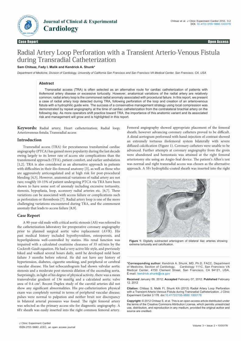
Femoral angiography showed appropriate placement of the femoral sheath; however advancing coronary catheters proved to be difficult. A distal aortogram performed with hand-injection of contrast showed an extremely tortuous iliofemoral system bilaterally with severe diffused calcification (Figure 1). Coronary catheters were unable to be advanced. Further attempts at coronary angiography from the groin were abandoned and hemostasis was attained at the right femoral arteriotomy site using an Angio-Seal device. The patient’s Allen’s test was normal and right transradial access was chosen as the alternative approach. A 5Fr hydrophilic-coated sheath was inserted into the right
*Corresponding author: Kendrick A. Shunk, MD, Ph.D, FACC, Department of Medicine, Section of Cardiology, Cardiology 111C, San Francisco VA Medical Center, 4150 Clement Street, San Francisco, CA 94121, USA, E-mail: [email protected]
Received January 09, 2012; Accepted February 07, 2012; Published February 12, 2012
Citation: Chitsaz S, Malik FI, Shunk KA (2012) Radial Artery Loop Perforation with a Transient Arterio-Venous Fistula during Transradial Catheterization. J Clinic Experiment Cardiol 3:178. doi:10.4172/2155-9880.1000178
Copyright: © 2012 Chitsaz S, et al. This is an open-access article distributed under the terms of the Creative Commons Attribution License, which permits unrestricted use, distribution, and reproduction in any medium, provided the original author and source are credited.
Radial Artery Loop Perforation with a Transient Arterio-Venous Fistula during Transradial CatheterizationSam Chitsaz, Fady I. Malik and Kendrick A. Shunk*
Department of Medicine, Division of Cardiology, University of California San Francisco and San Francisco VA Medical Center, San Francisco, CA, USA
AbstractTransradial access (TRA) is often selected as an alternative route for cardiac catheterization of patients with
iliofemoral artery disease or excessive tortuosity. However, anatomical variations of the radial artery are relatively common; radial artery loop is the commonest radial anomaly associated with procedural failure. In this report, we present a case of radial artery loop detected during TRA, following perforation of the loop and creation of an arteriovenous fistula with a hydrophilic guide wire. The success of a conservative management strategy using local compression was demonstrated by repeat angiography at the time of cardiac catheterization from the contralateral brachial artery on the following day. As more operators shift practice toward TRA, the importance of this anatomic variant and its associated risk and management will grow and is highlighted in this report.
Figure 1: Digitally subtracted arteriogram of bilateral iliac arteries showing extreme tortuosity and calcification.
Journal of Clinical & Experimental CardiologyJo
urna
l of C
linica
l & Experimental Cardiology
ISSN: 2155-9880
Citation: Chitsaz S, Malik FI, Shunk KA (2012) Radial Artery Loop Perforation with a Transient Arterio-Venous Fistula during Transradial Catheterization. J Clinic Experiment Cardiol 3:178. doi:10.4172/2155-9880.1000178
Page 2 of 3
Volume 3 • Issue 2 • 1000178J Clinic Experiment CardiolISSN:2155-9880 JCEC, an open access journal
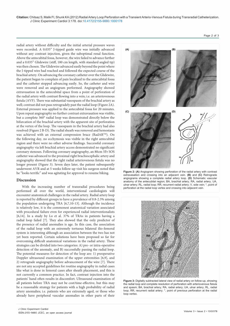
radial artery without difficulty and the initial arterial pressure waves were recorded. A 0.035” J-tipped guide wire was initially advanced without any contrast injection, given the suboptimal renal function. Above the antecubital fossa, however, the wire failed to advance further and a 0.035” Glidewire (stiff, 180 cm length, with standard angled tip) was then chosen. The Glidewire advanced easily beyond the point where the J-tipped wire had reached and followed the expected course of the brachial artery. On advancing the coronary catheter over the Glidewire, the patient began to complain of pain localized to the antecubital fossa and the catheter stopped advancing easily. So, the catheter and wire were removed and an angiogram performed. Angiography showed extravasation in the antecubital space from a point of perforation of the radial artery with contrast flowing into a vein; i.e. an arteriovenous fistula (AVF). There was substantial vasospasm of the brachial artery as well; contrast did not pass retrogradely past the radial loop (Figure 2A). External pressure was applied to the antecubital fossa for 20 minutes. Upon repeat angiography no further contrast extravasation was visible, but a complete 360° radial loop was demonstrated directly below the bifurcation of the brachial artery with the apparent site of perforation at the vertex of the loop. The vasospasm in the brachial artery had also resolved (Figure 2 B-D). The radial sheath was removed and hemostasis was achieved with an external compression brace (RadARTM). On the following day, no ecchymosis was visible in the right antecubital region and there were no other adverse findings. Successful coronary angiography via left brachial artery access demonstrated no significant coronary stenoses. Following coronary angiography, an 80cm 5Fr SOS catheter was advanced to the proximal right brachiocephalic artery and angiography showed that the right radial arteriovenous fistula was no longer present (Figure 3). Seven days later, the patient subsequently underwent AVR and at 5 weeks follow-up visit his surgeon noted that he “looks terrific” and was agitating for approval to resume biking.
DiscussionWith the increasing number of transradial procedures being
performed all over the world, interventional cardiologists will encounter anatomical challenges in the radial artery. Radial artery loop is reported by different groups to have a prevalence of 0.8-2.3% among the population undergoing TRA [6,7,10-13]. Although the incidence is relatively low, it is the commonest anatomical variation associated with procedural failure even for experienced radial interventionalists [6,14]. In a study by Lo et al. 37% of TRAs in patients having a radial loop failed [7]. They also showed that the only predictor of the presence of radial anomalies is age. In this case, the association of the radial loop with an extremely tortuous bilateral ilio-femoral system is interesting although an association between the two has not yet been reported. Certain solutions have been proposed so far for overcoming difficult anatomical variations in the radial artery. These strategies can be divided into two categories: A) pre- or intra-operative detection of the anomaly, and B) successfully passing the radial loop. The potential measures for detection of the loop are: 1) preoperative Doppler ultrasound examination of the upper extremities [6,9], and 2) retrograde angiography before advancement of the wire [7]. There are not any accepted guidelines for routine angiography in radial cases like what is done in femoral cases after sheath placement, and this is not currently a common practice. In fact, contrast injection into the patients’ hand often results in discomfort. Ultrasound examination of all patients before TRA may not be cost/time-effective, but this may be a reasonable strategy for patients with a high probability of radial artery anomalies; i.e. patients who are extremely aged, or those who already have peripheral vascular anomalies in other parts of their
(A) (B)
(C) (D)
Figure 2: (A) Angiogram showing perforation of the radial artery with contrast extravasation and crossing into an adjacent vein. (B) and (C) Retrograde angiograms showing a complete radial artery loop. (D) Schematic vascular anatomy of the antecubital region; BA, brachial artery; RA, radial artery; UA, ulnar artery; RL, radial loop; RR, recurrent radial artery; V, side vein; *, point of perforation at the radial loop vertex and crossing into adjacent vein.
Figure 3- Digitally subtracted lateral view of radial artery on follow-up, showing the radial loop and complete resolution of perforation with arteriovenous fistula and spasm; BA, brachial artery; RA, radial artery; UA, ulnar artery; RL, radial loop; RR, recurrent radial artery; *, point of previous perforation at the radial loop vertex.

Citation: Chitsaz S, Malik FI, Shunk KA (2012) Radial Artery Loop Perforation with a Transient Arterio-Venous Fistula during Transradial Catheterization. J Clinic Experiment Cardiol 3:178. doi:10.4172/2155-9880.1000178
Page 3 of 3
Volume 3 • Issue 2 • 1000178J Clinic Experiment CardiolISSN:2155-9880 JCEC, an open access journal
bodies. Retrograde angiography is more accurate than ultrasound for detection of radial anomalies. To successfully pass the radial loop, the techniques suggested are: 1) using hydrophilic wires for initial passing around the loop [10,15], although as this case illustrates, there is risk associated with that strategy, 2) loop straightening with either a steerable guidewire [16] or a diagnostic catheter [17], and 3) passing around the loop with a coronary wire and subsequent loop straightening with a 0.035” guide wire using a multipurpose shuttle catheter [18]. One key question to consider is how well these techniques are tolerated. Making a complete 360° loop with the catheter may limit any ability to torque or steer it once it reaches the aorta. In our case, it was difficult to imagine a means to pass around this loop in a useful and safe manner. The anatomy in this case would likely have precluded success with any of these techniques so the access site was abandoned and the contralateral brachial approach was selected on the following day. Loop straightening can be a useful technique for passing devices through a radial loop; nevertheless it may be difficult for loops detected incidentally during the procedure and those already complicated by perforation or spasm. Straightening maneuvers may cause spasm, pain, dissection or perforation of the loop in aged people whose arteries are often fragile. Hydrophilic devices may be associated with less radial artery spasm. However, our experience showed that even a hydrophilic device may not be capable of passing through a severely spasmodic radial loop and may in fact predispose to this complication. Interestingly, in a study of 3000 patients, perforation of anomalous radial artery was often caused by hydrophilic wires [11]. As we found in this case, radial loops commonly have a tiny remnant recurrent branch that may act as an accessory pathway particularly for the hydrophilic wire, mimicking the brachial artery route [16,18]. Thus, the operator must have a high degree of vigilance for this anomaly if there is any difficulty passing a wire or catheter past the antecubital region in order to prevent complications. The simplest and probably safest way to bypass the radial loop may be conversion to brachial access (if femoral access is not feasible).
In conclusion, we present a case of perforation of a radial artery loop with passage of the wire into an adjacent vein in a 90-year old patient with tortuous ilio-femoral arteries. We conclude that in patients with high probability of radial artery anomalies, assessment of radial anatomy with either ultrasound or retrograde angiography may prevent such complications, while utilization of hydrophilic wires, although sometimes helpful, is also not free of risk. In a case of difficult radial anatomy, the wire should be advanced very gently and if significant difficulty is encountered, switching to brachial artery access is recommended to bypass the anatomical obstacles in the radial artery. In the event of perforation and AVF formation, management with local external compression can result in complete resolution.
References
1. Freestone B, Nolan J (2010) Transradial cardiac procedures: The state of the art. Heart 96: 883-891.
2. Amoroso G, Kiemeneij F (2010) Transradial access for primary percutaneous coronary intervention: The next standard of care? Heart 96: 1341-1344.
3. Dahm JB, van Buuren F (2010) Transradial percutaneous coronary interventions: Indications, success rates & clinical outcome. Indian Heart J 62: 218-220.
4. Hildick-Smith DJ, Walsh JT, Lowe MD, Petch MC (2003) Coronary angiography in the fully anticoagulated patient: The transradial route is successful and safe. Catheter Cardiovasc Interv 58: 8-10.
5. Jolly SS, Amlani S, Hamon M, Yusuf S, Mehta SR (2009) Radial versus
femoral access for coronary angiography or intervention and the impact on major bleeding and ischemic events: A systematic review and meta-analysis of randomized trials. Am Heart J 157: 132-140.
6. Yokoyama N, Takeshita S, Ochiai M, Koyama Y, Hoshino S, et al. (2000) Anatomic variations of the radial artery in patients undergoing transradial coronary intervention. Catheter Cardiovasc Interv 49: 357-362.
7. Lo TS, Nolan J, Fountzopoulos E, Behan M, Butler R, et al. (2009) Radial artery anomaly and its influence on transradial coronary procedural outcome. Heart 95: 410-415.
8. Wiper A, Kumar S, MacDonald J, Roberts DH (2006) Day case transradial coronary angioplasty: A four-year single-center experience. Catheter Cardiovasc Interv 68: 549-553.
9. Louvard Y, Lefevre T (2000) Loops and transradial approach in coronary diagnosis and intervention. Catheter Cardiovasc Interv 51: 250-252.
10. Barbeau GR (2003) Radial loop and extreme vessel tortuosity in the transradial approach: Advantage of hydrophilic-coated guidewires and catheters. Catheter Cardiovasc Interv 59: 442-450.
11. Nie B, Zhou YJ, Li GZ, Shi DM, Wang JL (2009) Clinical study of arterial anatomic variations for transradial coronary procedure in chinese population. Chin Med J (Engl) 122: 2097-2102.
12. Fujii T, Masuda N, Tamiya S, Shima M, Toda E, et al. (2010) Angiographic evaluation of right upper-limb arterial anomalies: Implications for transradial coronary interventions. J Invasive Cardiol 22: 536-540.
13. Valsecchi O, Vassileva A, Musumeci G, Rossini R, Tespili M, et al. (2006) Failure of transradial approach during coronary interventions: Anatomic considerations. Catheter Cardiovasc Interv 67: 870-878.
14. Louvard Y, Lefevre T, Morice MC (1997) Radial approach: What about the learning curve? Cathet Cardiovasc Diagn 42: 467-468.
15. Caussin C, Gharbi M, Durier C, Ghostine S, Pesenti-Rossi D, et al. (2010) Reduction in spasm with a long hydrophylic transradial sheath. Catheter Cardiovasc Interv 76: 668-672.
16. Esente P, Giambartolomei A, Simons AJ, Levy C, Caputo RP (2002) Overcoming vascular anatomic challenges to cardiac catheterization by the radial artery approach: Specific techniques to improve success. Catheter Cardiovasc Interv 56: 207-211.
17. Wang HJ, Lee KW, Hsieh DJ (2006) Brachial loop: Transradial technique to overcome this rare anatomic variation. Catheter Cardiovasc Interv 68: 260-262.
18. Farman MT, Khan NU, Rizvi SN (2010) Successful transradial percutaneous coronary intervention with radial artery anomaly. J Pak Med Assoc 60: 593-595.