journal of anesthesia (invited) revised joan-d-18-00285
TRANSCRIPT
1
Journal of Anesthesia (invited) Revised JOAN-D-18-00285.
4447 words in text (excluding abstract and legends)=4515.
1 Table and 3 Figures.
109 References.
Opioids, gliosis and central immunomodulation
Salim Kadhim, John McDonald and David G Lambert*
Department of Cardiovascular Sciences, Division of Anaesthesia, Critical Care and Pain
Management, University of Leicester, Robert Kilpatrick Clinical Sciences Building,
Leicester Royal Infirmary, Leicester, LE1 5WW, United Kingdom.
*Address correspondence to Prof David G Lambert at the above address.
Tel: +44 (0)116 252 3161. Email: [email protected]
Fax: NONE
SK is funded by a scholarship from Higher Committee for Education Development in Iraq.
Abstract:
2
Neuropathic pain is a common health problem that affects millions of people worldwide.
Despite being studied extensively, the cellular and molecular events underlying the
central immunomodulation and the pathophysiology of neuropathic pain is still
controversial. The idea that ‘glial cells are merely housekeepers’ is incorrect and with
respect to initiation and maintenance of neuropathic pain microglia and astrocytes have
important roles to play. Glial cells differentially express opioid receptors and are thought
to be functionally modulated by the activation of these receptors. In this review, we
introduce evidence for glia-opioid modulation of pain by focusing on the pattern of
astrocyte and microglial activation throughout the progress of nerve injury/neuropathic
pain. Activation of astrocytes and microglia is a key step in central immunomodulation
in terms of releasing pro-inflammatory markers and propagation of a ‘central immune
response’. Inhibition of astrocytes before and after induction of neuropathic pain has
been found to prevent and reverse neuropathic pain, respectively. Moreover, microglial
inhibitors have been found to prevent (but not to reverse) neuropathic pain. As they are
expressed by glia, opioid receptors are expected to have a role to play in the treatment
of neuropathic pain.
Key Words: Neuropathic pain, immunomodulation, Glial cells, Astrocytes, Microglia,
Opioids, Cytokines, gliosis.
3
List of abbreviations ATP Adenosine Triphosphate BBB Blood Brain Barrier BDNF Brain-derived neurotrophic factor BrdU Bromodeoxyuridine (5-bromo-2'-deoxyuridine) CC5a Complement component 5a CC5aR Complement component 5a Receptor CCL2 Chemokine (C-C motif) ligand 2 CCR2 Chemokine (C-C motif) Receptor 2 CCR3 C-C chemokine receptor type 3 CD11B Cluster of differentiation molecule 11B CD14 Cluster of Differentiation Antigen 14 CNS Central Nervous System CNTF Ciliary neurotrophic factor COX-2 Cyclooxygenase-2 CVO Circumventricular Organs CX3CL1 CX3C chemokine CX3CR1 CX3C chemokine receptor DOP Delta (𝛿𝛿) Opioid Receptor ErbB2 Similar to ErbB (avian erythroblastosis oncogene B) GFAP Glial fibrillary acidic protein HIV Human immunodeficiency virus HPA Hypothalamic–Pituitary–Adrenal IASP International Association for the Study of Pain IBA1 Ionized calcium-binding adapter molecule 1 IFN-γ Interferon-γ IL-1 Interlukin-1 IL-13 Interlukin-13 IL-1β Interlukin-1β IL-4 Interlukin-4 IL-6 Interlukin-6 iNOS Inducible nitric oxide synthase ITGAM Integrin alpha M KOP Kappa (k) Opioid Receptor L5 5th Lumbar Vertebra LPS Lipopolysaccharide MAPK Mitogen-activated protein kinase
4
MCP-1 Monocyte Chemoattractant Protein-1 M-CSF Macrophage-Colony stimulating factor M-CSFR Macrophage-Colony stimulating factor Receptor MOP mu (μ) Opioid Receptor NO Nitric oxide NOP Nociceptin/orphanin FQ (N/OFQ) Opioid Receptor NRG-1 Neuregulin1 P2Y12 Platelet P2Y12 receptor PLA2 Phospholipase A2 RANTES Regulated on activation, normal T cell expressed and secreted ROS Reactive oxygen species RVM Rostral ventromedial medulla TGFβ Transforming growth factor 1β TLR-4 Toll-like receptor 4 TNF-α Tumour necrosis factor alpha
5
Introduction
With the central nervous system (CNS) no longer deemed as a passive immune-
privileged structure, it is recognised that the CNS can mount innate immune responses,
which when chronically activated potentially direct the pathophysiology of a number of
neurodegenerative disorders as well as having a central role to play in the development
of pathological pain states and opioid drug tolerance. Both neuronal and non-neuronal
components of the CNS are recognised as responsible for maintaining the physiologic
and pathologic state of the CNS. While the neuronal aspects of pathological pain
conditions have historically held the spotlight, attention is now being given to non-
neuronal cells, primarily glial cells, and discovering how these cells make fundamental
contributions in the development of chronic pain conditions. These same non-neuronal
glial cells are also being studied to delineate the mechanism underlying opioid tolerance
and opioid withdrawal-induced pain enhancement. New findings show that glial cells are
differentially activated to release a variety of signalling molecules, which can have
pathological actions, protective actions or both. However, the exact nature of the
association between opioids, pain, and alterations in immune functioning remain
unclear. In this mini review, we illustrate how glial cells are a focal point in the processes
underlying the development and maintenance of chronic pain conditions and the
retarded analgesic potency of opioids, and how this point of convergence has important
implications for future treatments in pain management.
Peripheral immune function and opioids
Immune regulation encompasses interactions between immune cells and mediators
that modulate a variety of stimuli including neuroendocrine modulation of stress
(corticosteroids and catecholamines), growth hormones, and opioids. The link between
immune function and opioids has been presumed from historical literature, which
observed an increase in the incidence and severity of infections in opioid addicts, and
from more recent findings, it has been highlighted that opioids affect the endocrine
system. Opioid immune modulatory effects, however, are dependent on a variety of
factors including the type of opioid drug, duration of use, as well as patient factors such
as genetic background.
6
The site (s) of action for opioid mediated immunomodulation is one of current research
and debate with potential sites of action including (a) peripheral immunocytes, (b) an
effect on the hypothalamic–pituitary–adrenal (HPA) axis and (c) effects on sympathetic
tone. A synopsis of the research evidence for these potential sites of immune
modulation by opioids is provided in Table 1 and we have reviewed this previously (Al-
Hashimi et al., 2013). Our interpretation of these findings leads us to conclude that a
direct action on immunocytes is doubtful, that the evidence supporting opioid
immunomodulation through the HPA axis is unclear (and species dependent) and it is
questionable that the opioid-mediated effect on sympathetic tone would be sufficient
to support the immunomodulation described. While the mechanisms for opioid-
mediated immunomodulation are not fully elucidated, what can be concluded from the
literature is that in MOP (mu :µ) receptor-knockout animals no opioid immune
modulation is seen, providing robust evidence that MOP is the biological target. In
addition, opioid drugs show variance in immunomodulatory effects and that there are
interspecies differences in the immunomodulatory actions of opioids. There is growing
evidence that glia are central in pain pathophysiology (Ji et al., 2013) and emerging
evidence that glia are opioid sensitive targets.
Central Immunity
Coupled with the expanding significance of glial cells is a gradual disappearance of the
idea that the CNS is an immune-privilege organ. It has been found that an innate immune
response can be propagated in the CNS (Nguyen et al., 2002) which may indicate that
the CNS has the ability to fight and recognise infectious and foreign bodies via pattern
recognition receptors. Evolving evidence suggests that the central nervous system is
able to process antigens and mount immune responses much like that utilised by
peripheral organs and exhibits co-ordinated innate immune reactions in response to
both cerebral injury, and systemic bacterial infection (Hendriks et al., 2005, Ransohoff
and Brown, 2012). Such innate immunity is an inflammatory response induced by the
detection of immunological proteins, released from microorganisms, and initially this
occurs in structures of the brain lacking the normal blood brain barrier (BBB), such as
the circumventricular organs (CVO) of the brain. These structures appear to act as
7
‘detector’ regions for immunological proteins by a way of constitutive expression of
CD14 (Cluster of Differentiation Antigen 14: a pattern recognition receptor) and TLR-4
(Toll-like receptor 4; which recognises pathogen-associated molecular patterns), the
activation of which leads to the pro-inflammatory events of innate immunity. Microglial
cells are located in these regions reacting to endotoxin in the initial innate responses,
along with an advancing effect on microglia across other regions of the brain that may
lead to the commencement of adaptive immunity in the CNS.
Central immunity and glia
Glial cells were initially thought to be merely supportive elements that surround,
protect, and shape the nervous system. In the past decade, glial cells have been
recognised to provide neurochemical precursors, supply energy to neurons, regulate the
environment of neurons, remove waste products, and control immunity. They are now
identified to contribute to the pathophysiology of different disease conditions such as
sleep disturbance, fever, disruption of memory and neuroinflamatory/
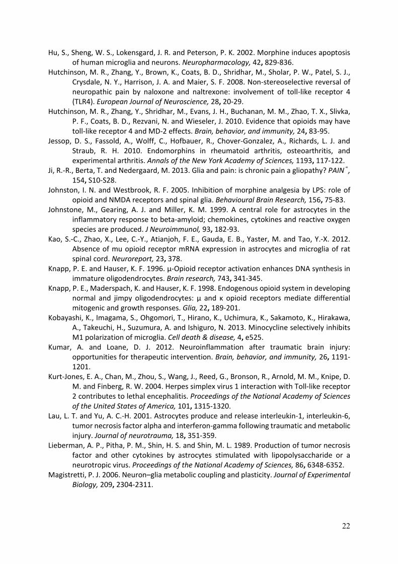
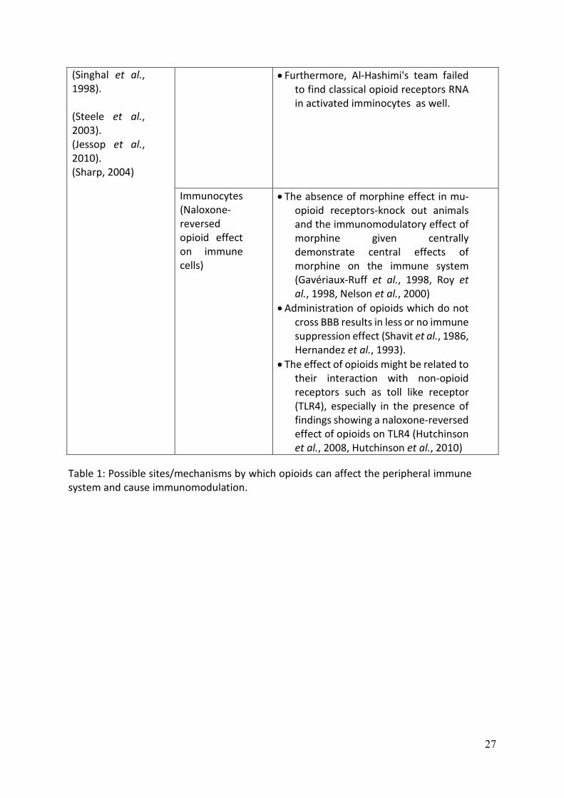
neurodegenerative diseases. Glial cells are classified into astrocytes, oligodendrocytes,
microglia, and ependymocytes, Figure 1. Polydendrocytes have also been categorised as
glial cells (Butt et al., 2005, Nishiyama, 2007).
Microglia are the mononuclear phagocytes of the CNS that have similar properties and
functions to peripheral macrophages (Streit, 2002). They are of mesodermal origin and
derived from myeloid precursor cells (Chan et al., 2007, Ransohoff and Perry, 2009,
Ginhoux et al., 2010). In a healthy mature CNS, microglia in a “resting state” will have a
small, ramified morphology with fine cellular processes and perform a surveillance
function. Microglial response or “microglial activation” is characterized by rapid and
intense changes in the cell morphology, function, and gene expression along with
several other events. These include; (i) migration towards the site of injury mediated by
various molecules including CCL2 (chemokine (C-C motif) ligand 2) via CCL2 receptor
(CCR2), fractalkine (CX3CL1) via CX3CR1, ATP via P2Y12 receptor, Neuregulin1( NRG1)
via ErbBR and possibly the complement CC5a (via CC5aR), (ii) proliferation in response
to the activation of ErbBR by NRG-1 and possibly Macrophage-Colony stimulating factor
8
(M-CSFR) by M-CSF, (iii) release of pro-inflammatory cytokines (such as Interlukine-1β
(IL-1β), Brain-derived neurotrophic factor (BDNF), Tumour necrosis factor alpha (TNF-
α), Nitric oxide (NO), Cyclooxygenase-2 (COX-2) and Interlukine-6 (IL-6)) (Van Rossum
and Hanisch, 2004, Streit et al., 2005, Block et al., 2007, Hanisch and Kettenmann, 2007,
Davoust et al., 2008, Colton and Wilcock, 2010, Graeber and Streit, 2010). Figure 3
summarises the possible events following microglial and astrocyte activation that are
expected to develop and/or maintain neuropathic pain.
Local activation of microglia is characterised by the production of signalling molecules
including various cytokines, proteases and reactive oxygen species (ROS) (Nimmerjahn
et al., 2005, Toklu and Tümer, 2015). The microenvironments in which microglia are
activated determine their phenotype. The classical activation is the early phase that is
induced by the presence of lipopolysaccharide and interferon-γ (IFN-γ) (Hernandez-
Ontiveros et al., 2013) and results in M1 phenotype. On the other hand, when microglia
are activated (alternative activation) in response to interlukin-4 (IL-4) or interlukin-13
(IL-13), they convert to the M2 phenotype. M1 microglia are characterised by a high
level of pro-inflammatory cytokines while the late M2 phenotype is characterised by the
production of anti-inflammatory molecules such as interlukin-1 (IL-1) and transforming
growth factor 1β (TGFβ) (Kumar and Loane, 2012, Chhor et al., 2013).
This activation of central innate immunity does not only occur in response to infection,
but also in response to other harmful conditions such as neuronal damage and ischemia.
The reactivity of microglial cells is advantageous permitting neuroprotection, brain
homeostasis, and possibly repairs through the release of neurotrophic factors. Despite
being a defensive mechanism of immune components, the central immune response is
associated with pathological conditions such as meningitis (Emonts et al., 2003,
Echchannaoui et al., 2002), encephalitis (Kurt-Jones et al., 2004, Wang et al., 2004),
multiple sclerosis (Bsibsi et al., 2002, Prinz et al., 2006), Alzheimer’s disease (Mcgeer and
Mcgeer, 2002) and Parkinson’s disease (Bonifati and Kishore, 2007). It is believed that
sustained microglial activation leads to such demyelinating and neurodegenerative
diseases, most likely through an excessive production of inflammatory mediators
modifying the function of structures such as the BBB.
9
As microglia are the central immune representatives, they were thought to be the only
player in the CNS immunity. It is now clear that astrocytes are important regulators of
central immune activity. Astroglia have multiple roles in the CNS and are known to
regulate local blood flow (Attwell et al., 2010), supply neurons with essential nutrients,
and control homeostasis (Mulligan and Macvicar, 2004, Magistretti, 2006, Araque and
Navarrete, 2010). They control the endothelial elements of BBB, and have important
implications on brain pathology (Bundgaard and Abbott, 2008). Astrocytes have been
found to be involved in a variety of neurological disorders through a process known as
reactive astrogliosis. This can have both beneficial and detrimental effects, and is
characterized by the upregulation of glial fibrillary acidic protein (GFAP) which is seen in
a range of neuropathologies including brain ischemia, brain hemorrhage, chronic CNS
infections, epilepsy, diabetic retinopathy, Alzheimer's disease, Parkinson's disease, and
multiple sclerosis. It is understood from rodent models that activation of astrocytes is
mediated by a variety of cytokines including transforming TGF-α, ciliary neurotrophic
factor (CNTF), and IL-6 (Merrill and Benveniste, 1996, Shrikant et al., 1995). Overall it is
believed that the extent of reactive astrogliosis, and subsequent changes in the
networks of astrocytes, is disease specific with different neuropathologies having
distinct molecular and cellular features. In addition, astrocytes are responsible for scar
formation, an event that protects fragile neurons (Faulkner et al., 2004), improves axon
regeneration (Anderson et al., 2016), and maintains BBB integrity (Bush et al., 1999).
However, this might result in neuronal damage or death as Tysseling-Mattiace and
colleagues found that the inhibition of scar formation promotes axon elongation after
spinal cord injury (Tysseling-Mattiace et al., 2008), Figure3.
Glial modifiers
Glial cells have been found to respond to several molecules. These compounds have
been used for in the study of glial cell behaviour in a range of disease conditions
experimentally and clinically. Minocycline is a member of tetracyclines, a group of
antibacterial agents. Minocycline has been found to act as a microglial inhibitor,
immunomodulatory, anti-inflammatory, and neuroprotective. The proposed
mechanism(s) are multifactorial and include the inhibition of key enzymes such as
10
inducible nitric oxide synthase (iNOS) (Amin et al., 1997) and Phospholipase A2 (PLA2)
(Pruzanski et al., 1992), ability to inhibit caspase-1 and caspase-3 (Chen et al., 2000),
peroxynitrite-scavenging activity (so it reduces protein tyrosine nitration) (Whiteman
and Halliwell, 1997), and inhibition of p38 MAPK activity.
Fluorocitrate is an astrocyte inhibitor that is considered as a fluoroacetate precursor. It has
the ability to inhibit aconitase, an enzyme that is responsible for isomerization of citrate to
isocitrate in the tricarboxylic acid cycle (TCA). Astrocytes have been described as an
acetate/glutamate specific compartment and have been found to be inhibited by this agent
[for more details about mechanism, see ref: (Fonnum et al., 1997)].
Pain and Glial Cells
Pain has been defined by The International Association for the Study of Pain (IASP) as
“an unpleasant sensory and emotional experience associated with actual or potential
tissue damage, or described in terms of such damage” (Loeser and Treede, 2008). It
involves both sensory and affective components. Pain is propagated by a process of
nociception, that includes the transmission of harmful stimuli by nociceptors (afferent
neurons) from the periphery to the CNS via the spinal cord. The transmitted stimulus is
processed centrally and results in pain sensation and reflex. In general, pain is classified
as acute and chronic; chronic pain is different from acute pain in terms of duration and
presence/absence of tissue damage. In general, when it is maintained for more than
three months after the disappearance of the causative tissue damage, pain is chronic.
Neuropathic pain is a chronic pain that is caused by somatosensory system damage
(Loeser and Treede, 2008). It is spontaneous pain with a low pain threshold
(hypersensitivity and allodynia) and a poor response to traditional analgesics.
The neuronal processes and plasticity underlying different chronic pain states are
historically known to involve both peripheral sensitisation – the hyperexcitability of
primary sensory neurons and central sensitisation, the increased excitatory transmission
at the level of spinal cord, brainstem, and cortex. However, there is growing
understanding that non-neural mechanisms are important in the commencement and
11
maintenance of chronic pain states with glial cells being recognised as central to these
non-neuronal processes. Pain researchers have been interested in the role of glia in the
regulation of pain since 1991, when Garrison and colleagues noticed an elevated GFAP
staining density in chronic pain state, an event that was correlated with hyperalgesia
and indicated astrocyte participation in peripheral nerve injury-induced neuropathy
(Garrison et al., 1991). Serious injuries and not minor acute pains are able to stimulate
dynamic alterations in glial cell functioning (Samikkannu et al., 2015).
Glia show a clear heterogeneity not only in receptor expression but also in their regional
response profiles. For example, cortical and cerebellar astrocytes are activated by a
profile of peptides which differ to those peptides stimulating spinal cord astrocytes
(Oberheim et al., 2012). Similarly, there are regional differences in microglial responses
between spinal and supraspinal sites following injury (Zhang et al., 2008). Such regional
differences in glial cell responses make interpreting findings from region to region and
from brain to spinal cord challenging.
Supraspinally, in the rostral ventromedial medulla (RVM) hyperactivation of microglia is
seen following chronic constriction injury of the infraorbital nerve, this occurs is 1-3 days
after injury, and is followed by a prolonged hyperactivation of astrocytes lasting for 28
days (Wei et al., 2008). The hyperactivation of glial cells in the rostral ventromedial
medulla (RVM) following nerve injury is known to release cytokines (IL-1B and TNF-α)
leading to subsequent glutamate receptor phosphorylation in descending pain-
modulating pathways leading to an overall facilitation in neuropathic pain. Cytokines
released from hyperperactivated RVM glial cells act as mediators leading to neuronal
hyperexcitability and the development of neuropathic pain (Wei et al., 2008). The
sequence of glial activation seen supraspinally in the RVM sees microglia cells as
important in the initiation phase and astrocytes in maintaining hyperalgesia following
nerve injury, similar to the activation chain seen spinally. However, Zhang and
colleagues found that microglial contribution in chronic pain conditions is limited to the
spinal cord but not all supraspinal region (Zhang et al., 2008)
Even in the spinal cord, microglial activation is variable. For example following peripheral
nerve injury, the activation and proliferation of microglia is seen on the ipsilateral dorsal
12
horn with the contralateral dorsal horn having weak activation (Tsuda et al., 2003). Glial
cells rarely divide under resting conditions and their proliferation in the spinal cord is a
crucial aspect of glial cell activation. Using a spared nerve injury model, cell proliferation
determined using Bromodeoxyuridine (5-bromo-2'-deoxyuridine, BrdU), was seen in the
dorsal and ventral horn of the spinal cord on the ipsilateral side, those cells positive for
BrdU were also labelled for IBA1 (microglial marker) demonstrating predominately
microglial propagation (Echeverry et al., 2008).
Surface marker expression following microglial and astroglial activation, at the
transcriptional level has been studied. Four hours following L5 nerve transection,
microglial activation was determined by an upregulation of microglial surface markers
integrin alpha M (ITGAM), TLR4 and CD14; this was followed later (4 days) by increased
and sustained upregulation of GFAP mRNA, indicative of astrocyte activation (Deleo et
al., 2004). In the same laboratory, the pre-emptive use of minocycline decreased the
increased expression of ITGAM and TLR4 and reduced nerve injury induced mechanical
allodynia. When minocycline was administered 5 days post injury its effect on
behavioural hypersensitivity and mRNA levels of ITGAM and TLR4 was limited
(Raghavendra et al., 2003), therefore both spinal microglia and astrocytes are
progressively involved in the spinal sensitisation following nerve injury.
Following painful injury glia go through a variety of activation states which include: (i)
up regulation of glial markers associated with glial cell activation CCR3 and CD11b, IBA1,
GFAP and related morphological changes such as hypertrophy, (ii) increased expression
and activation of TLRs involved in innate immunity and chemokine receptors on glial
cells (Mckimmie and Fazakerley, 2005, Carpentier et al., 2008), (iii) Stimulation of
intracellular MAPK cascades and (iv) subsequent increase in growth factors, cytokines,
and chemokines mediate the glial function.
Glia are non-axonal and cannot directly relay nociceptive signals to the brain from the
spinal cord, instead glial activation states are believed to shape pathological pain
conditions mediated through the release of glial pro-inflammatory products which have
a direct effect on nociceptive neurons to increase their excitability and hence firing.
Importantly, it has been shown that the inhibition of activated astrocytes and microglia
13
results in attenuated experimental neuropathic pain. Interestingly, minocycline has
been shown to prevent this process although it is unable to reverse neuropathic pain
once established, while the inhibition of astrocytes using fluorocitrate (an astrocyte
inhibitor) has been shown to reverse neuropathic pain. Together these findings suggest
that the tempo of microglia and astrocyte activation differ and is important in the
establishment and maintenance of neuropathic pain states.
Activation of microglia seems to occur during the early phase of pain (Hald et al., 2009)
which may further contribute to the activation of astrocytes (Giulian et al., 1994,
Retamal et al., 2007), (Figure 2). However, astrocytes release microglial activation
factors such as TNF-α, lymphotoxin, IL-6 (Lieberman et al., 1989, Aloisi et al., 1992, Lau
and Yu, 2001), alpha- and beta-interferons, Monocyte Chemoattractant Protein-1 (MCP-
1), CC5, RANTES (regulated on activation, normal T cell expressed and secreted)
(Johnstone et al., 1999). Therefore, it seems that astrocytes initially enhance the
activation of the already activated microglia during the early phase of neuropathic pain.
Neurons are not only influenced by these events but also they are able to exert their
own modulation on the orchestration of central immunity. Several actions are attributed
to the activation of neurons including the inhibition of microglia and induction of
microglial apoptosis along with other cellular components (Choi and Benveniste, 2004).
Unlike microglia, astrocytes are resistant to apoptosis (Song et al., 2006), which might
explain the effectiveness of astroglial but not microglial inhibitors in the reversal of
neuropathic pain (Figure 2).
Glia and opioids
Opioid receptors are GPCR members that are classified into classical (MOP, DOP, and
KOP) and are antagonised by naloxone, and non-classical (NOP) which has no affinity to
naloxone. They have been used extensively in the management of pain, diarrhoea, and
cough. However, their use is associated with unwanted effects including tolerance,
dependence, respiratory depression, and immunomodulation. Morphine is the
prototypical opioid. Beitner-Johnson and colleagues determined that the beneficial and
unwanted effects of opioids are not limited to neurons, and extend to non-neuronal glial
14
cells based on findings that showed an increased expression of GFAP in the ventral
tegmental area following prolonged systemic administration of morphine (Beitner-
Johnson et al., 1993). Subsequent studies have now shown that opioids are able to
activate glial cells leading to up regulation and release of pro-inflammatory
cytokines/chemokines and that repeated dosing of opioid drugs strengthens this
activation. In addition, how opioid-mediated activation of glia results in an enhancement
in nociceptive transmission that subsequently surpasses its analgesic actions
(Raghavendra et al., 2003, Deleo et al., 2004). Therefore, long-term exposure to opioids
results in processes that enable nociceptive transmission and it is believed that this
action contributes towards opioid drug tolerance.
Glial cell activation would appear to be a central mechanistic mediator in chronic pain
states and opioid drug tolerance, indeed there is strong evidence highlighting the
mechanistic parallels between morphine tolerance and neuropathic pain; both
conditions see glia increase extracellular concentrations of neuroexcitiatory substances
such as nitric oxide and prostaglandins. Both conditions result in the facilitation of pain
and reduction in morphine analgesia and see a reduction in glial glutamate transporters
in the dorsal horn leading to increased neuronal excitability. Furthermore, it has been
found that long term use of opioids is associated with increased microglial apoptosis and
reduced microglial activity (Hu et al., 2002). As mentioned before, microglial activation
throughout the progress of neuropathic pain involves an early “classical” pro-
inflammatory M1 phenotype and late (alternative) anti-inflammatory M2 phenotype.
Minocycline has been found to inhibit M1 but not M2 phenotype (Kobayashi et al.,
2013), findings that support the proposed explanation of the effectiveness of microglial
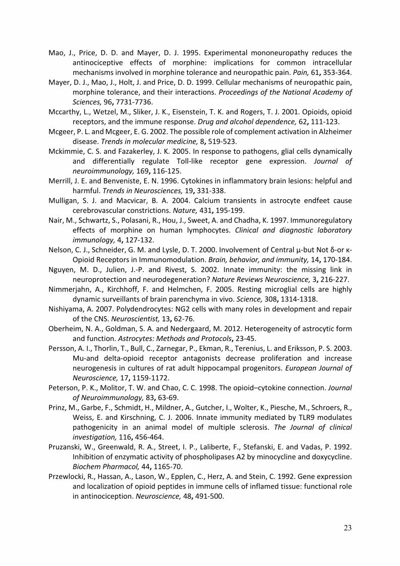
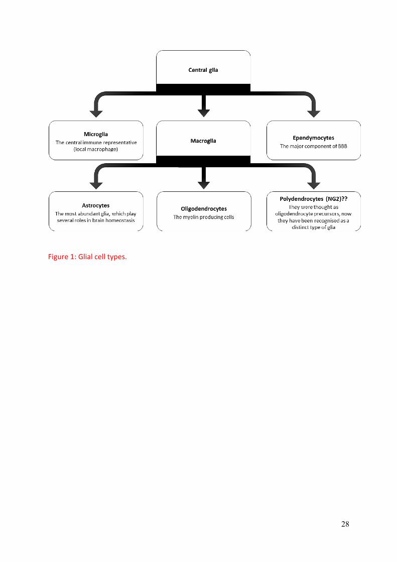
inhibitors in prevention but not reversion of neuropathic pain. Figure 2 shows the
progress of neuropathic pain and/or opioid use from the early phase until the
development of neuropathic pain and/or opioid tolerance along with the profile of
microglial, astroglial, and neuronal responses.
The findings presented here suggest that glial cell activation, whether induced by
opioids, inflammation or tissue injury leads to similar consequences including the
release of pro-inflammatory cytokines such as TNF-α, IL-1 and IL-6, which subsequently
15
lead to the release of neuroexcitiatory mediators including nitric oxide, prostaglandins,
and excitatory amino acids enhancing pain transmission. The recognition that opioid
tolerance and neuropathic pain could be attributed to similar mechanisms is
strengthened by the longstanding clinical awareness that opioid drugs poorly treat
neuropathic pain conditions (Mao et al., 1995, Mayer et al., 1999).
Glial cells and opioid receptor expression
Given that, opioid activation of glia leads to enhancement of tolerance, diminishing
opioid analgesia and augmentation of dependence and reward. It is important to
understand whether glial sensitivity to opioids is through a direct or indirect effect.
Some studies have indicated that MOP opioid receptor agonists may bind to MOP
receptors on spinal glial cells. Indeed numerous studies have shown that opioid receptor
expression on glia is variable from one cell line to another, from established cell lines to
primary cells, from in vitro to in vivo and from region to region in the CNS. For example,
MOP receptor mRNA detected in cultured cortical astrocytes was higher than in cultured
cerebellar and striatal astrocytes and was absent in hippocampal astrocytes (Ruzicka et
al., 1995). Low expression of MOP receptor was detected in vivo in a limited area of the
CNS under normal conditions (Stiene-Martin et al., 2001). Absence of MOP receptor
from astrocytes in rats was reported by Kao and colleagues (Kao et al., 2012). In regards
to the forskolin stimulated cAMP accumulation, Eriksson and colleagues found an
antagonist-reversed effect of DOP and KOP, but not MOP agonists in rat cerebral cortex
astrocytes (Eriksson et al., 1990, 1991). However, they found MOP-induced inhibition of
cAMP in other regions of brain (Eriksson et al., 1991).
Other studies have shown that MOP, DOP and KOP mRNA are differentially expressed in
rat primary astroglial cultures from different areas of the brain including cortical, striatal,
cerebellar, hippocampal and hypothalamic regions (Ruzicka et al., 1995). In five glial
cultures, Ruzicka and colleagues found that KOP and DOP expression is higher than MOP.
Although these receptors were expressed on astrocytes from all of these sites, it was
found that the abundance of opioid receptors was as follows: MOP in cortical, DOP in
cortical/hypothalamic, KOP in cortical/hypothalamic/ cerebellar astrocytes.
16
Other factors may affect cell behaviour and the expression of opioid receptors. Culture
confluence might influence the pattern of opioid receptor expression. Stiene-Martin and
colleagues found that cell confluence could change the expression of opioid receptors
on astroglia taken from the cerebral cortex, hippocampus, cerebellum, and striatum of
1-day-old mice (Stiene-Martin et al., 1998). Using flow cytometry and agonist-induced
changes in intracellular calcium, they found that low-density cultures resulted in greater
expression of MOP in the cerebral cortex and hippocampus and low expression of DOP.
At confluence, MOP expression was still the greatest while DOP expression declined in
the cerebellum but increased in the hippocampus. It was found that confluence did not
affect the expression of KOP and no difference was found between low-density and
confluent cultures. However, in confluent cortical cultures, the proportion of KOP
expressing cells is less than at low-density.
The expression of opioid receptors on oligodendrocytes is also differential. According to
Knapp and colleges, primary mouse oligodendrocytes express both MOP and KOP but
not DOP receptors. However, the expression of MOP and KOP was found to be
influenced by stage of development and level of stimulation (Knapp et al., 1998). Higher
expression was seen in the early stage of development; this decreased in mature cells.
Furthermore, they found a differential response of opioid receptor activation namely a
MOP-induced proliferation and KOP-induced growth and differentiation (Knapp and
Hauser, 1996). Progressive down-regulation of MOP receptor on primary mouse
oligodendrocytes was reported in relation to developmental stages (Tryoen-Toth et al.,
2000) that might indicate a direct effect of maturation on the expression patterns of
opioid receptors on oligodendrocytes. However, in rat primary oligodendrocytes, it was
found that MOP and DOP antagonists inhibit oligodendrocyte proliferation (Persson et
al., 2003)
Some uncertainty surrounds opioid expression on the central immune representative
cells, microglia. For example, in terms of RNA, radioligand binding assays and
immunofluorescence assays, it was found that primary neonatal human microglia
constitutively express KOP receptor (Chao et al., 1996). Inhibitory effect of MOP
receptor on microglial cell chemotaxis has been reported, suggesting MOP expression
17
(Chao et al. (1997). However, the absence of classical opioid receptors on microglia
and/or opioid receptor-independent actions has been reported by other studies (Qian
et al., 2007, Kao et al., 2012). A possible interpretation of morphine-induce microglial
response is the cross talk between opioid receptors and toll like receptors (TLRs; well
known to be expressed by microglia and other immune cells). Nevertheless, most
studies are reporting that morphine (as a prototypical opioid) has a naloxone reversible
effect on microglia and the question to be raised here is whether naloxone can reverse
the effect of morphine on TLR-4 receptors. Interestingly, it has been reported that
naloxone dose reverse the effect of morphine on TLR-4 (Hutchinson et al., 2008).
Given how the activation of glia by opioids essentially instigates a limiting factor in their
analgesic efficacy, and that chronic pain states and inflammation share a common
activation of glia, what are the possible clinical implications for the use of opioids in the
treatment of different conditions? In animal models intraperitoneal injection of the
bacterial endotoxin LPS can activate spinal glia and that a loss of analgesic efficacy for
morphine is seen (Johnston and Westbrook, 2005). Further studies have revealed how
neuropathic pain decreases morphine efficacy (Mao et al., 1995), a finding linked to the
increased production proinflammatory cytokines, by glia, when opioids are
administered to an established neuropathic environment.
Inhibitors of glial cell activity could therefore represent an approach for reducing opioid
tolerance, and it has been suggested that suppression of activated microglia attenuates
neuropathic pain. Indeed, both fluorocitrate and minocycline, which inhibit the actions
of pro-inflammatory cytokines, have been demonstrated to enhance morphine
analgesia and reduce morphine tolerance.
In conclusion, glia cells play central (and interacting) roles in several immune-related
actions including inflammation and neuropathic pain. Moreover, the use of opioids is
associated with addiction, tolerance, immunosuppression, astrogliosis, and microglial
apoptosis. Glial cells represent targets for use in the pain clinic and development of
novel selective drugs for both opioid receptors and glia is an exciting challenge for the
future.

18
19
Al-Hashimi, M., Mcdonald, J., Thompson, J. and Lambert, D. 2013. Classical opioid receptor mRNA is NOT present in whole human blood. British Journal of Anaesthesia, 110, 860-85P.
Al-Hashimi, M., Mcdonald, J., Thompson, J. P. and Lambert, D. G. 2016. Evidence for nociceptin/orphanin FQ (NOP) but not µ (MOP), δ (DOP) or κ (KOP) opioid receptor mRNA in whole human blood. British journal of anaesthesia, 116, 423-429.
Aloisi, F., Carè, A., Borsellino, G., Gallo, P., Rosa, S., Bassani, A., Cabibbo, A., Testa, U., Levi, G. and Peschle, C. 1992. Production of hemolymphopoietic cytokines (IL-6, IL-8, colony-stimulating factors) by normal human astrocytes in response to IL-1 beta and tumor necrosis factor-alpha. The Journal of Immunology, 149, 2358-2366.
Amin, A. R., Patel, R. N., Thakker, G. D., Lowenstein, C. J., Attur, M. G. and Abramson, S. B. 1997. Post-transcriptional regulation of inducible nitric oxide synthase mRNA in murine macrophages by doxycycline and chemically modified tetracyclines. FEBS Lett, 410, 259-64.
Anderson, M. A., Burda, J. E., Ren, Y., Ao, Y., O’shea, T. M., Kawaguchi, R., Coppola, G., Khakh, B. S., Deming, T. J. and Sofroniew, M. V. 2016. Astrocyte scar formation aids central nervous system axon regeneration. Nature, 532, 195.
Araque, A. and Navarrete, M. 2010. Glial cells in neuronal network function. Philosophical Transactions of the Royal Society of London B: Biological Sciences, 365, 2375-2381.
Attwell, D., Buchan, A. M., Charpak, S., Lauritzen, M., Macvicar, B. A. and Newman, E. A. 2010. Glial and neuronal control of brain blood flow. Nature, 468, 232-243.
Beck, M., Mirmohammadsadegh, A., Franz, B., Blanke, J. and Hengge, U. R. 2002. Opioid receptors on white blood cells: effect of HIV infection and methadone treatment. Pain, 98, 187-194.
Beitner-Johnson, D., Guitart, X. and Nestler, E. J. 1993. Glial Fibrillary Acidic Protein and the Mesolimbic Dopamine System: Regulation by Chronic Morphine and Lewis-Fischer Strain Differences in the Rat Ventral Tegmental Area. Journal of neurochemistry, 61, 1766-1773.
Bell, S. P., Sack, M. N., Patel, A., Opie, L. H. and Yellon, D. M. 2000. Delta opioid receptor stimulation mimics ischemic preconditioning in human heart muscle. Journal of the American College of Cardiology, 36, 2296-2302.
Block, M. L., Zecca, L. and Hong, J.-S. 2007. Microglia-mediated neurotoxicity: uncovering the molecular mechanisms. Nature Reviews Neuroscience, 8, 57-69.
Bonifati, D. M. and Kishore, U. 2007. Role of complement in neurodegeneration and neuroinflammation. Molecular immunology, 44, 999-1010.
Bsibsi, M., Ravid, R., Gveric, D. and Van Noort, J. M. 2002. Broad expression of Toll-like receptors in the human central nervous system. Journal of Neuropathology & Experimental Neurology, 61, 1013-1021.
Buckingham, J. C. and Cooper, T. A. 1984. Differences in hypothalamo-pituitary-adrenocortical activity in the rat after acute and prolonged treatment with morphine. Neuroendocrinology, 38, 411-7.
Bundgaard, M. and Abbott, N. J. 2008. All vertebrates started out with a glial blood-brain barrier 4–500 million years ago. Glia, 56, 699-708.
Bush, T. G., Puvanachandra, N., Horner, C. H., Polito, A., Ostenfeld, T., Svendsen, C. N., Mucke, L., Johnson, M. H. and Sofroniew, M. V. 1999. Leukocyte infiltration, neuronal degeneration, and neurite outgrowth after ablation of scar-forming, reactive astrocytes in adult transgenic mice. Neuron, 23, 297-308.
20
Butt, A. M., Hamilton, N., Hubbard, P., Pugh, M. and Ibrahim, M. 2005. Synantocytes: the fifth element. Journal of Anatomy, 207, 695-706.
Caldiroli, E., Leoni, O., Cattaneo, S., Rasini, E., Marino, V., Tosetto, C., Mazzone, A., Fietta, A. M., Lecchini, S. and Frigo, G. M. 1999. Neutrophil function and opioid receptor expression on leucocytes during chronic naltrexone treatment in humans. Pharmacol Res, 40, 153-8.
Carpentier, P. A., D’anne, S. D. and Miller, S. D. 2008. Glial toll-like receptor signaling in central nervous system infection and autoimmunity. Brain, behavior, and immunity, 22, 140-147.
Chan, W., Kohsaka, S. and Rezaie, P. 2007. The origin and cell lineage of microglia—new concepts. Brain research reviews, 53, 344-354.
Chao, C. C., Gekker, G., Hu, S., Sheng, W. S., Shark, K. B., Bu, D.-F., Archer, S., Bidlack, J. M. and Peterson, P. K. 1996. Kappa opioid receptors in human microglia downregulate human immunodeficiency virus 1 expression. Proceedings of the National Academy of Sciences, 93, 8051-8056.
Chao, C. C., Hu, S., Shark, K. B., Sheng, W. S., Gekker, G. and Peterson, P. K. 1997. Activation of mu opioid receptors inhibits microglial cell chemotaxis. Journal of pharmacology and experimental therapeutics, 281, 998-1004.
Chen, M., Ona, V. O., Li, M., Ferrante, R. J., Fink, K. B., Zhu, S., Bian, J., Guo, L., Farrell, L. A., Hersch, S. M., Hobbs, W., Vonsattel, J. P., Cha, J. H. and Friedlander, R. M. 2000. Minocycline inhibits caspase-1 and caspase-3 expression and delays mortality in a transgenic mouse model of Huntington disease. Nat Med, 6, 797-801.
Chhor, V., Le Charpentier, T., Lebon, S., Oré, M.-V., Celador, I. L., Josserand, J., Degos, V., Jacotot, E., Hagberg, H. and Sävman, K. 2013. Characterization of phenotype markers and neuronotoxic potential of polarised primary microglia in vitro. Brain, behavior, and immunity, 32, 70-85.
Choi, C. and Benveniste, E. N. 2004. Fas ligand/Fas system in the brain: regulator of immune and apoptotic responses. Brain Research Reviews, 44, 65-81.
Chuang, T. K., Killam, K. F., Jr., Chuang, L. F., Kung, H. F., Sheng, W. S., Chao, C. C., Yu, L. and Chuang, R. Y. 1995. Mu opioid receptor gene expression in immune cells. Biochem Biophys Res Commun, 216, 922-30.
Colton, C. A. and Wilcock, D. M. 2010. Assessing activation states in microglia. CNS & Neurological Disorders-Drug Targets (Formerly Current Drug Targets-CNS & Neurological Disorders), 9, 174-191.
Davoust, N., Vuaillat, C., Androdias, G. and Nataf, S. 2008. From bone marrow to microglia: barriers and avenues. Trends in immunology, 29, 227-234.
Deleo, J. A., Tanga, F. Y. and Tawfik, V. L. 2004. Neuroimmune activation and neuroinflammation in chronic pain and opioid tolerance/hyperalgesia. The Neuroscientist, 10, 40-52.
Echchannaoui, H., Frei, K., Schnell, C., Leib, S. L., Zimmerli, W. and Landmann, R. 2002. Toll-like receptor 2–deficient mice are highly susceptible to Streptococcus pneumoniae meningitis because of reduced bacterial clearing and enhanced inflammation. Journal of Infectious Diseases, 186, 798-806.
Echeverry, S., Shi, X. Q. and Zhang, J. 2008. Characterization of cell proliferation in rat spinal cord following peripheral nerve injury and the relationship with neuropathic pain. PAIN®, 135, 37-47.
21
Emonts, M., Hazelzet, J., De Groot, R. and Hermans, P. 2003. Host genetic determinants of Neisseria meningitidis infections. The Lancet infectious diseases, 3, 565-577.
Eriksson, P. S., Hansson, E. and Ronnback, L. 1990. Delta and kappa opiate receptors in primary astroglial cultures from rat cerebral cortex. Neurochem Res, 15, 1123-6.
Eriksson, P. S., Hansson, E. and Ronnback, L. 1991. Mu and delta opiate receptors in neuronal and astroglial primary cultures from various regions of the brain--coupling with adenylate cyclase, localisation on the same neurones and association with dopamine (D1) receptor adenylate cyclase. Neuropharmacology, 30, 1233-9.
Faulkner, J. R., Herrmann, J. E., Woo, M. J., Tansey, K. E., Doan, N. B. and Sofroniew, M. V. 2004. Reactive astrocytes protect tissue and preserve function after spinal cord injury. Journal of Neuroscience, 24, 2143-2155.
Fonnum, F., Johnsen, A. and Hassel, B. 1997. Use of fluorocitrate and fluoroacetate in the study of brain metabolism. Glia, 21, 106-113.
Franchimont, D. 2004. Overview of the actions of glucocorticoids on the immune response: a good model to characterize new pathways of immunosuppression for new treatment strategies. Annals of the New York Academy of Sciences, 1024, 124-137.
Garrison, C., Dougherty, P., Kajander, K. and Carlton, S. 1991. Staining of glial fibrillary acidic protein (GFAP) in lumbar spinal cord increases following a sciatic nerve constriction injury. Brain research, 565, 1-7.
Gavériaux-Ruff, C., Matthes, H. W., Peluso, J. and Kieffer, B. L. 1998. Abolition of morphine-immunosuppression in mice lacking the μ-opioid receptor gene. Proceedings of the National Academy of Sciences, 95, 6326-6330.
Ginhoux, F., Greter, M., Leboeuf, M., Nandi, S., See, P., Gokhan, S., Mehler, M. F., Conway, S. J., Ng, L. G. and Stanley, E. R. 2010. Fate mapping analysis reveals that adult microglia derive from primitive macrophages. Science, 330, 841-845.
Giulian, D., Li, J., Leara, B. and Keenen, C. 1994. Phagocytic microglia release cytokines and cytotoxins that regulate the survival of astrocytes and neurons in culture. Neurochemistry international, 25, 227-233.
Graeber, M. B. and Streit, W. J. 2010. Microglia: biology and pathology. Acta neuropathologica, 119, 89-105.
Guo, C.-J., Li, Y., Tian, S., Wang, X., Douglas, S. D. and Ho, W.-Z. 2002. Morphine enhances HIV infection of human blood mononuclear phagocytes through modulation of β-chemokines and CCR5 receptor. Journal of investigative medicine, 50, 435-442.
Hald, A., Nedergaard, S., Hansen, R. R., Ding, M. and Heegaard, A. M. 2009. Differential activation of spinal cord glial cells in murine models of neuropathic and cancer pain. European Journal of Pain, 13, 138-145.
Hanisch, U.-K. and Kettenmann, H. 2007. Microglia: active sensor and versatile effector cells in the normal and pathologic brain. Nature neuroscience, 10, 1387-1394.
Hendriks, J. J., Teunissen, C. E., De Vries, H. E. and Dijkstra, C. D. 2005. Macrophages and neurodegeneration. Brain Research Reviews, 48, 185-195.
Hernandez-Ontiveros, D. G., Tajiri, N., Acosta, S., Giunta, B., Tan, J. and Borlongan, C. V. 2013. Microglia activation as a biomarker for traumatic brain injury. Frontiers in neurology, 4.
Hernandez, M. C., Flores, L. R. and Bayer, B. M. 1993. Immunosuppression by morphine is mediated by central pathways. Journal of Pharmacology and Experimental Therapeutics, 267, 1336-1341.
22
Hu, S., Sheng, W. S., Lokensgard, J. R. and Peterson, P. K. 2002. Morphine induces apoptosis of human microglia and neurons. Neuropharmacology, 42, 829-836.
Hutchinson, M. R., Zhang, Y., Brown, K., Coats, B. D., Shridhar, M., Sholar, P. W., Patel, S. J., Crysdale, N. Y., Harrison, J. A. and Maier, S. F. 2008. Non-stereoselective reversal of neuropathic pain by naloxone and naltrexone: involvement of toll-like receptor 4 (TLR4). European Journal of Neuroscience, 28, 20-29.
Hutchinson, M. R., Zhang, Y., Shridhar, M., Evans, J. H., Buchanan, M. M., Zhao, T. X., Slivka, P. F., Coats, B. D., Rezvani, N. and Wieseler, J. 2010. Evidence that opioids may have toll-like receptor 4 and MD-2 effects. Brain, behavior, and immunity, 24, 83-95.
Jessop, D. S., Fassold, A., Wolff, C., Hofbauer, R., Chover-Gonzalez, A., Richards, L. J. and Straub, R. H. 2010. Endomorphins in rheumatoid arthritis, osteoarthritis, and experimental arthritis. Annals of the New York Academy of Sciences, 1193, 117-122.
Ji, R.-R., Berta, T. and Nedergaard, M. 2013. Glia and pain: is chronic pain a gliopathy? PAIN®, 154, S10-S28.
Johnston, I. N. and Westbrook, R. F. 2005. Inhibition of morphine analgesia by LPS: role of opioid and NMDA receptors and spinal glia. Behavioural Brain Research, 156, 75-83.
Johnstone, M., Gearing, A. J. and Miller, K. M. 1999. A central role for astrocytes in the inflammatory response to beta-amyloid; chemokines, cytokines and reactive oxygen species are produced. J Neuroimmunol, 93, 182-93.
Kao, S.-C., Zhao, X., Lee, C.-Y., Atianjoh, F. E., Gauda, E. B., Yaster, M. and Tao, Y.-X. 2012. Absence of mu opioid receptor mRNA expression in astrocytes and microglia of rat spinal cord. Neuroreport, 23, 378.
Knapp, P. E. and Hauser, K. F. 1996. μ-Opioid receptor activation enhances DNA synthesis in immature oligodendrocytes. Brain research, 743, 341-345.
Knapp, P. E., Maderspach, K. and Hauser, K. F. 1998. Endogenous opioid system in developing normal and jimpy oligodendrocytes: μ and κ opioid receptors mediate differential mitogenic and growth responses. Glia, 22, 189-201.
Kobayashi, K., Imagama, S., Ohgomori, T., Hirano, K., Uchimura, K., Sakamoto, K., Hirakawa, A., Takeuchi, H., Suzumura, A. and Ishiguro, N. 2013. Minocycline selectively inhibits M1 polarization of microglia. Cell death & disease, 4, e525.
Kumar, A. and Loane, D. J. 2012. Neuroinflammation after traumatic brain injury: opportunities for therapeutic intervention. Brain, behavior, and immunity, 26, 1191-1201.
Kurt-Jones, E. A., Chan, M., Zhou, S., Wang, J., Reed, G., Bronson, R., Arnold, M. M., Knipe, D. M. and Finberg, R. W. 2004. Herpes simplex virus 1 interaction with Toll-like receptor 2 contributes to lethal encephalitis. Proceedings of the National Academy of Sciences of the United States of America, 101, 1315-1320.
Lau, L. T. and Yu, A. C.-H. 2001. Astrocytes produce and release interleukin-1, interleukin-6, tumor necrosis factor alpha and interferon-gamma following traumatic and metabolic injury. Journal of neurotrauma, 18, 351-359.
Lieberman, A. P., Pitha, P. M., Shin, H. S. and Shin, M. L. 1989. Production of tumor necrosis factor and other cytokines by astrocytes stimulated with lipopolysaccharide or a neurotropic virus. Proceedings of the National Academy of Sciences, 86, 6348-6352.
Magistretti, P. J. 2006. Neuron–glia metabolic coupling and plasticity. Journal of Experimental Biology, 209, 2304-2311.
23
Mao, J., Price, D. D. and Mayer, D. J. 1995. Experimental mononeuropathy reduces the antinociceptive effects of morphine: implications for common intracellular mechanisms involved in morphine tolerance and neuropathic pain. Pain, 61, 353-364.
Mayer, D. J., Mao, J., Holt, J. and Price, D. D. 1999. Cellular mechanisms of neuropathic pain, morphine tolerance, and their interactions. Proceedings of the National Academy of Sciences, 96, 7731-7736.
Mccarthy, L., Wetzel, M., Sliker, J. K., Eisenstein, T. K. and Rogers, T. J. 2001. Opioids, opioid receptors, and the immune response. Drug and alcohol dependence, 62, 111-123.
Mcgeer, P. L. and Mcgeer, E. G. 2002. The possible role of complement activation in Alzheimer disease. Trends in molecular medicine, 8, 519-523.
Mckimmie, C. S. and Fazakerley, J. K. 2005. In response to pathogens, glial cells dynamically and differentially regulate Toll-like receptor gene expression. Journal of neuroimmunology, 169, 116-125.
Merrill, J. E. and Benveniste, E. N. 1996. Cytokines in inflammatory brain lesions: helpful and harmful. Trends in Neurosciences, 19, 331-338.
Mulligan, S. J. and Macvicar, B. A. 2004. Calcium transients in astrocyte endfeet cause cerebrovascular constrictions. Nature, 431, 195-199.
Nair, M., Schwartz, S., Polasani, R., Hou, J., Sweet, A. and Chadha, K. 1997. Immunoregulatory effects of morphine on human lymphocytes. Clinical and diagnostic laboratory immunology, 4, 127-132.
Nelson, C. J., Schneider, G. M. and Lysle, D. T. 2000. Involvement of Central μ-but Not δ-or κ-Opioid Receptors in Immunomodulation. Brain, behavior, and immunity, 14, 170-184.
Nguyen, M. D., Julien, J.-P. and Rivest, S. 2002. Innate immunity: the missing link in neuroprotection and neurodegeneration? Nature Reviews Neuroscience, 3, 216-227.
Nimmerjahn, A., Kirchhoff, F. and Helmchen, F. 2005. Resting microglial cells are highly dynamic surveillants of brain parenchyma in vivo. Science, 308, 1314-1318.
Nishiyama, A. 2007. Polydendrocytes: NG2 cells with many roles in development and repair of the CNS. Neuroscientist, 13, 62-76.
Oberheim, N. A., Goldman, S. A. and Nedergaard, M. 2012. Heterogeneity of astrocytic form and function. Astrocytes: Methods and Protocols, 23-45.
Persson, A. I., Thorlin, T., Bull, C., Zarnegar, P., Ekman, R., Terenius, L. and Eriksson, P. S. 2003. Mu-and delta-opioid receptor antagonists decrease proliferation and increase neurogenesis in cultures of rat adult hippocampal progenitors. European Journal of Neuroscience, 17, 1159-1172.
Peterson, P. K., Molitor, T. W. and Chao, C. C. 1998. The opioid–cytokine connection. Journal of Neuroimmunology, 83, 63-69.
Prinz, M., Garbe, F., Schmidt, H., Mildner, A., Gutcher, I., Wolter, K., Piesche, M., Schroers, R., Weiss, E. and Kirschning, C. J. 2006. Innate immunity mediated by TLR9 modulates pathogenicity in an animal model of multiple sclerosis. The Journal of clinical investigation, 116, 456-464.
Pruzanski, W., Greenwald, R. A., Street, I. P., Laliberte, F., Stefanski, E. and Vadas, P. 1992. Inhibition of enzymatic activity of phospholipases A2 by minocycline and doxycycline. Biochem Pharmacol, 44, 1165-70.
Przewlocki, R., Hassan, A., Lason, W., Epplen, C., Herz, A. and Stein, C. 1992. Gene expression and localization of opioid peptides in immune cells of inflamed tissue: functional role in antinociception. Neuroscience, 48, 491-500.
24
Qian, L., Tan, K. S., Wei, S.-J., Wu, H.-M., Xu, Z., Wilson, B., Lu, R.-B., Hong, J.-S. and Flood, P. M. 2007. Microglia-mediated neurotoxicity is inhibited by morphine through an opioid receptor-independent reduction of NADPH oxidase activity. The Journal of Immunology, 179, 1198-1209.
Raghavendra, V., Tanga, F. and Deleo, J. A. 2003. Inhibition of microglial activation attenuates the development but not existing hypersensitivity in a rat model of neuropathy. Journal of Pharmacology and Experimental Therapeutics, 306, 624-630.
Ransohoff, R. M. and Brown, M. A. 2012. Innate immunity in the central nervous system. The Journal of clinical investigation, 122, 1164.
Ransohoff, R. M. and Perry, V. H. 2009. Microglial physiology: unique stimuli, specialized responses. Annual review of immunology, 27, 119-145.
Retamal, M. A., Froger, N., Palacios-Prado, N., Ezan, P., Sáez, P. J., Sáez, J. C. and Giaume, C. 2007. Cx43 hemichannels and gap junction channels in astrocytes are regulated oppositely by proinflammatory cytokines released from activated microglia. Journal of Neuroscience, 27, 13781-13792.
Roy, S., Barke, R. A. and Loh, H. H. 1998. Mu-opioid receptor-knockout mice: role of μ-opioid receptor in morphine mediated immune functions. Molecular brain research, 61, 190-194.
Ruzicka, B. B., Fox, C. A., Thompson, R. C., Meng, F., Watson, S. J. and Akil, H. 1995. Primary astroglial cultures derived from several rat brain regions differentially express mu, delta and kappa opioid receptor mRNA. Brain Res Mol Brain Res, 34, 209-20.
Samikkannu, T., Rao, K. V., Salam, A. a. A., Atluri, V. S., Kaftanovskaya, E. M., Agudelo, M., Perez, S., Yoo, C., Raymond, A. D. and Ding, H. 2015. HIV subtypes B and C gp120 and methamphetamine interaction: dopaminergic system implicates differential neuronal toxicity. Scientific reports, 5.
Schultz, J. E. J., Hsu, A. K. and Gross, G. J. 1996. Morphine mimics the cardioprotective effect of ischemic preconditioning via a glibenclamide-sensitive mechanism in the rat heart. Circulation Research, 78, 1100-1104.
Schultz, J. J., Hsu, A. K. and Gross, G. J. 1997. Ischemic Preconditioning and Morphine-induced Cardioprotection Involve the Delta (δ)-opioid Receptor in the Intact Rat Heart. Journal of Molecular and Cellular Cardiology, 29, 2187-2195.
Sharp, B. M. 2004. Opioid receptor expression and function. Journal of neuroimmunology, 147, 3-5.
Shavit, Y., Depaulis, A., Martin, F. C., Terman, G. W., Pechnick, R. N., Zane, C. J., Gale, R. P. and Liebeskind, J. C. 1986. Involvement of brain opiate receptors in the immune-suppressive effect of morphine. Proceedings of the National Academy of Sciences, 83, 7114-7117.
Shrikant, P., Weber, E., Jilling, T. and Benveniste, E. N. 1995. Intercellular adhesion molecule-1 gene expression by glial cells. Differential mechanisms of inhibition by IL-10 and IL-6. The Journal of Immunology, 155, 1489-1501.
Singhal, P. C., Sharma, P., Kapasi, A. A., Reddy, K., Franki, N. and Gibbons, N. 1998. Morphine enhances macrophage apoptosis. The Journal of Immunology, 160, 1886-1893.
Song, J. H., Bellail, A., Margaret, C., Yong, V. W. and Hao, C. 2006. Human astrocytes are resistant to Fas ligand and tumor necrosis factor-related apoptosis-inducing ligand-induced apoptosis. Journal of Neuroscience, 26, 3299-3308.
Steele, A. D., Henderson, E. E. and Rogers, T. J. 2003. μ-opioid modulation of HIV-1 coreceptor expressionand HIV-1 replication. Virology, 309, 99-107.
25
Stiene-Martin, A., Zhou, R. and Hauser, K. F. 1998. Regional, developmental, and cell cycle-dependent differences in mu, delta, and kappa-opioid receptor expression among cultured mouse astrocytes. Glia, 22, 249-59.
Stiene-Martin, A., Knapp, P. E., Martin, K., Gurwell, J. A., Ryan, S., Thornton, S. R., Smith, F. L. and Hauser, K. F. 2001. Opioid system diversity in developing neurons, astroglia, and oligodendroglia in the subventricular zone and striatum: impact on gliogenesis in vivo. Glia, 36, 78-88.
Streit, W. J. 2002. Microglia and the Response to Brain Injury. In: KETTENMANN, H., BURTON, G. A. & MOENNING, U. J. (eds.) Neuroinflammation — From Bench to Bedside. Berlin, Heidelberg: Springer Berlin Heidelberg.
Streit, W. J., Conde, J. R., Fendrick, S. E., Flanary, B. E. and Mariani, C. L. 2005. Role of microglia in the central nervous system's immune response. Neurological research, 27, 685-691.
Suzuki, S., Chuang, T. K., Chuang, L. F., Doi, R. H. and Chuang, R. Y. 2001. Morphine upregulates kappa-opioid receptors of human lymphocytes. Adv Exp Med Biol, 493, 81-7.
Tasiemski, A., Verger-Bocquet, M., Cadet, P., Stefano, G. and Salzet, M. 2000. Proenkephalin and innate immunity in invertebrates: the antibacterial peptide, peptide B. Mol. Brain Res, 76, 237-252.
Toklu, H. Z. and Tümer, N. 2015. Oxidative Stress, Brain Edema, Blood–Brain Barrier Permeability, and Autonomic Dysfunction from Traumatic Brain Injury.
Tryoen-Toth, P., Gavériaux-Ruff, C. and Labourdette, G. 2000. Down-regulation of mu-opioid receptor expression in rat oligodendrocytes during their development in vitro. Journal of Neuroscience Research, 60, 10-20.
Tsuda, M., Shigemoto-Mogami, Y., Koizumi, S. and Mizokoshi, A. 2003. P2X4 receptors induced in spinal microglia gate tactile allodynia after nerve injury. Nature, 424, 778.
Tysseling-Mattiace, V. M., Sahni, V., Niece, K. L., Birch, D., Czeisler, C., Fehlings, M. G., Stupp, S. I. and Kessler, J. A. 2008. Self-Assembling Nanofibers Inhibit Glial Scar Formation and Promote Axon Elongation after Spinal Cord Injury. The Journal of Neuroscience, 28, 3814-3823.
Van Rossum, D. and Hanisch, U.-K. 2004. Microglia. Metabolic brain disease, 19, 393-411. Vuong, C., Van Uum, S. H. M., O'dell, L. E., Lutfy, K. and Friedman, T. C. 2010. The Effects of
Opioids and Opioid Analogs on Animal and Human Endocrine Systems. Endocrine Reviews, 31, 98-132.
Wang, T., Town, T., Alexopoulou, L., Anderson, J. F., Fikrig, E. and Flavell, R. A. 2004. Toll-like receptor 3 mediates West Nile virus entry into the brain causing lethal encephalitis. Nature medicine, 10, 1366-1373.
Wei, F., Guo, W., Zou, S., Ren, K. and Dubner, R. 2008. Supraspinal glial–neuronal interactions contribute to descending pain facilitation. Journal of Neuroscience, 28, 10482-10495.
Whiteman, M. and Halliwell, B. 1997. Prevention of peroxynitrite-dependent tyrosine nitration and inactivation of alpha1-antiproteinase by antibiotics. Free Radic Res, 26, 49-56.
Williams, J. P. 2008. Opioids and a neuro-vascular-immune axis. University of Leicester. Williams, J. P., Thompson, J. P., Mcdonald, J., Barnes, T. A., Cote, T., Rowbotham, D. J. and
Lambert, D. G. 2007. Human peripheral blood mononuclear cells express nociceptin/orphanin FQ, but not μ, δ, or κ opioid receptors. Anesthesia & Analgesia, 105, 998-1005.
26
Zhang, F., Vadakkan, K. I., Kim, S. S., Wu, L.-J., Shang, Y. and Zhuo, M. 2008. Selective activation of microglia in spinal cord but not higher cortical regions following nerve injury in adult mouse. Molecular Pain, 4, 15.
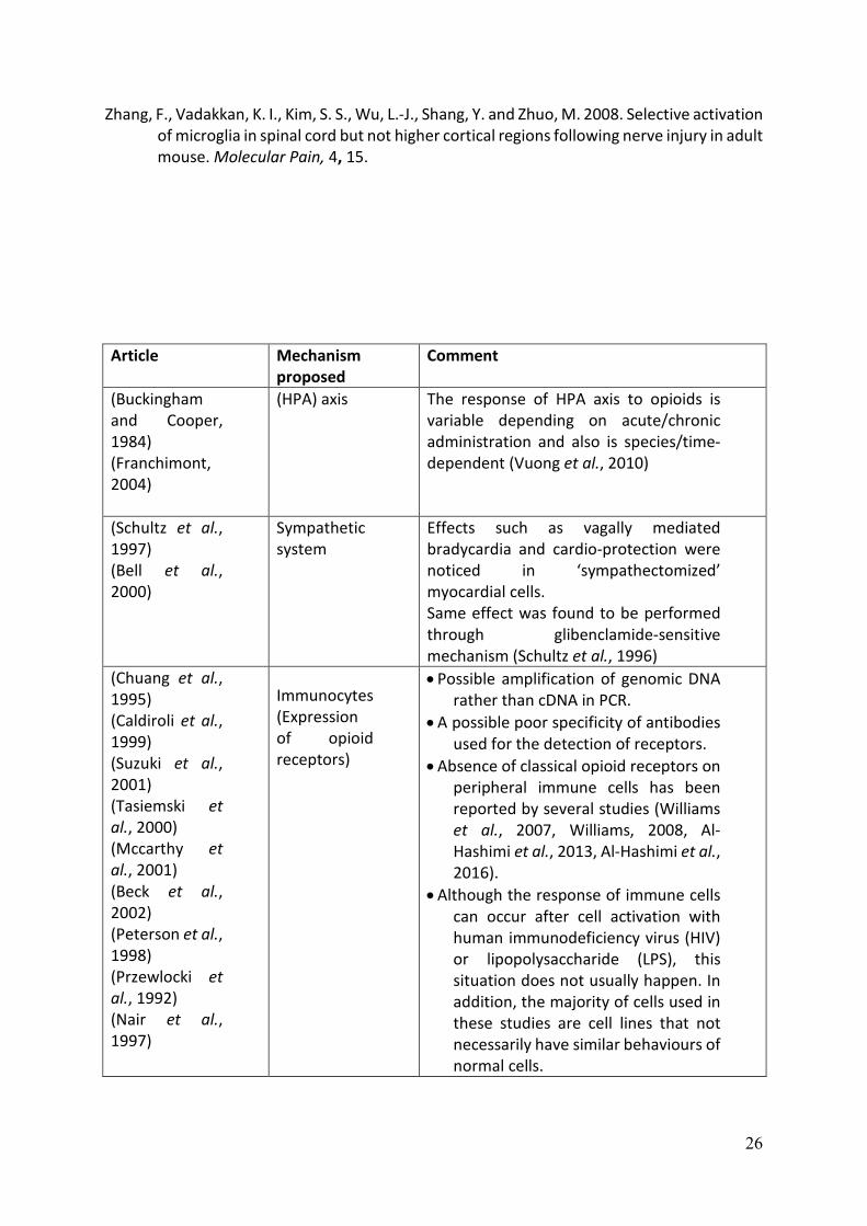
Article Mechanism
proposed Comment
(Buckingham and Cooper, 1984) (Franchimont, 2004)
(HPA) axis The response of HPA axis to opioids is variable depending on acute/chronic administration and also is species/time-dependent (Vuong et al., 2010)
(Schultz et al., 1997) (Bell et al., 2000)
Sympathetic system
Effects such as vagally mediated bradycardia and cardio-protection were noticed in ‘sympathectomized’ myocardial cells. Same effect was found to be performed through glibenclamide-sensitive mechanism (Schultz et al., 1996)
(Chuang et al., 1995) (Caldiroli et al., 1999) (Suzuki et al., 2001) (Tasiemski et al., 2000) (Mccarthy et al., 2001) (Beck et al., 2002) (Peterson et al., 1998) (Przewlocki et al., 1992) (Nair et al., 1997)
Immunocytes (Expression of opioid receptors)
• Possible amplification of genomic DNA rather than cDNA in PCR.
• A possible poor specificity of antibodies used for the detection of receptors.
• Absence of classical opioid receptors on peripheral immune cells has been reported by several studies (Williams et al., 2007, Williams, 2008, Al-Hashimi et al., 2013, Al-Hashimi et al., 2016).
• Although the response of immune cells can occur after cell activation with human immunodeficiency virus (HIV) or lipopolysaccharide (LPS), this situation does not usually happen. In addition, the majority of cells used in these studies are cell lines that not necessarily have similar behaviours of normal cells.
27
(Singhal et al., 1998).
(Steele et al., 2003). (Jessop et al., 2010). (Sharp, 2004)
• Furthermore, Al-Hashimi's team failed to find classical opioid receptors RNA in activated imminocytes as well.
Immunocytes (Naloxone-reversed opioid effect on immune cells)
• The absence of morphine effect in mu-opioid receptors-knock out animals and the immunomodulatory effect of morphine given centrally demonstrate central effects of morphine on the immune system (Gavériaux-Ruff et al., 1998, Roy et al., 1998, Nelson et al., 2000)
• Administration of opioids which do not cross BBB results in less or no immune suppression effect (Shavit et al., 1986, Hernandez et al., 1993).
• The effect of opioids might be related to their interaction with non-opioid receptors such as toll like receptor (TLR4), especially in the presence of findings showing a naloxone-reversed effect of opioids on TLR4 (Hutchinson et al., 2008, Hutchinson et al., 2010)
Table 1: Possible sites/mechanisms by which opioids can affect the peripheral immune system and cause immunomodulation.
28
Figure 1: Glial cell types.
29
Figure 2: Microglial, astroglial and neuronal responses during the progression of neuropathic pain and/or opioid use. A: Microglial activation is the early phase following nerve injury and characterised by classical M1 phenotype features including the release of astrocyte activating cytokines. B: Activation of astrocytes in response to microglial markers released. C: Further (peak) activation of microglia produced in response to activated astrocytes. D: Microglial activation includes the targeting of affected neurons (engulfment of injured neurons). E: The neurons themselves try to counteract the phagocytic and damaging activity of microglia by releasing microglial inhibitors and apoptotic factors. F: a possible event is that microglia switch to the anti-inflammatory M2 phenotype. Alternatively, the microglial activity declines in response to the neuron-induced apoptosis (G) and / or prolonged use of opioids (H).
30
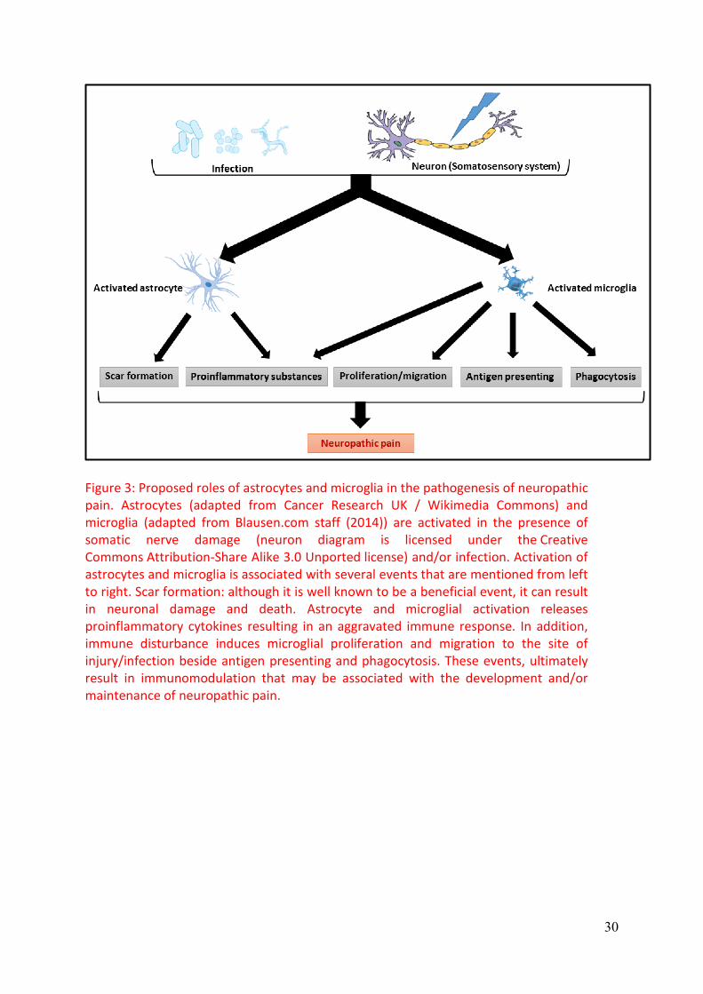
Figure 3: Proposed roles of astrocytes and microglia in the pathogenesis of neuropathic pain. Astrocytes (adapted from Cancer Research UK / Wikimedia Commons) and microglia (adapted from Blausen.com staff (2014)) are activated in the presence of somatic nerve damage (neuron diagram is licensed under the Creative Commons Attribution-Share Alike 3.0 Unported license) and/or infection. Activation of astrocytes and microglia is associated with several events that are mentioned from left to right. Scar formation: although it is well known to be a beneficial event, it can result in neuronal damage and death. Astrocyte and microglial activation releases proinflammatory cytokines resulting in an aggravated immune response. In addition, immune disturbance induces microglial proliferation and migration to the site of injury/infection beside antigen presenting and phagocytosis. These events, ultimately result in immunomodulation that may be associated with the development and/or maintenance of neuropathic pain.