jean-luc banks, ms3 journal club
TRANSCRIPT

Jean-Luc Banks, MS3
Journal Club

Learning Objectives
By the end of this journal club, participants will be able to:• Understand the epidemiology and etiology of developmental dysplasia of the
hip (DDH)
• Recognize the presentation of DDH in infants and children
• Understand the importance of the physical exam combined with imaging in the diagnosis of DDH
• Understand the significance of the alpha angle and acetabular angle in the diagnosis of DDH
• Be familiar with the Graf classification system in the diagnosis of DDH
• Understand the limitations of a single modality of imaging and the importance of follow-up in the diagnosis of DDH

Module Outline
I. Case
II. Background
III. Article Overview
IV. Clinical Questions
V. Key Points

Case Presentation7 week-old female referred to radiology for ”instability of the hips”
PMHx: Born to G1P1 mother at 39 weeks via uncomplicated C-section due to breach positioning. One- and five-minute APGAR scores were 8 and 9 respectively. Mother reports no complications during delivery. Baby girl is breastfed and gaining weight with no other reported health concerns. Mother states that they were referred for “instability in the hips” that was mentioned when the baby was born.
Fam Hx: Mother reports that maternal grandfather, great uncle, and a cousin were braced as children for “congenital hip problems.”

Case Questions
• What physical exam maneuvers do you think were performed to diagnose this “instability in the hip?”
• What imaging studies would you perform for the patient at this time?

2019 2018
Case Imaging - BilateralRadiopedia

2019 2018
Case Imaging – Right Hip
right alpha angle measures: 50 degrees
Radiopedia

2019 2018
Case Imaging – Left Hip
left alpha angle measures: 55 degrees
Radiopedia

Case Questions
• What do these ultrasound findings tell us about the diagnosis of the child?
• What are the imaging study recommendations for a patient with a normal hip ultrasound at 7 weeks? Should they be re-evaluated?
• If we find that a patient has DDH at 7-week ultrasound, what are the next steps in management?

Module Outline
I. Case
II. Background
III. Article Overview
IV. Clinical Questions
V. Key Points

Developmental Dysplasia of the Hip DDH
• Abnormal development of the acetabulum and proximal femur along with mechanical instability of the hip joint
• May affect 1 or both hips, but is more common in the left hip
• Affects 1-34:1000 depending on diagnostic evalulation2
• Risk factors
• First born status
• Female sex
• Breech positioning at > 34 weeks gestation
• Family history of DDH
• Oligohydramnios

Developmental Dysplasia of the Hip DDH• Presentation: Clinical features depend upon age of the child and severity
of the abnormality – from instability in newborn exam, to limited abduction in infant, to asymmetric gait in toddler, to activity related pain in adolescent, to osteoarthritis in adult1
• Etiology: multifactorial condition with several predisposing factors including ligament laxity, breech presentation, postnatal positioning, and primary acetabular dysplasia
• Diagnostic Evaluation:
• Physical Exam
• < 3 months – Ortalani, Barlow, and Galeazzi tests
• > 3 months – limited abduction, thigh-length discrepancy (in unilateral cases) and Galeazzi and Klisic tests
• Walking age children – positive Trendelenburg pelvic tilt test (in unilateral cases)
• Imaging
• < 4 months – ultrasound > 6 months – radiograph

Ortolani and Barlow Maneuvers

Ultrasound Findings and the Alpha Angle
Radiology Assistant
The normal value is greater than or equal to 60 degrees. Less than 60 degrees suggests dysplasia of the acetabulum.

Graf Classification System
Radiology Assistant
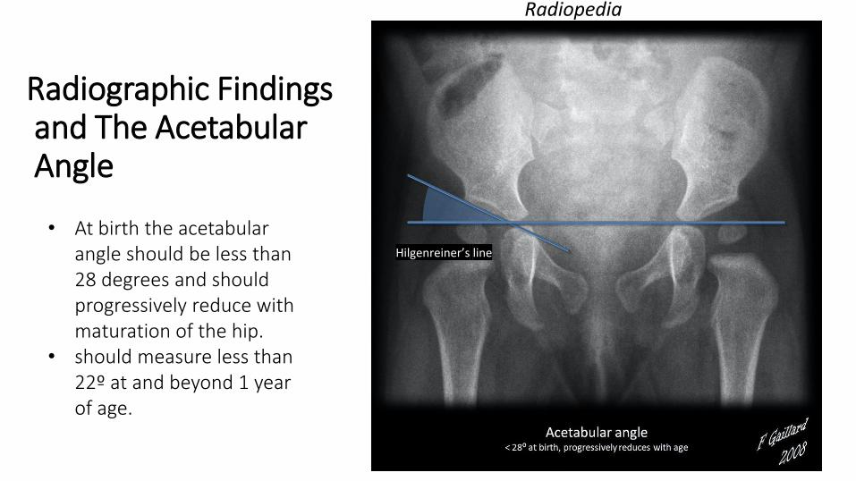
Radiographic Findingsand The AcetabularAngle
Hilgenreiner’s line
• At birth the acetabular angle should be less than 28 degrees and should progressively reduce with maturation of the hip.
• should measure less than 22º at and beyond 1 year of age.
Radiopedia

Module Outline
I. Case
II. Background
III. Article Overview
IV. Clinical Questions
V. Key Points

Article Specifics
Purpose: To evaluate the number of patients in their hospital network with a normal screening ultrasound at 6 weeks with evidence of DDH at the time of radiographic review at 6 months. Secondary aim: to determine the outcomes for these patients.
Journal: Irish Journal of Medical Science, July 2020
Study Type: Retrospective study done to infants undergoing DDH ultrasound screening between January and December 2015.
Number of Cases: 829 patients included for analysis
Data: ultrasound and radiographic reports

Study Cohort• 779 patients presenting after
normal US screening at 6 weeks + 50 patients presenting after normal US screening at 3 months due to initial immature(Graf IIa) hip = 829 patients


Material and Methods
• Retrospective review of patients presenting for DDH ultrasound screening in the study hospital between January-December 2015
• Medical charts and radiographic databases were searched for data including sex, risk factors, and other indications for referral
• Ultrasound and radiograph reports reviewed for findings consistent with DDH• Non-specific reports measurements taken from radiographs, using the PACS
software• An acetabular angle of more than than 27° was considered abnormal
• Radiographic diagnostic cases of DDH underwent logistic regression to determine risk of DDH for gender, presentation at birth, and family history of DDH

Results• 63 patients with abnormal radiographs at 6
months, • overall radiographic pick up rate of 8%
• 5/63 patients lost to follow-up. The rest diagnosed at 6 months were treated in Boston brace with regular follow-up until examination and radiographic parameters normalized and until walking age
• 4 infants had persistent dysplasia by walking age and were referred for tertiary pediatric orthopedic review. Surgery was performed in one patient in the radiograph diagnosed group

Results Continued
• 16 infants had unstable hips at birth, and a further 168 infants were diagnosed as a result of screening • 105 via US (33 at 6 weeks, 72 at 3 months)
• 63 via radiograph at 6 months
• Of the 184 DDH diagnoses for the year, 63 (34%) were diagnosed on radiograph at 6 months.

Results Continued

Results Continued
• Female gender was a strong risk factor for DDH in those with normal ultrasound examinations
• Family history of DDH showed an increased risk for DDH but this was not statistically significant

Discussion
• DDH screening continues to evolve
• Ultrasound techniques and population selection for screening may vary
• Follow up after initial normal ultrasound is not agreed upon internationally
• Patients diagnosed on radiograph at 6 months had milder pathological disease
• In comparison to other studies conducted, these findings reported higher pickup rates on radiograph than others
• Concern for radiation dosages in infants

Study Limitations
• Variability due to multiple reporters of radiographs from different institutions within the hospital network
• Learning curve associated with Graf ultrasonic method
• Retrospective design
• Natural history of DDH still not fully understood making it difficult to develop a universal screening program
• Spectrum of pathology in those with DDH limits generalizability
• No control group for treatment of radiograph-diagnosed group since all patient underwent bracing

ACR Appropriateness Criteria

ACR Appropriateness Criteria Continued

Module Outline
I. Case
II. Background
III. Article Overview
IV. Clinical Questions
V. Key Points

Clinical Questions
• What explains the DDH recognized on pelvic radiograph at 6 months that was not identified on US at 6 weeks?
• Should all infants with normal ultrasound images at 6 weeks be re-evaluated at 6 months of age for DDH?
• How do we adjust these criteria for:• Older children and adults?
• Children born in another country?
• Children who are unable to afford medical care?
• Do we follow ACR criteria at UNC? How might this change as we learn more about this condition?

Key Points• The alpha angle is an important diagnostic criterion for the
evaluation of DDH on ultrasound
• The acetabular angle is an important diagnostic criterion for the evaluation of DDH on radiograph
• Despite initial negative evaluation on ultrasound, DDH can present on 6-month radiograph
• Children who present later with DDH often have a less severe condition
• Current ACR guidelines exist for screening for hip dysplasia in children, but guidelines are not consistent internationally

Guess Who?
It’s Me!
2 months post bilateral femoral osteotomy for bilateral DDH

References
1. Shaw BA, Segal LS, SECTION ON ORTHOPAEDICS. Evaluation and Referral for Developmental Dysplasia of the Hip in Infants. Pediatrics 2016; 138.
2. Noordin S, Umer M, Hafeez K, Nawaz H. Developmental dysplasia of the hip. Orthop Rev (Pavia). 2010;2(2):e19. doi:10.4081/or.2010.e19
3. Lehmann HP, Hinton R, Morello P, Santoli J. Developmental dysplasia of the hip practice guideline: technical report. Committee on Quality Improvement, and Subcommittee on Developmental Dysplasia of the Hip. Pediatrics 2000; 105:E57.
4. American College of Radiology. ACR Appropriateness Criteria. Developmental dylsplasiaof the hip--child. www.acr.org/Quality-Safety/Appropriateness-Criteria
5. Phelan N, Thoren J, Fox C, O’Daly BJ, O’Beirne J (2015) Developmental dysplasia of the hip: incidence and treatment outcomes in the Southeast of Ireland. Ir J Med Sci
6. David M, Robb C, Jawanda S, Bache C, Bradish C. Late recurrence of developmental dysplasia of the hip following Pavlik harness treatment until normal ultrasound appearance. J Orthop. 2014;12(2):81-85. Published 2014 Jan 22. doi:10.1016/j.jor.2014.01.002