issue date - hokudai
TRANSCRIPT

Instructions for use
Title Aseptic Meningitis with Relapsing Polychondritis Mimicking Bacterial Meningitis
Author(s) Yaguchi, Hiroaki; Tsuzaka, Kazufumi; Niino, Masaaki; Yabe, Ichiro; Sasaki, Hidenao
Citation Internal Medicine, 48(20): 1841-1844
Issue Date 2009
Doc URL http://hdl.handle.net/2115/39596
Type article (author version)
File Information RP.pdf
Hokkaido University Collection of Scholarly and Academic Papers : HUSCAP

1
Aseptic meningitis with relapsing polychondritis
mimicking bacterial meningitis
Hiroaki Yaguchia),b), Kazufumi Tsuzakaa), Masaaki Niinob), Ichiro Yabeb)#,
Hidenao Sasakib).
a) Department of Neurology, Kushiro Rosai Hospital, Kushiro, Japan
b) Department of Neurology, Hokkaido University Graduate School of Medicine,
Sapporo, Japan
#Correspondence to Ichiro Yabe
Department of Neurology, Hokkaido University Graduate School of Medicine, N15 W7,
Kita-ku, Sapporo 060-8638, Japan
e-mail; [email protected]

2
Abstract
Relapsing polychondritis (RP) is a rare multisystem autoimmune disease. Though
meningitis in RP is not common, some cases with cerebrospinal fluid (CSF) pleocytosis
of lymphocyte cells have been reported. Of the 18 previously reported cases, two cases
demonstrated pleocytosis of polymorphonuclear leukocytes (PMN) in the CSF. In
addition, the cases whose glucose level in the CSF was decreased also were seen. Our
case also demonstrated pleocytosis of PMN in CSF mimicking bacterial meningitis. In
the clinical field, we cannot get the culture of CNF on the day. We regard the gulucose
level and cellular fraction as impotant. Therefore, we must look upon meningitis in RP as
a differential diagnosis of bacterial meningitis.
Key word: aseptic meningitis, relapsing polychondritis, polymorphonuclear leukocytes,
cerebrospinal fluid (CSF), bacterial meningitis

3
Introduction
Relapsing polychondritis (RP) is an episodic and progressive inflammatory disease of
cartilaginous structures, including the elastic cartilage of the ear and nose, hyaline
cartilage of the peripheral joints, fibrocartilage at axial sites, and cartilage of the
tracheobronchial tree [1]. Multiple neurological abnormalities including meningitis can
occur during the course of RP [2,3]. Recently attracts attention of the neurologist and
many meningitis patients had been reported. Some cases showed pleocytosis with a
predominance of polyphonuclear leukocytes. We must look upon meningitis in RP as a
differential diagnosis of bacterial meningitis .
Case report
A-56-old woman treated with the bronchodilating agent developed an acute onset of
headache, followed by bilateral ear swelling and diplopia. On admission, the patient’s
body temperature was 38.8℃, blood pressure 109/60 mmHg, and respiratory rate 22/min.
Her general examination revealed a saddle nose, bilateral ear swelling and a stenotic
sound of the upper respiratory tract. Laryngotracheal stenosis was observed with a
laryngoscope. Neurological examination revealed neck stiffness and left abducent nerve
palsy. Consciousness disturbance and pathological reflexes were not detected. Deep
tendon reflexes were present and symmetrical. Muscle tonus was normal. A superficial

4
and deep sensory disturbance was not detected. The patient’s cerebellar function and gait
were also normal. There was no muscular atrophy or involuntary movements.
The blood count showed 22,900 leukocytes with 92% polymorphonuclear leukocytes
(PMN), 2,870,000 red cells and 413,000 platelets per cubic millimeter. Serum
electrolytes, creatinine, glucose, coagulation tests, liver functional tests,
lacticodeshydrogenase, and creatine kinase were normal. C-reactive protein (CRP)(24.72
mg/dl) and the erythrocyte sedimentation rate (ESR) (160 mm/h) were elevated. Serum
IgG was 1,685 mg/dl. Laboratory data associated with collagen disease were all within
the normal range. Cultures of blood, urine and sputum were negative.
A CSF study showed 640 cells/mm3 with 94 % PMN, glucose 62 mg/dl (blood glucose
125 mg/dl), protein 114 mg/dl, and IgG index 0.47. The opening pressure was 200
mmH20. Adenosine deaminase (ADA) and angiotensin converting enzyme (ACE) were
within the normal range. Smear for acid-fast bacilli, Gram stain and Indian ink were
negative. In CSF cultures for bacteria, Mycobacterium tuberculosis and cytology were
also negative. And the other 2 times of CSF studies before PSL therapy showed
pleocytosis with a predominance of PML.
Brain MRI, MRA and Nerve conduction studies were normal. EEG demonstrated
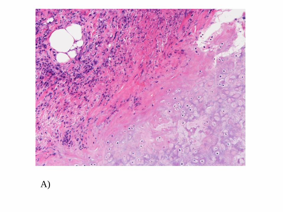
α-actives without a slow wave. A biopsy of the left ear cartilage before predonisone

5
therapy showed inflammation and infiltration of neutropils, plasma cells and
lymphocytes. (Fig. A,B)
We established a diagnosis of aseptic meningitis with RP. One thousand mg/day of
methylprednisolone was given intravenously for three days as a steroid pulse therapy,
followed by oral prednisolone (PSL) at 40 mg/day for four weeks ( which was then
tapered at rate of 10 mg every two weeks). After steroid therapy was initiated, her
symptoms improved considerably.
Two months later, her physical and neurological symptoms had returned to normal.
Serological data demonstrated that WBC, CRP and ESR also returned to normal. On CSF
study, 4 cells/mm3, glucose 67 mg/dl and protein 37 mg/dl were detected.
Two years later following the attack, she was still medicated with oral PSL(7.5 mg/day)
without recurrence.
Discussion
Relapsing polychondritis (RP) is a rare multisystem autoimmune disease of unknown
origin characterized by recurrent episodes of inflammation and a progressive destruction
of cartilaginous tissues. Elastic cartilage of the ears and nose, hyaline cartilage of the
peripheral joints, vertebral fibrocartilage and tracheobronchial cartilage, as well as the
proteoglycan-rich structures of the eye, heart, blood vessels or inner ear may all be
6
affected. In most patients RP manifests as a fluctuating but progressive course which
eventually results in a significant shortening of life expectancy [3]. Central and peripheral
nerve system involvement in RP occurs in approximately 3 % of patients [2]. Various
cranial neuropathies as well as headaches, encephalopathy, seizures, hemiplegia, and
cerebral aneurysms have been reported [2,3]. The diagnosis was usually established by
clinical features, whose criteria are proposed by McAdam et al [1], Daminani et al [4],
and Michet et al [5]. Modifications to the McAdam criteria were made by Daminani et al
due to the variability of clinical manifestations during the course of this disease. This
patient fulfilled both Damiani’s and Michet’s criteria.
In this case, the neurological symptoms were meningitis and abducens paralysis.
Abducens paralysis seemed to have been a cranial neuropathy because it was improved
by administration of PSL and there were no neurological sign of brain stem involvement
or increased intracranial pressure.
Our case showed marked pleocytosis with a predominance of PMN in the CSF. The
meningitis and encephalitis in patients with RP including our case has been noted in 17
cases previously reported in the literature (Table 1) [6-21]. In the initial reported cases,
pleocytosis with a predominance of lymphocytosis in the CSF were described. But in
more recent cases, pleocytosis with a predominance of PMN were reported. Eight cases

7
presented pleocytosis with a predominance of lymphocytosis in the CSF [6-16]. On the
other hand, 3 cases including our case presented pleocytosis with a predominance of
PMN leukocytes were reported [17,18]. In addition, in the 2 cases without our case the
level of glucose in the CSF was decreased, and therapy by immunosuppressors including
PSL made the level of glucose normalized. One case, at the first onset of meningitis,
showed pleocytosis of mononuclear cells in the CSF, but the recurrence showed
meningitis with pleocytosis of PMN in the CSF [19]. In the other case, PMN leukocytes
were nearly half of leukocytes [20]. The efficiency of therapy by immunosuppressors,
including PSL was good without one case[16]. In cases with more inflammation, a
predominance of PMN is observed.
Although pleocytosis of mononuclear cells in the CSF can be seen in meningitis with
most inflammatory diseases, patients with neuro-Behcet’s disease have increased levels
of PMN leukocytes in the CSF on acute exacerbation. In systemic lupus erythematosus
(SLE) patients with aseptic meningitis, significantly higher cell counts with neutorophil
predominance can occur rarely and suggests cerebral vasculitis with ischemia [22]. In
addition, the level of glucose in the CSF is rarely decreased in the neuropsychiatric
manifestation of SLE (NP-SLE), which has been reported at a low incidence between 3 %
and 8 % of patients [22].

8
Thirty percent of RP patients have a complication of autoimmune diseases e.g. Behcet’s
disease, SLE [23]. We speculate that the increase of PMN in the CSF is caused by RP as
well as other inflammatory diseases ( e.g. neuro-Behcet’s disease, SLE) .
One has to be aware that meningitis with RP can also be the differential diagnosis of
bacterial meningitis.

9
References
[1] McAdam LP, O'Hanlan MA, Bluestone R, Pearson CM. Relapsing polychondritis:
prospective study of 23 patients and a review of the literature. Medicine 55: 193-215,
1976.
[2] Letko E, Zafirakis P, Baltatzis S, Voudouri A, Livir-Rallatos C, Foster CS. Relapsing
polychondritis: a clinical review. Semin Arthritis Rheum 31: 384-395, 2002
[3] Gergely P Jr, Poor G. Relapsing polychondritis. Best Pract Res Clin Rheumatol 18:
723-738, 2004
[4] Damiani JM, Levine HL.Relapsing polychondritis--report of ten cases. Laryngoscope
89: 929-946, 1979.
[5] Michet CJ Jr, McKenna CH, Luthra HS, O'Fallon WM. Relapsing polychondritis.
Survival and predictive role of early disease manifestations. Ann Intern Med 104: 74-78,
1986.
[6] Nagashima T, Tanaka H, Ito M, Hirata K, Katayama S, Watanabe K. A case of aseptic
meningitis caused by relapsing polychondritis. Rinsho Shinkeigaku (Clin Neurol) 46:
40-44, 2006. (in Japanese, Abstract in English)
[7] Hsu KC, Wu YR, Lyu RK, Tang LM. Aseptic meningitis and ischemic stroke in
relapsing polychondritis.Clin Rheumatol 25: 265-267, 2006.

10
[8] Brod S, Booss J.Idiopathic CSF pleocytosis in relapsing polychondritis. Neurology
38: 322-323, 1988.
[9] Stewart SS, Ashizawa T, Dudley AW Jr, Goldberg JW, Lidsky MD. Cerebral
vasculitis in relapsing polychondritis. Neurology 38: 150-152, 1988.
[10] Hanslik T, Wechsler B, Piette JC, Vidailhet M, Robin PM, Godeau P. Central nervous
system involvement in relapsing polychondritis.Clin Exp Rheumatol 12: 539-541,1994.
[11] Ohta Y, Nagano I, Niiya D, Fujioka H, et al. Nonparaneoplastic limbic encephalitis
with relapsing polychondritis. J Neurol Sci 220: 85-88, 2004.
[12] Watanabe T, Yasuda Y, Tanaka H, Akiguchi I. Relapsing polychondritis with mental
disorders: a case report. Rinsho Shinkeigaku (Clin Neurol) 37: 243-248, 1997. (in
Japanese, Abstract in English)
[13] Yang SM, Chou CT. Relapsing polychondritis with encephalitis.J Clin Rheumatol
10: 83-85, 2004.
[14] Fujiki F, Tsuboi Y, Hashimoto K, Nakajima M, Yamada T. Non-herpetic limbic
encephalitis associated with relapsing polychondritis. J Neurol Neurosurg Psychiatry.
75:1646-1647, 2004.
[15] Kuwabara M, Shimono T, Toyomasu M, et al. "Prominent ear sign" on
diffusion-weighted magnetic resonance imaging in relapsing polychondritis. Radiat Med.
11
26:438-441, 2008.
[16] Imamura E, Yamashita H, Fukuhara T, Nagashima K, Kohriyama T, Tokinobu H. An
autopsy case of perivasculitic meningoencephalitis associated with relapsing
polychondritis presenting with central nervous system manifestation. Rinsho
Shinkeigaku (Clin Neurol) 49: 172-178, 2009. (in Japanese, Abstract in English)
[17] Ragnaud JM, Tahbaz A, Morlat P, Sire S, Gin H, Aubertin J. Recurrent aseptic
purulent meningitis in a patient with relapsing polychondritis. Clin Infect Dis 22: 374-375,
1996.
[18] Wasserfallen JB, Schaller MD. Unusual rhombencephalitis in relapsing
polychondritis. Ann Rheum Dis 51: 1184, 1992.
[19] Berg AM, Kasznica J, Hopkins P, Simms RW. Relapsing polychondritis and aseptic
meningitis. J Rheumatol 23: 567-569, 1996.
[20] Kothare SV, Chu CC, VanLandingham K, Richards KC, Hosford DA, Radtke RA.
Migratory leptomeningeal inflammation with relapsing polychondritis. Neurology 51:
614-617, 1998.
[21] Fujioka S, Tsuboi Y, Mikasa M, et al. A case of encephalitis lethargica associated
with relapsing polychondritis. Mov Disord
[22] West SG. Systemic lupus erythematosus and the nervous system. in: Duboi’s Lupus
23:2421-2423, 2008.

12
Erythematosus. 6th ed. Wallace DJ, Hahn BH Eds. Philadelpia, Lippincott Williams &
Wilkins, 2001: 708-717.
[23] Gilliland BC. Relapsing polychondritis. in: Harrison's Principles of Internal
Medicine. 16th ed. Kasper DL, Braunwald E, Fauci AS, Hauser SL, Longo D, Jameson JL
Eds. New York, McGraw-Hill, 2004: 2015-2017.
Legends
Table
PMN; polymorphonuclear cells, M; monocytes, L; lympocytes,
PSL; prednizolone, MTX; methotrexate, CyA; ciclosporin, AZP; azathioprine, CY;
cyclophosphamide, (-); not described
*no registry of cellular fraction
Figure
(A) Biopsy sample of the left ear cartilage shows perichondrial inflammation with
neutrophils, plasma cells and lymphocytes (hematoxylin-eosin stain ×10)
(B) Neutrophils in the areas of cartilage destruction (hematoxylin-eosin stain ×40)

Table Summary of relapsing polychondritis cases with meningitis
PMN; polymorphonuclear cells, M; monocytes, L; lymphocytes,
PSL; prednisolone, MTX; methotrexate, CyA; ciclosporin A, CY; cyclophosphamide, AZP;
azathioprine, (-); not describe *no registry of cellular fraction
References Age
(yrs)
and
gender
CSF profile Serum
glucose
(mg/dl)
Treatment Outcome
Leucocytes
(PMN/M/L)
(per mm3)
protein
(mg/dl)
glucose
(mg/dl)
Nagashima et al [6] 65 M 302/ 450 (M+L) 79 65 179 PSL improvement
Hsu et al [7] 71 F 0/ 110 (M+L) 116 57.6 (-) PSL improvement
Brod and Booss [8] 30 F 2/4/31 (-) (-) (-) (-) (-)
75 F 40/0/90 98-140 (-) (-) PSL improvement
Stewart et al [9] 52 M 140/0/200 176 normal (-) none improvement
Hanslik et al [10] 70 F 5/10/22 (-) (-) (-) PSL improvement
Ohta et al [11] 57 M 10/0/58 57 86 (-) PSL improvement
Watanabe et al [12] 60 M 5/0/44 51 normal (-) PSL improvement
Yang et al [13] 49 M 55/4/86 87 54 (-) PSL improvement
Fujiki et al[14] 45M 480(PMN+M)/7520 86 84 99 PSL improvement
62M 4080(PMN+M)/1992
0
46 77 107 PSL improvement
Kuwabara et al[15] 61 M 13/299(M+L) (-) (-) (-) PSL improvement
Imamura et al[16] 70 F 0/73(M) 123 (-) (-) PSL+MTX+
CyA
death
Ragnaud et al [17] 70 F 2340/ 260 (M+L) 40 37.2 (-) PSL improvement
Wasserfallen
and Schaller [18]
73 F 646/0/304 100 39.6 165.6 PSL+CY improvement
Berg et al [19] 60 M
onset 31/9/109 140 22 (-) PSL+AZP improvement
recurre
nce
190/10 (M+L) 80 20 (-) PSL improvement
Kothare et al [20] 66 M 14/4/8 59 68 (-) PSL improvement
Fujioka et al[21] 66 F 90* 147 (-) (-) PSL improvement
Our case 62 F 601/0/38 114 62 125 PSL improvement
A)

B)