isolation of a second form of calsequestrin* - jbc.org · the journal of biological chemistry vol....
TRANSCRIPT

THE JOURNAL OF BIOLOGICAL CHEMISTRY Vol. 249, No. 3, Issue of February 10, pp. 980-984, 1974
Printed in U.S.A.
Isolation of a Second Form of Calsequestrin*
(Received for publication, July 11, 1973)
DAVID H. MACLENNAN
From the Bunting and Best Department of Medical Research, Charles H. Best Institute, University of Toronto, Toronto, Ontario M5G lL6, Canada
SUMMARY
In the sarcoplasmic reticulum of individual rabbits, calse- questrin usually is observed as a single peptide in sodium dodecyl sulfate-polyacrylamide gels (Form 1). In some rabbits calsequestrin (Form 2) is found with a molecular weight 6% less than that of Form 1. In still other rabbits the two forms coexist in equal amounts. Form 2 is not a degradation product of Form 1 since mixing of muscle types before homogenization does not lead to breakdown of Form 1. Sarcoplasmic reticulum containing Form 2 calsequestrin is increased in its capacity for binding and transport of Ca2+. Form 2 calsequestrin has been highly purified and has Ca2+- binding capacity equal to Form 1. Amino acid analyses of Forms 1 and 2 indicate close similarity with differences in the content of tyrosine, cysteine, and methionine. Peptide maps of chymotryptic digests also show similarity but indi- cate two differences in peptide classes. Chicken antibodies against rabbit calsequestrin Form 1 also precipitate calseques- trin Form 2 and precipitin lines in Ouchterlony plates are confluent. Initial genetic studies indicate that inheritance of Form 2 calsequestrin is complex.
In 1971 we described the isolation from sarcoplasmic reticulum of calsequestrin, an acidic protein that binds calcium to the extent of 700 to 900 nmoles per mg with an apparent dissociation constant of 40 PM (1). This protein was believed to be lo- calized on the interior of the sarcoplasmic reticulum and to play a role in sequestering calcium transported to the interior of the vesicle. For further studies of calsequestrin, it became impera- tive to purify the protein on a large scale. However, we re- cently found that our preparations from pooled samples were increasingly contaminated with a protein of molecular weight ‘6% less than that of calsequestrin. In this paper evidence is presented that this protein is a second form of calsequestrin differing from the form already described by a 6% difference in mobility in sodium dodecyl sulfate gels and by a difference in amino acid content but possessing the same Ca2+-binding activity as calsequestrin.
* This research was supported by Grant MT-3399 from the Medical Research Councilof Canada and by a grant from the Muscular Dvstrouhv Association of Canada, This is the seventh paper in thi series “Resolution of Enzymes of Biological Trans- port.”
MATERIALS AND METHODS
Rabbits were obtained from Riemens Fur Ranch, St. Agatha, Ontario-. They were heterozygous strains obtained by crosses of New Zealand rabbits and New Zealand x California strains. Sarcoplasmic reticulum was isolated from individual rabbits or from muscle pooled from two rabbits by the method of i\lac- Lennan (2). Calsequestrin was isolated by the method of MacLennan and Wong (1) except that purification on hydroxyl- apatite preceded passage through Sephadex. Antibodies against calsequestrin were obtained by injecting mature hens intra- muscularly with 500 to 1000 pg of calsequestrin in Freund’s complete adjuvant, three times over a period of 30 days, with bleeding at 37 days.
Calcium binding studies were carried out by dialysis equilib- rium as described by MacLennan and Wong (1). Where indi- cated, 0.1 M KC1 was added to the dialysis media. Ca2+ trans- port was measured by the method of Sommer and Hasselbach (3). Protein was measured by the biuret method of Gornall et al. (4). Disc gel electrophoresis in sodium dodecyl sulfate was performed as described by Weber and Osborn (5). Samples were stained with Coomassie blue and densitometric scans were made with a Joyce-Loebl densitometer. Amino acid analyses were run following digestion in 6 x HCl on a Beckman 120C amino acid analyzer (6). Cysteine and methionine were ana- lyzed after performic oxidation (7). Glucosamine was measured with the amino acid analyzer following a 4.hour digestion in 4 N
HCl at 100”. For chymotryptic digestion 1 mg of calsequestrin and 50 pg of chymotrypsin were incubated in 1 ml of 0.01% NH4HC03 buffer, pH 7.8, for 6 hours at 37”. The samples were lyophilized and 400 pg of peptides were spotted in the center of a 25.cm strip of Whatman No. 3Mhl paper. High volt- age electrophoresis was carried out at 500 volts in pyridine- acetic acid-water (100 :4: 900) pH 6.5. Orange G was the marker for the acidic front.
RESULTS
In the heterozygous strains of rabbits obtained from a com- mercial breeder, we have detected three types of sarcoplasmic reticulum differing in protein composition as determined by their disc gel electrophoretic profiles. Fig. IA shows the usual electrophoretic profile in which proteins are recognizable by their relative mobilities. Fig. 1B is a protein profile of a second sarcoplasmic reticulum type in which calsequestrin was present together with an equivalent amount of a new protein with a greater mobility. Fig. 2B shows a third type of sarcoplasmic reticulum in which calsequestrin was absent and was replaced
980
by guest on August 17, 2019
http://ww
w.jbc.org/
Dow
nloaded from

FIG. 1 (top Zefl). Disc gel electrophoretic profiles of sarco- reticulum with calsequestrin Form 2; C, a mixture of A and B. plasmic reticulum obtained from individual rabbits. A, sarco- plasmic reticulum obtained from individual rabbits. A, sarco- FIG. 3 (bottom left). Disc gel electrophoretic profiles of sarco- FIG. 3 (bottom left). Disc gel electrophoretic profiles of sarco- plasmic reticulum containing Form 1 calsequestrin. R, sarco- plasmic reticulum containing Form 1 calsequestrin. R, sarco- coplasmic reticulum obtained by combining muscle tissue from coplasmic reticulum obtained by combining muscle tissue from plasmic reticulum containing Forms 1 and 2 calsequestrin in plasmic reticulum containing Forms 1 and 2 calsequestrin in two rabbits. two rabbits. A, Forms 1 and 2 in a 1:l ratio; B, Forms 1 and 2 A, Forms 1 and 2 in a 1:l ratio; B, Forms 1 and 2 equal quantity. equal quantity. in a 3:l ratio; C, Forms 1 and 2 in a 1:3 ratio. in a 3:l ratio; C, Forms 1 and 2 in a 1:3 ratio.
FIG. 2 (top right). Disc gel electrophoretic profiles of sarco- FIG. 2 (top right). Disc gel electrophoretic profiles of sarco- FIG. 4 (bottom right). FIG. 4 (bottom right). Disc gel electrophoretic profiles of puri- Disc gel electrophoretic profiles of puri- plasmic reticulum obtained from individual rabbits. A, sarco- plasmic reticulum obtained from individual rabbits. A, sarco- fied calsequestrin. A, Form 1; B, Form 2; C, mixture of Form fied calsequestrin. A, Form 1; B, Form 2; C, mixture of Form plasmic reticulum with calsequestrin Form 1; B, sarcoplasmic plasmic reticulum with calsequestrin Form 1; B, sarcoplasmic 1 and Form 2. 1 and Form 2.
by guest on August 17, 2019
http://ww
w.jbc.org/
Dow
nloaded from
982
by the new protein. Coelectrophoresis of the two types of sarcoplasmic reticulum (Fig. 2C) showed that the two forms of sarcoplasmic reticulum differed only in calsequestrin content. The difference in mobility indicated that the new protein had a molecular weight about 6y0 less than that of calsequestrin. Our
original estimate of the molecular weight of calsequestrin was 44,000. Others have estimated that it is 50,000 (8) and 55,000 (9) and even 63,000 (10). In repeated experiments we have measured molecular weights between 44,000 and 49,000 (11). We therefore suggest that the molecular weight of calsequestrin is 46,500 and the molecular weight of the new protein is 44,000. We have made densitometric scans of stained material in gels and have found that where the 46,500 and 44,000 molecular weight components coexist they are present in identical quanti- tics. Moreover, the sum of the staining material in the two coexisting proteins was approximately equal to the amount of staining material found in the 46,500 or 44,000 molecular weight component existing singly. These observations indicate that the new protein was not found in addition to calsequestrin but replaced it either completely or to the extent of 50%.
The possibility existed that the protein was a breakdown product of calsequestrin. I f calsequestrin were degraded by a cytoplasmic factor in certain rabbits, then mixing of muscle types before homogenization should result in breakdown of calscquestrin in a random fashion. On the other hand, if the two forms of calsequestrin were preformed and stable, then the two proteins should appear in the isolated sarcoplasmic reticu- lum in predictable ratios. If we assign a value of 2 to the nor- mal calsequestrin content, then the normal content of calseques- trin relative to the mutant form would be 2:0. If both forms were.present in the same rabbit the ratio would be 1:1 and if only the mutant form were present the ratio would be 0:2. Com- bining sarcoplasmic reticulum from two rabbits would make the total content of calscquestrin equal to 4. Therefore, if the two forms were present in individual rabbits in the ratios of 2 : 0, 1: 1, and 0:2, then the ratios in the mixtures from any two rabbits could be 4:0, 3:1, 2:2, 1:3, and 0:4. Fig. 3 shows gel electro- phoretic profiles of sarcoplasmic reticulum isolated after com- bining whole muscle tissue from two rabbits. Within the ex- perimental limits of measurement the predicted ratios were the only ones found after isolating sarcoplasmic reticulum from com- bined tissue from two rabbits. Therefore, it is extremely un- likely that the new protein arises from the breakdown of cal- sequestrin through the action of a tissue factor. In addition,
the fact that the two proteins are only found singly or coexisting in exactly equal amounts in individual rabbits is strongly indica- tive that the protein is not a breakdown product but exists in the tissue prior to isolation.
We have homogenized fresh muscle tissue in supernatants from which sarcoplasmic reticulum had been isolated. The eytosol was not found to influence the calsequestrin pattern in the resulting preparations. We would not expect proteases to affect calsequestrin in these membrane preparations since, in a companion paper (12), we have shown that the ATPase protein is the first target of proteolytic digestion and that the ATPase can be quantitatively cleaved by trypsin without affecting cal- sequestrin. In the presence of Ca2+ calsequestrin in the mem- brane is immune to proteolytic digestion. It is, therefore, highly unlikely that calsequestrin would be degraded under conditions which did not affect the molecular weight of the ATfase. The
possibility that one or both forms of calsequestrin are degradation products of the ATPase can be eliminated since digestion of the ATPase does not result in water-soluble products and antibodies
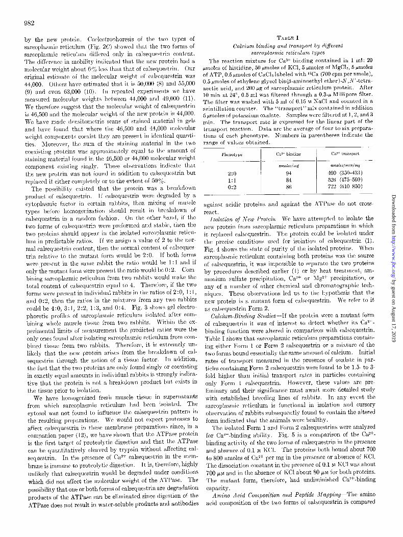
TABLE I Calcium binding and transport by different
sarcoplasmic reticulum types
The reaction mixture for Ca 2+ binding contained in 1 ml: 20 pmoles of histidine, 50 pmoles of KCl, 5 pmoles of &lgClz, 5 pmoles of ATP, 0.5 pmoles of CaClp labeled with 4SCa (700 cpm per nmole), 0.5 pmoles of ethylene glycol bis(p-aminoethyl ether)-N,N’-tetra- acetic acid, and 200 pg of sarcoplasmic reticulum protein. After 10 min at 24”, 0.5 ml was filtered through a 0.3-p lClillipore filter. The filter was washed with 5 ml of 0.15 M NaCl and counted in a scintillation counter. The “transport”mix contained in addition 5wmoles of potassium oxalate. Samples were filtered at 1,2, and 3 min. The transport rate is expressed for the linear part of the transport reaction. Data are the average of four to six prepara- tions of each phenotype. Numbers in parentheses indicate the range of values obtained.
Phenotype
2:o
1:l 0:2
CaZ+ binding ca2+ transport
nmoles/mg moles/min/mg
94 400 (350-433)
84 526 (475560)
86 722 (610-850)
against acidic proteins and against the ATPase do not cross- react.
Isolation of New Protein-We have attempted to isolate the new protein from sarcoplasmic reticulum preparations in which it replaced calsequestrin. The protein could be isolated under the precise conditions used for isolation of calsequestrin (1). Fig. 4 shows the state of purity of the isolated proteins. When sarcoplasmic reticulum containing both proteins was the source of calsequestrin, it was impossible to separate the two proteins by procedures described earlier (1) or by heat treatment, am- monium sulfate precipitation, Ca2+ or Mg2+ precipitation, or any of a number of other chemical and chromatographic tech- niques. These observations led us to the hypothesis that the new protein is a mutant form of calsequestrin. We refer to it as calsequestrin Form 2.
Calcium-Binding Studies-If the protein were a mutant form of calsequestrin it was of interest to detect whether its Ca2+- binding function were altered in comparison with calsequestrin. Table I shows that sarcoplasmic reticulum preparations contain- ing either Form 1 or Form 2 calsequestrin or a mixture of the two forms bound essentially the same amount of calcium. Initial rates of transport measured in the presence of oxalate in par- ticles containing Form 2 calsequestrin were found to be 1.5. to 3- fold higher than initial transport rates in particles containing only Form 1 calsequestrin. However, these values are pre- liminary and their significance must await more detailed study with established breeding lines of rabbits. In any event the sarcoplasmic reticulum is functional in isolation and cursory observation of rabbits subsequently found to contain the altered form indicated that the animals were healthy.
The isolated Form 1 and Form 2 calsequestrins were analyzed for Ca2+-binding ability. Fig. 5 is a comparison of the Ca2+- binding activity of the two forms of calsequestrin in the presence and absence of 0.1 M KCl. The proteins both bound about 700 to 800 nmoles of Ca2+ per mg in the presence or absence of KCl. The dissociation constant in the presence of 0.1 M KC1 was about 700 PM and in the absence of KC1 about 80 PM for both proteins. The mutant form, therefore, had undiminished Ca*+-binding capacity.
Amino Acid Composition and Peptide Mapping-The amino acid composition of the two forms of calsequestrin is compared
by guest on August 17, 2019
http://ww
w.jbc.org/
Dow
nloaded from

983
r 8600 - . z .
3 d600-
:
1 5 10 50 100 500
CALCIUM CONCENTRATION (Mxlo-5)
FIG. 5. Ca2+ binding by purified Forms 1 and 2 calsequestrin. Calcium binding was measured by dialysis equilibrium. Cal- sequestrin, 400 fig, in 0.4 ml was dialyzed for 40 hours at 4” against 100 ml of a solution containing either 5 mM Tris-HCl, pH 7.5, or 5 rnM Tris-HCl and 100 mM KCl, pH 7.5, to which was added 45Ca (3500 cpm per ml) and CaClz in the concentration indicated. 0, n , Form 1; 0, l , Form 2. KCl.
n , 0, represent the presence of
TABLE II
Amino acid composition of two forms of
calsequestrina
Amino acid Amount
Form 1 Form 2
g/100 8
Lysine. . . Histidine. Arginine
Aspartate Threonine Serine.............. .,
Glutamic acid. Proline. Glycine. Alanine.
Cysteic acid Valine Methionine sulfone Isoleucine Leucine
Tyrosine Phenylalanine Glucosamine
7.53 1.66 2.43
19.40 2.44 2.70
21.19
4.35 1.87
4.18 0.58 6.00 1.70 5.18 8.90
2.12 7.75 0.47
7.22 1.72 2.17
18.36 2.75 3.27
21.56 3.99 2.03
4.16 0.35 5.93 0.92 4.96 9.30
3.63 7.68 0.51
Form 1 Form 2
26 6 7
74 11 14 72
20 15
26 3
27 6
20
35 6
24 1
25 6 6
69 12
16 73 18
16 26
2 26
3 19 36 10
23 1
0 Data for calsequestrin Form 1 are from MacLennan and Wong (1). Form 2 data are calculated on the basis of a molecu- lar weight,of 44,000.
in Table II. The proteins have a very similar amino acid con-
tent. There is, however, a notable increase in the content of tyrosine (10 moles per mole in Form 2 versus 6 moles per mole in Form 1). On the other hand, there is a decrease in the content of both cysteine and methionine. There are minor differences throughout but the high content of aspartate and glutamate relative to the content of arginine and lysine remained as the salient feature of the amino acid compositions of the two pro- teins. The glucosamine content of the two forms of calseques- trin was approximately 1 mole per mole. We previously reported the presence of 1 mole of sialic acid per mole of calse-
FIG. 6. High voltage electriihorctic separation of 400 pg of peptides obtained from chymotryptic digests of calsequestrin Forms 1 and 2. The buffer was pyridine-acetic acid-water (100: 4:900), pH 6.5. Electrophoresis on Whatman No. 3MM paper was for 2 hours at 500 volts. Arrowheads indicate peptide classes differing in mobility between the two forms; shaded spots contained tyrosine; dots represent mobility of orange G marker.
questrin (1). Therefore, the carbohydrate content of the two proteins is not different.
The difference in tyrosine composition prompted us to analyze chymotryptic digests of the two forms of the protein and to spray the chromatograms with the Pauly reagent (13) in order to detect tyrosine-containing peptides. Fig. 6 shows peptide maps obtained after electrophoresis of chymotryptic digests of the two forms of calsequestrin. The peptide maps indicate a great similarity between the two proteins. This is even more evident on two dimensional chromatography (not shown). There are, however, two clear differences in the one dimensional chromatograms. Among the acidic peptidcs tyrosine-containing spots moved with different mobilities and among the basic pep- tides a difference in mobility of a nontyrosine-containing peptide class was also observed. All other peptide classes were identical in mobility. The data indicate that Form 2 calsequestrin is smaller in molecular weight and that the difference in molecular weight is accompanied by shifts in amino acid composition and by detectable differences in peptide fragments. We have not, as yet, determined whether the mutation consists of a deletion, an amino acid substitution or both.
Immunological Cross-Reactivity-We have obtained antibodies against Form 1 calsequestrin by injection into chickens. The antibodies against Form 1 calsequestrin also reacted with Form 2 calsequestrin. Fig. 7 shows that precipitin lines were formed in Ouchterlony double diffusion plates when Form 1 antiserum was used against either Form 1 or Form 2 calsequestrin and that the precipitin lines were confluent indicating that the antigenic sites of the two proteins were the same.
Genetic Studies-The calsequestrin mutant does not appear to be functionally altered and it is, therefore, not yet possible to detect the presence of the mutant protein other than by direct isolation. Since the mutation was widely spread in the rabbit population at hand we have made random matings and have killed five to seven littermates in order to determine the com- position of sarcoplasmic reticulum. We have found in a dozen analyses that all rabbits in seven litters were 2:0, all rabbits in three litters were 1: 1 and that the composition of individuals in two litters segregated. A simple prediction would be that those littermates cont,aining calsequestrin Forms 1 and 2 in a 1:l ratio were the result of a cross between rabbits of 2 :0 and 0:2 genotypes. This theory was tested by a second cross between parental types which were likely to be 2:0 (those giving rise only to 2 : 0 siblings) and parents whose genotypes were suspected to be either 2: 0 or 0: 2 (parents giving rise to 1: 1 genotype). In one set of crosses the sire of a 1: 1 litter, when crossed to two dams of 2:0 genotype, sired a 2:0 litter and a litter that segre- gated (Table III). The dam of the 1: 1 litter subsequently be- came infertile and was killed. Her phenotype was found to be
by guest on August 17, 2019
http://ww
w.jbc.org/
Dow
nloaded from

The mammalian membrane mutation which we have described appears to involve a deletion in the protein calsequestrin so that the molecular weight is reduced by 6% and the tyrosine content is increased while the cysteine and methionine contents are de- creased. The Ca*+-binding function of the protein is not af- fected. The mutation is detectable by analysis of the molecular weight of calsequestrin following isolation of the sarcoplasmic reticulum but is not detectable by examination of live animals. The mutant sarcoplasmic reticulum, however, appears to be more active in Ca*f transport than normal sarcoplasmic reticu- lum and, upon obtaining breeding lines of the mutant, it might be possible to detect subtle differences in muscle function. The mutant can provide useful information on the inheritance of extrinsic membrane proteins, on the structure of calsequestrin, and on the function of this protein in the Ca*+ transport system.
FIG. 7. Ouchterlony double diffusion plates. Chicken anti- serum against Form 1 calsequestrin was placed in the center well. Form 1 calsequestrin was in the wells marked 1 and Form 2 was
Acknowledgments-I thank Mr. Vijay Khanna, Mrs. Virginia
in wells marked 2. Videla and Mr. Girish Shah for expert technical assistance, Dr. Cecil Yip for carrying out the amino acid analyses, and
TABLE III Dr. John van der Meer and Dr. Ronald Subden for advice on
Genetic analysis of littermates genetic subjects. The cheerful cooperation of Mr. Don Riemens of Riemens Fur Ranch, St. Agatha, Ontario, is gratefully ac-
Crossing Sire Dam Lit&mat& knowledged.
First crosses P 14 35 1:l (7) REFERENCES 85 68 2:o (7) 43 42 2:0 (6) 1. MACLENNAN, D. H., AND WONG, P. T. S. (1971) Proc. Nat.
58 70 2:o (7) Acad. Sci. U. S. A. 68, 1231
44 51 2:0 (6) 2. MACLENNAN, D. H. (1970) J. BioZ. Chem. 246, 4508 3. SOMMER, J. H., AND HASSELBACH, W. (1967) J. Cell Biol. 34,
902 Second crosses P 14 68 2:0 (6) 4. GORNALL, A. G., BARDAWILL, C. J., AND DAVID, M. M. (1949)
P 14 42 2:o (4) 1:l (2) J. Biol. Chem. 177, 751 44 70 1:l (6) 5. WEBER, K., AND OSBORN, M. (1969) J. Biol. Chem. 244, 4406
6. SPACKMAN, D. H., STEIN, W. H., AND MOORE, S. (1958) Anal.
Phenotype of 35 2:o Chem. 30, 1190 7. MOORE, S. (1963) J. Biol. Chem. 238, 235
a Numbers in parentheses indicate the size of the litters.
9. IKEMOTO, N., BHATNAGAR, G. M., NAGY, B., AND GERGELY, J. 2:0. These results indicate that it is highly probable that in- (1972) J. Biol. Chem. 247, 7835
heritance of the 1: 1 and 0: 2 phenotypes is more complex than 10. MEISSNER, G., CONNOR, G. E., AND FLEISCHER, S. (1973)
was postulated above. A second series of crosses involving dam Biochim. Biophys. Acta 298, 246
number 70 (Table III) ruled out the possibility of maternal 11. OST~ALD, T. J., AND MACLENNAN, D. H. (1974) J. Biol. Chem.
249.974-979 determination of phenotype. Dam 70 bore sequential litters 12. STEWART, P. S., AND MACLENNAN, D. H. (1974) J. Biol. Chem. of 2:0 and 1 :l genotypes. 249,985-993
13. EASLEY, C. W. (1965) Biochim. Biophys. Acfa 107,38G
DISCUSSION 14. LIN, E. C. C. (1970) Annu. Rev. Genet. 4,225 15.
Biochemical genetics has proven to be one of the most useful approaches to the elucidation of transport systems in micro-
TILL, J. E., BAKER, R. M., BRUNETTE, D. M., LING, V., THOMPSON, L. H., AND WRIGHT, J. A. (1973) Fed. Proc. 32, 29
984
organisms (14) and applications of similar techniques to the study of mammalian transport systems is clearly a worthwhile objective. Mammalian cells have been selected for diminished sensitivity to ouabain, an inhibitor of Na+ and K+ transport, and these cells may prove useful in genetic studies utilizing so- matic cell hybrids (15).
8. MARTONOSI, A., AND HALPIN, It. A. (1971) Arch. Biochem. Biophys. 144, 66
by guest on August 17, 2019
http://ww
w.jbc.org/
Dow
nloaded from

David H. MacLennanIsolation of a Second Form of Calsequestrin
1974, 249:980-984.J. Biol. Chem.
http://www.jbc.org/content/249/3/980Access the most updated version of this article at
Alerts:
When a correction for this article is posted•
When this article is cited•
to choose from all of JBC's e-mail alertsClick here
http://www.jbc.org/content/249/3/980.full.html#ref-list-1
This article cites 0 references, 0 of which can be accessed free at
by guest on August 17, 2019
http://ww
w.jbc.org/
Dow
nloaded from