isolation and characterization of chinese hamster ovary
TRANSCRIPT

Isolation and Characterization of Chinese Hamster Ovary Cell Mutants Defective in Intracellular Low Density Lipoprotein-CholesterolTrafficking Kenne th M. Cadigan ,* Diane M. Spillane, and Ta-Yuan C h a n g
Department of Biochemistry, Dartmouth Medical School, Hanover, New Hampshire 03756
Abstract. This paper reports the isolation and charac- terization of Chinese hamster ovary cell mutants defec- tive in low density lipoprotein (LDL)-cholesterol trafficking. The parental cell line was 25-RA, which possesses LDL receptors and various cholesterogenic enzyme activities that are partially resistant to down regulation by exogenous sterols (Chang, T. Y., and J. S. Limanek. 1980. J. Biol. Chem. 255:7787-7795). Because these cells accumulate a large amount of in- tracellular cholesteryl ester when grown in medium containing 10% fetal calf serum, mutagenized popula- tions of 25-RA cells were grown in the presence of a specific inhibitor of acyl-coenzyme A: cholesterol acyltransferase (ACAT), which depleted their cholesteryl ester Stores. Without this cholesterol ester storage, 99 % of 25-RA cells die after 5-d growth in cholesterol starvation medium, while the mutant cells, which accumulate free cholesterol intracellularly, sur- vived. In two mutant clones chosen for characteriza- tion, activation of cholesteryl ester synthesis by LDL
was markedly reduced in the mutant cells compared with 25-RA cells. This lack of activation of choles- terol ester synthesis in the mutant cells could not be explained by defective uptake and/or processing of LDL or by a decreased amount of ACAT, as deter- mined by in vitro enzyme activity. Mutant cells grown in the presence of LDL contain numerous cytosolic particles that stain intensely with the fluorescent com- pound acridine orange, suggesting that they are acidic. The particles are also stained with filipin, a choles- terol-specific fluorescent dye. Indirect immunofluores- cence with a monoclonal antibody specific for a lysosomal/endosomal fraction revealed a staining pat- tern that colocalized with the filipin signal. The mu- tant phenotype was recessive. The available evidence indicates that the mutant cells can take up and process LDL normally, but the hydrolyzed cholesterol accumu- lates in an acidic compartment, probably the lyso- somes, where it can not be transported to its normal intracellular destinations.
I s mammalian cells, the enzyme responsible for intra- cellular cholesteryl ester synthesis, acylcoenzyme A: cholesterol acyltransferase (ACAT) ~ uses cholesterol
and fatty acyl coenzyme A as substrates (18). In rat liver, ACAT has been localized to the rough endoplasmic reticulum (4, 30). In Chinese hamster ovary (CHO) cells and primary cultures of human fibroblasts, ACAT activity is highly regu- lated by exogenous sources of cholesterol, such as low den- sity lipoprotein (LDL) (16, 23, 26, 35), and by endogenous cholesterol synthesis (15). All available evidence suggests that the enzyme is regulated by cholesterol at the posttransla- tional level (10, 16, 23, 24, 26). The major mechanism of regulation is probably the supply of the substrate cholesterol. In human fibroblasts, it has been shown that in order for LDL to exert its regulatory effects (i.e., down regulation of LDL receptors and cholesterol biosynthesis and activation of ACAT activity), the cholesteryl ester moiety of the liproprotein, which forms the majority of the LDL cholesterol, must be
1. Abbreviations used in this paper: ACAT, aeyl-coenzyme A: cholesterol acyltransferase; CT, cholesterol trafficking; DIC, differential interference contrast; LDL, low density lipoprotein; NP-C, Niemann-Pick, type C.
hydrolyzed by a specific acid lipase found in the lysosomes (26). Presumably, the free cholesterol then leaves the lyso- some, and in the case of ACAT, activates the enzyme by providing an increased amount of cholesterol as substrate.
The mechanism of transfer of the liberated cholesterol from lysosomes to the ACAT enzyme is unknown. There are cellular proteins capable of transferring hydrophobic mole- cules between membranes in vitro. One of these proteins, sterol carrier protein2, has been demonstrated to activate ACAT activity in vitro by stimulating the transfer of ex- ogenously added cholesterol to the ACAT enzyme (25, 48). However, the physiological role of this protein in cholesteryl ester synthesis has not been established.
Obtaining mutants in which cholesterol movement is defective would provide a valuable tool for defining intracel- lular cholesterol transport pathways and serve as a first step towards identifying the factors involved. It has been shown that fibroblasts isolated from patients with Niemann-Pick type C (NP-C) disease accumulate free cholesterol when in- cubated with LDL (36, 44). These fibroblasts bind and inter- nalize LDL at normal rates, and the LDL cholesteryl ester is hydrolyzed to free cholesterol. However, the normal
© The Rockefeller University Press, 0021-9525/90/02/295/14 $2.00 The Journal of Cell Biology, Volume 110, February 1990 295-308 295
Dow
nloaded from http://rupress.org/jcb/article-pdf/110/2/295/1059055/295.pdf by guest on 03 April 2022

regulatory responses elicted by LDL were much slower com- pared with unaffected fibroblasts (37, 45). This has been shown to be caused by a decrease in the rate of movement of LDL-derived cholesterol out of the NP-C lysosomes (38). Double fluorescence studies with the cholesterol-specific stain filipin and a monoclonal antibody directed against a lysosomal membrane antigen have demonstrated colocaliza- tion of the accumulated intracellular cholesterol with lyso- somes (7, 54). In addition, a strain of BALB/c mice with a neuropathological condition similar to NP-C disease has been described in which free cholesterol accumulates in the lysosomes (5, 40, 42, 52).
In this report, a highly efficient selection protocol for isolating CHO cell mutants resistant to cholesterol starvation is described. Biochemical analysis showed that the mutants possess a phenotype similar to the one described for NP-C fibroblasts (i.e., lack of activation of ACAT by LDL despite an intact LDL receptor pathway, and an accumulation of free cholesterol upon growth in medium containing LDL). Cell hybridization analysis demonstrated that the phenotype of the two mutant clones examined was recessive and that they belonged to the same complementation group. Whether the genetic locus affecting these CHO cell mutants is the same as the one(s) corresponding to the NP-C disease awaits fur- ther genetic analysis. These mutants should serve as unique tools for cloning and identifying the gene affected via DNA mediated gene transfer (1).
Materials and Methods
Reagents [10-3H]Oleic acid and ll,2,6,7-3H]chnlesteryl linoleate were purchased from Du Font Company-Biotechnoiogy Systems (Wilmington, DE). Oleyl coenzyme A was synthesized as described by Stadtman using oleyl anhy- dride (55) and [3H]oleyl cocnzyme A was synthesized by another method (6). Both preparations were quantitated as previously described (11, 12). Nal25I (carrier free in 0.1 N NaOH) and Ha [l-14C] acetate were from Amersham Corp. (Arlington Heights, IL). Compound 58-035 (3-[decyldi- methylsilyl-N-[2-(4-methylphenyl)-l-phenylethyi] propanamide) was pro- vided by Dr. John Heider (Sandoz Inc., East Hanover, NJ). Melvinolin was a girl from Alfred Alberts (Merck, Rahway, NJ). 25-hydroxycholesterol was purchased from Steraloids Inc. (Wilton, NH), dextran sulfate from Pharma- cia Inc. (Piscataway, NJ), and sodium taurocholate from Calbiochem Corp. (La Jolla, CA). Polyethylene glycol 4000 was obtained from EM Science (Cherry Hill, NJ). Chloroform, methanol, and isopropanol used for the cholesterol analysis were from Mallinckrodt Inc. (Paris, KY) or Fisher Scientific Co. (Pittsburgh, PA) and were nanograde and spectranalyzed grade, respectively. FrTC-conjugated goat anti-mouse antibody was ob- tained from Cappeii Laboratories (Malveru, PA) (No. 1211-0081). The anti- body lgp58 was generously provided by Dr. Ira Mellman (Yale University School of Medicine, Department of Cell Biology, New Havea, CT). MOPC- 21 antibody was a gift of Dr. Stanley C. Froehner (Department of Biochem- istry, Dartmouth Medical School, Hanover, NH). Cholesterol oxidase was generously provided by Dr. Albert Chen (Beckman Instruments, Inc., Fullerton, CA). Horseradish peroxidase (P-6140), cholesterol esterase (C- 1892), and phosphatidylcholine Type XI (P-2772, lot 67F-8410) and all other enzymes and biochemical reagents were from Sigma Chemical Co. (St. Louis, MO). Other organic solvents and chemicals were from Fisher Scientific Co. and were of reagent grade quality.
Cell Culture All CHO cell lines described in this paper were grown as monolayers in tissue culture flasks or dishes purchased from Costar (Data Packaging Corp., Cambridge, MA) or Falcon (Becton, Dickinson & Co., Lincoln Park, NJ). During the mutant selections all cells were maintained in F-12 medium minus linoleic acid supplemented with antibiotics as previously de-
scribed (11) plus 10% FCS (Sigma Chemical Co.). When delipidated FCS was used, it was prepared according to a published procedure (14), as modified by Chin and Chang (22). After the mutant selections, all of the CHO cell lines used in this report were maintained on a 1:1 mixture of the above mentioned F-12 medium and MEM (Gibco Laboratories, Grand Is- land, NY) supplemented with 2 mM glutamine and antibiotics, plus 10% bovine calf serum (Hyclone Laboratories, Sterile Systems, Inc., Logan UT). Delipidated bovine calf serum was made using Cab-o-sil (Eastman Kodak Co., Rochester, NY) as previously described (37).
In this report (with the exceptions of Figs. 2 and 6), all experiments were performed with CHO cells that had been adapted to continuous growth in 10% delipidated bovine calf serum in the F-12/MEM mixture. The adapta- tion (which took only one passage) and all subsequent platings were only successful when using tissue culture flasks or dishes from Falcon Labware. Upon plating of cells into the delipidated serum medium for the first time, most of the cells attached but looked unhealthy and grew very slowly. After a few days, however, some of the cells (,~,10%) began to grow more rapidly. When these cultures reached confluency, they could be plated into fresh flasks where they attached, appeared healthy, and grew rapidly.
Originally cultures were maintained in 10% delipidated serum. However, it was observed that occasionally the monolayers tended to clump together in patches. We subsequently found the clumping to occur less frequently when the concentration of delipidated serum was changed to 5 %. None of the data shown in this report were obtained from cultures with evident clumping. The human LDL (d -- 1.019-1.063 g/mi) used in this report was prepared from plasma by sequential flotation in the presence of protease in- hibitors as previously described (11).
Mutagenesis and Isolation of Mutants The CHO cell clone 25-RA and the ACAT mutant AC29 were mutagenized with 125 ~tg/ml N-nitroso-N-ethyhrea as previously described (11). 5 d after mutagenesis, AC29 ceils were plated at a low density (1.5 × 10⁵ cells per 20 × 100 mm dishes) in F-12 medium plus 10% FCS. Because large amounts of intracellular cholesterol ester interfered with the selection (see Results), mutagenized 25-RA cells were grown for 3 d in 10% FCS medium that contained the specific ACAT inhibitor, 58-035 (50) and then plated at low density in 10% FCS medium plus 58-035. The concentration of 58-035 used (200 ng/ml) inhibits cholesteryi ester synthesis >98% (11). 36-48 h after plating, the monolayers were washed twice with PBS and cultured in cholesterol starvation medium (F-12 medium plus 10% delipidated FCS, 35 /~M oleic acid, 50 #M mevinolin, and 230 #M mevalonate). Mevinolin, a competitive inhibitor of 3-hydroxy-3-methylglutaryl-coenzyme A reductase (2), blocks synthesis of mevalonate and thus inhibits synthesis of steroidal and nonsteroidal isoprenoids. The small amount of mevalonate permits the synthesis of nonsteroidal isoprenoids (51). Medium changes were given on the 2nd, 3rd, and 4th d of growth in the cholesterol starvation medium. Af- ter 5-6 d almost all of the mutagenized 25-RA cells were dead or dying. At this time, the starved cultures were washed once with PBS and grown in 10% FCS medium plus 230 ~tM mevaionate. After an additional 5 d of growth, the largest colonies of surviving cells were located and potential mutants were identified by their distinct morphological appearance (see Results). Colonies were removed using cloning rings and then recloned by limiting dilution in 10% FCS medium. Sometimes, depending on the amount of nonmutant cell contamination in the populations removed with the cloning ring, it was necessary to subject the cells to another round of starvation selection before recloning.
ACAT Assays Monnlayers were pulsed with a [3H]oleate/BSA solution and analyzed for incorporation of radiolabel into cholesteryi oleate as previously described (11, 12, 16). For the in vitro ACAT assays, cell homogenates were prepared by the hypotonic shock and scraping method (20) and used immediately. The microsomal assay has been described in detail previously (11, 16, 23). The reconstituted ACAT assay was performed as described in Cadigan and Chang (10).
LDL Pathway A nalysis LDL was iodinated using iodine monochloride as described by Goldstein et al. (29). After incubation of the monolayers with the indicated amounts of 1251-LDL, the cells were assayed for dextran sulfate releasable and cell- associated t25I-lipoprotein, and the medium was analyzed for ~25IoLDL degradation as previously described (11). Specific binding and cell uptake were calculated by subtracting the nonspecific value, determined by assay-
The Journal of Cell Biology, Volume 110, 1990 296
Dow
nloaded from http://rupress.org/jcb/article-pdf/110/2/295/1059055/295.pdf by guest on 03 April 2022

ing in the presence of 50-fold excess of unlabeled LDL (up to 500 #g/ml), from the total value (assayed with t25I-LDL alone). The nonspecific values ranged between 12 and 25% of the total value.
For measurement of the hydrolysis and reesterification of LDL-derived cholesteryl linoleate, LDL was labeled with [1,2,6,7-3H] cholesteryl iinole- ate by the method of Roberts et al. (49), using the lipoprotein free fraction (d >1.215) of fresh human serum as the source of cholesterol ester exchange protein. After incubation with medium containing the 3H-cholesteryl linoleate-LDL, the cells were harvested in 0.2 M NaOH, neutralized with HCI, and the lipids extracted exactly as previously described for the [3H]oleate poise (11, 16). Unlabeled cholesterol, cholesteryi oleate and cholesteryl linoleate were added as carriers. The extracted lipids were sepa- rated from one another essentially as described by Goldstein et al. (28). Briefly, the extracted lipids were spotted in 80 #1 ethyl acetate onto Silica G TLC plates (Analteeh Inc., Newark, DE) impregnated with AgNO3. The TLC plates were impregnated by dipping into a 6.7% solution (metha- nol/H20; 2:1) of AgNO3 for 2 rain followed by air drying for 30 rain. The plates were then baked at 80"C for 1 h, cooled, and spotted. Plates were run in a benzene/hexane (1:1) solvent system, allowed to air dry overnight, sprayed with dichlorofluorescine (0.2% solution in ethanol), air dried, and visualized with short wave ultraviolet light to localize the relevant lipid spe- cies. The Rf values were: cholesterol ,~0.1; cholesteryl linoleate '~0.3; and cholesteryl oleate ,~.6, respectively. Bands were scraped and counted in the presence of Betafluor scintillation fluid. An unidentified radioactive band (Rf 'x,0.75) was also found; its location on the plate made it likely that it was a cholesteryl ester with a saturated fatty acid moiety (28) and it was '~15% of the cpm found in the cholesterol oleate band. Since the ratio of the unknown band's cpm to the cholesterol oleate cpm remained roughly constant throughout the different conditions and different cell types, it was not included in the calculations of the data. Control experiments revealed an average recovery of 60% for cholesterol, cholesteryl oleate, and cholesteryl linoleate after extraction and TLC analysis.
Microscopy Cells were grown on glass coverslips at a density of 5 x 10⁴ cells per well in six well plates containing 1.5 mi of medium. It was found that the cells did not plate well on coverslips in medium containing delipidated bovine calf serum alone and it was necessary to plate in medium containing 0.5% bovine calf serum/4.5 % delipidated bovine calf serum. This concentration of bovine calf serum was not enough to induce the lipoprotein-dependent morphological changes described in this paper. I d after plating, the cultures were washed two times with PBS and changed to medium containing 5 % delipidated serum supplemented with 100 ttg/ml LDL (sometimes with 200 ng/ml 58-035) or washed medium containing only delipidated serum. A medium change was given 2 d after plating. On day 3, coverslips were stained as indicated, mounted, and kept moist with Hanks balanced salt so- lution containing no phenol red. Immediately thereafter, the cells were viewed using a Zeiss universal microscope with either a 67x achromat oil immersion (Carl Zeiss, Inc., Thoruwood, NY) phase-contrast objective with differential interference contrast (DIC) optics or a 40X achrornat water immersion (Cart Zeiss, Inc.) phase-contrast objective. All micrographs were taken with TMAX-400 film (Eastman Kodak Co.) and developed ac- cording to instructions provided by the manufacturer.
Acridine orange staining was carried out according to Poole (47). The monolayers were incubated for 30 min in the appropriate media containing 10 tLg/ml acridine orange. Cells were then incubated for 15 min in medium lacking acridine orange before mounting the coverslips as described above. During the staining process, cells were kept in a 37°C, 5 % CO2 incubator. A 1 mg/ml stoek solution of acridine orange in DMSO was made fresh each time before use and diluted 100-fold into media. Acridine orange fluores- cence was viewed with rhodamine fluorescence optics (No. 48 77 15; Carl Zeiss, Inc.).
For the filipin staining, cells were washed four times with PBS and fixed with 4 ml of 10% PBS formalin at room temperature for 1 h, rinsed four times with PBS, and stained with 1.5 ml of filipin solution for 1 h at room temperature (36) before washing and mounting for microscopy. The filipin solution was made fresh each time by dissolving 2.5 nag filipin in 1 ml dimethylformamide and then diluting into 50 ml of PBS. The fixed cells were examined with a UV filter package (No. 48 77 02; Carl Zeiss, Inc.).
When staining with the lysosomal/endosomal antibody, lgp58, cells were first stained with filipin as described above, lgp58 at 50--150 ~g/ml was diluted 50-fold into goat serum, spun in a microfuge for 30 s, and diluted another 50-fold into a 0.1% solution of saponin in PBS. This solution was used to label cells for 30 min at room temperature. Cells were washed exten- sively before labeling with an FITC-conjugated goat anti-mouse second an-
tibody solution for 30 min at room temperature. The FITC-conjugated anti- body was diluted 500-fold into goat serum, spun in a microfuge for 30 s, and then diluted another 500-fold into 0.1% saponin in PBS before adding to the cells. Cells were rinsed with PBS and mounted for microscopy. Im- munofluorescence was examined with FITC-fluorescence optics (No. 48 77 17; Carl Zeiss, Inc.). In control experiments, cells were labeled with lgG secreted by the myeloma cell line MOPC-21 at a similar concentfntion as with the antibody lgp58.
Other Analytical Procedures Cellular cholesterol was determined using the fluorometric assay of Heider and Boyett (31) as described previously (12).: Cholesteryl ester was calcu- lated by subtracting the free cholesterol value from the total cholesterol value (determined after incubation of cellular lipids with cholesterol ester- ase). Cell hybridizations (11) and the [14Clacetate pulse (11, 19) were per- formed as described previously. All protein determinations in this paper were made using the Peterson modification (46) of the method of Lowry et al. (39); TCA precipitation was performed only when determining the pro- tein content of the cell extracts used for the in vitro ACAT assays.
Results
Isolation of Cholesterol Starvation Resistant Mutants
The rationale for the ability of certain CHO cell mutants to survive cholesterol starvation is depicted in Fig. 1. The cho- lesterol starvation medium consisted of 10% delipidated FCS plus 50 ~M mevinolin (to inhibit endogenous choles- terol synthesis) and 230/~M mevalonate (to permit the syn- thesis of nonsteroidal isoprenoids essential for cell growth; see Materials and Methods). When grown in 10% FCS medium, 25-RA cells have previously been demonstrated to contain large amounts of cytosolic cholesterol ester droplets (11, 17). During cholesterol starvation, the hydrolysis of this cholesteryl ester apparently served as a reservoir of cellular cholesterol for the 25-RA cells, enabling them to survive (Fig.l a). As shown in Fig. 2 b, 25-RA cells were able to grow for at least 5 d in the cholesterol starvation medium. In contrast, 25-RA cells treated with the specific ACAT in- hibitor 58-035 (or ACAT deficient mutants derived from 25-RA cells) contain very little cholesteryl ester (11). Conse- quently, almost all of these cells die during cholesterol star- vation (Fig. 1 b and Fig. 2 b). The same result using the ACAT deficient mutant AC29 was found (data not shown).
This cholesterol starvation procedure was originally de- signed for the isolation of revertants of the ACAT deficient mutants. However, these attempts were not successful be- cause of the high frequency of a novel class of mutants that were resistant to cholesterol starvation. Grown in 10% FCS, these mutants (designated as cholesterol trafficking, or CT mutants) possess intracellular particles that are distinct from the cholesteryl ester droplets seen in 25-RA cells. As will be demonstrated in the following sections, the CT mutants ac- cumulate a large amount of unesterified cholesterol when grown in medium containing 10% FCS or LDL. The storage of free cholesterol in a distinct cellular component enabled the CT mutants to survive the cholesterol starvation selection (Fig. 1 c). Three independent CT clones were isolated from three dishes (4.5 x 10⁵ total cells) of mutagenized popula- tions of AC29 and 11 independent clones possessing the CT phenotype were isolated from 11 dishes (16.5 x 10⁵ total cells) of mutagenized populations of 25-RA cells (pretreated with 58-035 to remove intracellular cholesteryl esters). No CT mutants were found in four dishes (6 x 10⁵ total cells)
Cadigan et al. ClIO Cells Defective in Cholesterol Trafficking 297
Dow
nloaded from http://rupress.org/jcb/article-pdf/110/2/295/1059055/295.pdf by guest on 03 April 2022

10% Cholesterol FCS Starvat ion Medium
3 - 4 Days 5 - 6 Days
a(9 ,0
o@
~- Survival
Death
.~ Survival
F ) PM • ~ .J ) "~ Ester Cholesterol • Cholesteryl Intracellular
= Cholesterol
Figure 1. Depiction of 25-RA cells (a), ACAT- deficient mutants from 25-RA, or 25-RA cells pretreated with 58-035, a specific ACAT inhibi- tor (b), and CT mutants treated with 58-035 (c) during cholesterol starvation. After growth in 10% FCS medium, 25-RA cells contain a large amount of intracellular cholesteryl esters, while ACAT-deticient mutants or 25-RA cells treated with 58-035 contain very little. During cholesterol starvation, the cholesteryl esters in 25-RA hydrolyzed to free cholesterol can maintain a sufficient level of cholesterol for survival, Because they lack stored cholesterol, the ACAT-deficient mutants and 58-035-treated 25-RA ceils die in starvation medium. When grown in 10% FCS medium the CT mutants contain a high level of intracellular free choles- terol. This stored cholesterol pool apparently provides the mutants with enough cholesterol to survive the starvation. The ACAT inhibitor has no effect on the accumulation of this free cholesterol in the CT mutants.
of unmutagenized 25-RA cells. The biochemical character- ization of two cholesterol trafficking clones derived from 25- RA cells, CT52 and CT60, is described in this report.
As shown in Fig. 2 a, CT52 and CT60 pretreated with 58- 035 grow at comparable rates to 58-035 treated 25-RA in 10% delipidated serum, but unlike 25-RA they also grow in delipidated serum plus mevinolin and mevalonate (i.e., star- vation medium) (Fig. 2 b). These cell lines also retain resis- tance to the cytotoxic effects of 25-hydroxycholesterol (data not shown), a hallmark of 25-RA (19) and the 25-RA derived ACAT mutants (11).
As mentioned above, CT52 and CT60 have a distinct mor- phological appearance when grown in 10% FCS medium. The appearance of the various cell lines viewed with DIC op- tics is illustrated in Fig. 3. In delipidated serum (Fig. 3, a, d, and g) all three cell types had a similar appearance. When grown in delipidated serum plus 100/~g/ml LDL for 48 h, 25-RA (Fig. 3 b) accumulated perinuclear droplets. These particles were similar to the ones previously observed in 25- RA grown in FCS medium and have been identified as cho- lesteryl esters (11, 12). CT52 (Fig. 3 e) and CT60 (Fig. 3 h) ceils grown in LDL also have intracellular particles, which, unlike the droplets of 25-RA, extended beyond the perinu- clear area. Using the DIC microscopy, the particles in both cell types appeared similar. However, if the cells were grown in LDL plus 58-035, the particles in 25-RA (Fig. 3 c) disap- peared completely, while only a minor reduction occurred in the amount of particles in CT52 (Fig. 3 f ) and CT60 (Fig. 3 i). This indicates that the majority of particles in the CT mutants are not intracellular cholesteryl ester droplets de- rived as a product of the ACAT reaction.
Cholesteryl Ester Synthesis and Cell Hybridization Analysis
In pilot experiments (data not shown), it was found that the rate of cholesteryl ester synthesis in 25-RA cells grown in delipidated serum did not reach the low levels previously ob- served in this laboratory for wild-type CHO cells (16, 23). This was probably due to the high rate of endogenous choles- terol biosynthesis in 25-RA cells compared with wild-type
cells (11, 19). The addition of 10 #M mevinolin to the medium for 4 h before the pulse experiment using [3H]ole- ate reduced [3H]cholesteryl ester synthesis from a rate of 12.7 :t: 1.6 pmol/min/mg to 2.9 + 1.1 pmol/min/mg (data not shown). Therefore, all the experiments described in this paper used media containing 10 #M mevinolin (as well as 230 #M mevalonate) for the indicated times.
As shown in Fig. 4 a, CT52 and CT60 cells have a greatly reduced activation of cholesteryl ester synthesis compared to 25-RA cells as measured by [3H]oleate pulse after 6 h of in-
._c
o
0
600 300
500
400
300
200
1QO
J i i , i i 0 0 1 2 3 4 5 6
b
0 1 2 3 4 5 6
Time (days) Time (days)
250
200
150
100
50
0
Figure 2. Growth of 25-RA (solid circles), CT52 (open circles), and CT60 (open squares) cells in delipidated serum medium with or without an inhibitor of cholesterol biosynthesis. 2.5 x 10⁴ cells were plated in 9-cm 2 plates containing F-12 medium + 10% FCS and 200 ng/ml 58-035. After 48 h, the cultures were washed twice with sterile PBS and grown in F-12 medium supplemented with 10% delipidated FCS (a) or 10% delipidated FCS + 50 ttM mevinolin and 230 #M mevalonate (b). Growth of 25-RA cells without the 584)35 treatment are also indicated in b (solid squares). The FCS was delipidated using Cab-o-sil (see Materials and Methods). The cultures were given fresh medium every day and were harvested in 0.2 M NaOH at the indicated times and aliquots were taken for protein analysis as described in Materials and Methods. Values are the average of duplicate dishes and ranged within 5 % of the mean.
The Journal of Cell Biology, Volume 110, 1990 298
Dow
nloaded from http://rupress.org/jcb/article-pdf/110/2/295/1059055/295.pdf by guest on 03 April 2022
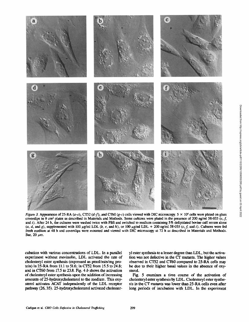
Figure 3. Appearance of 25-RA (a-c), CT52 (d-f), and CT60 (g-i) cells viewed with DIC microscopy. 5 x 10⁴ cells were plated on glass coverslips in 9 cm 2 plates as described in Materials and Methods. Some cultures were plated in the presence of 200 ng/ml 58-035 (c, f, and i). After 24 h, the cultures were washed twice with PBS and switched to medium containing 5% delipidated bovine calf serum alone (a, d, and g), supplemented with 100 #g/nil LDL (b, e, and h), or 100 #g/ml LDL + 200 ng/ml 58-035 (c,f, and i). Cultures were fed fresh medium at 48 h and coverslips were mounted and viewed with DIC microscopy at 72 h as described in Materials and Methods. Bar, 20 #m.
cubation with various concentrations of LDL. In a parallel experiment without mevinolin, LDL activated the rate of cholesteryl ester synthesis (expressed as pmol/min/mg pro- tein) in 25-RA from 11.1 to 51.6; in CT52 from 15.5 to 24.8; and in CT60 from 17.5 to 23.8. Fig. 4 b shows the activation of cholesteryl ester synthesis upon the addition of increasing amounts of 25-hydroxycholesterol to the medium. This oxy- sterol activates ACAT independently of the LDL receptor pathway (26, 35). 25-hydroxycholesterol activated cholester-
yl ester synthesis to a lesser degree than LDL, but the activa- tion was not defective in the CT mutants. The higher values observed in CT52 and CT60 compared to 25-RA cells may be due to their higher basal values in the absence of oxy- sterol.
Fig. 5 examines a time course of the activation of cholesteryl ester synthesis by LDL. Cholesteryl ester synthe- sis in the CT mutants was lower than 25-RA cells even after long periods of incubation with LDL. In the experiment
Cadigan et al. ClIO Cells Defective in Cholesterol Tra~icking 299
Dow
nloaded from http://rupress.org/jcb/article-pdf/110/2/295/1059055/295.pdf by guest on 03 April 2022

a
I 0 I I l I
2 40 60 80 100 I I I I
10 20 30 40 50
LDL Protein (l~g/ml)
/S o & ,Io ,is 21o
25-hydroxycholesterol (p.g/ml)
30
25
20
15
10
5
0
Figure 4. Effect of LDL and 25-hydroxycholesterol on the synthesis of cholesteryl esters in 25-RA (solid circles), CT52 (open circles), and CT60 (open squares) cells. Stock cultures were grown continu- ously in medium containing delipidated bovine calf serum for at least two passages as described in Materials and Methods. 4 x 10⁵ cells were plated into 9 cm e plates in medium containing 10% delipidated bovine calf serum. Medium was changed 48 h after plat- ing. 62 h after plating, the cultures were changed to 10% delipi- dated serum medium + 10 #M mevinolin and 230/zM mevalonate. At 66 h, the same medium supplemented with the indicated amounts of LDL (a) or 25-hydroxycholesterol (b) was added and 6 h later, the monolayers were pulsed with [3H]oleate for 30 min. After the pulse, the cells were harvested and analyzed for incorpo- ration of radiolabel into cholesteryl [3H]oleate as described in Materials and Methods. ValUes are the average of duplicate dishes and ranged within 15% of the mean.
shown in Fig. 5, the rate of incorporation of pH]oleate into triglycerides was found to be similar between all three cell types (data not shown).
The pH]oleate pulse provides an easy and quantitative method for distinguishing the 25-RA cell and the CT mutant phenotypes. All three cell lines were fused to a 25-RA cell clone bearing the azaguanine resistance (AG9 and ouabain resistance (oua9 markers. These genetic markers, one re- cessive (AG r) and one dominant (oua9 make these cells "universal fusion donors" that can be used to obtain hetero- karyons with any cell type (3, 32). The hybrids were kept in selective medium until plating for the pH]oleate pulse and >95 % of the cells in each population had the characteristic large size seen w i thCHO/CHO cell hybrids previously ob- tained in this laboratory (11). As seen in Fig. 6 a, the unfused controls grown in delipidated serum 5 :100 #g/ml LDL for 6 h had rates of cholesteryl ester formation similar to those seen in Fig. 4. In contrast, the data shown in Fig. 6 b demon- strate that the cholesterol esterification in the hybrids were all activated to a similar degree as was observed with unfused 25-RA cells. These data show that the phenotype of mutants CT52 and CT60 is recessive.
In addition, complementation analysis was performed using a CT60 cell line bearing the azaguanine- and ouabain- resistant markers as the fusion donor. When grown in 10% delipidated serum and then switched to medium containing 100 #g/ml LDL, the CT60/25-RA hybrids increased their cholesteryl ester synthesis from 11.1 to 46.2 pmol/min/mg. The CT60/CT60 hybrid's rate of synthesis went from 20.5 to 24.6 pmol/min/mg, and the CT60/CT52 hybrids from 13.2 to 18.4 pmol/min/mg. Thus CT60 and CT52 appear to be- long to the same complementation group.
140
120
loo
J ,o
4o
| 2o
o 0
6o
SO E
, o
3O
2o
0 0
0
Time (hours)
Figure 5. Time course of activation of cholesteryl ester synthesis by LDL in 25-RA (solid circles), CT52 (open circles), and CT60 (open squares) cells. Cells were plated as described in Fig. 4.4 h before the addition of LDL, the monolayers were switched to 10% delipidated serum medium + 10 #M mevinolin and 230 #M mevalonate. Cultures were switched to d¢lipidated serum medium plus mevinolin, mevalonate, and 100 #g/ml LDL at staggered times so that all the cultures could be pulsed with [3H]oleate 72 h after plating. All cells were fed the appropriate fresh medium at 48 and 60 h after plating. After the pulse, the cells were harvested and ana- lyzed for incorporation of radiolabel into cholestcryl [3H]oleatc as described in Materials and Methods. Values arc the average of duplicate dishes and ranged within 15% of the mean.
120
i 100
80
40
2O
0
25-RA CT52 CT60
b
25-RA CT52 CT60
120
100
80
60
40
20
0
Figure 6. Cholesteryl ester synthesis in unfused CHO cells (a) and cell hybrids (b) grown in delipidated serum (solid bars) or delipi- dated serum plus LDL (hatched bars). Hybrids were prepared by fusing the indicated cell clones with the 25-RA cells that bear the azaguanine resistant and ouabain resistant markers (11). 5 × 10⁴ unfused controls and 7.5 × 10⁴ hybrids were plated in 9 cm 2 dishes containing medium plus 10% FCS. After 48 h, the mono- layers were washed twice with PBS and switched to medium plus 10% delipidated FCS. Fresh medium was given 24 h later. At 86 h after plating, the cultures were changed to delipidated FCS medium plus 10 #M mevinolin and 230 #M mevalonate. At 90 h, the cells were given the above medium + 100 #g/ml LDL and grown for an additional 6 h before being pulsed for 30 min with [3H]ole- ate. Incorporation of radiolabel into cholesteryl [3H]oleate was de- termined as described in Materials and Methods. Values are the av- erage of duplicate dishes and ranged within 15% of the mean.
We next examined the in vitro ACAT activity in the rele- vant cell lines (Table I). The microsomal ACAT assay, like the [3H]oleate pulse, utilizes cellular cholesterol as one of the ACAT substrates. The values obtained from cell homog-
The Journal of Cell Biology, Volume I I0, 1990 300
Dow
nloaded from http://rupress.org/jcb/article-pdf/110/2/295/1059055/295.pdf by guest on 03 April 2022

Table I. Effect of Growth in LDL on the Microsomal and Reconstituted ACAT Activity of 25-RA, CT52, and CT60 Cell Extracts
ACAT specific activity
Cell type Time in LDL Microsomal Reconstituted
h pmol/min/mg
25-RA 0 18.9 82.3
2 115.3 104.0
48 131.5 98.7 CT52 0 25.1 136.6
2 45.3 142.6 48 98.0 154.2
CT60 0 19.8 78.6
2 29.4 87.1 48 174.4 94.0
3 x 10 ~ cells were plated in 75 cm ~ flasks in 12 ml of medium and grown un- der identical conditions as described in Fig. 5. LDL was added to the cultures at the indicated times, 72 h after plating, the monolayers were harvested and the microsomal and reconstituted ACAT activities of the cell extracts deter- mined as described in Materials and Methods. Values are the average of dupli- cate aliquots taken from each cell extract and ranged within 10% of the mean.
enates harvested after 2 h growth in LDL are reminiscent to those shown in Fig. 5, i.e., the CT mutants are defective in the activation of ACAT. However, the activities found from the extracts prepared from cultures grown in LDL containing medium for 48 h did not reflect the values obtained in the [3H]oleate pulse. Under these conditions, and when cells are grown continuously in FCS medium (data not shown), the ACAT activities are as high or higher in the CT mutants compared with 25-RA.
Since the [aH]oleate pulse and microsomal ACAT assay rely on cellular cholesterol, the results of these assays are influenced by the amount of cholesterol surrounding ACAT in addition to the absolute amount of enzyme. Therefore, reconstituted ACAT assays, where the enzyme activity is solubilized and then reconstituted into cholesterol-phospha- tidylcholine vesicles of a defined concentration (10) were performed. There was no difference found between recon- stituted ACAT activities in the cell extracts obtained from each cell line grown with or without LDL (Table I). This is consistent with previous work from this laboratory (10, 15, 16, 23) that suggested that changes in the ACAT protein con- tent are not responsible for the regulation of enzyme activity by LDL. The reconstituted ACAT activities in CT60 and 25- RA cell extracts were found to be similar but the recon- stituted ACAT activities in CT52 cell extracts were always '~50% greater than values obtained with 25-RA cell extracts. Since the mutants were isolated from mutagenized cells, it is possible that the CT52 mutant may contain a separate mu- tant locus, irrelevant to the CT locus addressed in this report, which may cause elevated ACAT activities in this particular cell line. Other possibilities can not be excluded. The data clearly show that in vitro ACAT activity is present in the CT mutants in normal or greater than normal amounts.
LDL Receptor Pathway Analysis
The binding, internalization, and degradation of '2~I-LDL was examined in 25-RA and the CT mutants (Fig. 7). The specific binding (Fig. 7 a) and internalization (Fig. 7 b) in the CT mutants were found to be ,x,l.6-2.0-fold higher than that of 25-RA cells. Total degradation of the radiolabeled
,0 f a J i 0.8
"~ 0.4
0.2
0.0
~ 3.0
= 2.5
¢" 2.0
~. ~n 0.5
0.0 I I I I I
I
0 | I I I 20 40 6 o 80 I o 0 1 2 0
12SI.LD L ixoteln (J~/ml)
Figure 7. The specific binding (a), specific internalization (b), and total degradation (c) of ~2~I-LDL in 25-RA (solid circles), CT52 (open circles), and CT60 (open squares) cells. Cells were plated and grown as described in Fig. 4, except that 5 % delipidated serum was used. At 66 h after plating, media containing the indicated amounts of ~25I-LDL (343 cpm/ng) were added to the cells. After 6 h, the cells were analyzed for specific binding and internalization, and total degradation of the ~I-LDL as described in Materials and Methods. Values are the average of duplicate dishes and ranged within 10% of the mean.
LDL was also measured (Fig. 7 c). Controls demonstrated that the addition of a large excess of unlabeled LDL (500 /zg/ml) inhibited the degradation of the ~2SI-lipoprotein by 82-93 %, and the presence of 100/zM chloroquine, an inhib- itor of lysosomal function (26, 28), did not block internaliza- tion but resulted in a 71 to 90% reduction in degradation (data not shown). Degradation of radiolabeled LDL in the CT mutants was similar or slightly higher than levels found in 25-RA cells.
Besides the degradation of the LDL protein moiety, the hy- drolysis of LDL-cholesterol ester also occurs in the lyso- somes, via a specific acid lipase (26, 28). Of the total choles- terol in LDL, 75-80% is estedfied (53), and the hydrolysis of this sterol ester is necessary for the regulatory effects of LDL to be observed in human fibtoblast (26, 28) and CHO cells (22). By labeling the LDL with [3H]cholesteryl linole- ate, it is possible to measure the hydrolysis of [3H]choles- teryl linoleate to free [3H]cholesterol.
The data in Fig. 8 illustrate the total LDL-[3H]choles - teryl linoleate hydrolyzed (Fig. 8 a) and the LDL derived [3H]cholesteml that is reesterified (Fig. 8 b) after addition of [3H]cholesteryl linoleate-LDL to the cultures. CT52 cells hy- drolyzed, on average, 2.1 times more LDL-[3H]cholesteryl
Cadigan et al. CHO Cells Defective in Cholesterol Trafficking 301
Dow
nloaded from http://rupress.org/jcb/article-pdf/110/2/295/1059055/295.pdf by guest on 03 April 2022

~ 8o
~ 6o
40
m .5 q o,-
a b
2O
3O
<o
o o
E .5 q
4 0 6 0 8 0 1 0 0 0 2 0 4 0 so 8 0
[3H] Choleste~l ~ a t o - L D L protein (pg/ml)
0 100 120
Figure 8. Hydrolysis (a) and reesterification (b) of LDL-[3H]cho - lesteryl linoleate in 25-RA (solid circles), CT52 (open circles), and CT60 (open squares) cells. Cells were plated and grown as de- scribed in Fig. 7, except that after 62 h, all media were supple- mented with 35 #M oleic acid. At 66 h after plating, medium con- mining the indicated amounts of [3H]cholesteryl linoleate-LDL (3,180 cpm/nmol cholesteryl linoleate) were added to the cells. Af- ter 6 h, the cells were harvested and the lipid extracts were analyzed for [3H]cholesterol, [3H] cholesteryl linoleate, and [3H]cholesteryl oleate as described in Materials and Methods. Hydrolysis of LDL- [3H]cholesteryl linoleate was calculated by addition of the [3H]cho- lesterol and [3H]cholesteryl oleate values. The amount of reesterifi- cation of LDL-[3H]cholesterol was calculated from the [3H]cho- lesteryl oleate values. Values are the average of duplicate plates and ranged within 10% of the mean.
linoleate than 25-RA cells, while CT60 hydrolyzed between 50 and 140% of the values found for 25-RA. Despite this rel- atively normal or greater than normal hydrolysis of LDL- [3H]cholesteryl linoleate in the CT mutants, they reesterified 11-19 times more LDL derived [3H]cholesterol than CT52 and CT60 cells.
Control experiments similar to the ones performed during the '2q-LDL experiment demonstrated that the amount of LDL-[3H]cholesteryl linoleate hydrolyzed by the cells was inhibited 66-87 % by excess unlabeled LDL. The addition of chloroquine to the medium caused a drastic increase in the amount of [3H]cholesterol linoleate found in the cells and a large decrease (73-88%) in the percentage of radiolabel found in the [3H]cholesterol and [3H]cholesteryl oleate bands (data not shown).
Sterol Analysis
The data shown in Fig. 8 indicates that the cholesteryl ester moiety of LDL is hydrolyzed normally in the CT mutants but unlike the parental cell line 25-RA, the cholesterol is reesterified at a much diminished rate. To address the ques- tion of what happens to this liberated cholesterol, sterol anal- ysis of 25-RA and the CT mutants was performed. Fig. 9 il- lustrates the changes in sterol content with increasing time (3-72 h) after the addition of 100 ~g/ml LDL. The un- esterified cholesterol level of the 25-RA cells remained rela- tively unchanged while the presence of LDL in the medium caused a large accumulation of cholesteryl ester in 25-RA cells and a smaller increase in the CT mutants, consistent with the activation of cholesteryl ester synthesis shown ear- lier (Figs. 4-6). In contrast, both CT mutants exhibited a dramatic time dependent increase in their cellular choles-
500 ,
E 400
300
0
y , t . i t
2 0 4 0 6 0
, i , , . , .
20 40 60
250
E 20O ~
150 -~
0 8O
Time (hour)
Figure 9. Time course of cholesterol (a) and cholesteryl ester (b) accumulation after addition of LDL. 25-RA (solid circles), CT52 (open circles), and CT60 (open squares) cells were plated and grown exactly as described in Fig. 5 except that 5 % delipidated se- rum was used throughout the experiment. At 72 h after plating, the cultures were harvested and analyzed for cholesterol and choles- teryl ester as described in Materials and Methods. Values are the average of duplicate aliquots taken from cell extracts and ranged within 5 % of the mean.
terol content, which reached at maximum 4.5-5.0-fold above the levels observed in the absence of LDL. This accumula- tion was also dependent on the concentration of LDL in the medium and was seen when the cells were grown in the pres- ence of the ACAT inhibitor 58-035 (data not shown).
In a parallel experiment to the one shown in Fig. 9, mevinolin was not included in the growth medium, and very similar results were obtained (data not shown), suggesting that endogenous synthesis of cholesterol did not contribute significantly to the accumulation of cellular cholesterol. Therefore, the large majority of accumulated cholesterol seen in the CT mutants must have come directly from LDL.
Localization o f the Accumulated IntraceUular Cholesterol
The accumulation of free cholesterol and the defective acti- vation of cholesteryl ester synthesis after LDL addition in the CT mutants is very similar to the observations made in fibroblasts isolated from a strain of BALB/c mice with a neu- rological disorder (43), and human fibroblasts isolated from patients with NP-C disease (36, 44). Cells from these affected mice and humans have been shown to accumulate free cholesterol in their lysosomes in response to exogenous sterols such as LDL (5, 7, 38, 52, 54). Both CT52 and CT60 have been shown to accumulate a large amount of dark intra- cellular particles (Fig. 3) and free cholesterol (Fig. 9) when grown in the presence of LDL. It seemed that these two phenomena were linked, and the similarity between the CT mutants and the NP-C and mouse fibroblasts suggested that the cytoplasmic particles in the CT mutants may be lysosomes full of cholesterol.
The fluorescent stain acridine orange is a membrane per- meant weak base that accumulates in acidic cellular com- partments (47) where it emits red light (>620 nm) upon exci- tation. As seen in Fig. 10, g and h, CT52 cells grown in LDL before acridine orange staining contain a fluorescence signal pattern that colocalized with the dark cytoplasmic particles
The Journal of Cell Biology, Volume 110, 1990 302
Dow
nloaded from http://rupress.org/jcb/article-pdf/110/2/295/1059055/295.pdf by guest on 03 April 2022

Figure 10. Localization of acidic compartments with acridine orange in 25-RA (a-d) and CT52 (e-h) cells. Cells were grown as described in Fig. 3 in medium containing 5% delipidated serum (a, b, e, and f ) or medium containing 5% delipidated serum supplemented with 100 #g/ml of LDL (c, d, g, and h). Cells were stained with acridine orange as described in Materials and Methods before viewing with phase-contrast (a, c, e, and g) or fluorescence (b, d, f, and h) microscopy. Bar, 20 t~m.
visualized with phase-contrast microscopy. In contrast, the lighter, perinuclear particles seen in the 25-RA cells treated with LDL did not colocalize with the acridine orange signal (Fig. 10, c and d). For CT52 cells, there was a large increase
in the amount of acridine orange signal in cells grown in LDL-containing medium compared to cells grown in delipi- dated serum medium alone (Fig. 10, f a n d h), while no in- crease was observed in 25-RA cells (Fig. 10, b and d).
Cadigan et al. ClIO Cells Defective in Cholesterol Trafficking 303
Dow
nloaded from http://rupress.org/jcb/article-pdf/110/2/295/1059055/295.pdf by guest on 03 April 2022
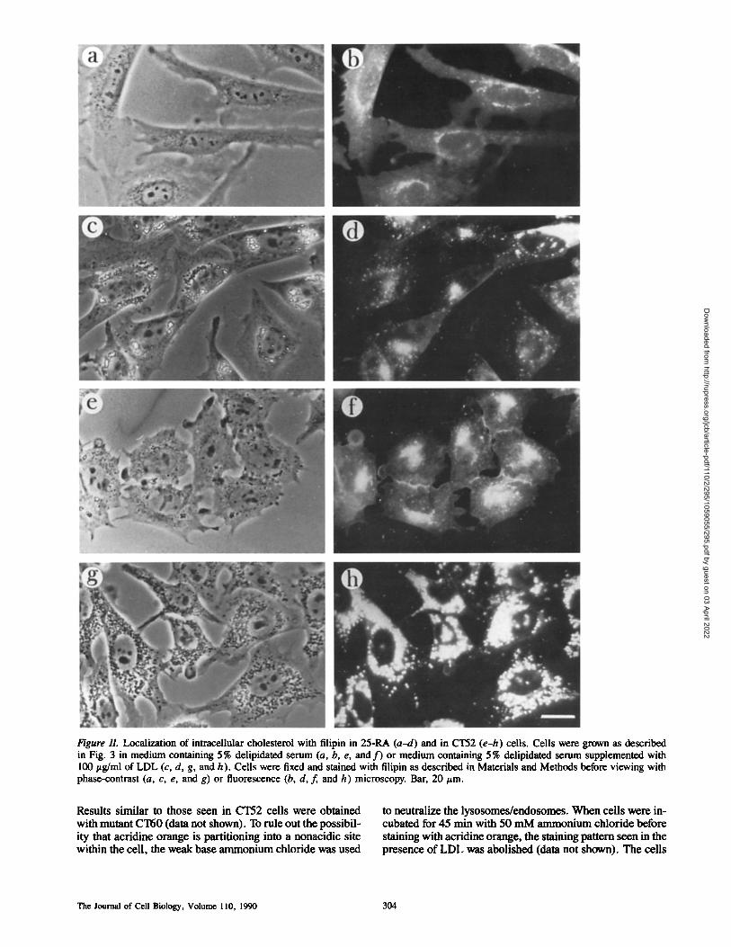
Figure 11. Localization of intracellular cholesterol with filipin in 25-RA (a-d) and in CT52 (e-h) cells. Cells were grown as described in Fig. 3 in medium containing 5% delipidated serum (a, b, e, and f ) or medium containing 5% delipidated serum supplemented with 100 ttg/ml of LDL (c, d, g, and h). ,Cells were fixed and stained with filipin as described in Materials and Methods before viewing with phase-contrast (a, c, e, and g) or fluorescence (b, d, f, and h) microscopy. Bar, 20/zm.
Results similar to those seen in CT52 cells were obtained with mutant CT60 (data not shown). To rule out the possibil- ity that acridine orange is partitioning into a nonacidic site within the call, the weak base ammonium chloride was used
to neutralize the lysosomes/endosomes. When cells were in- cubated for 45 rain with 50 mM ammonium chloride before staining with acridine orange, the staining pattern seen in the presence of LDL was abolished (data not shown). The cells
The Journal of Cell Biology, Volume 110, 1990 304
Dow
nloaded from http://rupress.org/jcb/article-pdf/110/2/295/1059055/295.pdf by guest on 03 April 2022

exhibited a uniform fluorescence that was faint and diffuse, thus the fluorescence pattern seen in Fig. 10 would appear to be from the accumulation of acridine orange in the cellular acidic compartments.
When CT52 cells were grown in LDL and stained with filipin, a fluorescent compound known to bind specifically to cholesterol (8), a strong filipin signal was seen that colocalized with dark perinuclear particles (Fig. 11, g and h). In contrast, the modest amount of filipin staining ob- served in 25-RA cells grown in LDL (Fig. 11, c and d) did not colocalize with its cytosolic, lipid droplet particles. As with the acridine orange staining experiment, after addition of LDL, there is a large increase in filipin fluorescence in CT52 mutants compared to CT52 grown in delipidated se- rum alone (Fig. 11, f and h). A much smaller increase was seen in 25-RA cells (Fig. 11, b and d). When these experi- ments were performed with CT60 cells, identical results were obtained (data not shown).
To further examine the intracellular location of the accu- mulated cholesterol, CT60 cells were costained with filipin and lgp58, a monoclonal antibody prepared against a CHO cell lysosomal/endosomal fraction (Mellman, I., unpub- lished data and personal communication). As seen in Fig. 12, a and b, the filipin (Fig. 12 a) signal colocalized with the signal obtained with the lysosmal/endosomal antibody via indirect immunofluorescence (Fig. 12 b). when the dou- ble staining experiment was performed with cells grown in delipidated serum medium alone, the filipin staining for 25- RA and CT60 resembled that shown in Fig. 11, while the an- tibody staining had a very similar and faint fluorescence for both cell types (data not shown). Results using CT52 cells were very similar to those shown using CT60 cells (data not shown). Control experiments with the nonspecific antibody MOPC-21 revealed faint uniform staining over the entire cell in all three cell types.
Discussion
This report describes a selection procedure for isolating mu- tant cells that are resistant to cholesterol starvation. These mutants, which we have termed cholesterol trafficking, or CT mutants, were shown to accumulate a large amount ofun- esterified cholesterol when grown in medium containing LDL (Fig. 9) or 10% FCS. The data indicate that the excess cholesterol is located in intracellular particles shown by DIC (Fig. 3) and phase-contrast microscopy (Fig. 11). These par- ticles are lysosomes and/or endosomes based on fluorescent microscopy studies with acridine orange (Fig. 11) and a lyso- somal/endosomal monoclonal antibody (Fig. 12). The high cholesterol levels seen in the CT mutants when they were grown in LDL were found to decrease dramatically after 2 d of growth in delipidated serum medium (data not shown). This observation suggests that the cholesterol was able to leave the lysosomes of the CT mutants, perhaps via a nonspecific pro- cess (i.e., monomer diffusion, membrane recycling, etc.). The ability of the cholesterol to escape from the lysosomes probably provided the CT cells with the cholesterol needed for survival during the starvation selection.
In the selection protocol, the number of mutagenized cells present in each dish at the start of the cholesterol starvation was ,x,l.5 x 105. Since every dish had at least one CT-like colony, the lowest possible frequency of this mutant pheno-
Figure 12. Indirect immunofluorescence of filipin-stained CT60 cells using a lysosomal/endosomal specific antibody. CT60 cells were grown as described in Fig. 3 in medium containing 5 % delipi- dated serum plus 100/zg/ml of LDL. Cells were fixed, stained with filipin, and then stained by indirect immunofuorescence with lgp58 as detailed in Materials and Methods. a and b show filipin and lgp58 fuorescence, respectively. Bar, 20 tzm.
type was •6.6 X 10 -6. However, more than one CT-like colony was always seen and our protocol only identified the largest and most obvious surviving colonies. The actual fre- quency was probably between 2 x 10 -5 (three independent CT mutants/dish) and 1 x 10 -4 (15 CT mutants/dish). These frequencies are 2-12 times higher than the frequency of obtaining ACAT revertants using an alternative method (12), which might explain why no ACAT revertants were found in the starvation selections.
The frequencies of mutation in mutagenized CHO cells have commonly been found to range from 1 x 10 -3 to 5 × 10- 5 for single genes (13, 21, 34, 41, 56), and 1 x 10⁻⁶ to 2 >( 10 -7 for genes that are functionally diploid (11, 21, 34, 41). Thus, it appears that the generation of the CT mutant phenotype may only have required the mutation of a single gene. The data in Fig. 6 indicated that the mutations in CT52 and CT60 are recessive, suggesting that there is only one ac- tive CT gene in the CHO cell line, 25-RA.
One phenomena which occurred frequently throughout the biochemical analysis of the CT mutants was the higher than normal (i.e., 25-RA) activities of some of the com- monly assayed processes of cholesterol metabolism, such as LDL receptor (Figs. 7 and 8) and ACAT activity (Table I), and the incorporation of labeled acetate into cholesterol (data not shown). The elevated reconstituted ACAT activity seen in
Cadigan et al. CHO Cells Defective in Cholesterol Trafficking 305
Dow
nloaded from http://rupress.org/jcb/article-pdf/110/2/295/1059055/295.pdf by guest on 03 April 2022

CT52 cell extracts (Table I) may be due to a secondary muta- tion since it is never observed in CT60 cells. Unlike ACAT, the LDL receptor has been demonstrated to be subject to stringent regulation at the level of transcription (27), and 3-hydroxy-3-methylgluCa-yl-coenzyme A reduetase, a rate lim- iting enzyme in cholesterol biosynthesis, has been shown to be highly regulated at both the transcriptional and posttranla- tional levels by cholesterol (27). It is possible that a mutation in a cholesterol transport process such as the one affected in the CT mutants may alter this complicated regulatory ap- paratus and cause an elevation of these activities. Once rever- tants of the CT mutants have been isolated and characterized, these possibilities can be more directly examined.
The microsomal ACAT specific activity of cell extracts normally reflects the values obtained in intact cells with the [3H]oleate pulse (15, 16). In Table I this was true for the cell extracts harvested after growth in delipidated serum medium or after 2 h in medium containing LDL. However, CT mutants grown for 48 h in LDL had microsomal ACAT specific activities as high or higher than 25-RA cell extracts, in contradiction to the [3H]oleate pulse data (Fig. 5). These high activities may be an artifact of the preparation of the cell extracts, which involved hypotonic shock (20) that may have disrupted the cholesterol loaded lysosomal/endosomal com- partments in the CT cells. This cholesterol could then act as a substrate for ACAT. This type of phenomena has been reported in NP-C fibroblasts (54).
Previously, it has been shown that the suppression of the LDL receptor and cholesterol biosynthesis by LDL occurred with a significant lag in NP-C fibroblasts compared to unaffected fibroblasts (37, 45), consistent with the defective movement of cholesterol out of the NP-C lysosomes (38). These experiments were attempted with the CT mutants but the results were ambiguous because the parental cell line, 25- RA, is partially resistant to suppression of cholesterol bio- synthesis and the LDL receptor pathway by exogenous sterols (19, and data not shown). Perhaps a significant lag in ~ e kinetics of LDL-mediated suppression of these pathways would be evident if the CT mutants were in a wild-type CHO cell genetic background. It should be pointed out that the LDL receptor pathway is two to three times more active in 25-RA cells treated with 58-035 than wild-type cells (11). Therefore, a CT mutant derived from a wild-type cell might not accumulate as much intracellular unesterified cholesterol and thus would not survive as well (if at all) during the star- vation selection.
The exact biochemical process defective in the CT mutants is not known. One obvious candidate would be an inactivated intracellular cholesterol transporter. Sterol carrier protein2 is a 13-kD protein originally identified in rat and bovine liver cytosol. This protein has been shown to transfer cholesterol between membranes in vitro and to activate ACAT activity in vitro (25, 48). Sterol carder protein2 has been localized predominantly to peroxisomes in liver (33, 57) but it is pres- ent in smaller amounts in the endoplasmic reticulum and cytosol (33). The development of a reliable assay for this pro- tein in CHO cells would allow direct testing of whether sterol carrier protein2 activity is present in CT mutant cell ex- tracts.
The defect in the CT mutants does not necessarily have to be in a cytosolic cholesterol transporter. There could be a de-
fect in the mobilization of the newly hydrolyzed cholesterol in the lysosomal interior, or a defect in a receptor molecule that receives the cholesterol after transport from the lyso- somes. More knowledge concerning the structure and poly- peptide composition of the cholesterol loaded lysosomes, and the normal pathways of LDL-derived cholesterol trans- port out of the lysosomes will be beneficial towards under- standing the exact molecular defect in the CT mutants.
In conclusion, this paper reports the isolation of CHO cells in which the intracellular movement of LDL-derived cholesterol is defective, leading to an accumulation of cho- lesterol in acidic compartments that are most likely lyso- somes. The phenotype of the CT mutants is almost identical to that of the NP-C fibroblasts and a strain of BALB/c mice with a similar neurological disorder. The value of the CT mutants is that CHO cells are much more amenable to genetic manipulation than primary cultures of human fibro- blasts or mice. The isolation of a large number of recessive CT mutants will allow complementation analysis to deter- mine if the inactivation of more than one gene can generate a CT phenotype. The ability to fuse CHO cells with human fibroblasts and the subsequent segregation of the human chromosomes could provide a means for identifying the chromosome where the human homologue of the CT gene (or genes) is located. In addition, fusions between the CT mutants and NP-C firboblasts and biochemical analysis of the hybrids could determine if the same locus is affected (no complementation observed). The final advantage of obtain- ing the cholesterol trafficking phenotype in a CHO genetic background, and perhaps the most important, is that exoge- nous DNA can be introduced into CHO cells via DNA medi- ated gene transfer (1). The complementation of the CT mu- tation via transfection of exogenous DNA should be possible provided a suitable selection system can be developed. Once transformants are obtained, the cloning of the complement- ing gene should be possible. Such work is currently under- way in our laboratory.
The authors would like to thank Drs. Stanley C. Froehner and Robert C. Jackson for their advice with the microscope work, and Dr. Donald L. Schneider for critically reading the manuscript. They would also like to thank Dr. Ira Meliman for generously providing the monoclonal antibody lgp58 and for advice with the immunofluorescence work.
Received for publication 1 August 1989 and in revised form 23 October 1989.
References
1. Abraham, I. 1985. DNA-mediated gene transfer. In Molecular Cell Genetics, M, M. Gottesman, editor. John Wiley & Sons, Inc., New York. 181-210.
2. Alberts, A. W., J. Chen, G. Kuron, V. Hunt, J. Huff, C. Hoffman, J. Rothrock, M. Lopez, H. Joshua, E. Harris, A. Patchett, R. Moneghan, S. Currie, E. Stapley, G. Albers-Schonberg, O. Hensons. J. Hirshfield, K. Hoogsteen, J. Liesch, and J. Springer. 1980. Mevinolin: a highly po- tent competitive inhibitor of hydroxymethylglutaryl-coenzyme A reduc- tase and a cholesterol-lowering agent. Proc. Natl. Acad. Sci. USA. 77:3957-3961.
3. Baker, R. M., D. M. Brunette, R. Mankovitz, L. H. Thompson, G. H. Whitmore, L. Siminoviteh, and J. E. Till. 1974. Ouabain-resistant mu- tants of mouse and hamster cells in culture. Cell. 1:9-21.
4. Balasnhramaniam, S., S. Venkatesan, K. A. Mitropoulus, and T. J. Peters. 1978. The submicrosomal localization of acyl-coenzyme A-cholesterol acyltransferase and its substrate, and ofcholesteryl esters in rat liver. Bio- chem. J. 174:863-872.
5. Bhuvaneswaran, C., M. D. Morris, H. Shio, and S. Fowler. 1982. Lyso- some lipid storage disorder in NCTR-BALB/c mice. llI. Isolation and
The Journal of Cell Biology, Volume ! 10, 1990 306
Dow
nloaded from http://rupress.org/jcb/article-pdf/110/2/295/1059055/295.pdf by guest on 03 April 2022

analysis of storage inclusions from liver. Am. J. Pathol. 102:160-170. 6. Bishop, J. E., and A. K. Hajra. 1980. A method for the chemical synthesis
of '4C-labeled fatty acyl coenzyme A's of high specific activity. Anal. Biochem. 106:344-350.
7. Blanchette-Mackie, E. J., N. K. Dwyer, L. M. Amende, H. S. Kruth, J. D. Butler, J. Sokol, M. E. Comiy, M. T. Vanier, J. T. August, R. O. Brady, and P. G. Pentchev. 1988. Type-C Niemann-Pick disease: low density lipoprotein uptake is associated with premature cholesterol accumulation in the Golgi complex and excessive cholesterol storage in lysosomes. Proc. Natl. Acad. Sci. USA. 85:8022-8026.
8. Bornig, H., and G. Geyer. 1974. Staining of cholesterol with the fluores- cent antibiotic "filipin. ~ Acta Histochem. 50:110-115.
9. Butler, J. D., M. E. Comly, H. S. Kruth, M, Vanier, M. Filling-Katz, J. Fink, N. Barton, H. Weintroub, J. M. Quirk, T. Tokoro, D. C. Marshall, R. O. Brady, and P. G. Pentchev. 1987. Niemann-Pick variant disorders: comparisons of errors of cellular cholesterol homeostasis in group D and group C fibroblasts. Proc. Natl. Acad. Sci. USA. 84:556-560.
10. Cadigan, K. M., and T. Y. Chang. 1988. A simple method for reconstitu- tion of CHO cell and human fibroblast ACAT activity into liposomes. J. Lipid Res. 29:1683-1692.
I 1. Cadigan, K. M., J. G. Heider, and T. Y. Chang. 1988. Isolation and char- acterization of Chinese hamster ovary cell mutants deficient in acyl- coenzyme A: cholesterol acyltransferase activity. J. Biol. Chem. 263: 274-282.
12. Cadigan, K. M., C. C. Y. Chang, and T. Y. Chang. 1989. Isolation of Chi- nese hamster ovary cell lines expressing human acyl-coenzymeA/choles- terol acyltransferase activity. J. Cell Biol. 108:2201-2210.
13. Campbell, C. E., and R. G. Wonon. 1979. Evidence obtained by induced mutation frequency analysis for functional hemizygosity at the emt locus in CHO cells. Somatic Cell Genet. 5:51-65.
14. Chain, B. E., and B. R. Knowles. 1976. A solvent system for delipidation of plasma or serum without protein precipitation. J. Lipid Res. 17:176-181.
15. Chang, C. C. Y., and T. Y. Chang. 1986. Cycloheximide sensitivity in regulation of acyl eoenzyme A: cholesterol acyltransferase activity in Chinese hamster ovary cells. IL Effect of sterol endogenously synthe- sized. Biochemistry. 25:1700-1706.
16. Chang, C. C. Y., G. M. Doolittle, and T. Y. Chang. 1986. Cycloheximide sensitivity in regulation of acyl coenzyme A: cholesterol acyltransferase activity in Chinese hamster ovary cells. I. Effect of exogenous sterols. Biochemistry. 25:1693-1699.
17. Chang, T. Y., and C. C. Y. Chang. 1982. Revertants of a Chinese hamster ovary cell mutant resistant to suppression by an analogue of cholesterol: isolation and partial biochemical characterization. Biochemistry. 21: 5316-5323.
18. Chang, T. Y., and G. M. Doolittle. 1983. Acyl coenzyme A: cholesterol O-acyltransferase. In The Enzymes. Vol. 16. P. D. Boyer, editor. Aca- demic Press, Inc., New York. 523-539.
19. Chang,T. Y., and J. S. Limanek. 1980. Regulation of cytosolic acetoacetyl coenzyme A thiolase, 3-hydroxy-3-methylglutaryl coenzyme A synthase, 3-hydroxy-3-methylglutaryl coenzyme A reductase, and mevalonate ki- nase by low density lipoprotein and by 25-hydroxycholesterol in Chinese hamster ovary cells. J. Biol. Chem. 255:7787-7795.
20. Chang, T. Y., J. S. Limanek, and C. C. Y, Chang. 1981. A simple and efficient procedure for the rapid homogenization of cultured animal cells grown in monolayer. Anal. Biochem. 116:298-302.
21. Chasin, L. A. 1974. Mutations affecting adenine phosphoribosyl transfer- ase activity in Chinese hamster cells. Cell. 2:37-41.
22. Chin, J., and T. Y. Chang. 1981. Evidence for coordinate expression of 3-hydroxy-3-methylglutaryl coenzyme A reductase and low density lipo- protein binding activity. J. Biol. Chem. 256:6304-6310.
23. Doolittle, G. M., and T. Y. Chang. 1982. Acyl-CoA: cholesterol acyltrans- ferase in Chinese hamster ovary cells. Enzyme activity determined after reconstitution in phospholipid/cholesterol liposomes. Biochim. Biophys. Acta. 713:529-537.
24. Drevon, C. A., D. B. Weinstein, and D. Steinberg. 1980. Regulation of cholesterol esterification and biosynthesis in monolayer cultures of nor- mal adult rat hepatocytes. J. Biol. Chem. 255:9128-9137.
25. Gavey, K. L., B. J. Noland, and T. J. Scallen. 1981. The participation of sterol carrier protein2 in the conversion of cholesterol to cholesterol es- ter by rat liver microsomes. J. Biol. Chem. 256:2993-2999.
26. Goldstein, J. L., and M. S. Brown. 1977. The low-density lipoprotein path- way and its relation to atherosclerosis, Annu, Rev. Biochem. 46:897-930,
27. Goldstein, J. L,, and M. S. Brown. 1984. Progress in understanding the LDL receptor and HMG-CoA reductase, two membrane proteins that regulate the plasma cholesterol. J. Lipid Res. 25:1450-1461.
28. Goldstein, J. L., S. E. Dana, J. R. Faust, A. L. Beaudet, and M. S. Brown. 1975. Role of lysosomal acid lipase in the metabolism of plasma low den- sity lipoprotein. J. Biol. Chem. 250:8487-8495.
29. Goldstein, J. L., S. K. Basu, and M. S. Brown. 1983. Receptor-mediated endocytosis of low-density lipoprntein in cultured cells. Methods En- zymol. 98:241-260.
30. Hashimoto, S., and A. M. Fogelman. 1980. Smooth microsomes. A trap for cholesteryl ester formed in hepatic microsomes. J. Biol. Chem.
255:8678-8684. 31. Heider, J. G., and R. L. Boyett. 1978. The picomole determination of free
and total cholesterol in cells in culture, J. Lipid Res. 19:514-518. 32. Jha, K. K., and H. L. Ozer. 1976. Expression of transformation in cell
hybrids. 1. Isolation and application of density-inhibited Balb/3T3 cells deficient in hypoxanthine phosphoribosyltransferase and resistant to oua- bain. Somatic Cell Genet. 2:215-223.
33. Keller, G. A., T. J. Scallen, D. Clarke, P. A. Maher, S. K. Krisans, and S. J. Singer. 1989. Subcellular localization of sterol carrier protein-2 in rat hepatocytes: its primary localization to peroxisomes. J. Cell Biol. 108:1353-1361.
34. Kingsley, D. M., and M. Krieger. 1984. Receptor-mediated endocytosis of low density lipoprotein: somatic cell mutants define multiple genes re- quired for expression of surface-receptor activity. Proc. Natl. Acad. Sci. USA. 81:5454-5458.
35. Krieger, M., M. S. Brown, and J. L. Goldstein. 1981. Isolation of Chinese hamster cell mutants defective in the receptor-mediated endocytosis of low density lipoprotein. J. Mol. Biol. 150:167-184.
36. Krath. H. S., M. E. Comly, J. D. Butler, M. T. Vanier, J. K. Fink, D. A. Wenger, S. Patel, and P. G. Pentchev. 1986. Type C Neimann-Pick dis- ease. Abnormal metabolism of low density lipoprotein in homozygous and heterozygous fibroblasts. J. Biol. Chem. 261 : 16769-16774.
37. Liscum, L., and J. R. Faust. 1987. Low density lipoprotein (LDL)- mediated suppression of cholesterol synthesis and LDL uptake is defec- tive in Neimann-Pick type C fibroblasts. J. Biol. Chem. 262:17002- 17008.
38. Liscum, L., R. M. Ruggietto, and J. R. Faust. 1989. The intracellular transport of low density lipoprotein-derived cholesterol is defective in Niemann-Pick type C fibroblasts. J. Cell Biol. 108:1625-1636.
39. Lowry, O. H., N. J. Rosebrough, A. L. Farr, and R. J. Randall. 1951. Pro- tein measurement with the folin phenol reagent. J. Biol. Chem. 193:265- 275.
40. Morris, M. D., C. Bhuvaneswaran, H. Shio, and S. Fowler. 1982. Lyso- some lipid storage disorder in NCTR-BALB/c mice. I. Description of the disease and genetics. Am. J. Pathol. 108:140-149.
41. Nalbantoglu, J., O. Goncalves, and M. Meuth. 1983. Structure of mutant alleles at the aprt locus of Chinese hamster ovary cells. J. Mol. Biol. 167:575-594.
42. Pentchev, P. G., A. E. Gal. A. D. Booth, F. Omodeo-Sale, J. Fouks, B. A. Leameyer, J. M. Quirk, G. Dawson, and R. O. Brady. 1980. A lyso- somal storage disorder in mice characterized by a dual deficiency of sphingomyelinase and glucocerebrosidase. Biochim. Biophys. Acta. 619:669-679.
43. Pentchev, P. G., A. D. Boothe, H. S. Kruth, H. Weintroub, J. Stivers, and R. O. Brady. 1984. A genetic storage disorder in BALB/c mice with a metabolic block in esterification of exogenous cholesterol. 3'. Biol. Chem. 259:5784-5791.
44. Pentchev, P. G., M. E. Comly, H. S. Kruth, M. T. Vanier, D. A. Wenger, S. Patel, and R. O. Brady. 1985. A defect in cholesterol esterification in Niemann-Pick disease (type C) patients. Proc. Natl. Acad. Sci. USA. 82:8247-825 I.
45. Pentchev, P. G., M. E. Comly, H. S. Kruth, T. Tokoro, J. Butler, J. Sokol, M. Filling-Katz, J. M. Quirk, D. C. Marshall, S. Patel, M. T. Vanier, and R. O. Brady. 1987. Group C Niemann-Pick disease: faulty regulation of low-density lipoprotein uptake and cholesterol storage in cultured fibroblasts. FASEB (Fed. Am. Soc. Exp. Biol.) J. 1:40-45.
46. Peterson, G. L. 1977. A simplification of the protein assay method of Lowry et al. which is more generally applicable. Anal. Biochem. 83:346-356.
47. Poole, A. R. 1977. The detection of lysosomes by vital staining with acri- dine orange. In Lysosomes: A Laboratory Handbook. 2nd Ed. J. T. Din- gle, editor. Elsevier/North-Holland, Amsterdam. 313-316.
48. Poorthuis, B, J. H. M., and K. W. A. Wirtz. 1982. Increased cholesterol esterification in rat liver microsomes by purified non-specific phospho- lipid transfer protein. Biochim. Biophys. Acta. 710:99-105.
49. Roberts, D. C. K., N. E. Miller, S. G. L. Price, D. Crook, C. Cortese, A. LaVille, L. Masana, and B. Lewis. 1985. An alternative procedure for incorporating radiolabeled cholesteryl ester into human plasma lipo- proteins in vitro. Biochem. J. 226:319-322.
50. Ross, A. C., K. J. Go, J. G. Heider, and G. H. Rothblat. 1984. Selective inhibition of acyl coenzyme A: cholesterol acyltransferase by compound 58-035. J. Biol. Chem. 259:815-819.
51. Sege, R. D., K. Kozarsky, D. L. Nelson, and M. Kreiger. 1984. Expres- sion and regulation of human low-density lipoprotein receptors in Chi- nese hamster ovary cells. Nature (Lond.). 307:742-745.
52. Shio, H., S. Fowler, C. Bhuvaneswaran, and M. D. Morris. 1982. Lyso- some lipid storage disorder in NCTR-BALB/c mice. II. Morphologic and cytochemical studies. Am. J. Pathol. 108:150-159.
53. Skipski, V. P. 1972. Lipid composition of lipoproteins in normal and dis- eased states. In Blood Lipids and Lipoproteins: Quantitation, Composi- tion and Metabolism. G. J. Nelson, editor. John Wiley & Sons, Inc., New York. 471-583.
54. Sokol, J., E. J. Blanchette-Mackie, H. S. Kruth, N. K. Dwyer, L. M. Amende, J. D. Buffer, E. Robinson, S. Patel, R. O. Brady, M. E. Comly,
Cadigan et at. ClIO Cells Defective in Cholesterol Trafficking 307
Dow
nloaded from http://rupress.org/jcb/article-pdf/110/2/295/1059055/295.pdf by guest on 03 April 2022

M. T. Vanier, and P. G. Pentchev. 1988. Type C Niemann-Pick disease. Lysosomai accumulation and defective intracellular mobilization of low density lipoprotein cholesterol. J. Biol. Chem. 263:3411-3417.
55. Stadtman, E. R. 1957. Preparation and assay of acylcoenzyme A and other thioi esters: use of hydmxylamine. Methods Enzymol. 3:931-941.
56. Urlaub, G., and L. A. Chasin. 1980. Isolation of Chinese hamster cell mu- tants deficient in dihydrofolate reductase activity. Proc. Natl. Acad. Sci.
USA. 77:4216-4220. 57. Van der Krit~, T. P., J. Leunissen, T. Teerlink, G. P. H. Van Heusden,
A. J. Verkleij, and K. W. A. Wirtz. 1985. Ultrastructural localization of a peroxisomal protein in rat liver using the specific antibody against the non-specific lipid transfer protein (sterol carrier protein 2). Biochim. Biophys. Acta. 812:387-392.
The Journal of Cell Biology, Volume 110, 1990 308
Dow
nloaded from http://rupress.org/jcb/article-pdf/110/2/295/1059055/295.pdf by guest on 03 April 2022